Embed Size (px)
Citation preview
BMP-2 Stimulated Non-Tenogenic Differentiation and PromotedProteoglycan Deposition of Tendon-Derived Stem Cells (TDSCs)In Vitro
Yun Feng Rui,1,2 Pauline Po Yee Lui,1,2,3 Yin Mei Wong,1,2 Qi Tan,1,2 Kai Ming Chan1,2,3
1Department of Orthopaedics and Traumatology, Faculty of Medicine, The Chinese University of Hong Kong, Hong Kong SAR, China, 2TheHong Kong Jockey Club Sports Medicine and Health Sciences Centre, Faculty of Medicine, The Chinese University of Hong Kong, Hong KongSAR, China, 3Program of Stem Cell and Regeneration, School of Biomedical Science, The Chinese University of Hong Kong, Hong Kong SAR,China
Received 7 September 2012; accepted 14 November 2012
Published online 13 December 2012 in Wiley Online Library (wileyonlinelibrary.com). DOI 10.1002/jor.22290
ABSTRACT: We hypothesized that BMP-2 might induce non-tenocyte differentiation and increase production of proteoglycans of ten-don-derived stem cells (TDSCs). This study investigated the effects of BMP-2 on the differentiation and production of proteoglycans inTDSCs in vitro. Rat patellar TDSCs were treated without or with BMP-2. The osteogenic, adipogenic, chondrogenic, and tenogenicdifferentiation of TDSCs were assessed by (1) Alizarin red-S staining assay; (2) Oil Red-O staining assay; (3) haematoxylin–eosin stain-ing, Safranin-O staining, immunohistochemical staining of Sox9, and collagen type II; and (4) qRT-PCR analysis of lineage-specificmarkers. The production of glycoaminoglycans (GAG) in the BMP-2-treated TDSCs was assessed by alcian blue staining. The mRNAexpression of aggrecan (Acan), decorin (Dcn), biglycan (Bgn), and fibromodulin (Fmod) in TDSCs after BMP-2 treatment was assessedby qRT-PCR. BMP-2 promoted the osteogenic, adipogenic, and chondrogenic differentiation but inhibited tenogenic marker expressionof TDSCs. GAG production and Acan increased while Dcn, Bgn, and Fmod decreased in TDSCs after BMP-2 stimulation. In conclusion,BMP-2 promoted GAG deposition, aggrecan expression, and enhanced non-tenocyte differentiation of TDSCs in vitro. The effect ofBMP-2 on TDSCs might provide insights into the histopathological changes of tendinopathy. � 2012 Orthopaedic Research Society.Published by Wiley Periodicals, Inc. J Orthop Res 31:746–753, 2013
Keywords: bone morphogenetic protein-2; tendon stem/progenitor cells; tendinopathy; proteoglycans; glycoaminoglycans
The pathogenesis of chronic tendinopathy remains un-clear and hence treatment is usually palliative. Histo-logically, tendinopathic tissue shows a failed healingresponse with an increase in cellularity, vascularity,matrix disturbance with an increase in proteoglycan(PG) deposition, particularly the oversulfated form,extracellular matrix (ECM) degradation, rounding ofcell nuclei, and acquisition of chondrocyte phenotypes,occasional adipose, and bony metaplasia, changes inthe expression profiles of matrix metallopreotinases(MMPs) and tissue inhibitor of metalloproteinases(TIMPs).1–4 Because of the degradation and changein the composition of the ECM, it is hypothesized thatthe affected tendon is weakened and hence is predis-posed to rupture or re-injury.
The production of abnormal matrix components(e.g., fatty degeneration, glycosaminoglycan accumula-tion with cell rounding, and acquisition of chondrocytephenotype and calcification) in tendinopathic tendonssuggested either that the differentiated non-tenocytesmigrated to the injury site; or that endogenous or ex-ogenous stem cells possessing multi-lineage differenti-ation potential differentiated into the non-tenocytes.
We supported the later hypothesis and tendon-derivedstem cells (TDSCs) might be a possible candidateas they were isolated from tendon tissue and possessmulti-lineage differentiation potential. Tenocytes werereported not to possess multi-lineage differentiationpotential in a previous study.5 TDSCs isolated fromthe ossified failed tendon healing animal modelshowed altered fate with higher osteogenic, chondro-genic, and adipogenic differentiation potential butlower tenogenic differentiation potential compared toTDSCs isolated from healthy tendons,6 suggestingthat these cells might be a possible player in the path-ogenesis in this animal model. There was ectopic ex-pression of chondro-osteogenic bone morphogeneticproteins (BMPs) including BMP-2, BMP-4, and BMP-7in clinical samples of tendinopathy7 and ossifiedfailed tendon healing animal model.8 No expressionof BMP-2, BMP-4, and BMP-7 was observed in theintact tendon.7,8 Repetitive cyclic tensile loading, oneof the suggested etiological factors of tendinopathy, in-creased the expression of BMP-2 in TDSCs in vitro.9
BMP-2, BMP-4, and BMP-7, were reported to induceadipogenic differentiation of stem cells besides theirwell-known effects in osteogenesis and chondrogene-sis.10 We reported the osteogenic effect of BMP-2 onTDSCs in vitro.9 Despite this finding, the effects ofchondro-osteogenic BMPs in the adipogenic, chondro-genic, and tenogenic differentiation of TDSCs as wellas the associated changes in ECM have not beenreported.
We hypothesized that BMP-2 might induce the non-tenocyte differentiation of TDSCs, resulting in theincreased production of proteoglycans in vitro. This
Additional supporting information may be found in the onlineversion of this article.Conflicts of interest: none.Yun Feng Rui and Pauline Po Yee Lui contributed equally thiswork.Grant sponsor: Hong Kong Jockey Club Charities Trust and theCUHK Direct Grant; Grant number: 2011.1.051.Correspondence to: Pauline Po Yee Lui (T: 1-852-2632-3072; F: 1-852-2646-3020; E-mail: [email protected])
� 2012 Orthopaedic Research Society. Published by Wiley Periodicals, Inc.
746 JOURNAL OF ORTHOPAEDIC RESEARCH MAY 2013
study therefore aimed to investigate the effects ofBMP-2 on the non-tenocyte differentiation and produc-tion of proteoglycans in TDSCs in vitro. BMP-2 waschosen in this study because it was the most studiedand well-known member of the chondro-osteogenicBMP family.11
MATERIALS AND METHODSIsolation and Culture of Rat TDSCsAll experiments were approved by the Animal ResearchEthics Committee of the authors’ institution. Four- to six-week-old male outbred Green Fluorescent Protein (GFP)Sprague–Dawley rats (SD-Tg (CAG-EGFP) Cz-004Osb),weighting 150–220 g, were used for TDSCs isolation as de-scribed previously12 and shown in Supplementary Appendix1. Cells at passage 3 were used for all experiments. The clo-nogenicity and multi-lineage differentiation potential of thesecells were confirmed before being used for the experiments inthis study using standard assays as described previously.12
TDSCs from GFP rats were used because we would like totrace the fate of these cells in future studies. There is no evi-dence that the expression of GFP would materially changethe biological characteristics of the cells, other than provid-ing a label for cell tracing.
Effect of BMP-2 on Osteogenic Differentiation of TDSCsTDSCs were plated at 4 � 103 cells/cm2 in a 24-well plateand cultured in complete culture medium until the cellsreached confluence. They were then incubated in completeculture medium with or without recombinant human BMP-2(rhBMP-2) (100 ng/ml; Wyeth, Cambridge, MA) at 378C, 5%CO2. Complete culture medium with or without rhBMP-2was changed every 3 days. At days 3 and 7, the calcium nod-ule formation in TDSCs was assessed by Alizarin red-S stain-ing and quantification as shown in Supplementary Appendix1. At days 1 and 3, the mRNA expression of alkaline phos-phatase (Alpl) and Runx2 (Runx2) was examined using quan-titative real-time RT-PCR (qRT-PCR) as described inSupplementary Appendix 1.
Effect of BMP-2 on Adipogenic Differentiation of TDSCsTDSCs were plated at 4 � 103 cells/cm2 in a 24-well plateand cultured in complete culture medium until the cellsreached confluence. They were then incubated in completeculture medium with or without rhBMP-2 (100 and 300 ng/ml) at 378C, 5% CO2 for 14 days. Complete culture mediumwith or without rhBMP-2 was changed every 3 days. The adi-pogenic differentiation of TDSCs was assessed at day 14 bythe presence of oil droplets as shown by Oil Red-O staining.The mRNA expression of peroxisome proliferator-activatedreceptor g (PPARg) and CCAAT/enhancer-binding protein a
(C/EBPa) in the monolayer cell culture at day 14 was alsoanalyzed using qRT-PCR as described in Supplementary Ap-pendix 1. To perform Oil Red-O staining, TDSCs werestained with 0.3% fresh Oil Red-O solution (Sigma–Aldrich,St Louis, MO) for 2 h after fixation with 70% ethanol for10 min.
Effect of BMP-2 on Chondrogenic Differentiation of TDSCs5 � 105 of TDSCs were pelleted into a micromass by centrifu-gation at 450g for 10 min in a 15-ml conical polypropylenetube and cultured in complete medium without or withrhBMP-2 (100 ng/ml) at 378C, 5% CO2. Complete culture me-dium with or without rhBMP-2 was changed every 3 days. At
day 21, the pellet was either fixed for haematoxylin and eosin(H&E) staining, Safranin O (SO)/fast green staining, orimmunohistochemical staining of Sox9 or collagen type II asdescribed in Supplementary Appendix 1. At days 14 and 21,the chondrogenic differentiation of TDSCs was also assessedby the mRNA expression of collagen type II (Col2a1), aggre-can (Acan), alkaline phosphatase (Alpl), and collagen type X(Col10a1) using qRT-PCR as described in SupplementaryAppendix 1.
Effect of BMP-2 on Tenogenic Marker Expression of TDSCsTDSCs were plated at 4 � 103 cells/cm2 in a 24-well plateand cultured in monolayer in complete culture medium untilthe cells reached confluence. They were then incubated incomplete culture medium with or without rhBMP-2 (100 ng/ml) at 378C, 5% CO2. At days 1 and 3, the mRNA expressionof collagen type I (Col1a1), scleraxis (Scx) and tenomodulin(Tnmd) was assessed by qRT-PCR as described in Supple-mentary Appendix 1.
Effect of BMP-2 on Proteoglycan Deposition in TDSCsTDSCs were seeded at 4 � 103 cells/cm2 in a 24-well plateand were treated without or with rhBMP-2 (100 ng/ml) afterconfluence. Complete culture medium without or withrhBMP-2 was changed every 3 days. At days 3 and 7, thedeposition of glycoaminoglycans (GAG) was assessed byalcian blue staining and quantification assay as described inSupplementary Appendix 1. At days 1 and 3, the mRNA ex-pression of aggrecan (Acan), biglycan (Bgn), decorin (Dcn),and fibromodulin (Fmod) was assessed by qRT-PCR as de-scribed in Supplementary Appendix 1.
Data AnalysisQuantitative data was presented in boxplots. The comparisonof two groups was done by Mann–Whitney U-test. The com-parison of more than two groups was done by Kruskal–Wallistest followed by post hoc comparison using Mann–WhitneyU-test. All the data analysis was done using SPSS analysissoftware (SPSS, Inc., Chicago, IL, version 16.0). p � 0.05 wasregarded as statistically significant.
RESULTS
BMP-2 on Osteogenic Differentiation of TDSCsMore Alizarin red S-positive calcium nodules were ob-served in the BMP-2-treated group compared to theuntreated group at both days 3 and 7 (Fig. 1A–D).Quantitative analysis showed that there was time-de-pendent increase in the amount of Alizarin red Sbound to the calcium nodules in both the BMP-2-treated TDSCs and the untreated TDSCs (bothp ¼ 0.004; Fig. 1E). The amount of Alizarin red Sbound to the calcium nodules was significantly higherin the BMP-2-treated group compared to that in theuntreated group at both days 3 and 7 (both p ¼ 0.004)(Fig. 1E). There was no significant difference in theexpression of Alpl and Runx2 with and without BMP-2treatment, probably due to small sample size as thevariation was big (Fig. 1F and G).
BMP-2 on Adipogenic Differentiation of TDSCsLipid droplet was not observed in both the BMP-2-treated and untreated groups at day 14 in repeated
DIFFERENTIATION/PROTEOGLYCANS IN BMP-2-TREATED TDSCS 747
JOURNAL OF ORTHOPAEDIC RESEARCH MAY 2013

experiments (results not shown). However, BMP-2dose-dependently increased the mRNA expression ofboth C/EBP (overall p ¼ 0.002; Fig. 2A) and PPARg(overall p ¼ 0.004; Fig. 2B) in TDSCs at day 14. ThemRNA expression of C/EBP was significantly higher
in TDSCs after treatment with 100ng/ml (post hocp ¼ 0.009) and 300 ng/ml (post hoc p ¼ 0.009) of BMP-2 (Fig. 2A). The expression of PPARg was alsoincreased significantly at day 14 after treatment ofTDSCs with 100 ng/ml (post hoc p ¼ 0.047) and
Figure 1. The effect of BMP-2 on the osteogenic differentiation of TDSCs in vitro. (A–D) Photomicrographs showing the Alizarin redS staining of TDSCs treated with or without BMP-2 for 3 and 7 days. (n ¼ 6); Representative results from two independent experimentswere reported. Scale bar: 100 mm; (E) Boxplot showing the quantization of bound Alizarin red S in TDSCs treated with or withoutBMP-2 at days 3 and 7. (n ¼ 6) (F–G) Boxplots showing the expression of (F) Alpl and (G) Runx2 in TDSCs treated with or withoutBMP-2 for 1 and 3 days. (n ¼ 3) ap � 0.050 when compared with BMP-2-untreated group. bp � 0.050 when compared with an earliertime point in the same group. ‘‘o’’ represents outliner value of the data set.
Figure 2. The effect of BMP-2 on the adipogenic differentiation of TDSCs in vitro. (A and B) Boxplots showing the mRNA expressionof (A) C/EBPa and (B) PPARg in TDSCs treated with or without BMP-2 for 14 days. (n ¼ 5); ap � 0.050 when compared with BMP-2-untreated group in post hoc analysis; bp � 0.050 when compared with BMP-2 (100 ng/ml) in post hoc analysis. ‘‘o’’ represents outlinervalue of the data set.
748 RUI ET AL.
JOURNAL OF ORTHOPAEDIC RESEARCH MAY 2013
300 ng/ml (post hoc p ¼ 0.009) of BMP-2 (Fig. 2B). Theexpression of C/EBPa and PPARg was also signifi-cantly higher in the 300 ng/ml BMP-2 treatment groupcompared to that in the 100 ng/ml BMP-2 treatmentgroup (both p ¼ 0.009; Fig. 2A and B).
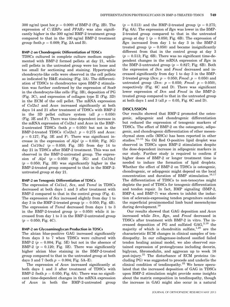
BMP-2 on Chondrogenic Differentiation of TDSCsTDSCs cultured in complete culture medium supple-mented with BMP-2 formed pellets at day 21, whilecell pellets in the untreated group were too loose andtoo small for sectioning and staining. Hypertrophicchondrocyte-like cells were observed in the cell pelletsas indicated by H&E staining (Fig. 3A). The differenti-ation of TDSCs to chondrocytes upon BMP-2 stimula-tion was further confirmed by the expression of Sox9in the chondrocyte-like cells (Fig. 3B), deposition of PG(Fig. 3C), and expression of collagen type II (Fig. 3D)in the ECM of the cell pellet. The mRNA expressionof Col2a1 and Acan increased significantly at bothdays 14 and 21 after treatment of TDSCs with BMP-2in the 3D pellet culture system (all p ¼ 0.050)(Fig. 3E and F). There was time-dependent increase inthe mRNA expression of Col2a1 and Acan in the BMP-2-untreated TDSCs (both p ¼ 0.050) but not in theBMP-2-treated TDSCs (Col2a1: p ¼ 0.275 and Acan:p ¼ 0.127; Fig. 3E and F). There was significant in-crease in the expression of Alpl (p ¼ 0.050; Fig. 3G)and Col10a1 (p ¼ 0.050; Fig. 3H) from day 14 today 21 in TDSCs after BMP-2 treatment. This was notobserved in the BMP-2-untreated group. The expres-sion of Alpl (p ¼ 0.050) (Fig. 3G) and Col10a1(p ¼ 0.050; Fig. 3H) was significantly higher in theBMP-2-treated group compared to that in the BMP-2-untreated group at day 21.
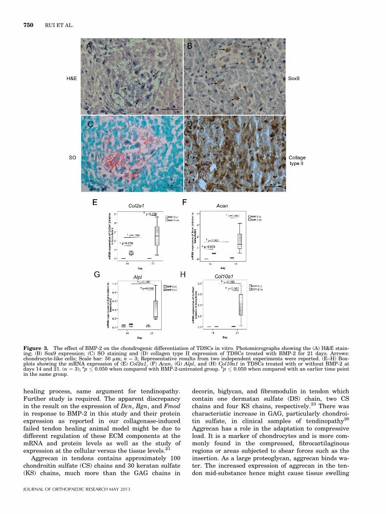
BMP-2 on Tenogenic Differentiation of TDSCsThe expression of Col1a1, Scx, and Tnmd in TDSCsdecreased at both days 1 and 3 after treatment withBMP-2 compared to that in the control group (Fig. 4).The expression of Scx increased slightly from day 1 today 3 in the BMP-2-treated group (p ¼ 0.050; Fig. 4B).The expression of Tnmd decreased from days 1 to 3in the BMP-2-treated group (p ¼ 0.050) while it in-creased from day 1 to 3 in the BMP-2-untreated group(p ¼ 0.050; Fig. 4C).
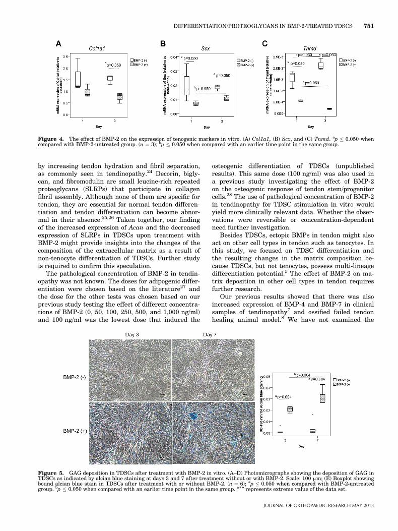
BMP-2 on Glycoaminoglycan Production in TDSCsThe alcian blue-positive GAG increased significantlyfrom days 3 to 7 when TDSCs were cultured withBMP-2 (p ¼ 0.004; Fig. 5E) but not in the absence ofBMP-2 (p ¼ 0.120; Fig. 5E). There was significantlyhigher alcian blue staining in the BMP-2-treatedgroup compared to that in the untreated group at bothdays 3 and 7 (both p ¼ 0.004; Fig. 5A–E).
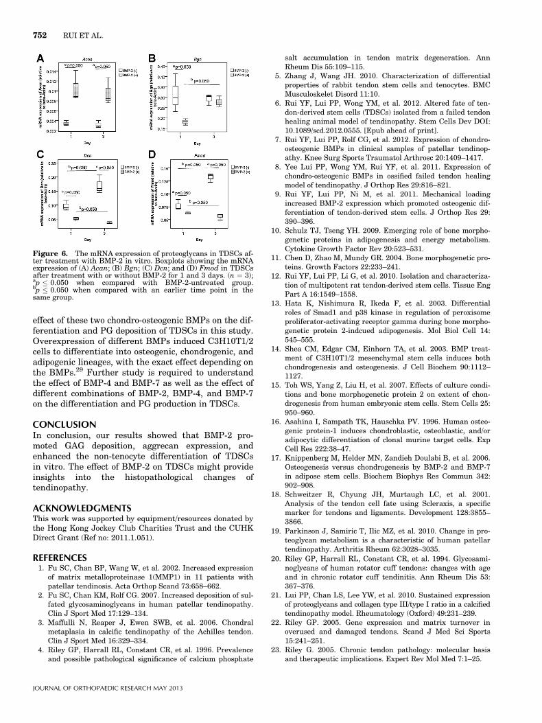
The expression of Acan increased significantly atboth days 1 and 3 after treatment of TDSCs withBMP-2 (both p ¼ 0.050; Fig. 6A). There was no signifi-cant time-dependent changes in the mRNA expressionof Acan in both the BMP-2-untreated group
(p ¼ 0.513) and the BMP-2-treated group (p ¼ 0.275;Fig. 6A). The expression of Bgn was lower in the BMP-2-treated group compared to that in the untreatedgroup at day 1 (p ¼ 0.050; Fig. 6B). The expression ofBgn increased from day 1 to day 3 in the BMP-2treated group (p ¼ 0.050) and became insignificantlydifferent from that in the control group at day 3(p ¼ 0.513; Fig. 6B). There was no significant time-de-pendent changes in the mRNA expression of Bgn inthe BMP-2-untreated group (p ¼ 0.827; Fig. 6B). Boththe expression of Dcn and Fmod decreased and in-creased significantly from day 1 to day 3 in the BMP-2-treated group (Dcn: p ¼ 0.050; Fmod: p ¼ 0.050) anduntreated group (Dcn: p ¼ 0.050; Fmod: p ¼ 0.050),respectively (Fig. 6C and D). There was significantlower expression of Dcn and Fmod in the BMP-2-treated group compared to that in the untreated groupat both days 1 and 3 (all p ¼ 0.05, Fig. 6C and D).
DISCUSSIONOur results showed that BMP-2 promoted the osteo-genic, adipogenic and chondrogenic differentiationbut reduced the expression of tenogenic markers ofTDSCs. The effect of BMP-2 on the osteogenic, adipo-genic, and chondrogenic differentiation of other mesen-chymal stem cells (MSCs) has been reported in otherstudies.13–15 No Oil Red O-positive lipid droplet wasobserved in TDSCs upon BMP-2 stimulation despitethe dose-dependent increase in adipogenic markers inour study. Further study is required to confirm ifhigher doses of BMP-2 or longer treatment time isneeded to induce the formation of lipid droplets.Whether the effect of BMP-2 on TDSCs is osteogenic,chondrogenic, or adiopgenic might depend on the localconcentration and duration of BMP stimulation.16,17
The differentiation of TDSCs to non-tenocytes mightdeplete the pool of TDSCs for tenogenic differentiationand tendon repair. In fact, BMP signaling (BMP-2,BMP-4, and BMP-7) was shown to inhibit the induc-tion of scleraxis-expressing tendon progenitors outsidethe superficial proximomedial limb bund mesenchymeduring development.18
Our results showed that GAG deposition and Acanincreased while Dcn, Bgn, and Fmod decreased inTDSCs after treatment with BMP-2 in vitro. The in-creased deposition of PG and sulfated GAG,2,19 themajority of which is chondroitin sulfate,1,20 are thecharacteristic ECM changes in clinical samples of ten-dinopathy. In our collagenase-induced ossified failedtendon healing animal model, we also observed sus-tained expression of proteoglycans including decorin,biglycan, fibromodulin, and aggrecan up to week 16post-injury.21 The disturbance of ECM proteins (in-cluding PG) was suggested to precede and underlie theclinical condition of tendinopathy.22 We hence specu-lated that the increased deposition of GAG in TDSCsupon BMP-2 stimulation might provide some insightsabout the matrix composition in tendinopathy, thoughthe increase in GAG might also occur in a natural
DIFFERENTIATION/PROTEOGLYCANS IN BMP-2-TREATED TDSCS 749
JOURNAL OF ORTHOPAEDIC RESEARCH MAY 2013
healing process, same argument for tendinopathy.Further study is required. The apparent discrepancyin the result on the expression of Dcn, Bgn, and Fmodin response to BMP-2 in this study and their proteinexpression as reported in our collagenase-inducedfailed tendon healing animal model might be due todifferent regulation of these ECM components at themRNA and protein levels as well as the study ofexpression at the cellular versus the tissue levels.21
Aggrecan in tendons contains approximately 100chondroitin sulfate (CS) chains and 30 keratan sulfate(KS) chains, much more than the GAG chains in
decorin, biglycan, and fibromodulin in tendon whichcontain one dermatan sulfate (DS) chain, two CSchains and four KS chains, respectively.23 There wascharacteristic increase in GAG, particularly chondroi-tin sulfate, in clinical samples of tendinopathy20
Aggrecan has a role in the adaptation to compressiveload. It is a marker of chondrocytes and is more com-monly found in the compressed, fibrocartilaginousregions or areas subjected to shear forces such as theinsertion. As a large proteoglycan, aggrecan binds wa-ter. The increased expression of aggrecan in the ten-don mid-substance hence might cause tissue swelling
Figure 3. The effect of BMP-2 on the chondrogenic differentiation of TDSCs in vitro. Photomicrographs showing the (A) H&E stain-ing; (B) Sox9 expression; (C) SO staining and (D) collagen type II expression of TDSCs treated with BMP-2 for 21 days. Arrows:chondrocyte-like cells; Scale bar: 50 mm; n ¼ 3; Representative results from two independent experiments were reported. (E–H) Box-plots showing the mRNA expression of (E) Col2a1, (F) Acan, (G) Alpl, and (H) Col10a1 in TDSCs treated with or without BMP-2 atdays 14 and 21. (n ¼ 3); ap � 0.050 when compared with BMP-2-untreated group. bp � 0.050 when compared with an earlier time pointin the same group.
750 RUI ET AL.
JOURNAL OF ORTHOPAEDIC RESEARCH MAY 2013
by increasing tendon hydration and fibril separation,as commonly seen in tendinopathy.24 Decorin, bigly-can, and fibromodulin are small leucine-rich repeatedproteoglycans (SLRPs) that participate in collagenfibril assembly. Although none of them are specific fortendon, they are essential for normal tendon differen-tiation and tendon differentiation can become abnor-mal in their absence.25,26 Taken together, our findingof the increased expression of Acan and the decreasedexpression of SLRPs in TDSCs upon treatment withBMP-2 might provide insights into the changes of thecomposition of the extracellular matrix as a result ofnon-tenocyte differentiation of TDSCs. Further studyis required to confirm this speculation.
The pathological concentration of BMP-2 in tendin-opathy was not known. The doses for adipogenic differ-entiation were chosen based on the literature27 andthe dose for the other tests was chosen based on ourprevious study testing the effect of different concentra-tions of BMP-2 (0, 50, 100, 250, 500, and 1,000 ng/ml)and 100 ng/ml was the lowest dose that induced the
osteogenic differentiation of TDSCs (unpublishedresults). This same dose (100 ng/ml) was also used ina previous study investigating the effect of BMP-2on the osteogenic response of tendon stem/progenitorcells.28 The use of pathological concentration of BMP-2in tendinopathy for TDSC stimulation in vitro wouldyield more clinically relevant data. Whether the obser-vations were reversible or concentration-dependentneed further investigation.
Besides TDSCs, ectopic BMPs in tendon might alsoact on other cell types in tendon such as tenocytes. Inthis study, we focused on TDSC differentiation andthe resulting changes in the matrix composition be-cause TDSCs, but not tenocytes, possess multi-lineagedifferentiation potential.5 The effect of BMP-2 on ma-trix deposition in other cell types in tendon requiresfurther research.
Our previous results showed that there was alsoincreased expression of BMP-4 and BMP-7 in clinicalsamples of tendinopathy7 and ossified failed tendonhealing animal model.8 We have not examined the
Figure 4. The effect of BMP-2 on the expression of tenogenic markers in vitro. (A) Col1a1, (B) Scx, and (C) Tnmd. ap � 0.050 whencompared with BMP-2-untreated group. (n ¼ 3); bp � 0.050 when compared with an earlier time point in the same group.
Figure 5. GAG deposition in TDSCs after treatment with BMP-2 in vitro. (A–D) Photomicrographs showing the deposition of GAG inTDSCs as indicated by alcian blue staining at days 3 and 7 after treatment without or with BMP-2. Scale: 100 mm; (E) Boxplot showingbound alcian blue stain in TDSCs after treatment with or without BMP-2. (n ¼ 6); ap � 0.050 when compared with BMP-2-untreatedgroup. bp � 0.050 when compared with an earlier time point in the same group. ‘‘�’’ represents extreme value of the data set.
DIFFERENTIATION/PROTEOGLYCANS IN BMP-2-TREATED TDSCS 751
JOURNAL OF ORTHOPAEDIC RESEARCH MAY 2013
effect of these two chondro-osteogenic BMPs on the dif-ferentiation and PG deposition of TDSCs in this study.Overexpression of different BMPs induced C3H10T1/2cells to differentiate into osteogenic, chondrogenic, andadipogenic lineages, with the exact effect depending onthe BMPs.29 Further study is required to understandthe effect of BMP-4 and BMP-7 as well as the effect ofdifferent combinations of BMP-2, BMP-4, and BMP-7on the differentiation and PG production in TDSCs.
CONCLUSIONIn conclusion, our results showed that BMP-2 pro-moted GAG deposition, aggrecan expression, andenhanced the non-tenocyte differentiation of TDSCsin vitro. The effect of BMP-2 on TDSCs might provideinsights into the histopathological changes oftendinopathy.
ACKNOWLEDGMENTSThis work was supported by equipment/resources donated bythe Hong Kong Jockey Club Charities Trust and the CUHKDirect Grant (Ref no: 2011.1.051).
REFERENCES1. Fu SC, Chan BP, Wang W, et al. 2002. Increased expression
of matrix metalloproteinase 1(MMP1) in 11 patients withpatellar tendinosis. Acta Orthop Scand 73:658–662.
2. Fu SC, Chan KM, Rolf CG. 2007. Increased deposition of sul-fated glycosaminoglycans in human patellar tendinopathy.Clin J Sport Med 17:129–134.
3. Maffulli N, Reaper J, Ewen SWB, et al. 2006. Chondralmetaplasia in calcific tendinopathy of the Achilles tendon.Clin J Sport Med 16:329–334.
4. Riley GP, Harrall RL, Constant CR, et al. 1996. Prevalenceand possible pathological significance of calcium phosphate
salt accumulation in tendon matrix degeneration. AnnRheum Dis 55:109–115.
5. Zhang J, Wang JH. 2010. Characterization of differentialproperties of rabbit tendon stem cells and tenocytes. BMCMusculoskelet Disord 11:10.
6. Rui YF, Lui PP, Wong YM, et al. 2012. Altered fate of ten-don-derived stem cells (TDSCs) isolated from a failed tendonhealing animal model of tendinopathy. Stem Cells Dev DOI:10.1089/scd.2012.0555. [Epub ahead of print].
7. Rui YF, Lui PP, Rolf CG, et al. 2012. Expression of chondro-osteogenic BMPs in clinical samples of patellar tendinop-athy. Knee Surg Sports Traumatol Arthrosc 20:1409–1417.
8. Yee Lui PP, Wong YM, Rui YF, et al. 2011. Expression ofchondro-osteogenic BMPs in ossified failed tendon healingmodel of tendinopathy. J Orthop Res 29:816–821.
9. Rui YF, Lui PP, Ni M, et al. 2011. Mechanical loadingincreased BMP-2 expression which promoted osteogenic dif-ferentiation of tendon-derived stem cells. J Orthop Res 29:390–396.
10. Schulz TJ, Tseng YH. 2009. Emerging role of bone morpho-genetic proteins in adipogenesis and energy metabolism.Cytokine Growth Factor Rev 20:523–531.
11. Chen D, Zhao M, Mundy GR. 2004. Bone morphogenetic pro-teins. Growth Factors 22:233–241.
12. Rui YF, Lui PP, Li G, et al. 2010. Isolation and characteriza-tion of multipotent rat tendon-derived stem cells. Tissue EngPart A 16:1549–1558.
13. Hata K, Nishimura R, Ikeda F, et al. 2003. Differentialroles of Smad1 and p38 kinase in regulation of peroxisomeproliferator-activating receptor gamma during bone morpho-genetic protein 2-indcued adipogenesis. Mol Biol Cell 14:545–555.
14. Shea CM, Edgar CM, Einhorn TA, et al. 2003. BMP treat-ment of C3H10T1/2 mesenchymal stem cells induces bothchondrogenesis and osteogenesis. J Cell Biochem 90:1112–1127.
15. Toh WS, Yang Z, Liu H, et al. 2007. Effects of culture condi-tions and bone morphogenetic protein 2 on extent of chon-drogenesis from human embryonic stem cells. Stem Cells 25:950–960.
16. Asahina I, Sampath TK, Hauschka PV. 1996. Human osteo-genic protein-1 induces chondroblastic, osteoblastic, and/oradipocytic differentiation of clonal murine target cells. ExpCell Res 222:38–47.
17. Knippenberg M, Helder MN, Zandieh Doulabi B, et al. 2006.Osteogenesis versus chondrogenesis by BMP-2 and BMP-7in adipose stem cells. Biochem Biophys Res Commun 342:902–908.
18. Schweitzer R, Chyung JH, Murtaugh LC, et al. 2001.Analysis of the tendon cell fate using Scleraxis, a specificmarker for tendons and ligaments. Development 128:3855–3866.
19. Parkinson J, Samiric T, Ilic MZ, et al. 2010. Change in pro-teoglycan metabolism is a characteristic of human patellartendinopathy. Arthritis Rheum 62:3028–3035.
20. Riley GP, Harrall RL, Constant CR, et al. 1994. Glycosami-noglycans of human rotator cuff tendons: changes with ageand in chronic rotator cuff tendinitis. Ann Rheum Dis 53:367–376.
21. Lui PP, Chan LS, Lee YW, et al. 2010. Sustained expressionof proteoglycans and collagen type III/type I ratio in a calcifiedtendinopathy model. Rheumatology (Oxford) 49:231–239.
22. Riley GP. 2005. Gene expression and matrix turnover inoverused and damaged tendons. Scand J Med Sci Sports15:241–251.
23. Riley G. 2005. Chronic tendon pathology: molecular basisand therapeutic implications. Expert Rev Mol Med 7:1–25.
Figure 6. The mRNA expression of proteoglycans in TDSCs af-ter treatment with BMP-2 in vitro. Boxplots showing the mRNAexpression of (A) Acan; (B) Bgn; (C) Dcn; and (D) Fmod in TDSCsafter treatment with or without BMP-2 for 1 and 3 days. (n ¼ 3);ap � 0.050 when compared with BMP-2-untreated group.bp � 0.050 when compared with an earlier time point in thesame group.
752 RUI ET AL.
JOURNAL OF ORTHOPAEDIC RESEARCH MAY 2013
24. Smith MM, Sakurai G, Smith SM, et al. 2008. Modulationof aggrecan and ADAMTS expression in ovine tendinopathyinduced by altered strain. Arthritis Rheum 58:1055–1066.
25. Ameye L, Aria D, Jepsen K, et al. 2002. Abnormal collagenfibrils in tendons of biglycan/fibromodulin-deficient micelead to gait impairment, ectopic ossification, and osteoarthri-tis. FASEB J 16:673–680.
26. Danielson KG, Baribault H, Holmes DF, et al. 1997.Targeted disruption of decorin leads to abnormal collagenfibril morphology and skin fragility. J Cell Biol 136:729–743.
27. Kato S, Kawabata N, Suzuki N, et al. 2009. Bone morphoge-netic protein-2 induces the differentiation of a mesenchymalprogenitor cell line, ROB-C26, into mature osteoblasts andadipocytes. Life Sci 84:302–310.
28. Bi Y, Ehirchiou D, Kilts TM, et al. 2007. Identification oftendon stem/progenitor cells and the role of the extracellularmatrix in their niche. Nat Med 13:1219–1227.
29. Ahrens M, Ankenbauer T, Schroder D, et al. 1993. Expres-sion of human bone morphogenetic proteins-2 or -4 inmurine mesenchymal progenitor C3H10T1/2 cells inducesdifferentiation into distinct mesenchymal cell lineages. DNACell Biol 12:871–880.
DIFFERENTIATION/PROTEOGLYCANS IN BMP-2-TREATED TDSCS 753
JOURNAL OF ORTHOPAEDIC RESEARCH MAY 2013