Embed Size (px)
Citation preview

Biomarker Methods in Drug Discoveryand Development
METHODS IN PHARMACOLOGY AND TOXICOLOGYTM
Y. James Kang, SERIES EDITOR
Biomarker Methods in Drug Discovery and Developmentedited by Feng Wang, 2008
Pharmacogenomics and Personalized Medicineedited by Nadine Cohen, 2008
Cytokines in Human Health: Immunotoxicology, Pathology,and Therapeutic Applications
edited by Robert V. House and Jacques Descotes, 2007
Drug Metabolism and Transport: Molecular Methods and Mechanismsedited by Lawrence Lash, 2005
Optimization in Drug Discovery: In Vitro Methodsedited by Zhengyin Yan and Gary W. Caldwell, 2004
In Vitro Neurotoxicology: Principles and Challengesedited by Evelyn Tiffany-Castiglioni, 2004
Cardiac Drug Development Guideedited by Michael K. Pugsley, 2003
Methods in Biological Oxidative Stressedited by Kenneth Hensley and Robert A. Floyd, 2003
Apoptosis Methods in Pharmacology and Toxicology:Approaches to Measurement and Quantification
edited by Myrtle A. Davis, 2002
Ion Channel Localization: Methods and Protocolsedited by Anatoli N. Lopatin and Colin G. Nichols, 2001
METHODS IN PHARMACOLOGY AND TOXICOLOGYTM
Biomarker Methodsin Drug Discoveryand Development
Edited by
Feng Wang, PhDProcter & Gamble Pharmaceutical, Inc., Mason, Ohio
EditorFeng WangProcter & Gamble Pharmaceutical, Inc.8700 Mason-Montgomery RoadMason OH, USA
Series EditorY. James KangDepartment of Medicine, Pharmacology & ToxicologySchool of MedicineUniversity of LouisvilleLouisville, KY, USA
ISBN: 978-1-934115-23-7 e-ISBN: 978-1-59745-463-6
Library of Congress Control Number: 2007943120
©2008 Humana Press, a part of Springer Science+Business Media, LLCAll rights reserved. This work may not be translated or copied in whole or in part without the writtenpermission of the publisher (Humana Press, 999 Riverview Drive, Suite 208, Totowa, NJ 07512 USA),except for brief excerpts in connection with reviews or scholarly analysis. Use in connection with anyform of information storage and retrieval, electronic adaptation, computer software, or by similar ordissimilar methodology now known or hereafter developed is forbidden.The use in this publication of trade names, trademarks, service marks, and similar terms, even if theyare not identified as such, is not to be taken as an expression of opinion as to whether or not they aresubject to proprietary rights.While the advice and information in this book are believed to be true and accurate at the date of goingto press, neither the authors nor the editors nor the publisher can accept any legal responsibility forany errors or omissions that may be made. The publisher makes no warranty, express or implied, withrespect to the material contained herein.
Printed on acid-free paper
9 8 7 6 5 4 3 2 1
springer.com

Preface
The pharmaceutical industry has faced many significant challenges sincethe early 1990s. The fundamental issue that needs to be addressed is howto improve the efficiency of drug discovery and development. The currentresearch and development (R&D) cost for developing a new therapeutic drugis greater than $800 million. Additionally, it takes an average of 12 years toget a new drug to market with an attrition rate greater than 90%. Reviewingthe overall pharmaceutical R&D process, it has become clear that many ofthe drug failures are due to our lack of knowledge in population diversity,which is responsible for differences in drug efficacy and toxicity. In fact,not a single approved drug is 100% safe and efficacious for all patients. Forresearchers, the key question is: How can we discover biomarkers that canbe used to distinguish patients who will respond to the drug without adverseeffects from those who will not respond and/or will have adverse effects?There is tremendous urgency to address this question. Biomarkers also fitperfectly with the vision of personalized medicine, the new expectationof medical practice. This is why biomarker research has been a centralfocus in many research labs across academia, government agencies, and thepharmaceutical industry.
There are many different ways to define biomarkers based on molecularproperties, applications, and methods. The National Institutes of Health(NIH) suggested an inclusive definition for biomarker as “a characteristicthat is objectively measured and evaluated as an indicator of normal biologicprocesses, pathogenic processes, or pharmacologic responses to a therapeuticintervention.” A biomarker can be DNA, protein, metabolite, mRNA, orlipid. This certainly increases the complexity of biomarker research. Anarray of technologies is needed for biomarker research to increase the successrate. Fortunately, many existing biological and analytical technologies canbe, and have already been, directly applied to biomarker research. The“omics” technologies, including genomics, proteomics, and metabonomics,have been developed and can be used for identifying potential biomarkersat several different molecular levels. The unbiased nature of these “omics”
v
vi Preface
technologies is well suited for biomarker research. The goal of BiomarkerMethods in Drug Discovery and Development is to provide a tool box forthose who have a general interest in biomarker research and also for thosewho are currently specializing in certain technologies but want to gain anunderstanding of other available methodologies. Many technologies coveredin this book are well validated and mature methods, whereas others are rathernew but with huge promise. This book also covers some specific issuesrelated to clinical biomarker research, such as clinical sample handling. Atotal of 17 chapters contributed by many experts in their research areasprovide detailed descriptions of biomarker methodologies. This book isintended to be used as a guideline and a protocol reference for biomarkerresearchers.
Clinical biomarker research often uses patient samples and specimens.This creates significant challenges in sample collection and handlingcompared with tissue and animal experiments. There are many aspects ofsample collection and preservation that need to be considered in orderto control and reduce experimental variability. Chapter 1 provides criticalinsights into careful study planning to ensure that robust data can begenerated from clinical samples. The examples given in this chapter coversample collection and handling for DNA, RNA, and protein analyses fromperipheral blood. This chapter also highlights many parts of the assay underdevelopment that need to be examined so that the performance character-istics can be well understood. It is important to ensure that the biomarkerdata generated from the assay will be solid so that accurate conclusions canbe reached.
One significant outcome of the Human Genome Project was the devel-opment of numerous genomic technologies over the past decade. Manyareas of research such as drug target identification, target validation,pharmacogenomics, pharmacogenetics, as well as biomarkers have benefitedgreatly from these new technologies. In fact, genomics has become amajor tool used in biomarker research. This book highlights six genomicmethodologies including gene expression, single nucleotide polymorphism(SNP), DNA methylation, and laser capture microdissection, a very usefultissue sample retrieval technique. Adverse effects and toxicity are stillbeing predominately identified at clinical stages of drug developmentusing lengthy and costly approaches. Good toxicity biomarkers with highpredictive value are highly desired in drug R&D. Because microarrayplatforms offer unique advantages in identifying novel mechanism-basedbiomarkers, it is a powerful method to interrogate perturbations inducedby experimental drugs and to pinpoint individual genes or gene setsregulated in parallel with a toxic reaction. Chapter 2 uses three examples of
Preface vii
gene expression–based biomarkers for hepatotoxicity, nephrotoxicity, andgeneral toxicity signatures in blood to describe applications for microarrayplatform technology in toxicity biomarkers. The detailed methodology ofthe fluorescent microspheres (microbeads) gene expression platform isdescribed in Chapter 3. Using a panel of the “signature” gene expressionpattern, the microsphere approach offers advantages in flexibility over thetraditional whole-genome microarrays. Real-time PCR is another valuableand widely used gene expression methodology. High-throughput whole-genome microarrays enable screening of large numbers of genes to identifypotential biomarkers whose expression levels are correlated with diseasestate, clinical outcome, and treatment regimens. These candidate biomarkersneed to be validated with different sets of samples and, preferably, differentmethods. Real-time PCR technology fits extremely well for this purposewith excellent design flexibility and fast turnaround. Chapter 4 provides anoverview of real-time PCR and practical assay protocols.
Gene expression analysis platforms are also useful in SNP identification.Chapter 5 describes the serial analysis of gene expression (SAGE) method-ology and the bioinformatics approach for the applications of SNP analysis.The uniqueness of SAGE is that any molecular biology lab can easilyperform the protocol without relying on specialized, expensive equipment.This chapter provides detailed methods and notes so one can readily followthe experimental procedures. Another important genomic biomarker toolis Pyrosequencing, which is covered in Chapter 6. Pyrosequencing is agenotyping method based on sequencing by synthesis. This technique offersaccurate and quantitative analysis of DNA sequences without the presence ofa restriction enzyme site. It can also be used to identify triallelic, indel, andshort repeat polymorphisms, as well as to determine allele percentages forDNA methylation. Chapter 6 provides an overview for the Pyrosequencingmethod and assay details for commonly analyzed and clinically relevantpolymorphisms such as SNPs in the cytochrome P450, as well as assayprotocols for DNA methylation measurement.
Biological tissues have high degrees of cell heterogeneity. For somestudies aimed at identifying specific biological pathways, analysis ispreferably done using the targeted cell type. In Chapter 7, a relatively newtissue selection and retrieval method, laser capture microdissection (LCM),is described. This chapter demonstrates the utility of LCM when it is coupledwith gene expression analyses using primate endometrium tissue. The LCM-collected samples certainly can be used for analysis utilizing other biomarkerplatform technologies.
Six protein biomarker analysis methodologies are described in this book(Chapters 8 to 13) in order to represent a variety of commonly used

viii Preface
technologies. As in genomics, there are numerous protein analysis methodsthat have been used in biomarker research. Traditional two-dimensional gelelectrophoresis (2-DE) is a powerful protein separation method that hasbeen constantly improved in reproducibility and ease of use. Chapter 8 usesa clinical biomarker study as an example to illustrate the application of2-DE/mass spectrometry (MS) method. A specific drug adverse effect wasinvestigated using 2-DE/MS with patient plasma samples. Human plasmais a convenient sample source for clinical biomarker research. However,the large dynamic range of plasma protein concentration poses a significantchallenge for analysis of medium- or low-abundance proteins, which aremost likely more biologically relevant. Prefractionation step(s) are oftennecessary before any type of proteomic analysis. Chapter 8 also describes adetailed immunochemistry method that is commonly used in many labs todeplete the most abundant plasma proteins. It demonstrates that the 2-DE/MSapproach can be used for complex samples to reveal potential proteinbiomarkers. In an attempt to simplify the 2-DE process, the differencein-gel electrophoresis (DIGE) method was developed. As an adaptation ofconventional 2-DE, DIGE uses novel fluorescent labels so that two to threesamples can be resolved on a single gel under identical electrophoreticconditions. It offers advantages in simplifying image analysis, increasingsample throughput, and reducing 2-DE experimental variation. Chapter 9provides an overview and easy-to-follow protocols for the DIGE technology.
The rapid advancement in MS technologies has resulted in manysignificant developments in proteomics and protein biomarker research.Currently, there are two MS-based strategies commonly used in quanti-tative global proteomics. Chapter 10 covers the bottom-up strategy with theshotgun tryptic peptide liquid chromatography–mass spectrometry (LC-MS)approach, and Chapter 11 describes the top-down strategy using a set ofnovel protein labels to identify and quantify the differences between multipleprotein samples. In the bottom-up approach, proteins are first digested byan enzyme into peptide fragments that are analyzed by LC–tandem massspectrometry (MS/MS) and then identified by database searching. Proteinquantification is achieved by measuring chromatographic peak intensity.Chapter 10 provides a comprehensive review of the technique along withthe author’s insights pertaining to data analysis and bioinformatics. Thischapter also provides a case study as a practical example to highlight theexperiment design details and data interpretation. This approach is highlysensitive and can be easily automated, offering great feasibility for large-scale protein biomarker analysis. As the top-down proteomics approachinvolves the analysis of intact proteins, it promises the ability to characterizeposttranslational modifications and to reduce false-positive identifications
Preface ix
because of its multiplexing capabilities. The top-down approach is wellillustrated in Chapter 11 with a newly developed isobaric mass taggingtechnology, ExacTag Labeling system. The concept of this technology isvery similar to isotope code affinity tag (ICAT). But this method labelswhole proteins rather than proteolytic peptide fragments, and the subsequentprotein mixtures can be enriched under the identical conditions. Experi-mental variation can be significantly reduced as multiple samples are taggedand mixed before any processing steps.
Other than the traditional 2-DE/MS and extensive proteomic profilingtechnologies introduced previously, Biomarker Methods in Drug Discoveryand Development also covers two unique methods that are well suitedfor specific protein biomarker analysis needs. The surface-enhanced laserdesorption ionization mass spectrometry (SELDI-MS) described in Chapter12 is a high-throughput screening platform especially applicable for largenumbers of solution samples such as plasma, urine, and conditioned media.It is a good method to quickly identify the differences among samples basedon MS patterns. Further identification of the molecular entities responsiblefor the differences requires extra effort and most likely other technologies.When a protein biomarker is identified through any approach, validationof the biomarker with a large number of samples is required. The enzyme-linked immunosorbent assay (ELISA) array is the method of choice forvalidation as well as a diagnostic platform. Chapter 13 provides excellenttechnical details of how to generate high-quality ELISA microarrays witheasy-to-follow directions and notes.
Metabolic biomarkers are also attractive in pharmaceutical biomarkerresearch. This is because metabolite levels can be regarded as the finalprocess readout of biological systems combining both internal factors(genetic) and external factors (disease or drug treatment). Many minorchanges at the transcript and protein levels are significant enough to bebiologically meaningful. These minor changes themselves may not bedetectable. However, after the biological and metabolic process, they canlead to major changes at the metabolite level, which makes analysis morefeasible. As an important component of systems biology and biomarkerdiscovery, metabonomics (or metabolomics) technologies have been contin-ually improved. MS and nuclear magnetic resonance (NMR) spectroscopyare the two core technologies used in metabolite analysis. Coupled withliquid chromatographic separation, the MS technique offers advantages inautomation and high resolution. Chapter 14 discusses LC-MS methodologyand its biomarker applications. It provides detailed descriptions on eachcomponent of the LC-MS platform and the multivariate statistical dataanalysis methods. NMR methodology is covered in Chapter 15 where a
x Preface
variety of NMR analysis approaches and applications are demonstrated.Chapter 16 addresses methodology for a specific group of metabolites,nonpolar analytes, using gas chromatography–MS (GC-MS). TraditionalLC-MS and NMR metabolite analyses do not cover molecules with lowpolarities because of the differences in separation and ionization mecha-nisms. Cellular lipids play important roles in membrane biology andmetabolic dysfunctions. Quantitative lipid analysis, as a biomarker tool, is ofinterest in the pharmaceutical industry. Chapter 17 introduces MS method-ologies in analyzing glycerophospholipids, the main constituents of cellularmembranes.
Biomarkers offer tremendous potential and promises for transformingpharmaceutical research and development processes. Biomarker researchwill positively impact not only new therapeutics but also diagnostics, aswell as our overall understanding in the general life sciences. We need toapply multiple technologies at different molecular levels to work on thismonumental and complex task. I hope this book will be used as a biomarkertechnical guideline and reference to stimulate more exciting biomarkerresearch and more technology development.
I sincerely thank all the chapter authors who tirelessly took the extra timebeyond their busy daily research activities to contribute to this book, and Iexpress my genuine gratitude to all the authors for their expert knowledgeand for their efforts.
Feng Wang, PhD
Contents
Preface . . . . . . . . . . . . . . . . . . . . . . . . . . . . . . . . . . . . . . . . . . . . . . . . . . . . . vContributors . . . . . . . . . . . . . . . . . . . . . . . . . . . . . . . . . . . . . . . . . . . . . . . xiiiColor Plates . . . . . . . . . . . . . . . . . . . . . . . . . . . . . . . . . . . . . . . . . . . . . . . . xvii
1 Biomarker Sample Collection and Handling in the ClinicalSetting to Support Early-Phase Drug Development
Chris B. Russell, Sid Suggs, Kristina M. Robson,Keith Kerkof, Lisa D. Kivman, Kimberly H. Notari,William A. Rees, Natalia Leshinsky,and Scott D. Patterson. . . . . . . . . . . . . . . . . . . . . . . . . . . . . . . . . . . 1
2 Gene Expression-Based Biomarkers of Drug SafetyEric A.G. Blomme and Scott E. Warder . . . . . . . . . . . . . . . . . . . . . 27
3 Profiling Gene Expression SignaturesUsing Fluorescent Microspheres
Suzanne M. Torontali, Jorge M. Naciff, Kenton D. Juhlin,and Jay P. Tiesman . . . . . . . . . . . . . . . . . . . . . . . . . . . . . . . . . . . . . . 51
4 Real-Time Polymerase Chain Reaction Gene ExpressionAssays in Biomarker Discovery and Validation
Yulei Wang, Catalin Barbacioru, David Keys, Pius Brzoska,Caifu Chen, Kelly Li, and Raymond R. Samaha . . . . . . . . . . 63
5 SAGE Analysis in Identifying PhenotypeSingle Nucleotide Polymorphisms
Sandro J. de Souza, I-Mei Siu, Janete M. Cerutti,and Gregory J. Riggins . . . . . . . . . . . . . . . . . . . . . . . . . . . . . . . . . . . 87
6 Single Nucleotide Polymorphisms and DNA MethylationAnalysis Using Pyrosequencing Methods
Jinsheng Yu and Sharon Marsh . . . . . . . . . . . . . . . . . . . . . . . . . . . . . 1197 The Application of Laser Capture Microdissection for
the Analysis of Cell-Type-Specific Gene Expressionin a Complex Tissue: The Primate Endometrium
William C. Okulicz. . . . . . . . . . . . . . . . . . . . . . . . . . . . . . . . . . . . . . . . . 141
xi
xii Contents
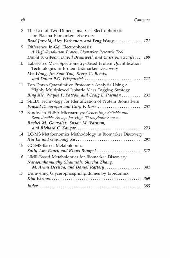
8 The Use of Two-Dimensional Gel Electrophoresisfor Plasma Biomarker Discovery
Brad Jarrold, Alex Varbanov, and Feng Wang . . . . . . . . . . . . . . 1719 Difference In-Gel Electrophoresis:
A High-Resolution Protein Biomarker Research ToolDavid S. Gibson, David Bramwell, and Caitriona Scaife . . . 189
10 Label-Free Mass Spectrometry-Based Protein QuantificationTechnologies in Protein Biomarker Discovery
Mu Wang, Jin-Sam You, Kerry G. Bemis,and Dawn P.G. Fitzpatrick . . . . . . . . . . . . . . . . . . . . . . . . . . . . . . 211
11 Top-Down Quantitative Proteomic Analysis Using aHighly Multiplexed Isobaric Mass Tagging Strategy
Bing Xie, Wayne F. Patton, and Craig E. Parman . . . . . . . . . . 23112 SELDI Technology for Identification of Protein Biomarkers
Prasad Devarajan and Gary F. Ross . . . . . . . . . . . . . . . . . . . . . . . 25113 Sandwich ELISA Microarrays: Generating Reliable and
Reproducible Assays for High-Throughput ScreensRachel M. Gonzalez, Susan M. Varnum,
and Richard C. Zangar . . . . . . . . . . . . . . . . . . . . . . . . . . . . . . . . . . . 27314 LC-MS Metabonomics Methodology in Biomarker Discovery
Xin Lu and Guowang Xu . . . . . . . . . . . . . . . . . . . . . . . . . . . . . . . . . . . 29115 GC-MS-Based Metabolomics
Sally-Ann Fancy and Klaus Rumpel . . . . . . . . . . . . . . . . . . . . . . . . 31716 NMR-Based Metabolomics for Biomarker Discovery
Narasimhamurthy Shanaiah, Shucha Zhang,M. Aruni Desilva, and Daniel Raftery . . . . . . . . . . . . . . . . . . . 341
17 Unraveling Glycerophospholipidomes by LipidomicsKim Ekroos. . . . . . . . . . . . . . . . . . . . . . . . . . . . . . . . . . . . . . . . . . . . . . . . . 369
Index . . . . . . . . . . . . . . . . . . . . . . . . . . . . . . . . . . . . . . . . . . . . . . . . . . . . . . . 385

Contributors
Catalin Barbacioru, phd • Molecular Biology Division, AppliedBiosystems, Foster City, California
Kerry G. Bemis, phd • Indiana Centers for Applied Protein Sciences(INCAPS), Indianapolis, Indiana
Eric A.G. Blomme, dvm, phd • Abbott Laboratories, Global Pharmaceu-tical Research and Development, Department of Cellular, Molecular andExploratory Toxicology, Abbott Park, Illinois
David Bramwell, phd • Nonlinear Dynamics Limited, Newcastle uponTyne, United Kingdom
Pius Brzoska, phd • Molecular Biology Division, Applied Biosystems,Foster City, California
Janete M. Cerutti, phd • Laboratory of Molecular Endocrinology,Department of Medicine, Federal University of São Paulo, São Paulo,Brazil
Caifu Chen, phd • Molecular Biology Division, Applied Biosystems,Foster City, California
M. Aruni Desilva, ms • Department of Chemistry, Purdue University,West Lafayette, Indiana
Prasad Devarajan, md • Nephrology and Hypertension, CincinnatiChildren’s Hospital Medical Center, University of Cincinnati,Cincinnati, Ohio
Kim Ekroos, phd • Department of Molecular Pharmacology, AstraZenecaR&D, Mölndal, Sweden
Sally-Ann Fancy, phd • Pfizer Global Research and Development,Department of Exploratory Medicinal Sciences, Sandwich, Kent, UnitedKingdom
Dawn P.G. Fitzpatrick, bs • Department of Biochemistry and MolecularBiology, Indiana University School of Medicine, Indianapolis, Indiana
David S. Gibson, phd • Queen’s University Belfast, Arthritis ResearchGroup, Musculoskeletal Education and Research Unit, Belfast, UnitedKingdom
xiii
xiv Contributors
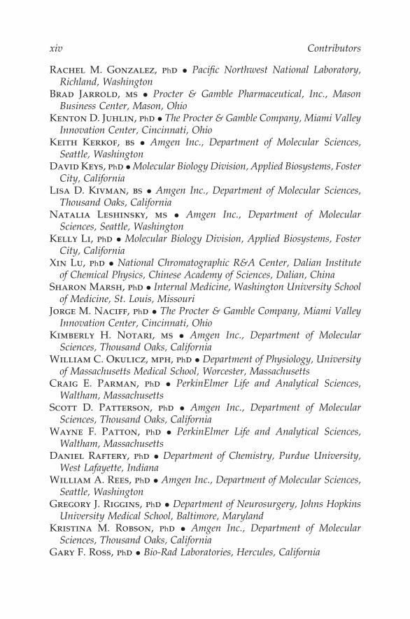
Rachel M. Gonzalez, phd • Pacific Northwest National Laboratory,Richland, Washington
Brad Jarrold, ms • Procter & Gamble Pharmaceutical, Inc., MasonBusiness Center, Mason, Ohio
Kenton D. Juhlin, phd • The Procter & Gamble Company, Miami ValleyInnovation Center, Cincinnati, Ohio
Keith Kerkof, bs • Amgen Inc., Department of Molecular Sciences,Seattle, Washington
David Keys, phd • Molecular Biology Division, Applied Biosystems, FosterCity, California
Lisa D. Kivman, bs • Amgen Inc., Department of Molecular Sciences,Thousand Oaks, California
Natalia Leshinsky, ms • Amgen Inc., Department of MolecularSciences, Seattle, Washington
Kelly Li, phd • Molecular Biology Division, Applied Biosystems, FosterCity, California
Xin Lu, phd • National Chromatographic R&A Center, Dalian Instituteof Chemical Physics, Chinese Academy of Sciences, Dalian, China
Sharon Marsh, phd • Internal Medicine, Washington University Schoolof Medicine, St. Louis, Missouri
Jorge M. Naciff, phd • The Procter & Gamble Company, Miami ValleyInnovation Center, Cincinnati, Ohio
Kimberly H. Notari, ms • Amgen Inc., Department of MolecularSciences, Thousand Oaks, California
William C. Okulicz, mph, phd • Department of Physiology, Universityof Massachusetts Medical School, Worcester, Massachusetts
Craig E. Parman, phd • PerkinElmer Life and Analytical Sciences,Waltham, Massachusetts
Scott D. Patterson, phd • Amgen Inc., Department of MolecularSciences, Thousand Oaks, California
Wayne F. Patton, phd • PerkinElmer Life and Analytical Sciences,Waltham, Massachusetts
Daniel Raftery, phd • Department of Chemistry, Purdue University,West Lafayette, Indiana
William A. Rees, phd • Amgen Inc., Department of Molecular Sciences,Seattle, Washington
Gregory J. Riggins, phd • Department of Neurosurgery, Johns HopkinsUniversity Medical School, Baltimore, Maryland
Kristina M. Robson, phd • Amgen Inc., Department of MolecularSciences, Thousand Oaks, California
Gary F. Ross, phd • Bio-Rad Laboratories, Hercules, California

Contributors xv
Klaus Rumpel, phd • Pfizer Global Research and Development,Department of Exploratory Medicinal Sciences, Sandwich, Kent, UnitedKingdom
Chris B. Russell, phd • Amgen Inc., Department of Molecular Sciences,Seattle, Washington
Raymond R. Samaha, phd • Molecular Biology Division, AppliedBiosystems, Foster City, California
Caitriona Scaife, msc • University College Dublin, Conway Instituteof Biomolecular and Biomedical Research, Proteome Research Centre,Belfield, Dublin, Ireland
Narasimhamurthy Shanaiah, phd • Department of Chemistry, PurdueUniversity, West Lafayette, Indiana
I-Mei Siu, phd • Department of Neurosurgery, Johns Hopkins UniversityMedical School, Baltimore, Maryland
Sandro J. de Souza, phd • Ludwig Institute for Cancer Research, SaoPaulo Branch, Hospital Alemão Oswaldo Cruz, São Paulo, Brazil
Sid Suggs, phd • Amgen Inc., Department of Molecular Sciences,Thousand Oaks, California
Jay P. Tiesman, phd • The Procter & Gamble Company, Miami ValleyInnovation Center, Cincinnati, Ohio
Suzanne M. Torontali, bs • The Procter & Gamble Company, MiamiValley Innovation Center, Cincinnati, Ohio
Alex Varbanov, phd • Procter & Gamble Pharmaceutical, Inc., MasonBusiness Center, Mason, Ohio
Susan M. Varnum, phd • Pacific Northwest National Laboratory,Richland, Washington
Feng Wang, phd • Procter & Gamble Pharmaceutical, Inc., MasonBusiness Center, Mason, Ohio
Mu Wang, phd • Department of Biochemistry and Molecular Biology,Indiana University School of Medicine, Indianapolis, Indiana
Yulei Wang, phd • Molecular Biology Division, Applied Biosystems,Foster City, California
Scott E. Warder, phd • Abbott Laboratories, Global PharmaceuticalResearch and Development, Department of Cellular, Molecular andExploratory Toxicology, Abbott Park, Illinois
Bing Xie, phd • PerkinElmer Life and Analytical Sciences, Waltham,Massachusetts
Guowang Xu, phd • National Chromatographic R&A Center, DalianInstitute of Chemical Physics, Chinese Academy of Sciences, Dalian,China

xvi Contributors
Jin-Sam You, phd • Indiana Centers for Applied Protein Sciences(INCAPS), Indianapolis, Indiana
Jinsheng Yu, md, phd • Departments of Pathology & Immunology,Washington University School of Medicine, St. Louis, Missouri
Richard C. Zangar, phd • Pacific Northwest National Laboratory,Richland, Washington
Shucha Zhang, ms • Department of Chemistry, Purdue University, WestLafayette, Indiana
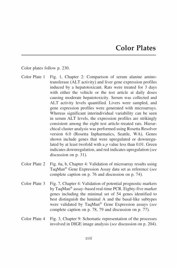
Color Plates
Color plates follow p. 230.
Color Plate 1 Fig. 1, Chapter 2: Comparison of serum alanine amino-transferase (ALT activity) and liver gene expression profilesinduced by a hepatotoxicant. Rats were treated for 3 dayswith either the vehicle or the test article at daily dosescausing moderate hepatotoxicity. Serum was collected andALT activity levels quantified. Livers were sampled, andgene expression profiles were generated with microarrays.Whereas significant interindividual variability can be seenin serum ALT levels, the expression profiles are strikinglyconsistent among the eight test article–treated rats. Hierar-chical cluster analysis was performed using Rosetta Resolverversion 6.0 (Rosetta Inpharmatics, Seattle, WA). Genesshown include genes that were upregulated or downregu-lated by at least twofold with a p value less than 0.01. Greenindicates downregulation, and red indicates upregulation (seediscussion on p. 31).
Color Plate 2 Fig. 6a, b, Chapter 4: Validation of microarray results usingTaqMan® Gene Expression Assay data set as reference (seecomplete caption on p. 76 and discussion on p. 74).
Color Plate 3 Fig. 7, Chapter 4: Validation of potential prognostic markersby TaqMan® assay–based real-time PCR. Eighty-five markergenes including the minimal set of 54 genes identified tobest distinguish the luminal A and the basal-like subtypeswere validated by TaqMan® Gene Expression assays (seecomplete caption on p. 78, 79 and discussion on p. 77).
Color Plate 4 Fig. 3, Chapter 9: Schematic representation of the processesinvolved in DIGE image analysis (see discussion on p. 204).
xvii

xviii Color Plates
Color Plate 5 Fig. 1a, b, Chapter 11: Composition of isobaric mass tags.(A) Schematic structure of the isobaric mass tag describedin this chapter. The tag contains four elements: (1) areactive group with specificity toward a thiol (cysteine) or anamine (lysine, �-amino) group; (2) a cleavage enhancementmoiety, which is an aspartic acid (Asp, D) proline (Pro, P)scissile bond group. Distributed around the DP sequenceare the (3) low mass signal reporter and its (4) corre-sponding balancer sequence. (B) Detailed composition ofseven isobaric mass tags. The tag contains a common aminoacid composition. G represents glycine. The red bold “G” isheavy glycine and plain “G” is normal glycine. In MS/MS,the tags generate two sets of signals: low mass signals andhigh mass signals (see discussion on p. 234).
Color Plate 6 Fig. 5, Chapter 11: Hierarchical clustering of 440 proteins.The protein list was clustered by dynamic changes inexpression levels (the most downregulated in dark-greenand the most upregulated in dark-red) (see discussion onp. 242).
Color Plate 7 Fig. 6a, b, Chapter 11: Nucleolar protein profiling bythe described isobaric mass tagging technology. (A)Changes in the abundance of various ribosomal proteinsubunits after actinomycin D treatment. Relative ratios ofquantified ribosomal proteins are shown. (B) Changes inthe abundances of DEAD box domain containing proteinsafter actinomycin D treatment. Relative fold changes ofDEAD/H domain containing proteins are shown. HLA-B,HLA-B associated transcript (see discussion on p. 243).