Embed Size (px)
Citation preview
www.afm-journal.de
© 2013 WILEY-VCH Verlag GmbH & Co. KGaA, Weinheim4454
www.MaterialsViews.com
wileyonlinelibrary.com
FEATU
RE
ARTI
CLE
Javier G. Fernandez and Donald E. Ingber *
1. Introduction
Nature is abundant with materials that possess extraordi-nary mechanical characteristics. This has not gone unnoticed as man has long sought to harness this knowledge, starting with the fabrication of the fi rst tool implements from bones, teeth, skin, hair, wood and shells. Development of extraction
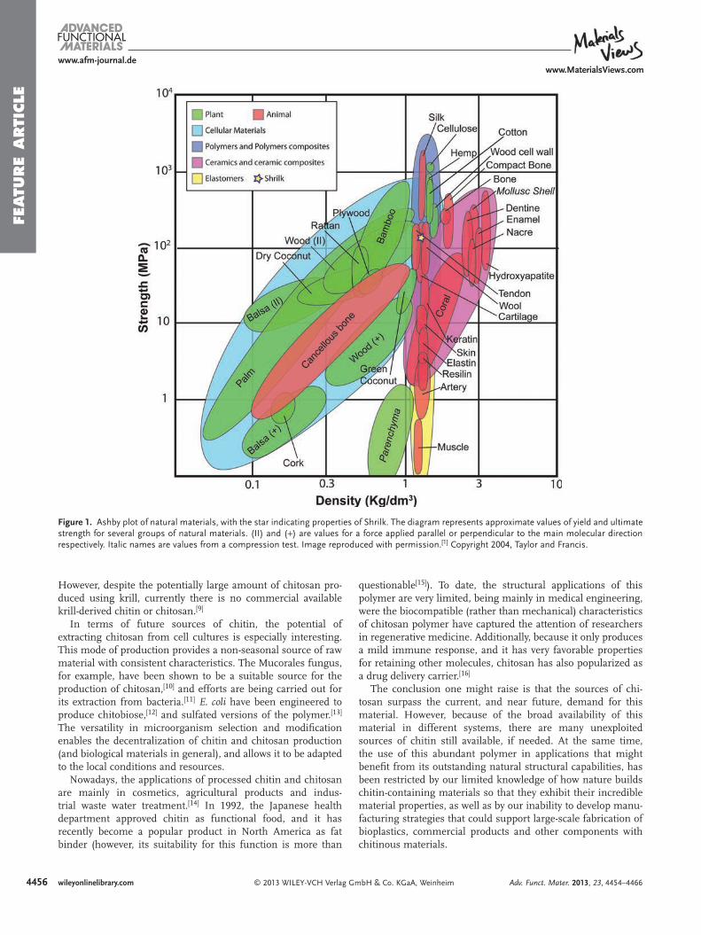
methods enabled isolation of plant-based cellulose polymers from wood and other sources that have been used for construc-tion of paper, cardboard and other struc-tural materials. In contrast, even though chitinous materials in structures, such as shell nacre and insect cuticles, that pro-vide even greater strength and similar toughness than wood [ 1 ] ( Figure 1 ) are equally ubiquitous, they have been largely ignored in materials engineering because their novel material properties are largely based on their biological ultrastructure and hierarchical organization, which have been diffi cult to recapitulate.
This failure to regenerate the hierar-chical designs of biological materials has not impeded the development and appli-cation of other biological polymers, and especially proteins. The use of egg white as a binding agent in the fl oor of the Pad-manabhapuram Palace is an impressive example of the use of a naturally derived protein polymer in civil engineering. The milk protein casein also was used for structural applications, such as the fabri-cation of buttons, starting over a century ago, and it has found a modern applica-
tion in the textile industry with QMilch, a casein-based woven fi ber produced entirely from milk.
With the discovery of the fi rst synthetic polymers, the whole fi eld of materials changed drastically. If the use of stone, bronze and iron defi ned past periods of human history, the 20th cen-tury might be called the “Plastic Age”, and it still dominates to this day. [ 2 ] Indeed, plastics production has increased from 0.5 to 380 million tons per year since 1950, [ 3 ] and with the rise in the dependences on synthetic polymers, the use of biolog-ical materials was pushed aside. This is because plastics are incredibly versatile materials, and they are inexpensive, strong, and durable. The diversity of synthetic polymers, and the ver-satility of their properties, facilitates the production of a vast array of plastic products that bring technological advances and numerous other benefi ts to society. In contrast, biologically-derived materials have may drawbacks, including cost issues, diffi culty in processing, and the seasonal nature of most of their sources. Thus, it is easy to understand why the use of regener-ated biological materials was mostly abandoned.
On the other hand, the increasing use of plastics, which in most cases are prepared by polymerization of monomers derived from a nonrenewable source, creates major waste
Bioinspired Chitinous Material Solutions for Environmental Sustainability and Medicine
Chitin—the second most abundant organic material on earth—is a polysac-charide that combines with proteinaceous materials to form composites that provide the structural backbone of insect cuticles, crustacean exoskeletons, cephalopod shells and covering surfaces of many other living organisms. Although chitin and its related chitosan materials have been used in various industrial and medical applications based on their chemical properties, their unique mechanical functions have not date been leveraged for commercial applications. The use of chitinous materials for structural applications has been limited by our inability to reproduce, or even fully understand, the com-plex hierarchical designs behind naturally occurring chitin composites. In this article, an example of engineered chitinous materials is used to introduce the reader to the potential value that bioinspired materials offer for engineering of synthetic and biological materials. The nature of chitin and its general characteristics are fi rst reviewed, and examples of chitinous structures are presented that are designed to perform very different functions, such as nacre and the insect cuticle. Investigation of the structural organization of these materials leads to understanding of the principles of natural materials design that are beginning to be harnessed to fabricate biologically-inspired com-posites for materials engineering with tunable properties that mimic living materials, which might provide useful for environmental challenges, as well as medical applications.
Dr. J. G. Fernandez, Dr. D. E. IngberWyss Institute for Biologically Inspired Engineering at Harvard University Boston, MA 02115, USA E-mail: [email protected] Dr. J. G. Fernandez, Dr. D. E. IngberHarvard School of Engineering and Applied Sciences Harvard University Cambridge, MA 02139, USA Dr. D. E. IngberVascular Biology ProgramDepartments of Surgery and Pathology Children’s Hospital and Harvard Medical School Boston, MA 02115, USA
DOI: 10.1002/adfm.201300053
Adv. Funct. Mater. 2013, 23, 4454–4466
4455
www.afm-journal.dewww.MaterialsViews.com
wileyonlinelibrary.com© 2013 WILEY-VCH Verlag GmbH & Co. KGaA, Weinheim
FEATU
RE A
RTIC
LE
management and environmental problems. Most of the plastic produced is used to make disposable items or other short-lived products that are discarded within a year of manufacture. These objects account for approximately 10 per cent of the waste we generate, [ 4 ] which accumulate in landfi lls or contaminates large quantities of marine habitats, from remote shorelines and heavily populated coastlines to areas of the deep sea that were previously thought to be virtually inaccessible. [ 5 ] These factors highlight the unsustainability of the current use of plastics, which is driving a rebirth of interest in biological materials that are fully biocompatible and recyclable. Moreover, these mate-rials also offer the possibility of providing entirely new material properties while retaining these critical features for long-term sustainability.
However, this renewed interest in biological materials does not mean a return to ancient technologies since biomaterials science has advanced dramatically during the last century. New techniques in biochemistry and advances in microelectronic engineering are boosting our knowledge of biological mate-rials, and providing tools to fabricate at the scale at which these materials are made in nature. Recent efforts in biologically inspired engineering not only aim to replicate natural struc-tures with industrial manufacturing technologies, but also pro-vide the opportunity to use natural components and biological (e.g., water-based) fabrication strategies.
When manufacturing synthetic polymers (and the few exam-ples of regenerated biological polymers mentioned above), the macroscopic properties are defi ned at one level of interaction. On the other hand, biological materials are complex composite structures that are constructed from the bottom-up and dis-play hierarchical design across several scales. These microscale design features enable the combination of a small number of abundant natural components to form structures with a huge range of mechanical properties that are manufactured under aqueous conditions, in the absence of large sources of energy.
Chitin is an excellent example of a biological material that exhibits superb structural properties when employed in the right conditions, although we have been unable to reproduce these conditions. This polysaccharide plays a major role in some of the most outstanding biological structural materials, such as shrimp shells, insect wings, fungus walls, and nacre. Surprisingly, the industrial uses of chitinous materials (chitin and chitosan, which is a chemically modifi ed form obtained after extraction and isolation) have been largely based on its to chemical characteristics (e.g., Its extremely high nitrogen con-tent), rather than on its ability to form structural composites (e.g., insect cuticle or nacre) with novel mechanical properties. In addition, chitin is a very abundant material: one class of small crustaceans (Copepods) alone may synthesize 10 9 tons of chitin annually, which makes chitin the second most abundant polymer on earth, only surpassed by cellulose.
There is, however, an obvious difference in the development of the cellulose and chitin industries. Past emphasis on the use of cellulose is largely based on two advantages: cellulose can be obtained much more in industrial amounts, and the character-istics of chitin that convey its unique properties in living mate-rials are not well understood.
Although cellulose is easy to obtain, chitin (or its iso-lated derivative, chitosan) also is commonly isolated in large
Javier G. Fernandez, PhD , is Associate Researcher at the Wyss Institute for Bioinspired Engineering, and at the Harvard School of Engineering and Applied Sciences. His research is focused on the broad study, development, and applica-tion of biological materials in science and technology. He has developed, published, and patented a number of
new methods and materials for fi elds such as microelec-tronics, biomedicine, or sustainable engineering. Javier received his PhD (2008) from the University of Barcelona, in Nanobioengineering.
Donald E Ingber, MD, PhD , is the Founding Director of the Wyss Institute for Biologically Inspired Engineering at Harvard University, the Judah Folkman Professor of Vascular Biology at Harvard Medical School and Children’s Hospital Boston, and Professor of Bioengineering at the Harvard School of
Engineering and Applied Sciences. He is a founder of the emerging fi eld of biologically inspired engineering.
quantities from the shells of crustaceans during processing of seafood. The processing of 1 kg of shrimp, for example, pro-duces 0.75 kg of waste (e.g., chitin-containing shells) and only 0.25 kg of fi nal food product. [ 6 ] This large amount of chitin-rich byproduct, if not reutilized, is a source of environmental con-tamination and an economic burden for processing factories. [ 7 ] Additionally, chitosan production can be more economically effi cient when combined with the extraction of astaxanthin, a carotenoid found in considerable quantities in the shells, that has so far not been synthesized artifi cially, and which is mar-keted as a fi sh food additive. [ 8 ]
Another alternative source to crustacean chitin is the squid pen, which requires fewer resources to be processed due to the different nature of the chitin. For example, the squid pen does not require a demineralization step because the low content of inorganic components in its structure. Also, the removal of the protein is simpler than for the chitin of crustaceans. However, the amount of squid pen-derived chitosan on the market is still negligible compared to the production from crustacean shells. Krill has also been considered as a source of chitin. The krill shell contains 34–40% chitin, which has motivated the con-struction of some pilot plants to extract chitin from this source.
Adv. Funct. Mater. 2013, 23, 4454–4466
4456
www.afm-journal.dewww.MaterialsViews.com
wileyonlinelibrary.com © 2013 WILEY-VCH Verlag GmbH & Co. KGaA, Weinheim
FEATU
RE
ARTI
CLE
questionable [ 15 ] ). To date, the structural applications of this polymer are very limited, being mainly in medical engineering, were the biocompatible (rather than mechanical) characteristics of chitosan polymer have captured the attention of researchers in regenerative medicine. Additionally, because it only produces a mild immune response, and it has very favorable properties for retaining other molecules, chitosan has also popularized as a drug delivery carrier. [ 16 ]
The conclusion one might raise is that the sources of chi-tosan surpass the current, and near future, demand for this material. However, because of the broad availability of this material in different systems, there are many unexploited sources of chitin still available, if needed. At the same time, the use of this abundant polymer in applications that might benefi t from its outstanding natural structural capabilities, has been restricted by our limited knowledge of how nature builds chitin-containing materials so that they exhibit their incredible material properties, as well as by our inability to develop manu-facturing strategies that could support large-scale fabrication of bioplastics, commercial products and other components with chitinous materials.
However, despite the potentially large amount of chitosan pro-duced using krill, currently there is no commercial available krill-derived chitin or chitosan. [ 9 ]
In terms of future sources of chitin, the potential of extracting chitosan from cell cultures is especially interesting. This mode of production provides a non-seasonal source of raw material with consistent characteristics. The Mucorales fungus, for example, have been shown to be a suitable source for the production of chitosan, [ 10 ] and efforts are being carried out for its extraction from bacteria. [ 11 ] E. coli have been engineered to produce chitobiose, [ 12 ] and sulfated versions of the polymer. [ 13 ] The versatility in microorganism selection and modifi cation enables the decentralization of chitin and chitosan production (and biological materials in general), and allows it to be adapted to the local conditions and resources.
Nowadays, the applications of processed chitin and chitosan are mainly in cosmetics, agricultural products and indus-trial waste water treatment. [ 14 ] In 1992, the Japanese health department approved chitin as functional food, and it has recently become a popular product in North America as fat binder (however, its suitability for this function is more than
Figure 1 . Ashby plot of natural materials, with the star indicating properties of Shrilk. The diagram represents approximate values of yield and ultimate strength for several groups of natural materials. (II) and ( + ) are values for a force applied parallel or perpendicular to the main molecular direction respectively. Italic names are values from a compression test. Image reproduced with permission. [ 1 ] Copyright 2004, Taylor and Francis.
Adv. Funct. Mater. 2013, 23, 4454–4466
4457
www.afm-journal.dewww.MaterialsViews.com
wileyonlinelibrary.com© 2013 WILEY-VCH Verlag GmbH & Co. KGaA, Weinheim
FEATU
RE A
RTIC
LE
2. Chitin Chemistry and Structural Organization
From a molecular point of view, chitin is very similar to cellu-lose, with the difference of an acetylamide or an amine group replacing the C-2 hydroxyl group of each glucose unit. Chitin (at any degree of deacetylation) takes a 2/1-helical symmetry with a repeating period of about 10 Å. In the case of the deacet-ylated version of the polymer (i.e., chitosan), several crystalline polymorphic forms have been reported, with the differences being limited to the water content and packing density.
From a biological point of view, chitin played a very impor-tant role during the Cambrian explosion, being the structural material that conveyed stiffness and mechanical stability to the hard structures of many organisms. Currently, it appears in such different organisms as crustaceans, mollusks, and fungi, in three different polymorphic forms; β -chitin, α -chitin, and γ -chitin, which are β and α polymorphs analogues to cellulose types I and II, respectively.
α -Chitin is by far, the most abundant crystalline form of chitin in nature. It is present in fungal and yeast walls, as well as arthropod exoskeletons. In this polymorphic form, the chitin chains are organized in sheets of antiparallel chains (a sort of molecular zipper), which are held by a large number of intra-sheet hydrogen bonds. In this polymorph, the sheets are also bonded together, a feature not present in β -chitin. This differ-ence motivates the reference to α -chitin as a “3D” polymorph, in contrast with the “2D” nature of the β -chitin.
β -Chitin is formed by parallel aligned chains. The lack of inter-sheet bonds mentioned above, makes it more susceptible to swelling, and its natural form is a crystalline hydrate. While lacking the stability of α -chitin, β -chitin has a unique ability to incorporate small molecules other than water in the crystal lat-tice, and it is able to form crystal complexes with other com-ponents, such as alcohols or aliphatic amines. [ 17 ] β -Chitin is found, for example, in the organic matrix of nacre, in the spines of diatoms, and in the squid pen.
The third polymorph, γ -chitin, is the rarest and least char-acterized form. Initially discovered in the stomach of some squids, it also was found later in the cocoons of some beetles. This chitin form is usually described as a combination of the previous two, where each sheet is composed of repeats con-taining two parallel chains separated by an antiparallel chain. Some authors have suggested that it may be a distorted version of one of the other polymorphs rather than a true polymorphic form. [ 18 ] Both γ - and β -chitin can be converted to α -chitin by treatment with lithium thiocyanate or hydrochloric acid, respec-tively. [ 19 ] Alternatively, α -chitin has been reported to convert to β -chitin with a simple treatment with NaOH. [ 20 ]
The crystal structure of chitin is just the fi rst of several levels of order. In the next level of organization, the crystallized chitin chains aggregate in the form of microfi brils (sometimes referred as “nanofi brils”), which are composed of around 20 chitin chains. Microfi bril formation has been observed at the time of the polymer synthesis in the deposition zone, in a pro-cess which seems to be independent of the biological species producing it.
While the structure of chitin is of great importance for its application as a structural polymer, equally important is the knowledge of how these structures are formed. In general, the
formation of chitin-based composites is a complex interaction between self-assembly and well-ordered biosynthesis, where it is very diffi cult to mark the boundaries of both mechanisms. This is a central fact for the application of chitinous materials in biomimetic engineering: it is unlikely that the fully artifi cial reproduction of such complex structures can be achieved by the use of one of these processes alone.
Self-assembly is likely to be the main driving force in the formation of the chitin microfi brils. This structure is repeat-edly found in natural and synthetic materials, such as regen-erated cellulose (i.e., rayon) [ 21 ] and polyethylene. [ 22 ] In the case of chitin, similar results have been obtained when the polymer is synthesized in vitro in the absence of cells. [ 23 ] On the other hand, the nucleation point of those fi brils as well as their ori-entation, which are critical factors for the mechanical charac-teristics of chitin-based structures, are defi ned by the surface membrane of living cells.
Chitin is, in general, a stiff component of the extracellular environment, with only a few known exceptions, such as the chitin granules inside the skin and stomach cells of some Nudibranch Mollusc. [ 24 ] This general absence of chitin fi brils inside cells is a logical result of the observed synthesis at the epidermal cell plasma membrane, a process broadly reported in systems from fungi to arthropods. A particularity of this syn-thesis mechanism in insects is the production of the chitinous structure in a sheet (i.e., 2D) of adjacent epidermal cells, in con-trast to the formation of 3D tissues in vertebrates.
Cells forming the epidermis in chitin-producing organisms have a strong lateral affi nity, which keep them bound together during the secretion of the cuticle. This cellular geometry and the self-assembled nature of the synthesized chitin, enables the formation of continuous and non-compartmented struc-tures, even though the chitin is produced in separate cells. This method of fabrication gives rise to structures with outstanding mechanical stability because of the lack of interfaces. However, this leads to diffi culties in the healing and biological remode-ling of such systems once they are formed.
During chitin synthesis, the orientation of the microfi brils is controlled by the cell membrane, in a manner dependent on the organism. It is known, for example, that chitin synthetase in fungi is a highly oriented enzyme of the cell membrane, defi ning the direction of chitin fi ber formation. [ 25 ] In some arthropods, chitin fi bers are synthesized at the tip of micro-villi, which orient the chitin microfi brils by oscillation. [ 26 ] In Drosophila , the formation of a stable topography on the plasma membrane has been reported, which may be related to the ori-entation of the chitin microfi brils. [ 27 ]
In the next level of the hierarchical organization, chitin microfi brils usually aggregate into larger bundles. Most chi-tinous structural systems require another component to pro-duce this, and subsequent levels of hierarchical design. In most cases, the chitin bundles are aggregated by small quantities of proteins situated in between the microfi brils, the nature and spatial distribution of these proteins being the determining fac-tors in the fi nal conformation. This conformation is, however, rarely unique over the whole biological specimen, which usually shows a combination of different areas, with different confor-mations depending of their specialized function. Interestingly, it has been observed that both circadian [ 28 ] and non-circadian [ 29 ]
Adv. Funct. Mater. 2013, 23, 4454–4466
4458
www.afm-journal.dewww.MaterialsViews.com
wileyonlinelibrary.com © 2013 WILEY-VCH Verlag GmbH & Co. KGaA, Weinheim
FEATU
RE
ARTI
CLE muscles. [ 32 ] A similar process has been also observed in bees.
The reaction of chitin microfi brils to force also can be easily observed in butterfl y wings: unexpanded wings have a random chitin orientation, while their expansion results in the orienta-tion of the chitin in the direction of the unfolded wing venation.
2.1. Light and Strong: Chitin and Proteins in the Insect Cuticle
Chitin is commonly not found on its own in biological struc-tures, with a few rare exceptions (e.g., oral grasping spines of some marine worms [ 33 ] and some algal fi laments [ 34 ] ). Instead, in most cases, chitin is embedded in composites that also con-tain proteins, lipids and minerals. A paradigmatic and highly studied example of these composites is the insect exoskeleton or cuticle.
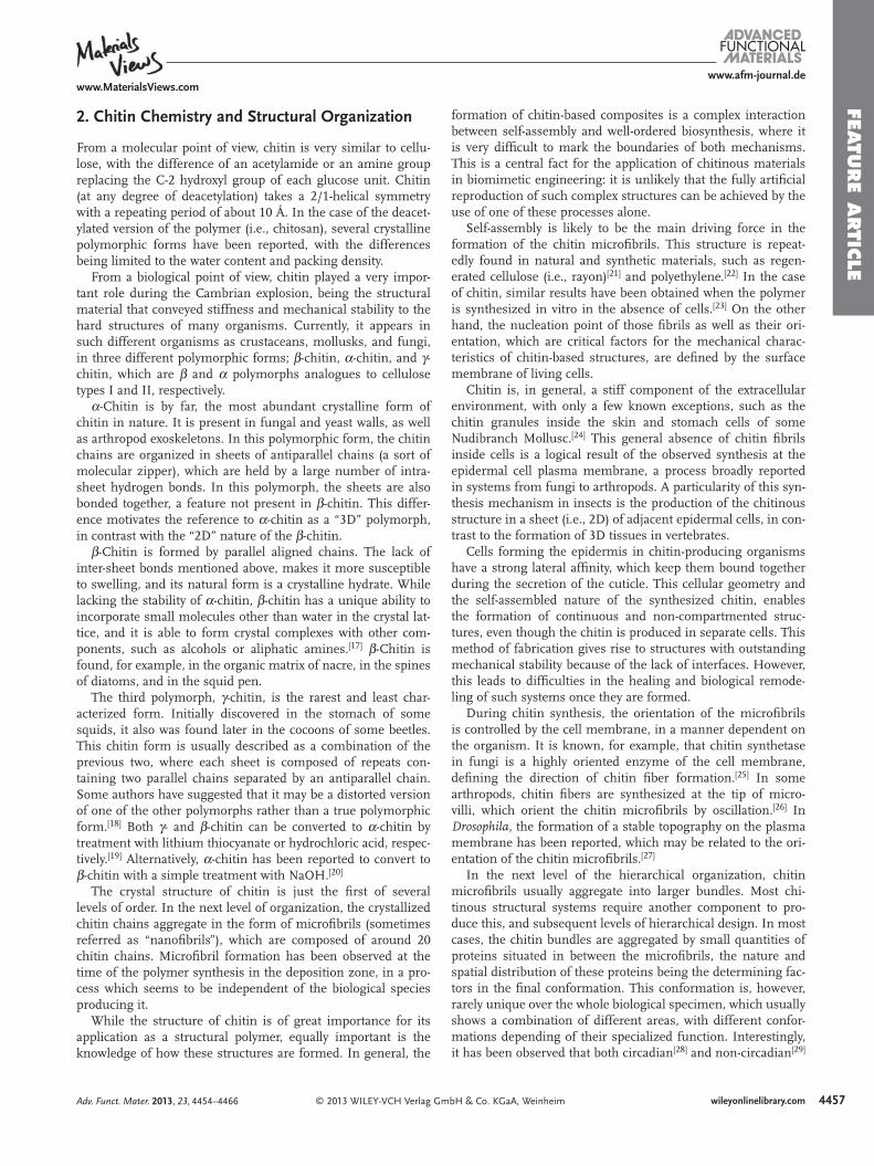
The insect cuticle is an extracellular layer that covers the outer surface of the animal and rests on a layer of epidermal cells. It is mainly composed of chitin microfi laments in a pro-tein matrix, with smaller amounts of lipids and phenols present as well ( Figure 2 ). Historically, cuticles have been divided into “hard” and “soft” (with some exceptions), but as our knowledge of these structures advances, we have realized that this divi-sion is too simplistic. Although the cuticle contains the same
clocks regulate the alternation of these different supramolec-ular chitin conformations in animals.
The formation of chitinous systems is, therefore, a bottom-up process driven by a combination of self-assembly and cell-regulated ordering of microfi brils. However, in some chi-tinous system, large-scale external factors may also become an important contributor to the organization of the chitin micro-fi brils. [ 30 ] Molecular rearrangement due to an external force is well known in polymer glasses, that exhibit strain-hardening associated with an anisotropic stress. [ 31 ] This effect, which is well known in artifi cial systems, has also been observed in the arthropod cuticle, where the areas under stress show a greater alignment of the chitin microfi brils. Interestingly, as cell-generated (cytoskeletal-based) contractile forces align collagen bundles in this manner, similar forces also might contribute to chitin microfi bril alignment as well.
Realignment of chitin microfi brils to improve mechanical properties in a particular direction can be the result of stresses generated by the normal use of the chitinous structure (e.g., a moving articulation). However, in some cases, the macro-scopic biomechanical force can be induced by the system itself during its construction. For example, the long apodermes of dragonfl ies, composed of highly oriented chitin, result from the action on the untanned cuticle of the extension of the fl ight
Figure 2 . A) Diagram of a simplifi ed insect cuticle. The epidermal cells secrete a chitinous layered structure. Each layer is represented as seen under a polarized light, where dark and clear layers appear because the different alignments of the microfi brils respect the polarizing plane. B) Helical structure of the chitin microfi brils. Chitin microfi brils (yellow) in a protein matrix (blue) are arranged parallel to each other forming a sheet (i.e., laminae). Each sheet is rotated with respect the one below, in a plywood structure.
Adv. Funct. Mater. 2013, 23, 4454–4466
4459
www.afm-journal.dewww.MaterialsViews.com
wileyonlinelibrary.com© 2013 WILEY-VCH Verlag GmbH & Co. KGaA, Weinheim
FEATU
RE A
RTIC
LE
of the cuticle requires the presence of water, which begins to be absorbed about 1 h before the ecdysis, and then water is purged out of the cuticle in large quantities after the cuticle has been secreted. An exemplary case of the infl uence of water in the mechanical properties of the cuticle is the Rhodnius Prolixus , an important spreader of Chagas disease. This hematophagous insect has the ability to alter the water content of its cuticle by modulating pH locally. This ability, which is controlled by the nervous system, enables the insect to plasticize its abdomen during feeding, thereby creating an elastic reservoir that can accommodate large amounts of blood. Moreover, this plasticiza-tion is reversible as the cuticle returns to its stiffer, and more protective state, after feeding.
Based on this evidence, the mechanical characteristics of the cuticle have been suggested to be a function of their internal concentration of protein, chitin, and water. In soft cuticles that contain 40–75% water, chitin and protein are thought to con-tribute equally to its mechanical properties. In contrast, hard cuticles contain only about 12% water and 15–30% chitin, with the protein serving as the dominant structural component. [ 44 ]
Similar to the tanning hypothesis, the evidence does not yet exist to suggest that dehydration alone is the sole process regu-lating cuticle hardening. It is thought that the fi nal mechanical properties of the cuticle probably result from the additive effect of both hardening methods and, therefore, its artifi cial replica-tion in the fabrication of bioinspired chitinous materials would need to take account of both of these mechanisms.
Regardless of whether the formation of the cuticle requires sclerotized proteins or dehydration, questions still remain regarding how the proteins and chitin interact with each other, or if they are bonded at all. The treatment of cuticles with chitinase results in extraction of only 30% of the total chitin present, which has led to some to suggest that the remaining chitin is unextractable because it is physically bonded to the protein. Also, the treatment of the some hard cuticles with lithium bromide gives rise to chitin-protein complexes, which would be expected to be disrupted by the treatment, suggesting the existence of a strong bond between them. [ 45 ] This evidence is supported by IR data, which shows that all the functional groups of chitin are chemically bonded. This is an expected result inside the chitin microfi brils, because the crystal confor-mation of the chitin involves bonding between adjacent chains. However, those polymer chains situated on the surface of the microfi brils should have free groups available for bonding. Hence, since these free groups do not appear in the FTIR data, they are thought to be bonded to surrounding protein matrix. This interpretation is also supported by the fi nding that the exposed groups of chitin chains are laterally spaced at the same (in α -chitin) or a proportional ( β -chitin) distance between adja-cent protein chains of an antiparallel β -sheet protein, such as fi broin. [ 30 ]
While many chitin-binding proteins have been character-ized in vitro, [46 ] to our knowledge, there is no direct observa-tion of the bonding between protein and chitin in the cuticle. Although many models have been proposed to explain these different observations, this kind of direct bonding interaction between protein and chitin molecules does not appear to hold generally in living materials. For example, while hard cuti-cles fi t well with models where chitin and protein are related
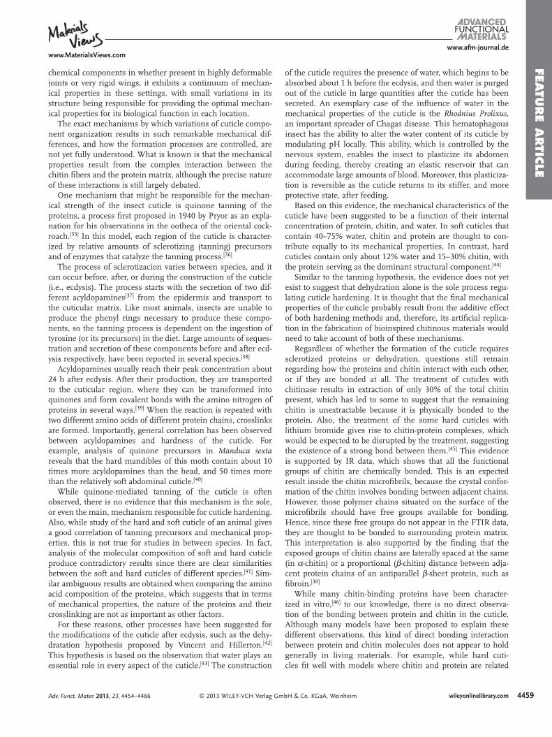
chemical components in whether present in highly deformable joints or very rigid wings, it exhibits a continuum of mechan-ical properties in these settings, with small variations in its structure being responsible for providing the optimal mechan-ical properties for its biological function in each location.
The exact mechanisms by which variations of cuticle compo-nent organization results in such remarkable mechanical dif-ferences, and how the formation processes are controlled, are not yet fully understood. What is known is that the mechanical properties result from the complex interaction between the chitin fi bers and the protein matrix, although the precise nature of these interactions is still largely debated.
One mechanism that might be responsible for the mechan-ical strength of the insect cuticle is quinone tanning of the proteins, a process fi rst proposed in 1940 by Pryor as an expla-nation for his observations in the ootheca of the oriental cock-roach. [ 35 ] In this model, each region of the cuticle is character-ized by relative amounts of sclerotizing (tanning) precursors and of enzymes that catalyze the tanning process. [ 36 ]
The process of sclerotizacion varies between species, and it can occur before, after, or during the construction of the cuticle (i.e., ecdysis). The process starts with the secretion of two dif-ferent acyldopamines [ 37 ] from the epidermis and transport to the cuticular matrix. Like most animals, insects are unable to produce the phenyl rings necessary to produce these compo-nents, so the tanning process is dependent on the ingestion of tyrosine (or its precursors) in the diet. Large amounts of seques-tration and secretion of these components before and after ecd-ysis respectively, have been reported in several species. [ 38 ]
Acyldopamines usually reach their peak concentration about 24 h after ecdysis. After their production, they are transported to the cuticular region, where they can be transformed into quinones and form covalent bonds with the amino nitrogen of proteins in several ways. [ 39 ] When the reaction is repeated with two different amino acids of different protein chains, crosslinks are formed. Importantly, general correlation has been observed between acyldopamines and hardness of the cuticle. For example, analysis of quinone precursors in Manduca sexta reveals that the hard mandibles of this moth contain about 10 times more acyldopamines than the head, and 50 times more than the relatively soft abdominal cuticle. [ 40 ]
While quinone-mediated tanning of the cuticle is often observed, there is no evidence that this mechanism is the sole, or even the main, mechanism responsible for cuticle hardening. Also, while study of the hard and soft cuticle of an animal gives a good correlation of tanning precursors and mechanical prop-erties, this is not true for studies in between species. In fact, analysis of the molecular composition of soft and hard cuticle produce contradictory results since there are clear similarities between the soft and hard cuticles of different species. [ 41 ] Sim-ilar ambiguous results are obtained when comparing the amino acid composition of the proteins, which suggests that in terms of mechanical properties, the nature of the proteins and their crosslinking are not as important as other factors.
For these reasons, other processes have been suggested for the modifi cations of the cuticle after ecdysis, such as the dehy-dratation hypothesis proposed by Vincent and Hillerton. [ 42 ] This hypothesis is based on the observation that water plays an essential role in every aspect of the cuticle. [ 43 ] The construction
Adv. Funct. Mater. 2013, 23, 4454–4466
4460
www.afm-journal.dewww.MaterialsViews.com
wileyonlinelibrary.com © 2013 WILEY-VCH Verlag GmbH & Co. KGaA, Weinheim
FEATU
RE
ARTI
CLE nacre is 3000-fold tougher and has 1000-fold more fracture
resistance than this crystal form of calcium carbonate. These exceptional properties are the consequence of the combination of aragonite with a softer chitinous organic matrix, enabling the use of a brittle, but extremely abundant, material (i.e., arago-nite) in structures with great mechanical performance.
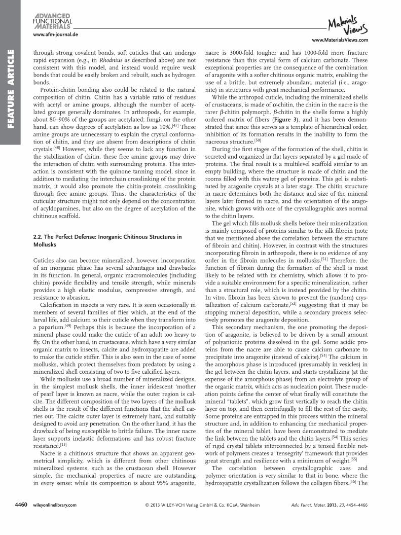
While the arthropod cuticle, including the mineralized shells of crustaceans, is made of α -chitin, the chitin in the nacre is the rarer β -chitin polymorph. β -chitin in the shells forms a highly ordered matrix of fi bers ( Figure 3 ), and it has been demon-strated that since this serves as a template of hierarchical order, inhibition of its formation results in the inability to form the nacreous structure. [ 50 ]
During the fi rst stages of the formation of the shell, chitin is secreted and organized in fl at layers separated by a gel made of proteins. The fi nal result is a multilevel scaffold similar to an empty building, where the structure is made of chitin and the rooms fi lled with this watery gel of proteins. This gel is substi-tuted by aragonite crystals at a later stage. The chitin structure in nacre determines both the distance and size of the mineral layers later formed in nacre, and the orientation of the arago-nite, which grows with one of the crystallographic axes normal to the chitin layers.
The gel which fi lls mollusk shells before their mineralization is mainly composed of proteins similar to the silk fi broin (note that we mentioned above the correlation between the structure of fi broin and chitin). However, in contrast with the structures incorporating fi broin in arthropods, there is no evidence of any order in the fi broin molecules in mollusks. [ 51 ] Therefore, the function of fi broin during the formation of the shell is most likely to be related with its chemistry, which allows it to pro-vide a suitable environment for a specifi c mineralization, rather than a structural role, which is instead provided by the chitin. In vitro, fi broin has been shown to prevent the (random) crys-tallization of calcium carbonate, [ 52 ] suggesting that it may be stopping mineral deposition, while a secondary process selec-tively promotes the aragonite deposition.
This secondary mechanism, the one promoting the deposi-tion of aragonite, is believed to be driven by a small amount of polyanionic proteins dissolved in the gel. Some acidic pro-teins from the nacre are able to cause calcium carbonate to precipitate into aragonite (instead of calcite). [ 53 ] The calcium in the amorphous phase is introduced (presumably in vesicles) in the gel between the chitin layers, and starts crystallizing (at the expense of the amorphous phase) from an electrolyte group of the organic matrix, which acts as nucleation point. These nucle-ation points defi ne the center of what fi nally will constitute the mineral “tablets”, which grow fi rst vertically to reach the chitin layer on top, and then centrifugally to fi ll the rest of the cavity. Some proteins are entrapped in this process within the mineral structure and, in addition to enhancing the mechanical proper-ties of the mineral tablet, have been demonstrated to mediate the link between the tablets and the chitin layers. [ 54 ] This series of rigid crystal tablets interconnected by a tensed fl exible net-work of polymers creates a ‘tensegrity’ framework that provides great strength and resilience with a minimum of weight. [ 55 ]
The correlation between crystallographic axes and poly mer orientation is very similar to that in bone, where the hydroxyapatite crystallization follows the collagen fi bers. [ 56 ] The
through strong covalent bonds, soft cuticles that can undergo rapid expansion (e.g., in Rhodnius as described above) are not consistent with this model, and instead would require weak bonds that could be easily broken and rebuilt, such as hydrogen bonds.
Protein-chitin bonding also could be related to the natural composition of chitin. Chitin has a variable ratio of residues with acetyl or amine groups, although the number of acety-lated groups generally dominates. In arthropods, for example, about 80–90% of the groups are acetylated; fungi, on the other hand, can show degrees of acetylation as low as 10%. [ 47 ] These amine groups are unnecessary to explain the crystal conforma-tion of chitin, and they are absent from descriptions of chitin crystals. [ 48 ] However, while they seems to lack any function in the stabilization of chitin, these free amine groups may drive the interaction of chitin with surrounding proteins. This inter-action is consistent with the quinone tanning model, since in addition to mediating the interchain crosslinking of the protein matrix, it would also promote the chitin-protein crosslinking through free amine groups. Thus, the characteristics of the cuticular structure might not only depend on the concentration of acyldopamines, but also on the degree of acetylation of the chitinous scaffold.
2.2. The Perfect Defense: Inorganic Chitinous Structures in Mollusks
Cuticles also can become mineralized, however, incorporation of an inorganic phase has several advantages and drawbacks in its function. In general, organic macromolecules (including chitin) provide fl exibility and tensile strength, while minerals provides a high elastic modulus, compressive strength, and resistance to abrasion.
Calcifi cation in insects is very rare. It is seen occasionally in members of several families of fl ies which, at the end of the larval life, add calcium to their cuticle when they transform into a paparium. [ 49 ] Perhaps this is because the incorporation of a mineral phase could make the cuticle of an adult too heavy to fl y. On the other hand, in crustaceans, which have a very similar organic matrix to insects, calcite and hydroxyapatite are added to make the cuticle stiffer. This is also seen in the case of some mollusks, which protect themselves from predators by using a mineralized shell consisting of two to fi ve calcifi ed layers.
While mollusks use a broad number of mineralized designs, in the simplest mollusk shells, the inner iridescent ‘mother of pearl’ layer is known as nacre, while the outer region is cal-cite. The different composition of the two layers of the mollusk shells is the result of the different functions that the shell car-ries out. The calcite outer layer is extremely hard, and suitably designed to avoid any penetration. On the other hand, it has the drawback of being susceptible to brittle failure. The inner nacre layer supports inelastic deformations and has robust fracture resistance. [ 13 ]
Nacre is a chitinous structure that shows an apparent geo-metrical simplicity, which is different from other chitinous mineralized systems, such as the crustacean shell. However simple, the mechanical properties of nacre are outstanding in every sense: while its composition is about 95% aragonite,
Adv. Funct. Mater. 2013, 23, 4454–4466
4461
www.afm-journal.dewww.MaterialsViews.com
wileyonlinelibrary.com© 2013 WILEY-VCH Verlag GmbH & Co. KGaA, Weinheim
FEATU
RE A
RTIC
LE
same effect has been also observed in other organisms that uti-lize mineralized chitinous systems, such as crustaceans, [ 57 ] in which there seems to be a correlation between calcite crystal-lographic axes and the orientation of the chitin fi bers.
In summary, the formation of the shell in mollusks can be described in three phases: the synthesis of the chitinous struc-ture, the fi lling of the structure with the fi broin-like gel phase, and fi nally the nucleation of the CaCO 3 crystals in association to the secreted chitinous matrix.
3. Applications of Chitinous Materials
One important step in the processing of chitin for technical and commercial applications is its artifi cial deacetylation. Chitin appears in nature at many degrees of acetylation, with highly acetylated forms predominating. Unfortunately, its current technological uses in this state are very limited, mainly because it is insoluble in common solvents without degradation. This limitation has been overcome by its artifi cial deacetylation in an alkaline solution, which transforms it into a closely related molecule known as chitosan. The chitosan polymer contains (protonable) free amine groups that enable the introduction of repulsive forces between chains and, therefore, molecular disso-lution. [ 58 ] While there is not a sharp distinction between chitin and chitosan, it is commonly accepted to talk about chitosan when at least one half of the functional groups in the structure of the polysaccharide are amines. In general, from this degree of de-acetylation and beyond, dissolution is possible. [ 59 ]
Many researchers have explored the possibilities of regener-ating chitosan from solution, with disparate results depending
of the process used. [ 60 ] These methods give rise to a chitosan polymer with mechanical properties vastly different from those measured in chitin within natural materials. Many researchers therefore explored ways to revert the de-acetylation process of chitosan to produce chitin; however, these efforts were not fruitful. Actually, the smaller size of the amine groups enables shorter bonds between chains than the acetyl form, and thus, the strength of artifi cial chitinous structures is generally pro-portional to the degree of deacetylation of the polymer. [ 61 ] The disparity of the results between natural and artifi cial systems, is more likely to be related with the molecular weight of chi-tosan, which is much lower than that of natural chitin, and with the inability to reproduce the hierarchical design of natural chi-tinous materials.
In addition to be fully compostable, chitosan has been dem-onstrated to be a growth enhancer in plants. [ 62 ] This plant growth enhancement effect is usually associated with the large amount of nitrogen provided by the degraded polymer. Addi-tionally, chitosan has been reported to be a fungicidal agent in Lisianthus , where chitinase enzymes are produced as defense mechanism to phytopathogenic infections. In the presence of chitosan, the defense mechanism of the seed is triggered ear-lier than the normal cycle, protecting the plant from infections before they happen. [ 63 ]
While the natural functions of chitin and chitosan are mostly structural, their applications in medicine have largely focused on exploiting their chemical properties, such as their ability to capture other molecules, rather than their ability to form mechanically strong structures. Chitosan also has an intrinsic antibacterial activity as its cationic amino groups associate with anions on the bacterial cell wall, which accelerates bacterial
Figure 3 . A) Structure of nacre. The (yellow) chitinous matrix defi nes the boundaries and layers of the interconnected cavities where the future arago-nite tablets will grow. The (blue) polymer gel is mainly composed of silk-like proteins. B) Diagram of the growing aragonite tablet. The chitin fi bers are oriented in a unidirectional fashion and the axis of the crystallites will defi ne the orientation of the aragonite crystals.
Adv. Funct. Mater. 2013, 23, 4454–4466
4462
www.afm-journal.dewww.MaterialsViews.com
wileyonlinelibrary.com © 2013 WILEY-VCH Verlag GmbH & Co. KGaA, Weinheim
FEATU
RE
ARTI
CLE (and hence the pore size) is known to be
dependent on heat and mass transfer rates. At higher temperatures the system is domi-nated by heat transfer, but when the tem-perature is lowered, it is governed by mass transfer and the pore radius becomes almost independent of the temperature change. [ 68 ] The main application of chitosan beads is found in drug delivery because their release rate is dependent on pH. [ 69 ] However, the beads are also good absorbents for phthalate ester (PAEs) contaminants by selectively cap-turing those compounds from solution. [ 70 ]
Chitosan also can be formed into fi bers using an electro-spinning process. In this method, the polymer solution is fi rst placed in a pipette connected to a positive electrode. When an electric fi eld is applied, a charged
jet of chitosan is emitted from the droplet on the pipette tip. The electrifi ed liquid jet of polymer solution is then driven to the collector, which neutralizes the charge. Finally, the fl uid stream solidifi es into a nanofi ber through the evaporation of the solvent.
As with chitin, the main problem of working with chitosan is related to the selection of the solvent, and making micro-fi bers is no exception. Among solvents tested for electrospin-ning of chitosan are acetic acid, formic acid, hydrochloric acid, or dichloroacetic acid, but none of these produce a visible fl uid stream when the electric fi eld is applied. Only trifl uoro-acetic acid deposits chitosan fi bers onto the collector, and its concentration determines the morphology of the resulting structures. [ 65 ] For better results, chitosan can be mixed with a polymer known for its capacity to be conveniently electrospun from aqueous medium, such as poly(ethylene oxide) [ 71 ] or chi-tosan derivates. [ 72 ] The drawback of both compromises is that the resulting blend will have different properties than pure chi-tosan, which may limit its use for biomedical applications.
Recently, electrospun micro- and nanofi bers of chitosan have attracted attention in microelectronics for their use in actu-ator [ 73 ] and sensor applications. [ 74 ] For this reason, changes of chitosan morphology produced by electrical charge have been studied extensively during the last few years. [ 75 ] These studies revealed that chitosan undergoes a shape change that corre-lates directly with the ion concentration of its surroundings (i.e., pH), and this property has been exploited to impart topo-graphical and chemical patterns to 3D chitosan structures for the controlled release of biomolecules. [ 76 ]
While these basic examples of fi lms, fi bers and foams, are indeed a starting point for the fabrication of chitosan, they do not exploit the real structural potential of the polymer. Impor-tantly, recent studies have revealed that this chitinous polymer can form complex structures at much smaller scales, which facilitate better interactions with living cells.
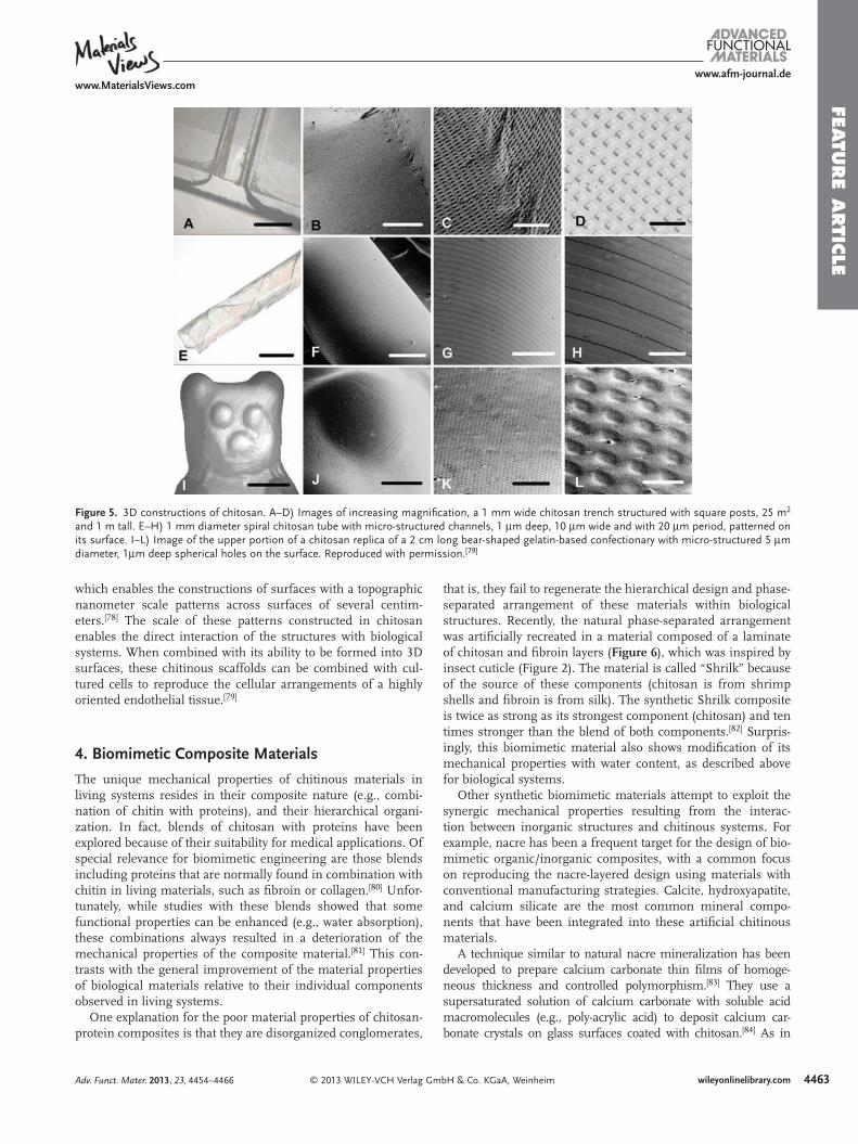
Chitosan can be structured to the micro-scale using soft lithography, a technique that enables the formation of large surfaces with a topography defi ned with a precision of few micrometers [ 77 ] ( Figure 5 ). A modifi cation of this technique called “forced soft lithography” has been used to address the problem of bubble nucleation for polymers in volatile solvents,
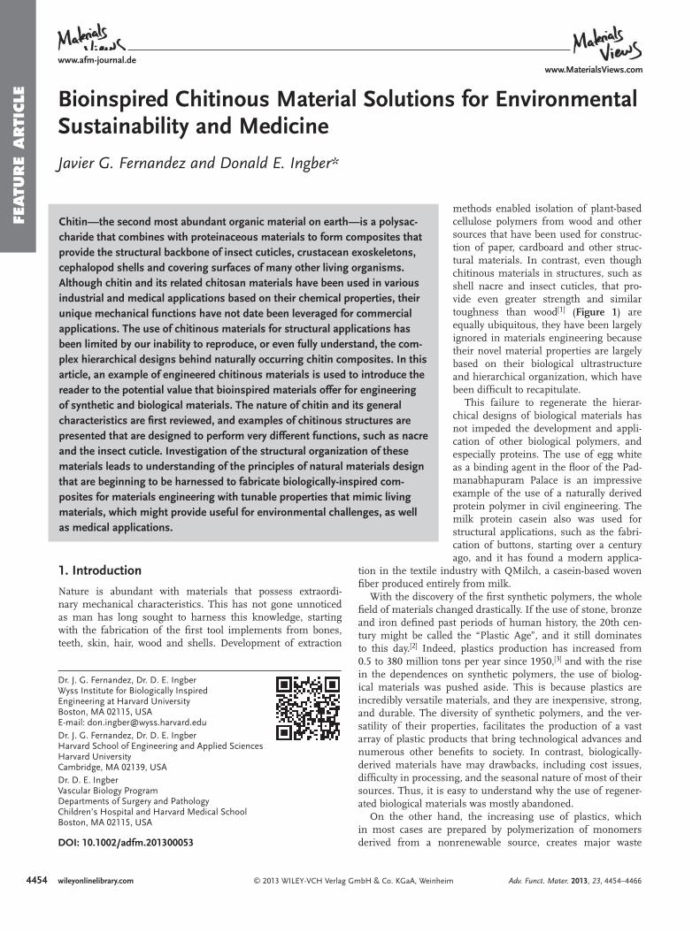
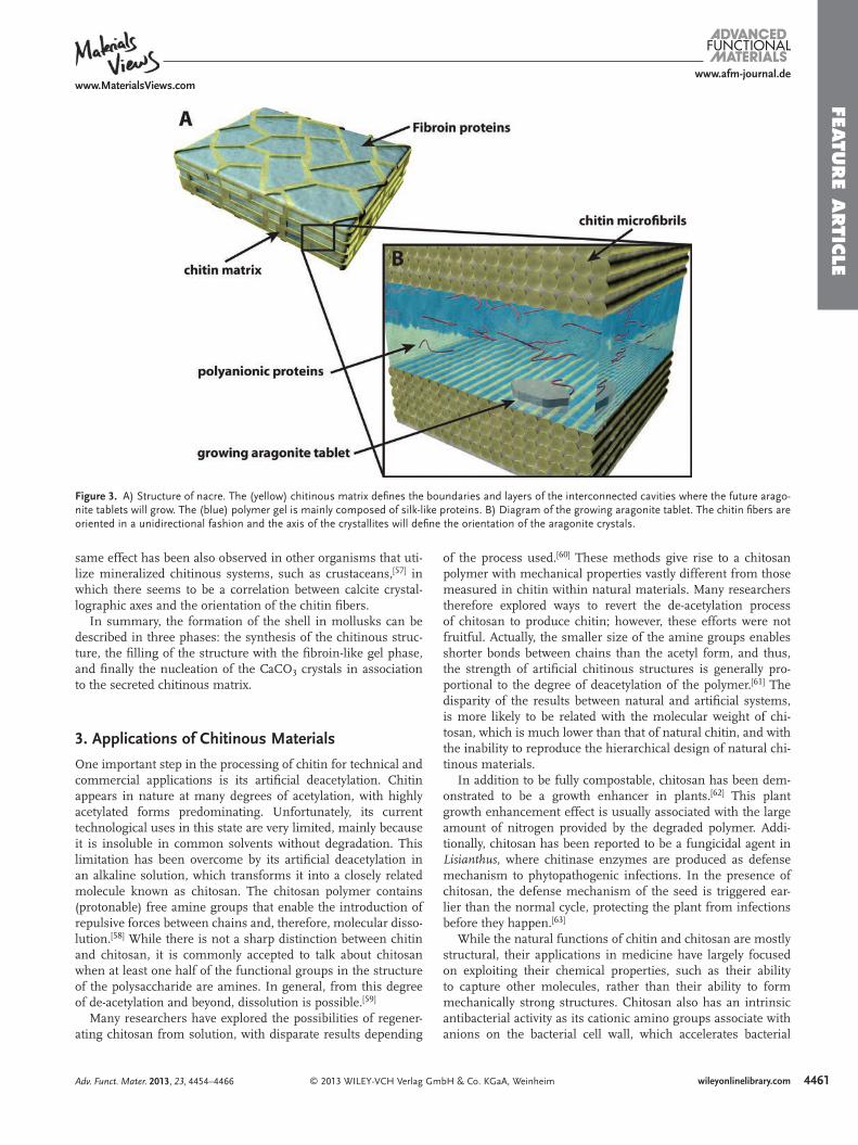
death. This cationic nature also means that chitosan interacts electrostatically with anionic glycosaminoglycans (GAGs), proteoglycans, and other negatively charged molecules in the extracellular matrix. These properties have motivated the utili-zation of chitosan for tissue engineering applications, and the development of methods for its use as a structural material in regenerative medicine. These methods include the formation of porous scaffolds, fi bers, and beads ( Figure 4 ).
The formation of porous chitosan scaffolds was fi rst pro-posed by Mandihally and co-workers in 1999, [ 64 ] and has con-sequently become the most promising application for chitosan in tissue engineering, especially for its use in bone grafts. Bulk chitosan scaffolds are prepared by freezing and lyophilizing (i.e., drying in a frozen state under high vacuum) chitosan dissolved in acetic acid. The process generates an open pore microstructure with a high degree of interconnectivity. Control of the pore size is made by the selection of the temperature, which in the range between − 20 ° C and − 196 ° C produces a mean pore diameter from 40 to 250 µ m.
The scaffolds also can be formed inside a mold that gives the fi nal form to the bulk polymer. Alternatively, the resulting porous chitosan piece can be cut or carved to produce a desired shape. [ 66 ] This technique is also used to form porous beads, which are popular as drug carriers. Depending on the condi-tions in which the beads are formed, the properties are very different. [ 67 ] In the case of direct freezing, the beads are clearly porous and those pores are aligned with a radial orientation, having a nearly homogeneous diameter. Beads with a reason-able quality and radii ranging from 20 to 50 µ m can be pro-duced by this technique.
Freezing chitosan after its gelation in NaOH produces even more interesting results. These beads apparently have a smooth surface except for a few cracks (produced by thermal expan-sion-contraction). But when the beads are observed at higher magnifi cation, a highly porous, fi brous structure is revealed. The average pore size is about 1–5 µ m and this fi brous struc-ture has been interpreted to result from the formation of small ice crystals during freezing. In the beads, the dependence of pore size on the freezing temperature has a very strong cor-relation between − 5 and − 15 ° C, but the dependence decays between − 15 ° C and − 196 ° C. This is because the crystal growth
Figure 4 . Basic constructions of chitosan. Image at the left is a scanning electron microscope (SEM) image of chitosan microfi bers fabricated by electro-spinning, employing Trifl uoroacetic Acid as solvent. Reproduced with permission. [ 65 ] The central images are SEM images of porous chitosan microcarriers (beads). The chitosan droplets can be directly frozen in liquid nitrogen (top) or from a gel before being frozen (bottom). The foam (right) is a SEM image of a bulk porous Chitosan scaffold. The image corresponds to a 2% (w/v) solution of Chitosan frozen in dry ice ( − 20 ° C). Reproduced with permission. [ 64 ] Copyright 1999, Elsevier.
Adv. Funct. Mater. 2013, 23, 4454–4466
4463
www.afm-journal.dewww.MaterialsViews.com
wileyonlinelibrary.com© 2013 WILEY-VCH Verlag GmbH & Co. KGaA, Weinheim
FEATU
RE A
RTIC
LE
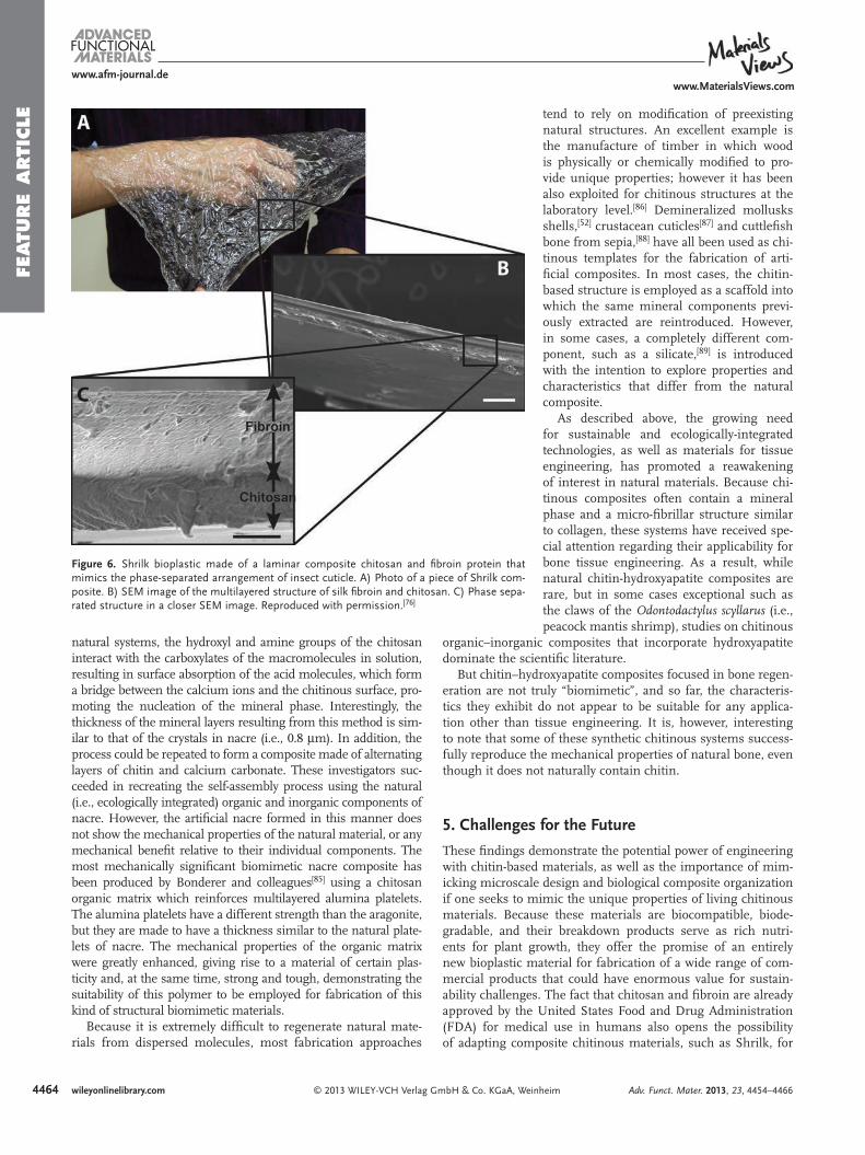
that is, they fail to regenerate the hierarchical design and phase-separated arrangement of these materials within biological structures. Recently, the natural phase-separated arrangement was artifi cially recreated in a material composed of a laminate of chitosan and fi broin layers ( Figure 6 ), which was inspired by insect cuticle (Figure 2 ). The material is called “Shrilk” because of the source of these components (chitosan is from shrimp shells and fi broin is from silk). The synthetic Shrilk composite is twice as strong as its strongest component (chitosan) and ten times stronger than the blend of both components. [ 82 ] Surpris-ingly, this biomimetic material also shows modifi cation of its mechanical properties with water content, as described above for biological systems.
Other synthetic biomimetic materials attempt to exploit the synergic mechanical properties resulting from the interac-tion between inorganic structures and chitinous systems. For example, nacre has been a frequent target for the design of bio-mimetic organic/inorganic composites, with a common focus on reproducing the nacre-layered design using materials with conventional manufacturing strategies. Calcite, hydroxyapatite, and calcium silicate are the most common mineral compo-nents that have been integrated into these artifi cial chitinous materials.
A technique similar to natural nacre mineralization has been developed to prepare calcium carbonate thin fi lms of homoge-neous thickness and controlled polymorphism. [ 83 ] They use a supersaturated solution of calcium carbonate with soluble acid macromolecules (e.g., poly-acrylic acid) to deposit calcium car-bonate crystals on glass surfaces coated with chitosan. [ 84 ] As in
which enables the constructions of surfaces with a topographic nanometer scale patterns across surfaces of several centim-eters. [ 78 ] The scale of these patterns constructed in chitosan enables the direct interaction of the structures with biological systems. When combined with its ability to be formed into 3D surfaces, these chitinous scaffolds can be combined with cul-tured cells to reproduce the cellular arrangements of a highly oriented endothelial tissue. [ 79 ]
4. Biomimetic Composite Materials
The unique mechanical properties of chitinous materials in living systems resides in their composite nature (e.g., combi-nation of chitin with proteins), and their hierarchical organi-zation. In fact, blends of chitosan with proteins have been explored because of their suitability for medical applications. Of special relevance for biomimetic engineering are those blends including proteins that are normally found in combination with chitin in living materials, such as fi broin or collagen. [ 80 ] Unfor-tunately, while studies with these blends showed that some functional properties can be enhanced (e.g., water absorption), these combinations always resulted in a deterioration of the mechanical properties of the composite material. [ 81 ] This con-trasts with the general improvement of the material properties of biological materials relative to their individual components observed in living systems.
One explanation for the poor material properties of chitosan-protein composites is that they are disorganized conglomerates,
Figure 5 . 3D constructions of chitosan. A–D) Images of increasing magnifi cation, a 1 mm wide chitosan trench structured with square posts, 25 m 2 and 1 m tall. E–H) 1 mm diameter spiral chitosan tube with micro-structured channels, 1 µ m deep, 10 µ m wide and with 20 µ m period, patterned on its surface. I–L) Image of the upper portion of a chitosan replica of a 2 cm long bear-shaped gelatin-based confectionary with micro-structured 5 µ m diameter, 1 µ m deep spherical holes on the surface. Reproduced with permission. [ 79 ]
Adv. Funct. Mater. 2013, 23, 4454–4466
4464
www.afm-journal.dewww.MaterialsViews.com
wileyonlinelibrary.com © 2013 WILEY-VCH Verlag GmbH & Co. KGaA, Weinheim
FEATU
RE
ARTI
CLE tend to rely on modifi cation of preexisting
natural structures. An excellent example is the manufacture of timber in which wood is physically or chemically modifi ed to pro-vide unique properties; however it has been also exploited for chitinous structures at the laboratory level. [ 86 ] Demineralized mollusks shells, [ 52 ] crustacean cuticles [ 87 ] and cuttlefi sh bone from sepia, [ 88 ] have all been used as chi-tinous templates for the fabrication of arti-fi cial composites. In most cases, the chitin-based structure is employed as a scaffold into which the same mineral components previ-ously extracted are reintroduced. However, in some cases, a completely different com-ponent, such as a silicate, [ 89 ] is introduced with the intention to explore properties and characteristics that differ from the natural composite.
As described above, the growing need for sustainable and ecologically-integrated technologies, as well as materials for tissue engineering, has promoted a reawakening of interest in natural materials. Because chi-tinous composites often contain a mineral phase and a micro-fi brillar structure similar to collagen, these systems have received spe-cial attention regarding their applicability for bone tissue engineering. As a result, while natural chitin-hydroxyapatite composites are rare, but in some cases exceptional such as the claws of the Odontodactylus scyllarus (i.e., peacock mantis shrimp), studies on chitinous
organic–inorganic composites that incorporate hydroxyapatite dominate the scientifi c literature.
But chitin–hydroxyapatite composites focused in bone regen-eration are not truly “biomimetic”, and so far, the characteris-tics they exhibit do not appear to be suitable for any applica-tion other than tissue engineering. It is, however, interesting to note that some of these synthetic chitinous systems success-fully reproduce the mechanical properties of natural bone, even though it does not naturally contain chitin.
5. Challenges for the Future
These fi ndings demonstrate the potential power of engineering with chitin-based materials, as well as the importance of mim-icking microscale design and biological composite organization if one seeks to mimic the unique properties of living chitinous materials. Because these materials are biocompatible, biode-gradable, and their breakdown products serve as rich nutri-ents for plant growth, they offer the promise of an entirely new bioplastic material for fabrication of a wide range of com-mercial products that could have enormous value for sustain-ability challenges. The fact that chitosan and fi broin are already approved by the United States Food and Drug Administration (FDA) for medical use in humans also opens the possibility of adapting composite chitinous materials, such as Shrilk, for
Figure 6 . Shrilk bioplastic made of a laminar composite chitosan and fi broin protein that mimics the phase-separated arrangement of insect cuticle. A) Photo of a piece of Shrilk com-posite. B) SEM image of the multilayered structure of silk fi broin and chitosan. C) Phase sepa-rated structure in a closer SEM image. Reproduced with permission. [ 76 ]
natural systems, the hydroxyl and amine groups of the chitosan interact with the carboxylates of the macromolecules in solution, resulting in surface absorption of the acid molecules, which form a bridge between the calcium ions and the chitinous surface, pro-moting the nucleation of the mineral phase. Interestingly, the thickness of the mineral layers resulting from this method is sim-ilar to that of the crystals in nacre (i.e., 0.8 µ m). In addition, the process could be repeated to form a composite made of alternating layers of chitin and calcium carbonate. These investigators suc-ceeded in recreating the self-assembly process using the natural (i.e., ecologically integrated) organic and inorganic components of nacre. However, the artifi cial nacre formed in this manner does not show the mechanical properties of the natural material, or any mechanical benefi t relative to their individual components. The most mechanically signifi cant biomimetic nacre composite has been produced by Bonderer and colleagues [ 85 ] using a chitosan organic matrix which reinforces multilayered alumina platelets. The alumina platelets have a different strength than the aragonite, but they are made to have a thickness similar to the natural plate-lets of nacre. The mechanical properties of the organic matrix were greatly enhanced, giving rise to a material of certain plas-ticity and, at the same time, strong and tough, demonstrating the suitability of this polymer to be employed for fabrication of this kind of structural biomimetic materials.
Because it is extremely diffi cult to regenerate natural mate-rials from dispersed molecules, most fabrication approaches
Adv. Funct. Mater. 2013, 23, 4454–4466
4465
www.afm-journal.dewww.MaterialsViews.com
wileyonlinelibrary.com© 2013 WILEY-VCH Verlag GmbH & Co. KGaA, Weinheim
FEATU
RE A
RTIC
LE
[ 23 ] J. Ruiz-Herrera , V. O. Sing , W. J. Van der Woude , S. Bartnicki-Garcia , Proc. Natl. Acad. Sci. USA 1975 , 72 , 2706 .
[ 24 ] R. Martin , S. Hild , P. Walther , K. Ploss , W. Boland , K.-H. Tomaschko , Biol. Bull. 2007 , 213 , 307 .
[ 25 ] A. Durán , B. Bowers , E. Cabib , Proc. Natl. Acad. Sci. USA 1975 , 72 , 3952 .
[ 26 ] B. Shillito , J. P. Lechaire , F. Gaill , J. Struct. Biol. 1993 , 111 , 59 . [ 27 ] B. Moussian , C. Seifarth , U. Müller , J. Berger , H. Schwarz , Arthropod.
Struct. Dev. 2006 , 35 , 137 . [ 28 ] A. C. Neville , Q. J. Microsc. Sci. 1965 , s3-106 , 315 . [ 29 ] B. Zelazny , A. C. Neville , J. Insect Physiol. 1972 , 18 , 1967 . [ 30 ] A. C. Neville , Biology of fi brous composites: development beyond the
cell membrane , Cambridge University Press , New York 1993 . [ 31 ] I. M. Ward , J. Sweeney , Mechanical Properties of Solid Polymers ,
John Wiley & Sons, Ltd , New York 2012 . [ 32 ] A. C. Neville , J. E. Treherne, J. W. L. Beament , V. B. Wigglesworth ,
Adv. Insect. Physiol. 1967 , 4 , 213 . [ 33 ] Y. Saito , T. Okano , H. Chanzy , J. Sugiyama , J. Struct. Biol. 1995 , 114 ,
218 . [ 34 ] M.-J. Chrétiennot-Dinet , M.-M. Giraud-Guille , D. Vaulot , J.-L. Putaux ,
Y. Saito , H. Chanzy , J. Phycol. 1997 , 33 , 666 . [ 35 ] M. G. M. Pryor , Proc. R. Soc. London, Ser. B 1940 , 128 , 378 . [ 36 ] H. S. Mason , Nature 1955 , 175 , 771 . [ 37 ] P. Karlson , C. E. Sekeris , Nature 1962 , 195 , 183 . [ 38 ] R. F. Chapman , The insects: structure and function Cambridge Uni-
versity Press , New York 1998 . [ 39 ] S. O. Andersen , Insect Biochem. Molec. 2010 , 40 , 166 . [ 40 ] T. D. Morgan , T. L. Hopkins , K. J. Kramer , C. R. Roseland ,
T. H. Czapla , K. B. Tomer , F. W. Crow , Insect Biochem. 1987 , 17 , 255 . [ 41 ] D. E. Bliss , L. H. Mantel , The Biology of Crustacea: Integument, Pig-
ments, and Hormonal Processes , Vol. 9 , Academic Press , New York 1985 .
[ 42 ] J. E. Hillerton , S. E. Reynolds , J. F. V. Vincent , J. Exp. Biol. 1982 , 96 , 45 .
[ 43 ] H. R. Hepburn , I. Joffe , N. Green , K. J. Nelson , Comp. Biochem. Phys. A 1975 , 50 , 551 .
[ 44 ] J. F. V. Vincent , U. G. K. Wegst , Arthropod. Struct. Dev. 2004 , 33 , 187 . [ 45 ] L. Richards , Integument of Arthropods , University of Minnesota
Press , Minneapolis 1951 . [ 46 ] a) S.-I. Kawabata , R. Nagayama , M. Hirata , T. Shigenaga ,
K. L. Agarwala , T. Saito , J. Cho , H. Nakajima , T. Takagi , S. Iwanaga , J. Biochem. 1996 , 120 , 1253 ; b) L. Tang , J. Liang , Z. Zhan , Z. Xiang , N. He , Insect Biochem. Molec. 2010 , 40 , 228 .
[ 47 ] T. Wu , S. Zivanovic , F. A. Draughon , W. S. Conway , C. E. Sams , J. Agr. Food Chem. 2005 , 53 , 3888 .
[ 48 ] P. Sikorski , R. Hori , M. Wada , Biomacromolecules 2009 , 10 , 1100 . [ 49 ] G. Fraenkel , C. Hsiao , J. Insect Physiol. 1967 , 13 , 1387 . [ 50 ] V. Schonitzer , I. Weiss , BMC Struct. Biol. 2007 , 7 , 71 . [ 51 ] G. Falini , S. Weiner , L. Addadi , Calcif. Tissue Int. 2003 , 72 , 548 . [ 52 ] G. Falini , S. Albeck , S. Weiner , L. Addadi , Science 1996 , 271 , 67 . [ 53 ] B.-A. Gotliv , L. Addadi , S. Weiner , ChemBioChem 2003 , 4 , 522 . [ 54 ] M. Suzuki , K. Saruwatari , T. Kogure , Y. Yamamoto , T. Nishimura ,
T. Kato , H. Nagasawa , Science 2009 , 325 , 1388 . [ 55 ] a) B. Ji , H. Gao , K. Jimmy Hsia , Phil. Mag. Lett. 2004 , 84 , 631 ;
b) D. E. Ingber , Sci. Am. 1998 , 278 , 48 . [ 56 ] S. Weiner , W. Traub , FEBS Lett. 1986 , 206 , 262 . [ 57 ] A. Al-Sawalmih , C. Li , S. Siegel , H. Fabritius , S. Yi , D. Raabe ,
P. Fratzl , O. Paris , Adv. Funct. Mater. 2008 , 18 , 3307 . [ 58 ] C. K. S. Pillai , W. Paul , C. P. Sharma , Prog. Polym. Sci. 2009 , 34 , 641 . [ 59 ] D. Liu , Y. Wei , P. Yao , L. Jiang , Carbohydr. Res. 2006 , 341 , 782 . [ 60 ] K. Ogawa , T. Yui , M. Miya , Biosci. Biotechnol. Bio. 1992 , 56 , 858 . [ 61 ] J. Nunthanid , S. Puttipipatkhachorn , K. Yamamoto , G. E. Peck , Drug
Dev. Ind. Pharm. 2001 , 27 , 143 . [ 62 ] F. Burrows , C. Louime , M. Abazinge , O. Onokpise , Am. Eurasian J.
Agric. Environ. Sci. 2007 , 2 , 103 .
medical applications that require high strength and tough-ness as well as biocompatibility. But to advance these materials into widespread use, it will be necessary to adapt conventional large-scale manufacturing methods for production of chitinous materials. It also will be important to develop even more effi -cient methods of producing chitosan as well as gaining an even deeper understanding of how microscale interactions among biological chitinous composites convey their distinct material properties. With this knowledge, and these methods in hand, it should be possible to develop approaches for more generalized integration of biomimetic chitinous materials in all forms engi-neering and technologies. [ 90 ]
Acknowledgements This work was supported by funding from the Wyss Institute for Biologically Inspired Engineering at Harvard University. The authors also would like to thank Sandeep T. Koshy for his helpful comments.
Received: January 7, 2013 Revised: May 22, 2013
Published online: July 1, 2013
[ 1 ] U. G. K. Wegst , M. F. Ashby , Philos. Mag. 2004 , 84 , 2167 . [ 2 ] R. C. Thompson , S. H. Swan , C. J. Moore , F. S. vom Saal , Philos.
Trans. R. Soc. London, Ser. B 2009 , 364 , 1973 . [ 3 ] A. L. Andrady , M. A. Neal , Philos. Trans. R. Soc. London, Ser. B 2009 ,
364 , 1977 . [ 4 ] J. Hopewell , R. Dvorak , E. Kosior , Philos. Trans. R. Soc. London, Ser.
B 2009 , 364 , 2115 . [ 5 ] Y. Mato , T. Isobe , H. Takada , H. Kanehiro , C. Ohtake , T. Kaminuma ,
Environ. Sci. Technol. 2000 , 35 , 318 . [ 6 ] H. Teftal , Master’s Thesis , McGill University, Montreal 2000 . [ 7 ] S. Andree , Alternatives for Wakulla County management of blue crab
processing solid waste , Vol. 53 , Sea Grant Extension Program, Univer-sity of Florida 1988 .
[ 8 ] M. N. V. Ravi Kumar , React. Funct. Polym. 2000 , 46 , 1 . [ 9 ] E. Budzinski , P. Bykowski , D. Dutkiewicz , Possibilities of processing
and marketing of products made from antarctic krill , Food & Agricul-ture Org ., Rome 1985 .
[ 10 ] R. V. D. S. Amorim , W. D. Souza , K. Fukushima , G. M. D. Campos-Takaki , Braz. J. Microbiol. 2001 , 32 , 20 .
[ 11 ] T. Endo , S. Koizumi , Curr. Opin. Struct. Biol. 2000 , 10 , 536 . [ 12 ] S. Cottaz , E. Samain , Metab. Eng. 2005 , 7 , 311 . [ 13 ] E. Samain , S. Drouillard , A. Heyraud , H. Driguez , R. A. Geremia ,
Carbohydr. Res. 1997 , 302 , 35 . [ 14 ] P. K. Dutta , J. Dutta , V. Tripathi , J. Sci. Ind. Res. India 2004 , 63 , 20 . [ 15 ] A. B. Jull , C. Ni Mhurchu , D. A. Bennett , C. A. Dunshea-Mooij ,
A. Rodgers , Cochrane Db. Syst. Rev. 2008 , 16 . [ 16 ] O. Felt , P. Buri , R. Gurny , Drug Dev. Ind. Pharm. 1998 , 24 , 979 . [ 17 ] Y. Noishiki , Y. Nishiyama , M. Wada , S. Okada , S. Kuga , Biomacro-
molecules 2003 , 4 , 944 . [ 18 ] a) J. F. Kennedy , Z. M. B. Figueiredo , Carbohyd. Polym. 1994 , 23 , 75 ;
b) E. Atkins , J. Biosci. 1985 , 8 , 375 . [ 19 ] K. M. Rudall , W. Kenchington , Biol. Rev. 1973 , 48 , 597 . [ 20 ] Y. Noishiki , H. Takami , Y. Nishiyama , M. Wada , S. Okada , S. Kuga ,
Biomacromolecules 2003 , 4 , 896 . [ 21 ] a) E. Ribi , Nature 1951 , 168 , 1082 ; b) A. M. Hindeleh , Text. Res. J.
1980 , 50 , 581 . [ 22 ] V. A. Marikhin , L. P. Myasnikova , N. L. Viktorova , Polym. Sci.
U.S.S.R. 1976 , 18 , 1493 .
Adv. Funct. Mater. 2013, 23, 4454–4466
4466
www.afm-journal.dewww.MaterialsViews.com
wileyonlinelibrary.com © 2013 WILEY-VCH Verlag GmbH & Co. KGaA, Weinheim
FEATU
RE
ARTI
CLE [ 63 ] K. Ohta , A. Taniguchi , N. Konishi , T. Hosoki , HortScience 1999 , 34 ,
233 . [ 64 ] S. V. Madihally , H. W. T. Matthew , Biomaterials 1999 , 20 , 1133 . [ 65 ] K. Ohkawa , D. Cha , H. Kim , A. Nishida , H. Yamamoto , Macromol.
Rapid Commun. 2004 , 25 , 1600 . [ 66 ] H. H. K. Xu , C. G. Simon Jr ., Biomaterials 2005 , 26 , 1337 . [ 67 ] G. Lu , L. Zhu , L. Kong , L. Zhang , Y. Gong , N. Zhao , X. Zhang , Tsin-
ghua Sci. Technol. 2006 , 11 , 427 . [ 68 ] a) H. Jiankang , L. Dichen , L. Yaxiong , Y. Bo , L. Bingheng , L. Qin ,
Polymer 2007 , 48 , 4578 ; b) I. J. Roh , I.-C. Kwon , J. Biomater. Sci.-Polym. E 2002 , 13 , 769 .
[ 69 ] a) R. Jayakumar , R. L. Reis , J. F. Mano , J. Bioact. Compat. Polym. 2006 , 21 , 327 ; b) E. B. Denkbas , R. M. Ottenbrite , J. Bioact. Compat. Polym. 2006 , 21 , 351 .
[ 70 ] C.-Y. Chen , C.-C. Chen , Y.-C. Chung , Bioresource Technol. 2007 , 98 , 2578 .
[ 71 ] B. Duan , C. Dong , X. Yuan , K. Yao , J. Biomater. Sci., Polym. E 2004 , 15 , 797 .
[ 72 ] H. Jiang , D. Fang , B. Hsiao , B. Chu , W. Chen , J. Biomater. Sci., Polym. E 2004 , 15 , 279 .
[ 73 ] K. J. Pawlowski , H. L. Belvin , D. L. Raney , J. Su , J. S. Harrison , E. J. Siochi , Polymer 2003 , 44 , 1309 .
[ 74 ] G. M. Spinks , S. R. Shin , G. G. Wallace , P. G. Whitten , S. I. Kim , S. J. Kim , Sens. Actuators B 2006 , 115 , 678 .
[ 75 ] a) C. K. Lee , S. J. Kim , S. I. Kim , B.-J. Yi , S. Y. Han , Smart Mater. Struct. 2006 , 15 , 607 ; b) B.-M. Min , S. W. Lee , J. N. Lim , Y. You ,
T. S. Lee , P. H. Kang , W. H. Park , Polymer 2004 , 45 , 7137 ; c) A. M. A. Nada , M. El-Sakhawy , S. Kamel , M. A. M. Eid , A. M. Adel , Carbohyd. Polym. 2006 , 63 , 113 .
[ 76 ] J. G. Fernandez , J. Samitier , C. A. Mills , J. Biomed. Mater. Res. A 2011 , 98A , 229 .
[ 77 ] J. G. Fernandez , C. A. Mills , E. Martinez , M. J. Lopez-Bosque , X. Sisquella , A. Errachid , J. Samitier , J. Biomed. Mater. Res. A 2008 , 85A , 242 .
[ 78 ] J. G. Fernandez , C. A. Mills , M. Pla-Roca , J. Samitier , Adv. Mater. 2007 , 19 , 3696 .
[ 79 ] J. G. Fernandez , C. A. Mills , J. Samitier , Small 2009 , 5 , 614 . [ 80 ] L. Ma , C. Gao , Z. Mao , J. Zhou , J. Shen , X. Hu , C. Han , Biomaterials
2003 , 24 , 4833 . [ 81 ] K. Haeyong , H. Hyun Chul , U. In Chul , P. Young Hwan , J. Appl.
Polym. Sci. 2001 , 80 , 928 . [ 82 ] J. G. Fernandez , D. E. Ingber , Adv. Mater. 2012 , 24 , 480 . [ 83 ] A. Sugawara , T. Kato , Chem. Commun. 2000 , 0 , 487 . [ 84 ] T. Kato , T. Suzuki , T. Irie , Chem. Lett. 2000 , 29 , 186 . [ 85 ] L. J. Bonderer , A. R. Studart , L. J. Gauckler , Science 2008 , 319 , 1069 . [ 86 ] H. Ehrlich , Int. Geol. Rev. 2010 , 52 , 661 . [ 87 ] M. E. Gunthorpe , C. S. Sikes , A. P. Wheeler , Biol. Bull. 1990 , 179 , 191 . [ 88 ] W. Ogasawara , W. Shenton , S. A. Davis , S. Mann , Chem. Mater.
2000 , 12 , 2835 . [ 89 ] V. Pedroni , P. C. Schulz , M. E. Gschaider de Ferreira , M. A. Morini ,
Coll. Polym. Sci. 2000 , 278 , 964 . [ 90 ] K. E. Tanner , Science 2012 , 336 , 1237 .
Adv. Funct. Mater. 2013, 23, 4454–4466