Embed Size (px)
Citation preview

Biochimica et Biophysica Acta 1860 (2016) 2017–2030
Contents lists available at ScienceDirect
Biochimica et Biophysica Acta
j ourna l homepage: www.e lsev ie r .com/ locate /bbagen
O-GlcNAcylation is a key modulator of skeletal muscle sarcomericmorphometry associated to modulation of protein–protein interactions
Matthias Lambert a, Elodie Richard b, Sophie Duban-Deweer c, Frederic Krzewinski d, Barbara Deracinois a,Erwan Dupont a, Bruno Bastide a, Caroline Cieniewski-Bernard a,⁎a Univ.Lille, EA7369-URePSSS, Unité de Recherche Pluridisciplinaire Sport, Santé, Société, Equipe « Activité Physique, Muscle, Santé », F-59000 Lille, Franceb BiCeL (BioImaging Center of Lille — Campus Lille 1), Univ.Lille, FR3688 CNRS FRABio, F-59000 Lille, Francec Laboratoire de la Barrière Hémato-Encéphalique (LBHE), EA2465, Université d'Artois, Faculté Jean Perrin, 62307 Lens, Franced PAGés (Plateforme d'Analyses des Glycoconjugués), Univ.Lille, CNRS, UMR 8576, UGSF, Unité de Glycobiologie Structurale et Fonctionnelle, F-59000 Lille, France
⁎ Corresponding author at: EA7369-URePSSS, Unité dSport, Santé, Société, Equipe « Activité Physique, Muscle59650 Villeneuve d'Ascq, France.
E-mail address: caroline.cieniewski-bernard@univ-lille
http://dx.doi.org/10.1016/j.bbagen.2016.06.0110304-4165/© 2016 Elsevier B.V. All rights reserved.
a b s t r a c t
a r t i c l e i n f oArticle history:Received 14 March 2016Received in revised form 18 May 2016Accepted 6 June 2016Available online 11 June 2016
Background: The sarcomere structure of skeletalmuscle is determined throughmultiple protein–protein interac-tionswithin an intricate sarcomeric cytoskeleton network. Themolecularmechanisms involved in the regulationof this sarcomeric organization, essential to muscle function, remain unclear. O-GlcNAcylation, a post-translational modification modifying several key structural proteins and previously described as a modulatorof the contractile activity, was never considered to date in the sarcomeric organization.Methods: C2C12 skeletal myotubes were treated with Thiamet-G (OGA inhibitor) in order to increase the globalO-GlcNAcylation level.Results: Our data clearly showed a modulation of the O-GlcNAc level more sensitive and dynamic in themyofilament-enriched fraction than total proteome. This fine O-GlcNAc level modulation was closely related tochanges of the sarcomeric morphometry. Indeed, the dark-band and M-line widths increased, while the I-bandwidth and the sarcomere length decreased according to the myofilament O-GlcNAc level. Some structuralproteins of the sarcomere such as desmin, αB-crystallin, α-actinin, moesin and filamin-C have been identifiedwithin modulated protein complexes through O-GlcNAc level variations. Their interactions seemed to bechanged, especially for desmin and αB-crystallin.Conclusions: For the first time, our findings clearly demonstrate that O-GlcNAcylation, through dynamic regula-tions of the structural interactome, could be an importantmodulator of the sarcomeric structure andmay providenew insights in the understanding of molecular mechanisms of neuromuscular diseases characterized by a dis-organization of the sarcomeric structure.General significance: In the present study, we demonstrated a role of O-GlcNAcylation in the sarcomeric structuremodulation.
© 2016 Elsevier B.V. All rights reserved.
Keywords:Sarcomere structureSkeletal muscle cellsO-GlcNAcylationProtein–protein interactionsDesminαB-crystallin
1. Introduction
The sarcomere is the functional unit of the skeletal muscle, present-ing an accurate striated organization essential for force generation.While contractile and regulatory proteins are the main actors ofsarcomere shortening andmuscle contraction, structural proteinsmain-tain this highly ordered organization termed sarcomeric structure.These constitutive proteins are closely arranged in various intercon-nected complexes, leading to an intricate network described nowadays
e Recherche Pluridisciplinaire, Santé », Université de Lille 1,
1.fr (C. Cieniewski-Bernard).
as a “sarcomeric cytoskeleton” [1]. The sarcomeric structure is dynamic[2], combining coordinated changes in structural protein homeostasis[3] as well as in its assembly and maintenance [4]. This dynamism,required for sarcomere equilibrium, is dependent and regulated bykinetic of protein–protein interactions [5,6], in particular at two nodalpoints of the sarcomere: the M-line and the Z-line [7]. For instance,phosphorylation is clearly known to be involved in the maintenanceof this sarcomeric structure by modulating some protein–protein inter-actions on myomesin [8], telethonin [9], or desmin [10,11].
However,while phosphorylation regulates awide field of themusclephysiology, another post-translational modification that interplayswith phosphorylation (for reviews [12,13,14]) is important as well. O-linked N-acetyl-glucosaminylation, termed O-GlcNAcylation, is anatypical, reversible and dynamic glycosylation. Occurring exclusively

2018 M. Lambert et al. / Biochimica et Biophysica Acta 1860 (2016) 2017–2030
on nucleocytoplasmic and mitochondrial proteins [15,16,17], O-GlcNAcylation is mediated by a couple of antagonist enzymes:the OGT (uridine diphospho-N-acetylglucosamine: peptide beta-N-ace-tyl-glucosaminyl-transferase) transfers the monosaccharide fromthe donor UDP-GlcNAc to the serine or threonine hydroxyl group ofa protein through a beta linkage [18,19], while OGA (beta-N-acetylglucosaminidase) hydrolyses the O-GlcNAc moieties from O-GlcNAcylated proteins [20]. Like phosphorylation, the O-GlcNAcylationis involved in almost all if not all intracellular processes [14] and itsdysregulation can play a crucial role in the etiology of several diseasessuch as type II diabetes [21], cancer [22,23], neurodegenerativedisorders [24,25], heart [26,27] and skeletal muscle diseases [28,29,30,31].
Indeed, over the last ten years, more and more accumulated datademonstrate the involvement of O-GlcNAcylation in skeletal musclephysiology (for review [32]). In our lab, it has recently been shownthat OGA and OGT are located at the nodal Z-line [33] and manymyofi-brillar proteins have been identified to be O-GlcNAc modified. Amongthem, some contractile and regulatory proteins that can be related tothe fact that O-GlcNAcylation is a modulator of skeletal muscle contrac-tile activity, in particular on the calcium activation properties [34,35].Interestingly, several key structural proteins of the sarcomere are alsoO-GlcNAcylated, including αB-crystallin, α-actinin, desmin [36,37,35],ZASP [38], filamin-C (published data in this present paper). Moreover,some studies strongly suggest the involvement of O-GlcNAcylation inprotein–protein interactions. Indeed, some O-GlcNAc sites are locatedclose to the interaction domain betweenmyosin, and titin ormyomesin,and to the polymerization domain on myosin [39]. In this same study,some of the O-GlcNAc sites corresponded to mutated sites closelyassociated with the development of muscle pathologies such as Laingmyopathy [39]. In addition, O-GlcNAcylation seems to play a role inthe polymerization state of some intermediate filaments bymodulatingphosphorylation [40,41,11]. Finally, it is noteworthy that desminpresents lectin properties toward O-GlcNAc moieties [42], whichreinforce the role of O-GlcNAc in protein–protein interactions.
However, to date and despite these observations, no scientificevidence shows that O-GlcNAcylation modulates the sarcomereorganization. The purpose of this study is to determine whether O-GlcNAcylation could be involved in the regulation of the sarcomericstructure, and in the modulation of its structural interactome. To thisaim, we increased the global O-GlcNAcylation level in C2C12 myotubes,through the inhibition of OGA by pharmacological treatments, to definewhether sarcomeric structure could bemodulated through the variationof O-GlcNAcylation level. Secondly, we attempted to determine if O-GlcNAcylation variation could modulate the structural interactome inC2C12 myotubes. In the present paper, we identified some structuralproteins within some modulated protein complexes, and determinedwhether their protein–protein interactions have been modulated aftervariation of global O-GlcNAcylation level, in particular the interactionbetween desmin and its molecular chaperone, i.e. the αB-crystallin.
2. Experimental procedures
2.1. C2C12 cells culture
2.1.1. Myoblasts proliferation and myotubes differentiationC2C12 mouse myoblasts (ATCC: American Type Culture Collection,
Manassas, VA) were grown in proliferation medium (PM), correspond-ing to Dulbecco's Modified Eagle Medium (DMEM, Gibco) supplement-ed with 10% fetal calf serum (Gibco) and 1% antibiotics/antimycotics.Cells were plated at a density of 2 × 10⁵ cells/ml in PM. Whenreaching 80–90% confluence, myoblasts were induced to differentiateinto myotubes by switching the PM to DMEM containing 2% heat-inactivated horse serum (Gibco) and antibiotics/antimycotics(corresponding to DM, Differentiation Medium). The shifting time toDMwas assigned to day 0 of differentiation.
All cultures were performed at 37 °C in a 5% CO2-humidifiedatmosphere; media were changed every 48 h, and myotubes formationwas monitored daily.
2.1.2. Pharmacological treatmentsAfter 5 days of differentiation,myotubeswere submitted to pharma-
cological treatments in order to modulate the global O-GlcNAc level.Thiamet-G (Thiazoline amino-ethyl gluco-configured, Caymanchemicals), stocked in 1 M HEPES pH 7.5 (as reported by others [43]),was applied for 4 h at different concentrations (0.1, 0.5 and 1.0 μM) inDM containing a final concentration of 1 mM HEPES. Thiamet-Gtreatments had a control corresponding to DM containing HEPES for4 h (without pharmacological molecule). Reversion treatmentscorresponding to post-treatment with DM and HEPES without 0.5 μMof Thiamet-G for 2 and 4 h were also performed. Agent concentrationsand time treatments were determined from the literature [44] andpreliminary experiments. All experiments were carried out after treat-ments on 5-days differentiated myotubes. At this stage of differentia-tion, C2C12 myotubes were fully differentiated and developed maturesarcomeres.
2.1.3. Immunofluorescence labeling and sarcomeric morphometryC2C12myoblasts were plated in Lab-Tek chamber slides at a density
of 2 × 10⁵ cells/ml in PM, and then differentiated as described above.Cells were washed three-times with cold PBS (Phosphate-BufferedSaline) and fixed in 4% paraformaldehyde (PFA) in PBS overnight at4 °C. PFA was removed by 3 rinses with PBS and cells were perme-abilized in 0.2% Triton-X100 in PBS for 30 min at room temperature(RT). Cells were rinsed with PBS and incubated in blocking solution(1% BSA in PBS) for 30 min at RT. Primary antibody (Anti-MHC my32,Sigma-Aldrich) at a concentration of 0.5 μg/ml was added in blockingsolution for 2 h at RT. Cells were rinsed with PBS and the secondaryantibody labeled with Alexa Fluor 555 (Life Technologies) at a finalconcentration of 20 μg/μl was added in blocking solution for 2 h, in thedark, at RT. Lab-Tek chambers were rinsed with PBS and slides weremounted with VectaShield mounting medium (Vector laboratories)containing DAPI.
Sarcomeric organization (resulting from MHC labeling) was visual-ized by confocal microscopy (LSM 780, Cark Zeiss MicroImagingGmbH). Fluorophore excitations were performed simultaneouslyusing 405 nm diode for DAPI imaging and 514 nm laser line for AlexaFluor 555. Images were acquired with a Plan Apochromat 40X/1.3 nu-merical aperture oil immersion objective, using Zen software (CarlZeiss MicroImaging GmBH). For each cell culture, seven images fromtwo different Lab-Tek chamberswere acquired per condition. Fourmor-phometric parameters were determined: theM-line, the dark band andI-band widths and the sarcomere length (μm). For each parameter, 1myotube per image was selected, and ten measurements were carriedout per myotubes; in total 70 measurements were determined perparameter. Measurements were realized with ImageJ software.
2.2. Protein extractions
2.2.1. Whole cellular extractC2C12 myotubes were rinsed three-times in cold PBS. They
were then scrapped in cold RipA lysis buffer (10 mM Tris/HCl,pH 7.4; 150 mM NaCl; 1 mM EDTA; 1% Triton X-100; 0.5% sodiumdeoxycholate; 0.1% SDS) containing anti-proteases (Complete EDTA-free, Roche Diagnostic), anti-phosphatases (Phos-Stop, Roche Diagnos-tic), and 50 μM PUGNAc (O-(2-acetamido-2-deoxy-D-glucopyrano-silidene) amino-N-phenyl-carbamate, Sigma-Aldrich). Protein extractswere sonicated using Ultra-sonic Cell Disruptor, and then homogenizedwith gentle agitations for 1 h at 4 °C. Protein concentration of thesewhole cellular extracts was done using Bradford assay (Biorad).

2019M. Lambert et al. / Biochimica et Biophysica Acta 1860 (2016) 2017–2030
2.2.2. Protein subfractionationMyotubes proteins were subfractionated as previously described
[45]. Briefly, myotubes were scrapped in cold lysis buffer (50 mM Tris/HCl, pH 7.5; 5 mM EGTA; 2 mM EDTA; 5 mM DTT; 0.05% saponin)containing inhibitors as described just above. Cell lysates were rapidlysonicated and then centrifuged at 9500 rpm for 30 min at 4 °C.Supernatants (corresponding to the cytosol-enriched fraction) werediscarded. The resulting pellets were solubilized in cold lysis buffer con-taining 1% Triton X-100, homogenized and centrifuged at 9500 rpm for30min at 4 °C. Supernatants (corresponding to themembrane-enrichedfraction) were removed and remaining pellets (corresponding to themyofilament-enriched fraction)were solubilized in cold RipA lysis buff-er. The protein content of the myofilament-enriched fraction wasassayed using the Bradford's method.
2.2.3. Native extractC2C12 myotubes were rinsed three-times in cold PBS. They were
then scrapped in cold native buffer (20 mM Bis-Tris, pH 7.0; 500 mM6-amino-n-caproic acid; 20 mM NaCl; 2 mM EDTA; 10% glycerol)containing inhibitors. Native-proteins extracts were gently and rapidlysonicated, and then homogenized with gentle agitation for 1 h at 4 °C.Protein estimation was done using Bradford assay.
2.2.4. OGA activity assayO-GlcNAcase was assayed as reported by Zachara and colleagues
[43] using 50 μg of whole extracts (extraction with RipA buffer withoutPUGNAc) were incubated in 96 well-plates in OGA buffer (50 mMsodium cacodylate, pH 6.4; 5 mM N-acetylgalactosamine (GalNAc),anti-phosphatases and anti-proteases); 50 mM Na2CO3; 10 mM p-nitrophenol N-acetylglucosaminide (pNP-GlcNAc, Sigma-Aldrich)) in afinal volume of 100 μl. A blank reaction without whole extract wasincluded to remove the background. Plates were then incubated 2 h at37 °C. The reaction was stopped by the addition of 100 μl of 500 mMNa2CO3 were added to each well to stop the reaction. Absorbance wasread at 400 nm on a micro-plate reader. OGA activity was calculatedaccording to the following equation: mM of pNP-GlcNAc released =A400 / (17.4 × 10 mM−1·cm−1 × pathlength). The molar extinctioncoefficient for pNP-GlcNAc is 17.4 × 10 mM−1·cm−1 at pH 10. Thepathlength for 200 μl on a 96-well plate is 0.71 cm.
2.2.5. Immunoprecipitation experimentsImmunoprecipitation was performed on 100 μg of proteins
extracted in RipA buffer. Samples were pre-cleared with protein Gcoupled on magnetic beads (Millipore). Non-retained samples were in-cubated with the RL-2 antibody (MA1-072, Thermo Scientific, at a finalconcentration of 20 μg/ml) overnight at 4 °C with gentle agitation,followed by 2 h incubation at 4 °C with magnetic beads (1:5; v/v).Beads were washed sequentially using RipA; RipA + 0.5 M NaCl;RipA / TNE (TNE: 10 mM Tris/HCl, pH 7.4; 150 mM NaCl; 1 mM EDTA)(50:50, v/v), and the last one with TNE. Beads were finally resuspendedin Laemmli buffer (62.5 mM Tris/HCl, pH 6.8; 10% glycerol; 2% SDS; 5%β-mercaptoethanol; 0.02% bromophenol blue) and boiled for 10 min.The remaining soluble fractions, corresponding to immunoprecipitatedproteins were analyzed using SDS-PAGE and western blot as describedbelow.
2.2.6. Co-immunoprecipitation experimentsCo-immunoprecipitation was performed on 100 μg of proteins
extracted in native buffer. Samples were pre-cleared with protein Gcoupled on magnetic beads. Non-retained samples were incubatedwith primary antibody (at a final concentration of 10 μg/ml) overnightat 4 °C with gentle agitation, followed by 2 h incubation at 4 °C withmagnetic beads (1:5; v/v). Beads were washed five times using thenative buffer with strong agitations. Beads were finally resuspended inLaemmli buffer and boiled for 10 min. The remaining soluble fractions,
corresponding to co-immunoprecipitated proteins were analyzedusing SDS-PAGE and western blot as described below.
2.2.7. SDS-PAGE and western blot analysisCo- and immunoprecipitated samples, whole extracts and
myofilament-enriched fractions (previously boiled in Laemmlibuffer) were resolved by SDS-PAGE. Proteins were separated on Mini-PROTEAN TGX Stain-Free (SF) 7.5% precast polyacrylamide gels(Biorad); an internal standard was loaded on each gel. Stain-Free tech-nology contains a proprietary trihalo compounds which react withproteins, rendering them detectable with UV exposures. SF imagingwas performed using ChemiDoc MP Imager and Image Lab 4.0.1 soft-ware (Biorad) with a 5-min stain activation time, and total protein im-ages were therefore obtained. Proteins were then transferred to 0.2 μmnitrocellulose sheet using the Trans-Blot Turbo Transfer System(Biorad). The quality of transfer was controlled by imaging membranesusing the SF technology. The membranes were then washed in TBST(15 mM Tris/HCl, pH 7.6; 140 mM NaCl; 0.05% Tween-20) and blockedin 5% non-fat dry milk or BSA in TBST. Membranes were then blottedwith primary antibodies (α-actinin, ab9465, Abcam; αB-crystallin,ab13497, Abcam; desmin, ab6322, Abcam; filamin-C, HPA006135,Sigma-Aldrich; MHC, my32, Sigma-Aldrich; moesin, ab52490, Abcam;RL-2, MA1-072, Thermo Scientific) in blocking solution overnight at4 °C or 2 h at room temperature. After 3 × 10 min washes in TBST,membranes were probed with secondary antibodies (IgG, HRP linked,7076 or 7074S, Cell signaling) in blocking solution for 2 h at RT, and fi-nally extensively washed in TBST. All experimental procedures, suchas the blocking solutions aswell as the dilutions of primary and second-ary antibodies were optimized for each antibody. Chemiluminescencedetectionwas carried out using ECL Clarity (Biorad), and images capturewere done with ChemiDoc MP. All the images were analyzed using theImage Lab 4.0.1 software. Normalization of protein signal intensitieswas carried out following the quantification of respective total proteinlevel on SF images and internal standards.Whethermembranes neededto be reprobed to use another antibody, stripping membrane wasperformed using Western reprobe (G-Biosciences).
2.2.8. Red Native-PAGE (RN-PAGE): western blot and colloidal blue-stainRN-PAGE was performed according to previously Native-PAGE
protocolswithminor changes [46,47] on Biorad Protein II XL Cell. Nativeextracts were separated on 5% acrylamide resolving gel. The stackinggel was composed of 4% acrylamide/bisacrylamide (37.5:1), 0.125 MTris/HCl pH 6.8, 0.034% APS, 0.06% TEMED, 0.012% Red ponceau S, andthe separating gel of 5% acrylamide/bisacrylamide (37.5:1), 0.375 MTris/HCl, pH 8.8, 0.034% APS, 0.06% TEMED, 0.012% Red ponceau S.Electrophoresis was performed at 150 V in the stacking gel and at100 V in the separation gel at 4 °C for a total duration of 27 h. The runwas performed with the cathode buffer (15 mM Bis-Tris, pH 7.0;50 mM Tricine; 0.012% Red Ponceau S) and the anode buffer (50 mMBis-Tris, pH 7.0).
Following RN-PAGE, proteins were transferred to 0.2 μm nitrocellu-lose sheet (Hybond, GEHealthcare) in transfer buffer (20mMTris base;150 mM glycine; 20% methanol; 0.025% SDS). Quality of transfer wasverified by Ponceau staining. Antibody incubations and detection ofprotein signals were carried out as described above. Quantification ofprotein level was done using the Image Lab 4.0.1 software.
In other cases, gels were colloidal blue-stained. Briefly, gels werefixed in 50% ethanol and 2% phosphoric acid overnight at RT, and thenwashed 2 × 45 min in 2% phosphoric acid. They were incubated in asolution of 15% ammonium sulfate, 2% phosphoric acid, 17% ethanolfor 20 min, and finally in the same buffer containing 0.1% CoomassieBrillant Blue G-250 (Sigma-Aldrich). After 3 days of staining, gels werewashed in water, image captures were done with ChemiDoc MP andgel bands were analyzed using Image Lab 4.0.1 software.

2020 M. Lambert et al. / Biochimica et Biophysica Acta 1860 (2016) 2017–2030
2.2.9. “In-gel” digestion of proteinsDifferential bands were excised from native gels. Gel bands were
destained in 0.1 M ammonium bicarbonate (NH4HCO3) for 15 min,and an equal volume of acetonitrile (ACN) was added for 20 min.These steps were repeated until bands were completely destained,and gels pieces were then dehydrated and shrunk in a vacuum centri-fuge. Proteinswere reduced at 56 °C for 30min with 10mMdithiothre-itol in 0.1 M NH4HCO3 and submitted to alkylation in 55 mMiodoacetamide in 0.1 M NH4HCO3 for 30 min in the dark. Gel pieceswere washed with 0.1 M NH4HCO3 for 15 min, and then dehydratedand dried as described previously.
For “in-gel” digestion, gel pieces were rehydrated in the digestionbuffer containing 0.1 M NH4HCO3, 5 mM CaCl2, and 12.5 ng/μl trypsin,for 15min at 4 °C. The excess of buffer was removed, and the gel pieceswere covered with the digestion buffer without trypsin. Digestion wasperformed overnight at 37 °C.
After “in gel” trypsic digestion, peptides were extracted by theaddition of 50 μl of 25 mM NH4HCO3, gel pieces were shaken for15 min, and the supernatant was collected. Two successive extractionswere performed with 45% ACN/0.1% TFA for 15 min. The last extractionwas realized with 95% ACN/0.1% TFA for 15 min. Extracts were pooled,dried in a vacuum centrifuge, and stored at −20 °C.
2.2.10. Nano-LCFollowing trypsin digestion, the peptides were separated on an
U3000 nano-LC system (Dionex-LC-Packings, Sunnyvale, CA, USA).After a standard pre-concentration step (C18 cartridge, 300 μm,1mm), the peptide sampleswere separated on an Acclaim PepMap100,C18 column (75 μm i.d. × 15 cm, 3 μm, 100Å) using anACN gradient (noACN over 3min, followed by gradient from 0% to 15% over 7min, 15% to65% over 42min, 65% to 90%over 5min and, lastly, 6min in 90% of ACN).The flow was set to 300 nl/min and a total of 110 fractions were auto-matically collected (one every 30 s) on anAnchorChip™ 600MALDI tar-get by using a Proteineer™ fraction collector (Bruker Daltonics). TheCHCA (α-Cyano-4-hydroxycinnamic acid) matrix (2 μl of 0.3 mg/ml inacetone: ethanol: 0.1% TFA-acidified water, 3:6:1 v/v/v) was added toeach deposit during the collection process.
2.2.11. MALDI-TOF/TOFThe MS (reflectron mode) and MS/MS (lift mode) measurements
were performed off-line in automatic mode on an Ultraflex™ II TOF/TOF mass spectrometer (Bruker Daltonics) running FlexControl™ 3.3software (Bruker Daltonics). External calibration over the 1000–3500mass range was performed using the [M + H]+ mono-isotopic ionsfrom bradykinin 1–7, angiotensin I, angiotensin II, substance P,bombesin and adrenocorticotropic hormone (clips 1–17 and clips18–39) from a peptide calibration standard kit (Bruker Daltonics).Briefly, each MS spectrum was acquired by accumulating data from500 laser shots with a 25 kV accelerating voltage, a 26.3 kV reflectorvoltage and a 160 ns pulsed ion extraction. Peptide fragmentation wasdriven by Warp-LC software 1.2 (Bruker Daltonics), according to thefollowing parameters: signal-to-noise ratio N 15, N3 MS/MS per spot ifthe MS signal was available, 0.15 Da of MS tolerance for peak mergeand the elimination of peaks that appeared in over 35% of the fractions.Precursor ionswere accelerated to 8 kV and selected in a timed ion gate.Metastable ions generated by laser-induced decomposition werefurther accelerated by 19 kV in the lift cell and their masses were mea-sured in reflectron mode. For precursor and daughter ions, each MS/MSspectrum was produced by accumulating data from 200 and 1000 lasershots, respectively. Peak lists were generated from MS and MS/MSspectra using Flexanalysis™ 3.3 software (Bruker Daltonics). Proteinswere identified on the basis of peptide fragmentation fingerprints,according to published guidelines. Database searches with Mascot2.3.02 (Matrix Science Ltd., London, UK)were performed in theUnitProtdatabase via ProteinScape 2.1 (Bruker Daltonics). Taxonomy wasrestricted to Rodentia; a mass tolerance of 75 ppm and 1 missing
cleavage site for PMF and an MS/MS tolerance of 0.5 Da and 1 missingcleavage site for MS/MS searching were allowed. Carbamidomethyla-tion of cysteine and oxidation of methionine residues were consideredas fixed and variable modifications, respectively. The relevance ofprotein identities was judged according to the probability-basedMolecular Weight Search score MOWSE (calculated with p b 0.05).
2.2.12. Statistical analysisAll treatments were performed in biological triplicates from 3
independent cultures. All quantitative results were presented asmeans ± SEM. Significance of intergroup differences was examinedusing Mann–Whitney tests and t-tests (GraphPad Prism software).Differences were considered statistically significant when p b 0.05.
3. Results
3.1. Modulation of the global level of O-GlcNAcylation in C2C12 myotubes:O-GlcNAc level variations are more sensitive and dynamic on myofilamentproteins than whole extract
To determine the effects of O-GlcNAcylation variations on the sarco-meric structure in mature C2C12 myotubes, described as a suitableskeletal muscle model for this study [48], we modulated the global O-GlcNAcylation level using a pharmacological molecule. Thus, to increasethe O-GlcNAc level, Thiamet-G, an OGA inhibitor, was used [44].
For the increase of O-GlcNAc level, three concentrations of Thiamet-G were chosen (0.1, 0.5 and 1.0 μM) for 4 h. For each treatment, themyotube and sarcomere maturations determined using fusion indexand western blot of myosin chain respectively, as well as the cell viabil-ity, by using MTT assay, were not altered (data not shown).
To quantify the global O-GlcNAc level variations, western blots wereperformed using RL-2 antibody on whole protein extract and onmyofilament-enriched fraction. The whole protein pattern (Stain-free)and the RL-2 profiles were presented on the top of Fig. 1, while thequantification of the variation of O-GlcNAc level were presentedabove. As shown on whole extract (Fig. 1A), Thiamet-G led to a signifi-cant increase of O-GlcNAc level (1.33 ± 0.10 a.u., p b 0.05 and 1.50 ±0.08 a.u., p b 0.001, for 0.1 μM and 0.5 μM of Thiamet-G, respectively);we also observed that the increase of O-GlcNAc pattern with 0.5 μM or1.0 μM of Thiamet-G was similar. The inhibitory effect of Thiamet-Gwas not reversible on whole protein extract, at least for 4 h post-treatment (no significant difference: NSD between 0.5 μM of Thiamet-G and 4 h of post-treatment without 0.5 μM of Thiamet-G). On themyofilament-enriched fraction (Fig. 1B), we measured a significantand dose-dependent increase of O-GlcNAc level with Thiamet-G treat-ments (1.52 ± 0.04, 1.74 ± 0.09 and 2.07 ± 0.14 a.u., for 0.1, 0.5 and1 μM of Thiamet-G, respectively, p b 0.001). The O-GlcNAc levelreturned gradually to control value during the reversion protocol after2 h (1.56 ± 0.09 a.u., p b 0.01 compared to control), and 4 h (1.25 ±0.08 a.u., NSD compared to control) when Thiamet-G was removedfrom culture medium. In particular, a significant difference (p b 0.05)between treatment with 0.5 μM of Thiamet-G and 4 h post-treatmentwithout 0.5 μM of Thiamet-G was observed. So, as shown in Fig. 1B,the modulation of O-GlcNAc levels was more sensitive and dynamicon the myofilament-enriched fraction than total proteome in C2C12myotubes. Similar results were obtained using western blot WGA-HRPto detect O-GlcNAc modified proteins (data not shown).
To demonstrate the inhibition of OGA by Thiamet-G, wemeasured the OGA activity in each experimental condition (Fig. 1,supplemental data). A dose-dependent decrease of the OGA activitywas observed with Thiamet-G (supp. Fig. 1A), from 0.074 ±0.005 nmol·μg−1·proteins·min−1 in untreated myotubes, to 0.065 ±0.005, 0.051 ± 0.005 (p b 0.01) and 0.040 ± 0.004 nmol·μg−1
proteins·min−1 (p b 0.001) for Thiamet-G concentrations of 0.1, 0.5and 1 μM respectively. The OGA expression was not altered (data notshown). Moreover, after two (p b 0.05) and 4 h (NSD) without 0.5 μM

Fig. 1. Quantification of the global O-GlcNAcylation level in C2C12 myotubes by western blot. Proteins from (A) whole cellular extract and (B) myofilament-enriched fraction were sep-arated on 7.5% Stain-free gel. Whole proteins profiles corresponding to stain-free images were presented above, while the western blot using RL-2 antibody were presented below. ForThiamet-G treatments and reversions (Rev.), histograms corresponding to the variation of the relative O-GlcNAc level were presented at the bottom of fig. RL-2 signal values were nor-malized to stain-free signal, an internal standard and to respective control. Data were expressed asmean± SEM andwere representative of n=9 (fromN=3 independent cell cultures)for each group. Significant differences compared to their respective control (*) or between groups (* above a horizontal bar) were referred as follows: *p b 0.05; **p b 0.01; ***p b 0.001(Mann–Whitney test).
2021M. Lambert et al. / Biochimica et Biophysica Acta 1860 (2016) 2017–2030
of Thiamet-G, OGA activity tended to return gradually to control value(supp. Fig. 1B). The OGA expression was not altered (data not shown).To sum up, Thiamet-G allowed a reversible decrease of the OGA activityin C2C12 myotubes.
3.2. Changes in sarcomeric morphometry through the variation of O-GlcNAc level on myofilament-enriched fraction
In order to measure the impact of the O-GlcNAc level variations onthe sarcomeric structure, we investigated, using confocal microscopy,the sarcomeric morphometry after the immunofluorescent labeling ofMyosin Heavy Chain (Fig. 2A). Four essential parameters characterizingthe sarcomeric morphometry were measured: the dark band, I-bandand M-line widths and, in fine, the sarcomere length (Fig. 2B). Thedifferent distances variations were presented, according to the O-GlcNAc level variations on myofilament-enriched fraction determinedabove (Fig. 2C). The corresponding values were presented in supple-mental data as table 1. The average length for sarcomere for untreatedmyotubes was 2.74 μm, while the width for dark band, M-line, and I-band were 0.58 μm, 0.23 μm and 1.21 μm, respectively (dotted line inFig. 2C).
Interestingly, linear variations between increased dark band, M-line widths as well as decreased I-band width and sarcomere lengthand O-GlcNAcylation level were measured (Fig. 2C and supp. table 1).Indeed, a 50% increase of the global O-GlcNAc level led to the increaseof the dark-band and M-line widths (0.60 ± 0.01 μm, p b 0.05 and0.26 ± 0.01 μm, p b 0.001 respectively), while the I-band width andthe sarcomere length decreased at 1.01 ± 0.01 μm, p b 0.001 and2.59 ± 0.02 μm, p b 0.001, respectively. The increase of the dark-bandand M-line widths reached 0.62 ± 0.01 μm, p b 0.001 and 0.29 ±0.01 μm, p b 0.001, respectively, while the decrease of the I-bandwidth and the sarcomere length reached 0.88 ± 0.01 μm, p b 0.001and 2.52 ± 0.01 μm, p b 0.001 for 75% increase of the global O-GlcNAclevel. Finally, a two-fold increase of the global O-GlcNAc level led to
the increase of the dark-band and M-line widths to 0.62 ± 0.01 μm,p b 0.001 and 0.30 ± 0.01 μm, p b 0.001, respectively, while the I-bandwidth (0.80 ± 0.01 μm, p b 0.001) and the sarcomere length (2.49 ±0.02 μm, p b 0.01) decreased. Interestingly, the sarcomericmorphometry(whatever the width or length considered) tended to return to controlvalues after 4 h without 0.5 μM of Thiamet-G (supp. table 1). As canbe seen on the curves (Fig. 2C), these increased and decreased distancesvaried linearly to the myofilament O-GlcNAc level.
We schemed on Fig. 2D the variations of sarcomeric morphometryaccording to the global O-GlcNAcylation variations; as shown on theschematic sarcomere, the sarcomeric structure was modulated propor-tionally to the O-GlcNAc level variations of the myofilament-enrichedfraction.
3.3. Protein complexes, including structural proteins, were modulatedthrough O-GlcNAc variations
In view of the morphometric changes of sarcomere according to theO-GlcNAc level, we determined if some protein complexes withinsarcomere structureweremodulated, and sowhich could be potentiallyinvolved in the sarcomeric structure changes.
Red-Native PAGE was performed to detect protein complexes. Thisoriginal method used Ponceau-S to help the electrophoretic separationof proteins complexes, in their native form, according to their molecularweight (as described Dráb and colleagues [47]). Without denaturingagents, it allowed separating proteins in native form to maintain proteincomplexes through the maintenance of protein–protein interactions.After colloidal blue-staining, native protein bands were visualized in gel,corresponding to different protein complexes. Native protein profilesfrom whole extracts visualized on the Red-Native gel were presented inFig. 3. As illustrated in the expanded regions of the presented gel inFig. 3, six regions presented dose-dependent pattern variations corre-sponding to changes in their electrophoreticmobility. In particular,within

Fig. 2.Changes of the sarcomeremorphometry depending on the variations of the global O-GlcNAc level in themyofilament-enriched fraction. (A)MHC striations (Alexa Fluor 555, in red)and nucleus (DAPI, in blue), were visualized by confocal microscopy. (B) Fourmorphometric parameters of the sarcomerewere determined: the dark band (green), theM-line (blue) andI-bandwidths (purple) and the sarcomere length (red). (C) Depending on theO-GlcNAc level variations in themyofilament-enriched fraction quantified in Fig. 1,morphometricmeasure-mentswere carried out, and represented by curves on the graphs. n=70measurementswere determined per parameter (fromN=3 independent cell cultures). Datawere expressed asmean ± SEM. Significant differences compared to control (represented by the vertical dotted line on graphs) were referred as follows: *p b 0.05; **p b 0.01; ***p b 0.001 (t-test). (D) Thesummary diagram of the sarcomere stressed on the changes in sarcomere organization due to the variations of the global O-GlcNAc level.
2022 M. Lambert et al. / Biochimica et Biophysica Acta 1860 (2016) 2017–2030
regions 2 to 6, electrophoretic mobility seemed to be reduced when theO-GlcNAcylation level increased (Thiamet-G treatments of myotubes).
These bands of interest were cut from the gel and submitted totrypsin digestion in order to identify the constitutive proteins of thesecomplexes (complete list of identified protein is provided in supple-mented table 2). A large number of proteins have been identifiedwithineach area, suggesting that several protein complexes might co-migratewithin the same bands in this one-dimensional gel electrophoresis.Interestingly, the identified proteins belong to different families ofproteins, corresponding so to a wide range of functions. Indeed, major-ity of identifications was structural proteins, heat-shock proteins,glycolytic proteins, and proteins related to degradation systems.Among them, some proteins could be more particularly candidates to
explain sarcomeric structure changes, in particular: filamin-C, desmin,αB-crystallin,moesin andα-actinin (Table 1). These results have clearlyshown that some key proteins of the sarcomeric structure have beenidentified within protein complexes finely modulated by O-GlcNAclevel variations.
3.4. Protein–protein interactions: focus on candidate proteins in complexes
The results described just above suggested that some protein–protein interactions, involving key structural proteins, were modulatedconsecutively to the global O-GlcNAcylation variations on C2C12myotubes. To define the involvement of theses structural proteins inthe remodeling of sarcomere, we analyzed the non-retained proteins

Fig. 3.Modulation of some protein complexes byusing Red-NativePAGE. Native proteinswere separatedon5%native polyacrylamidegel containing RedPonceau S.Gelwas then colloidal-blue stained. Results from Thiamet-G treatments for 4 h (at 0.1 and 0.5 μM) and its control were represented. The differential proteins regions compared to control were characterized byvertical lanes, then enlarged at the right side of the figure, and numbered. Experiment was performed three times.
2023M. Lambert et al. / Biochimica et Biophysica Acta 1860 (2016) 2017–2030
complexes after co-immunoprecipitation of these protein candidates.As indicated by full arrows in Fig. 4B–F, when structural proteins wereco-immunoprecipitated with their proteins partners, we observedon Red-native PAGE the disappearance of the bands 1 to 3 (as shownin Fig. 3) which were modulated consecutively to the global O-GlcNAcylation variations. These data supported the involvement offilamin-C, moesin, αB-crystallin, desmin and α-actinin in the com-plexes modulated consecutively of a global O-GlcNAcylation variation.
Interestingly, someof these proteins presented amodulation of theirO-GlcNAc level following the Thiamet-G treatments of C2C12myotubes.Indeed, we determined the O-GlcNAcylation variation on filamin-C,
Table 1Short list of identified proteins within modulated protein complexes though O-GlcNAc level varganization changes. Protein identifications were detailed using the protein name, the UniProtKnumber of identified peptides. Mascot score was obtained fromMascot database (www.matrixon Fig. 3.
Protein nameAccessionno. (UniProtKB)
PredictedMW (kDa)
Sc
Filamin-C Q8VHX6 290.9 2Moesin P26041 67.7 1Desmin P31001 53.5 1Alpha-crystallin B chain P05811 20.1 2Alpha-actinin-1 Q7TPR4 103
desmin and αB-crystallin, by using RL-2 immunoprecipitation. Resultswere presented in Fig. 5. For the first time, filamin-C was shown to beO-GlcNAcylated. This data is strengthened by the reciprocal experiment,i.e. IP filamin-C/WB RL-2 (Fig. 2, supplemental data), since the signalcorresponded to the O-GlcNAc modified filamin-C. However, filamin-Cdid not present any change in its O-GlcNAc level following Thiamet-Gtreatments despite a decrease tendency (p = 0.08, Fig. 5A). Moreover,we have also quantified any change in the O-GlcNAcylation level ofαB-crystallin in Thiamet-G-treated myotubes compared with control(Fig. 5B). In contrast, as shown on Fig. 5C, the O-GlcNAc level decreasedon desmin in Thiamet-G-treated myotubes (0.71 ± 0.09, p b 0.05).
iations on RN-PAGE, by Nano-LC MS/MS: candidate proteins to explain the sarcomeric or-B accession number, the predicted MW (Molecular Weight), the sequence coverage, thescience.com). Bands numbers were assigned to the identified proteins from the RN-PAGE
equenceoverage (%)
No. of identifiedpeptides Mascot score Band
2 35 2431 1-21 6 407 1-2-36 4 326 1-2-38 4 265 1-2-36 4 141 3

Fig. 4. The structural proteins desmin, αB-crystallin, α-actinin, filamin-C and moesin belong to the complexes modulated consecutively to the global O-GlcNAcylation variations. Fromnative proteins (A), non-retained fraction of desmin (B), αB-crystallin (C), α-actinin (D), filamin-C (E), moesin (F) after co-immunoprecipitation of these proteins of interest were sep-arated using the RN-PAGE protocol. Gel was then colloidal-blue stained. The empty arrows correspond to IgG bands. The full arrows indicated changes of complexes profile comparedto the native protein extract (A).
2024 M. Lambert et al. / Biochimica et Biophysica Acta 1860 (2016) 2017–2030
Lastly, while moesin was described to be O-GlcNAcylated in pancreaticβ-cell [49], such as α-actinin in soleus [35], we did not detect theseproteins in these experiments.
According to the western blot experiments following Red-NativePAGE presented in Fig. 6, native-desmin profile possessed threepredominant bands (Fig. 6A). The intensity of the third band changedwith O-GlcNAcylation level, and increased (p b 0.05) in Thiamet-Gtreated cells; it is worth to note that the total expression of desminwas not modified consecutively to pharmacological treatments(Fig. 6F). We also observed that several bands were detected in thenative profile of αB-crystallin. In opposite to desmin, the intensity ofthe αB-crystallin signal in the third band decreased with Thiamet-G(p b 0.01, Fig. 6B), while the global expression of αB-crystallin was notmodified as measured in western blot performed after SDS-PAGE(Fig. 6G). Interestingly, both described native-bands were almostcompletely superposed.
We also analyzed the expression ofα-actinin, moesin and filamin-C.The α-actinin and moesin signals seemed to be changed in somecharacteristic native bands (Fig. 6C and D). Their signal intensitiesdecreased in Thiamet-G treated cells. Following SDS-PAGE, α-actininwas not changed (Fig. 6H). However,moesin expressionwasmodulated(Fig. 6I). Indeed, moesin expression decreased with Thiamet-G(p b 0.05) and could partially explain the decrease the signal measuredin native conditions (Fig. 6D). Subsequently to RN-PAGE (Fig. 6E) andSDS-PAGE (Fig. 6J), there were no changes in filamin-C expressions.
Thus, in particular for desmin and αB-crystallin, the distribution ofthese proteins in some native bands seemed to be modulated followingThiamet-G treatments, without changes of its total expression, andcould mean a modulation of protein–protein interactions.
Finally, we focused on protein–protein interactions betweendesmin and αB-crystallin; indeed, modulated native-bands werealmost completely superposed (Fig. 6A and B). Interactions betweendesmin and the heat shock protein αB-crystallin have been describedto play a crucial role on desmin aggregation and localization [50]. Co-immunoprecipitations (Co-IP) of αB-crystallin (Fig. 7A), and desmin(Fig. 7B), followed by western blot (WB) were performed. Co-IP ofαB-crystallin, followed by its proper detection on western blot,showed that the expression of αB-crystallin did not change afterC2C12myotubes treatment, which is in agreement with data presentedjust above (Fig. 6G). Similar data were obtained for desmin, as expected(Fig. 6F). Co-IP αB-crystallin/WB desmin experiments (Fig. 7A)showed that interactions between αB-crystallin and desmin increased(1.40 ± 0.16 a.u., p b 0.05) when C2C12 myotubes were treatedwith Thiamet-G. The reciprocal experiment (i.e Co-IP desmin/WB αB-crystallin, Fig. 7B) showed similar variations. Indeed, the desmin/αB-crystallin interaction increased with Thiamet-G (1.35 ± 0.24 a.u.,p = 0.07).
Taken together, these results showed that some protein–protein in-teractions could be modulated consecutively to global O-GlcNAcylationvariation, especially the interaction between desmin and αB-crystallin.
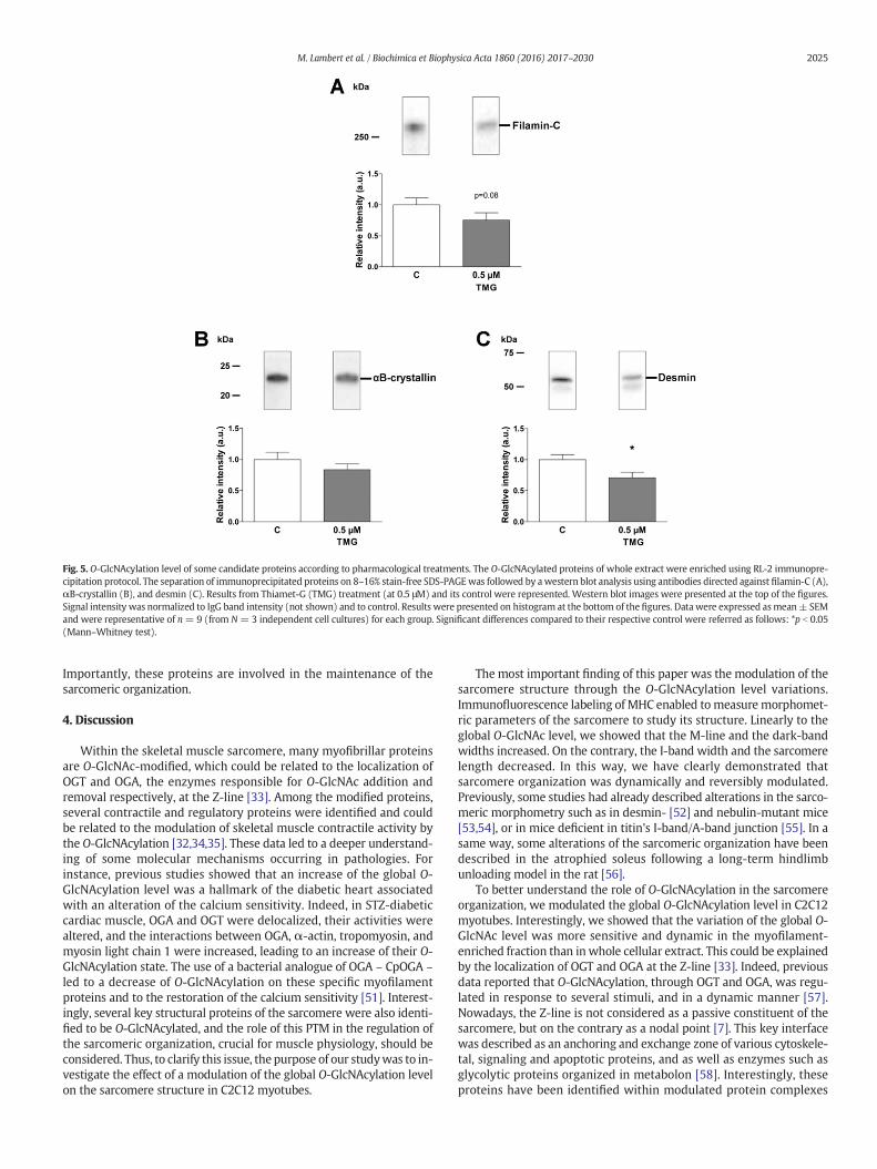
Fig. 5. O-GlcNAcylation level of some candidate proteins according to pharmacological treatments. The O-GlcNAcylated proteins of whole extract were enriched using RL-2 immunopre-cipitation protocol. The separation of immunoprecipitated proteins on 8–16% stain-free SDS-PAGEwas followed by awestern blot analysis using antibodies directed against filamin-C (A),αB-crystallin (B), and desmin (C). Results from Thiamet-G (TMG) treatment (at 0.5 μM) and its control were represented. Western blot images were presented at the top of the figures.Signal intensity was normalized to IgG band intensity (not shown) and to control. Results were presented on histogram at the bottom of the figures. Data were expressed asmean± SEMand were representative of n= 9 (from N= 3 independent cell cultures) for each group. Significant differences compared to their respective control were referred as follows: *p b 0.05(Mann–Whitney test).
2025M. Lambert et al. / Biochimica et Biophysica Acta 1860 (2016) 2017–2030
Importantly, these proteins are involved in the maintenance of thesarcomeric organization.
4. Discussion
Within the skeletal muscle sarcomere, many myofibrillar proteinsare O-GlcNAc-modified, which could be related to the localization ofOGT and OGA, the enzymes responsible for O-GlcNAc addition andremoval respectively, at the Z-line [33]. Among the modified proteins,several contractile and regulatory proteins were identified and couldbe related to the modulation of skeletal muscle contractile activity bythe O-GlcNAcylation [32,34,35]. These data led to a deeper understand-ing of some molecular mechanisms occurring in pathologies. Forinstance, previous studies showed that an increase of the global O-GlcNAcylation level was a hallmark of the diabetic heart associatedwith an alteration of the calcium sensitivity. Indeed, in STZ-diabeticcardiac muscle, OGA and OGT were delocalized, their activities werealtered, and the interactions between OGA, α-actin, tropomyosin, andmyosin light chain 1 were increased, leading to an increase of their O-GlcNAcylation state. The use of a bacterial analogue of OGA – CpOGA –led to a decrease of O-GlcNAcylation on these specific myofilamentproteins and to the restoration of the calcium sensitivity [51]. Interest-ingly, several key structural proteins of the sarcomere were also identi-fied to be O-GlcNAcylated, and the role of this PTM in the regulation ofthe sarcomeric organization, crucial for muscle physiology, should beconsidered. Thus, to clarify this issue, the purpose of our studywas to in-vestigate the effect of a modulation of the global O-GlcNAcylation levelon the sarcomere structure in C2C12 myotubes.
The most important finding of this paper was the modulation of thesarcomere structure through the O-GlcNAcylation level variations.Immunofluorescence labeling of MHC enabled tomeasure morphomet-ric parameters of the sarcomere to study its structure. Linearly to theglobal O-GlcNAc level, we showed that the M-line and the dark-bandwidths increased. On the contrary, the I-band width and the sarcomerelength decreased. In this way, we have clearly demonstrated thatsarcomere organization was dynamically and reversibly modulated.Previously, some studies had already described alterations in the sarco-meric morphometry such as in desmin- [52] and nebulin-mutant mice[53,54], or in mice deficient in titin's I-band/A-band junction [55]. In asame way, some alterations of the sarcomeric organization have beendescribed in the atrophied soleus following a long-term hindlimbunloading model in the rat [56].
To better understand the role of O-GlcNAcylation in the sarcomereorganization, we modulated the global O-GlcNAcylation level in C2C12myotubes. Interestingly, we showed that the variation of the global O-GlcNAc level was more sensitive and dynamic in the myofilament-enriched fraction than in whole cellular extract. This could be explainedby the localization of OGT and OGA at the Z-line [33]. Indeed, previousdata reported that O-GlcNAcylation, through OGT and OGA, was regu-lated in response to several stimuli, and in a dynamic manner [57].Nowadays, the Z-line is not considered as a passive constituent of thesarcomere, but on the contrary as a nodal point [7]. This key interfacewas described as an anchoring and exchange zone of various cytoskele-tal, signaling and apoptotic proteins, and as well as enzymes such asglycolytic proteins organized in metabolon [58]. Interestingly, theseproteins have been identified within modulated protein complexes

Fig. 6. Protein interactions of some candidate proteins were modulated though global O-GlcNAcylation variations. Native proteins were separated on 5% Red-Native PAGE. Proteins werethen transferred and analyzed on desmin (A), αB-crystallin (B), α-actinin (C), moesin (D) and filamin-C (E) western blot. Quantification of signals from Thiamet-G (TMG) treatment (at0.5 μM)and its controlwere represented.Western blot images and profile lanes (in red)were presented at the left of thefigures; protein bands have been described andnumbered. Proteinexpression of the numbered band in boldwith an asterisk was quantified and normalized to control; results were presented on histogram at the right side of the figures. Then, proteins ofwhole cellular extracts were separated on 8–16% linear gradient stain-free SDS-PAGE, transferred and analyzed on desmin (F), αB-crystallin (G), α-actinin (H), moesin (I) and filamin-C(J) western blot. Western blot images were presented at the top of the figures. Respective signal intensity was normalized to total protein (stain-free signal described in Fig. 1), to controland to an internal standard; results were presented on histogram at the bottom of the figures. Data were expressed as mean ± SEM andwere representative of n=9 (from N=3 inde-pendent cell cultures) for each group. Significant differences compared to their respective control were referred as follows: *p b 0.05; **p b 0.01 (Mann–Whitney test).
2026 M. Lambert et al. / Biochimica et Biophysica Acta 1860 (2016) 2017–2030
belonging to the Z-line. Therefore, by the varied nature of itscomposition, the Z-line was often sensitive to a multitude of intra-and extracellular stimuli, and was constantly remodeled [59]. In thisway, the sarcomere could be quickly and locally modulated. Takentogether, these evidences could explain a fine modulation of the O-GlcNAcylation on the sarcomere, which could lead to a finemodulationof the sarcomere remodeling.
Since several structural myofilament proteins were O-GlcNAcmodified, and due to the modulation of the sarcomere organizationthrough O-GlcNAcylation variations, we assume that an increase of theglobal O-GlcNAcylation level induced by Thiamet-G could lead to amodulation of protein–protein interactions resulting to a potent physi-ological consequence on the sarcomeric organization. Using Red-Native
PAGE, we demonstrated that some protein complexes were modulatedconsecutively to the variation of the global O-GlcNAc level. A multitudeof proteins have been identified in these protein complexes includingmyofibrillar proteins. Among them, there were some structuralproteins, key regulators of the sarcomere structure that have beenidentified previously to be O-GlcNAc-modified. Moreover and exceptfor moesin, the total expression of these proteins didn't changefollowing treatments, but the O-GlcNAc level varied on some proteins,in particular desmin. Interestingly, desmin presented a decrease ofits O-GlcNAcylation level in Thiamet-G treated myotubes. We alsoobserved variations in the amount of these proteins in some of the pro-teins complexes. Thus, we concluded that protein–protein interactionschanged, reflected by the modulation of these protein complexes

Fig. 7.Protein interactions between desmin andαB-crystallinweremodulated though globalO-GlcNAcylation variations. Co-immunoprecipitation (Co-IP) ofαB-crystallin (A) and desmin(B) were performed, followed by electrophoretic separation on stain-free SDS-PAGE, and immunodetection of αB-crystallin and desmin using western blot for each co-IP. Results fromThiamet-G (TMG) treatment (at 0.5 μM) and its control were represented. Western blot images were presented at the top of the figures. (A) Desmin signal intensity was normalizedto αB-crystallin signal and control. (B) αB-crystallin signal intensity was normalized to desmin signal and control. Results were presented on histogram at the bottom of the figures.Datawere expressed asmean±SEMandwere representative of n=12(fromN=3 independent cell cultures) for each group. Significant differences compared to their respective controlwere referred as follows: *p b 0.05 (Mann–Whitney test).
2027M. Lambert et al. / Biochimica et Biophysica Acta 1860 (2016) 2017–2030
observed in native-PAGE, which strongly supported that O-GlcNAcylation modulated the structural interactome. A simplifiedscheme of the interactome of the present identified “candidate”proteins, that could explain the modulation of the sarcomeric structurefollowing O-GlcNAc variations, was presented Fig. 8. It is worth to notethat among the “candidate” structural proteins, almost all being O-GlcNAcylated, belonging to an intricate network involved in thestructure and the organization of the sarcomere. Interestingly, a num-ber of them are located to the Z-line, a nodal point for the sarcomereorganization stability.
One of the identified proteins in complexes wasα-actinin. Based onourwestern blot from RN-PAGE, someα-actinin interactions seemed tobe altered. This protein is fundamental for sarcomere organizationbecause it is the major component of the nodal Z-line [60]. By theway, α-actinin was described as the major multivalent platform for anumber of protein–protein interactions with cytoskeletal and regulato-ry proteins. Indeed, it bound sarcomeric actin to stabilize the contractileapparatus and connected it to diverse signaling pathways [61]. Interest-ingly, moesin operated in both mechanisms. Indeed, this cytoskeletonprotein changed it conformation to associate the sarcomere to the sar-colemma. For example, after phosphorylation on threonine 558, theconformation of moesin changed, leading to its binding to F-actin toactivate other binding partners [62]. Moreover, abnormalmoesin, local-izedmainly to themembrane, increased actin polymerization leading tocardiopathy, and altered RhoA/ROCK pathway leading to diabetescomplications [63].
We also identified desmin as a component of modulated proteincomplexes. Mainly located around the Z-line and at the M-line, thisintermediate filament protein is essential for the structural integrity ofthe sarcomere [64]. Desmin anchored myofibril to the sarcolemmaand other structural elements of the cell, but also integrated manyprotein complexes within the contractile apparatus and finely main-tained its organization [65]. Moreover, desmin structure was highlydynamic [66]. In this present study, we showed that desmin wasmodulated in some protein complexes according to the global level ofO-GlcNAcylation. This data could be supported by twohypotheses. First-ly, several recent studies described desmin as a substrate of awide spec-trum of post-translational modifications (for review [67,68]). Its degreeof polymerization was dependent on its state of phosphorylation [11,
40], but could also depend on its level of O-GlcNAcylation since it wasdemonstrated that polymerization of cytokeratins 8 and 18 [69] ortubulin [70] was modified through O-GlcNAcylation. Secondly, desminintegrity was also regulated by numerous heat shock proteins.Phosphorylation of HSP25 at ser15 seemed to change the localizationof desmin at the Z-line and its colocalization with alpha-actinin. Inter-estingly, this relocalization was correlated with the formation of disor-ganized Z-lines [71]. Then, interactions between αB-crystallin, anotherheat shock protein, and desmin filaments played a crucial role on itsaggregation and localization [50]. To date, there are no available dataon O-GlcNAcylation affecting such interactions although desminpossessed lectin domains [42] and its interactome was highly complex[72]. However, some works showed O-GlcNAcylation [73] of αB-crystallin played a crucial role on its translocation and stabilization tocytoskeletal elements. Interestingly, from our native western blot aswell as co-IP/WB experiments, we showed herein that interactionsbetween desmin and αB-crystallin were modulated consecutively toglobal O-GlcNAcylation changes in C2C12 myotubes. However, theeffects of αB-crystallin toward desmin filaments depending on the O-GlcNAcylation remained to be elucidated. Indeed, changes to anyinteracting region ofαB-crystallin can produce positive as well as nega-tive effects on desmin [74]. According to the interaction domain of theαB-crystallin, its binding with desmin, the self-assembly and the fila-ment–filament interactions of the desmin were incredibly modified[74], and could explain more precisely some effects on the sarcomerestructure.
To conclude, our study focused on a modulation of the global O-GlcNAcylation level in particular on myofilament proteins in C2C12myotubes. These variations affected the sarcomere structure and someprotein complexes including structural proteins that regulated thesarcomere organization. Finally, some interactions of these proteinsseemed to be altered and emphasized the role of O-GlcNAcylation inthe dynamic sarcomere remodeling. Interestingly, key structuralproteins such as desmin, αB-crystallin, and filamin-C, belong tostructural complexes modulated by O-GlcNAcylation, their locationwithin protein complexes being themselves modified. Importantly,these proteins are affected by mutations which are directly involvedin myofibrillar myopathies [75], characterized by a dramatic sarcomeredisorganization. In the same way, α-actinin mutations are often

Fig. 8. Simplified interactome of “candidate proteins” that could explain the modulation of the sarcomere structure following O-GlcNAcylation variations in C2C12 myotubes. The local-ization of “candidate proteins” was indicated, and main roles of these proteins were provided below the scheme.
2028 M. Lambert et al. / Biochimica et Biophysica Acta 1860 (2016) 2017–2030
associatedwithmyopathies, showing the importance of the nodal Z-line[76]. Finally, moesin modifications could associate muscular diseasesand diabetes complications [63].
Previously, O-GlcNAcylation was described to be a regulator ofmyocardial contractile function [77] and a modulator of contractileactivity in striated muscle [37]. Number of studies (for review [32])had identified some O-GlcNAcylation sites in contractile/regulatorymyofilament proteins leading to new insights in the understanding ofthe calcium sensitivity/affinity alterations following O-GlcNAc varia-tions in sarcomere. We demonstrated herein that O-GlcNAcylationcould be also involved in the structural organization of the sarcomerein skeletal muscle. Altogether, this paper clearly argues in favor of thekey role of O-GlcNAcylation in the sarcomeric organization in skeletalmuscle, through protein localization within protein complexes, and/orprotein–protein interactions through the O-GlcNAc level variations. Inorder to better understand O-GlcNAcylation as a key modulator of thesarcomeric organization, future investigations must identify O-GlcNAcsites in structural myofilament proteins. Finally, it would be essential
in the future to consider O-GlcNAcylation as an important actor ofsarcomeric remodeling resulting from several disorders such as diabe-tes, myopathies and cardiomyopathies.
Conflict of interest
The authors declare that they have no conflict of interest.
Author contributions
Matthias Lambert: Performed the experiments, analyzed thedata and wrote the paper; Elodie Richard, Sophie Duban-Deweer,Frederic Krzewinski: contributed to acquisition and analysis of thedata; Barbara Deracinois and Erwan Dupont: Performed the experi-ments; Bruno Bastide: conceived the experiments, and wrote thepaper; Caroline Cieniewski-Bernard: conceived and designed the exper-iments, performed the experiments, analyzed the data, and wrote thepaper.

2029M. Lambert et al. / Biochimica et Biophysica Acta 1860 (2016) 2017–2030
Supplementary data to this article can be found online at http://dx.doi.org/10.1016/j.bbagen.2016.06.011.
Transparency document
The Transparency document associated with this article can befound, in the online version.
Acknowledgments
We thank Johan Hachani (Plateforme de spectrométrie de masseet d'analyse de l'Artois) for his help with the mass spectrometryanalysis.
This work was supported by grant from the Région Nord-Pas-de-Calais 2011 (Emergent Research Project, n° 12003803) and the Cen-tre National d'Etudes Spatiales (N° 4800000784). Matthias Lambertis a recipient from the French Ministry for Research and TertiaryEducation.
References
[1] M. Gautel, The sarcomeric cytoskeleton: who picks up the strain? Curr. Opin. CellBiol. 23 (2011) 39–46, http://dx.doi.org/10.1016/j.ceb.2010.12.001.
[2] J.W. Sanger, J. Wang, Y. Fan, J. White, J.M. Sanger, Assembly and dynamics ofmyofibrils, J. Biomed. Biotechnol. 2010 (2010) http://dx.doi.org/10.1155/2010/858606.
[3] O. Boonyarom, K. Inui, Atrophy and hypertrophy of skeletal muscles: structural andfunctional aspects, Acta Physiol. 188 (2006) 77–89, http://dx.doi.org/10.1111/j.1748-1716.2006.01613.x.
[4] K.a. Clark, A.S. McElhinny, M.C. Beckerle, C.C. Gregorio, Striated musclecytoarchitecture: an intricate web of form and function, Annu. Rev. Cell Dev. Biol.18 (2002) 637–706, http://dx.doi.org/10.1146/annurev.cellbio.18.012502.105840.
[5] Y. Au, Themuscle ultrastructure: a structural perspective of the sarcomere, Cell. Mol.Life Sci. 61 (2004) 3016–3033, http://dx.doi.org/10.1007/s00018-004-4282-x.
[6] S. Lange, E. Ehler, M. Gautel, From a to Z and back? Multicompartment proteins inthe sarcomere, Trends Cell Biol. 16 (2006) 11–18, http://dx.doi.org/10.1016/j.tcb.2005.11.007.
[7] D. Frank, C. Kuhn, H.a. Katus, N. Frey, The sarcomeric Z-disc: a nodal point in signal-ling and disease, J. Mol. Med. 84 (2006) 446–468, http://dx.doi.org/10.1007/s00109-005-0033-1.
[8] W.M. Obermann, M. Gautel, K. Weber, D.O. Fürst, Molecular structure of the sarco-meric M band: mapping of titin and myosin binding domains in myomesin andthe identification of a potential regulatory phosphorylation site in myomesin,EMBO J. 16 (1997) 211–220, http://dx.doi.org/10.1093/emboj/16.2.211.
[9] T. Sadikot, C.R. Hammond, M.B. Ferrari, Distinct roles for telethonin N-versus C-terminus in sarcomere assembly and maintenance, Dev. Dyn. 239 (2010)1124–1135, http://dx.doi.org/10.1002/dvdy.22263.
[10] X. Huang, J. Li, D. Foster, S.L. Lemanski, D.K. Dube, C. Zhang, L.F. Lemanski, Proteinkinase C-mediated desmin phosphorylation is related to myofibril disarray incardiomyopathic hamster heart, Exp. Biol. Med. (Maywood) 227 (2002) 1039–1046.
[11] R.K. Sihag, M. Inagaki, T. Yamaguchi, T.B. Shea, H.C. Pant, Role of phosphorylation onthe structural dynamics and function of types III and IV intermediate filaments, Exp.Cell Res. 313 (2007) 2098–2109, http://dx.doi.org/10.1016/j.yexcr.2007.04.010.
[12] Q. Zeidan, G.W. Hart, The intersections between O-GlcNAcylation and phosphoryla-tion: implications for multiple signaling pathways, J. Cell Sci. 123 (2010) 13–22,http://dx.doi.org/10.1242/jcs.053678.
[13] S. Mishra, S.R. Ande, N.W. Salter, O-GlcNAc modification: why so intimately associ-ated with phosphorylation? Cell Commun. Signal 9 (2011) 1, http://dx.doi.org/10.1186/1478-811X-9-1.
[14] G.W. Hart, C. Slawson, G. Ramirez-Correa, O. Lagerlof, Cross talk between O-GlcNAcylation and phosphorylation: roles in signaling, transcription, and chronicdisease, Annu. Rev. Biochem. 80 (2011) 825–858, http://dx.doi.org/10.1146/annurev-biochem-060608-102511.
[15] C.R. Torres, G.W. Hart, Topography and polypeptide distribution of terminal N-acetylglucosamine residues on the surfaces of intact lymphocytes. Evidence for O-linked GlcNAc, J. Biol. Chem. 259 (1984) 3308–3317.
[16] G. Hart, M. Housley, C. Slawson, Cycling of O-linked β-N-acetylglucosamine onnucleocytoplasmic proteins, Nature 446 (2007) 1017–1022, http://dx.doi.org/10.1038/nature05815.
[17] Y. Gu, S.R. Ande, S. Mishra, Altered O-GlcNAc modification and phosphorylation ofmitochondrial proteins in myoblast cells exposed to high glucose, Arch. Biochem.Biophys. 505 (2011) 98–104, http://dx.doi.org/10.1016/j.abb.2010.09.024.
[18] R.S. Haltiwanger, G.D. Holt, G.W. Hart, Enzymatic addition of O-GlcNAc to nuclearand cytoplasmic proteins, J. Biol. Chem. 265 (1990) 2563–2568.
[19] J.a. Hanover, M.W. Krause, D.C. Love, The hexosamine signaling pathway: O-GlcNAccycling in feast or famine, Biochim. Biophys. Acta 1800 (2010) 80–95, http://dx.doi.org/10.1016/j.bbagen.2009.07.017.
[20] D.L. Dong, G.W. Hart, Purification and characterization of an O-GlcNAc selective N-acetyl-beta-D-glucosaminidase from rat spleen cytosol, J. Biol. Chem. 269 (1994)19321–19330.
[21] Y. Akimoto, Y. Miura, T. Toda, M.a. Wolfert, L. Wells, G.-J. Boons, G.W. Hart, T. Endo,H. Kawakami, Morphological changes in diabetic kidney are associated with in-creased O-GlcNAcylation of cytoskeletal proteins including α-actinin 4, Clin. Proteo-mics 8 (2011) 15, http://dx.doi.org/10.1186/1559-0275-8-15.
[22] C. Slawson, G.W. Hart, O-GlcNAc signalling : implications for cancer cell biology, 11(2012) 678–684, http://dx.doi.org/10.1038/nrc3114.O-GlcNAc.
[23] Y. Fardini, V. Dehennaut, T. Lefebvre, T. Issad, O-GlcNAcylation: a new cancer hall-mark? Front. Endocrinol. (Lausanne) 4 (2013) 1–14, http://dx.doi.org/10.3389/fendo.2013.00099.
[24] B. Lazarus, D. Love, J. Hanover, O-GlcNAc cycling: implications for neurodegenera-tive disorders, J. Biochem. Cell Biol. 41 (2009) 2134–2146, http://dx.doi.org/10.1016/j.biocel.2009.03.008.O-GlcNAc.
[25] S. Förster, A.S. Welleford, J.C. Triplett, R. Sultana, B. Schmitz, D.A. Butterfield,Increased O-GlcNAc levels correlate with decreased O-GlcNAcase levels inAlzheimer disease brain, Biochim. Biophys. Acta Mol. basis Dis. 1842 (2014)1333–1339, http://dx.doi.org/10.1016/j.bbadis.2014.05.014.
[26] G.a. Ngoh, H.T. Facundo, A. Zafir, S.P. Jones, O-GlcNAc signaling in the cardiovascularsystem, Circ. Res. 107 (2010) 171–185, http://dx.doi.org/10.1161/CIRCRESAHA.110.224675.
[27] S. Dassanayaka, S.P. Jones, O-GlcNAc and the cardiovascular system, Pharmacol.Ther. 142 (2014) 62–71, http://dx.doi.org/10.1016/j.pharmthera.2013.11.005.
[28] C. Cieniewski-Bernard, Y. Mounier, J.-C. Michalski, B. Bastide, O-GlcNAc levelvariations are associated with the development of skeletal muscle atrophy, J. Appl.Physiol. 100 (2006) 1499–1505, http://dx.doi.org/10.1152/japplphysiol.00865.2005.
[29] P. Huang, S.-R. Ho, K.Wang, B.C. Roessler, F. Zhang, Y. Hu, D.B. Bowe, J.E. Kudlow, A.J.Paterson, Muscle-specific overexpression of NCOATGK, splice variant of O-GlcNAcase, induces skeletal muscle atrophy, Am. J. Physiol. Cell Physiol. 300(2011) C456–C465, http://dx.doi.org/10.1152/ajpcell.00124.2010.
[30] S. Nakamura, S. Nakano, M. Nishii, S. Kaneko, H. Kusaka, Localization of O-GlcNAc-modified proteins in neuromuscular diseases, Med. Mol. Morphol. 45 (2012)86–90, http://dx.doi.org/10.1007/s00795-011-0542-7.
[31] L. Stevens, B. Bastide, J. Hedou, C. Cieniewski-Bernard, V. Montel, L. Cochon, E.Dupont, Y. Mounier, Potential regulation of human muscle plasticity by MLC2post-translational modifications during bed rest and countermeasures, Arch.Biochem. Biophys. 540 (2013) 125–132, http://dx.doi.org/10.1016/j.abb.2013.10.016.
[32] C. Cieniewski-Bernard, M. Lambert, E. Dupont, V. Montel, L. Stevens, B. Bastide, O-GlcNAcylation, contractile protein modifications and calcium affinity in skeletalmuscle, Front. Physiol. 5 (2014) 1–7, http://dx.doi.org/10.3389/fphys.2014.00421.
[33] C. Cieniewski-Bernard, E. Dupont, E. Richard, B. Bastide, Phospho-GlcNAc modula-tion of slow MLC2 during soleus atrophy through a multienzymatic and sarcomericcomplex, Pflugers Arch. - Eur. J. Physiol. 466 (2014) 2139–2151, http://dx.doi.org/10.1007/s00424-014-1453-y.
[34] J. Hedou, C. Cieniewski-Bernard, Y. Leroy, J.-C. Michalski, Y. Mounier, B. Bastide, O-linked N-acetylglucosaminylation is involved in the Ca2+ activation properties ofrat skeletal muscle, J. Biol. Chem. 282 (2007) 10360–10369, http://dx.doi.org/10.1074/jbc.M606787200.
[35] C. Cieniewski-Bernard, V. Montel, S. Berthoin, B. Bastide, Increasing O-GlcNAcylationlevel on organ culture of soleus modulates the calcium activation parameters ofmuscle fibers, PLoS One 7 (2012), e48218 http://dx.doi.org/10.1371/journal.pone.0048218.
[36] C. Cieniewski-Bernard, B. Bastide, T. Lefebvre, J. Lemoine, Y. Mounier, J.-C. Michalski,Identification of O-linked N-acetylglucosamine proteins in rat skeletal muscle usingtwo-dimensional gel electrophoresis and mass spectrometry, Mol. Cell. Proteomics3 (2004) 577–585, http://dx.doi.org/10.1074/mcp.M400024-MCP200.
[37] C. Cieniewski-Bernard, V. Montel, L. Stevens, B. Bastide, O-GlcNAcylation, an originalmodulator of contractile activity in striated muscle, J. Muscle Res. Cell Motil. 30(2009) 281–287, http://dx.doi.org/10.1007/s10974-010-9201-1.
[38] M.C. Leung, P.G. Hitchen, D.G. Ward, A.E. Messer, S.B. Marston, Z-band alternativelyspliced PDZ motif protein (ZASP) is the major o-linked beta-N-acetylglucosamine-substituted protein in human heart myofibrils, J. Biol. Chem. 288 (2013)4891–4898, http://dx.doi.org/10.1074/jbc.M112.410316.
[39] J. Hédou, B. Bastide, A. Page, J.-C. Michalski,W. Morelle, Mapping ofO-linked beta-N-acetylglucosamine modification sites in key contractile proteins of rat skeletalmuscle, Proteomics 9 (2009) 2139–2148, http://dx.doi.org/10.1002/pmic.200800617.
[40] A.M. Farach, D.S. Galileo, O-GlcNAc modification of radial glial vimentin filaments inthe developing chick brain, Brain Cell Biol. 36 (2008) 191–202, http://dx.doi.org/10.1007/s11068-008-9036-5.
[41] C. Slawson, T. Lakshmanan, S. Knapp, G.W. Hart, A mitotic GlcNAcylation/phosphor-ylation signaling complex alters the posttranslational state of the cytoskeletalprotein vimentin, Mol. Biol. Cell 19 (2008) 4130–4140, http://dx.doi.org/10.1091/mbc.E07-11-1146.
[42] H. Ise, S. Kobayashi, M. Goto, T. Sato, M. Kawakubo, M. Takahashi, U. Ikeda, T. Akaike,Vimentin and desmin possess GlcNAc-binding lectin-like properties on cell sur-faces, Glycobiology 20 (2010) 843–864, http://dx.doi.org/10.1093/glycob/cwq039.
[43] N.E. Zachara, K. Vosseller, G.W. Hart, Detection and analysis of proteins modified byO-linked N-acetylglucosamine, Curr. Protoc. Protein Sci. (2011) http://dx.doi.org/10.1002/0471140864.ps1208s66 (Chapter 12, Unit12.8).
[44] S.A. Yuzwa, M.S. Macauley, J.E. Heinonen, X. Shan, R.J. Dennis, Y. He, G.E. Whitworth,K.A. Stubbs, E.J. McEachern, G.J. Davies, D.J. Vocadlo, A potent mechanism-inspiredO-GlcNAcase inhibitor that blocks phosphorylation of tau in vivo, Nat. Chem. Biol.4 (2008) 483–490, http://dx.doi.org/10.1038/nchembio.96.
2030 M. Lambert et al. / Biochimica et Biophysica Acta 1860 (2016) 2017–2030
[45] X. Yin, F. Cuello, U. Mayr, Z. Hao, M. Hornshaw, E. Ehler, M. Avkiran, M. Mayr,Proteomics analysis of the cardiac myofilament subproteome reveals dynamic alter-ations in phosphatase subunit distribution, Mol. Cell. Proteomics 9 (2010) 497–509,http://dx.doi.org/10.1074/mcp.M900275-MCP200.
[46] W.W.A. Schamel, Two-dimensional blue native polyacrylamide gel electropho-resis, Curr. Protoc. Cell Biol. (2008) http://dx.doi.org/10.1002/0471143030.cb0610s38 (Chapter 6, Unit 6.10).
[47] T. Dráb, J. Kračmerová, I. Tichá, E. Hanzlíková, M. Tichá, J. Liberda, Native polyacryl-amide electrophoresis in the presence of Ponceau Red to study oligomeric states ofprotein complexes, J. Sep. Sci. 34 (2011) 1692–1695, http://dx.doi.org/10.1002/jssc.201000869.
[48] S. Burattini, R. Ferri, M. Battistelli, R. Curci, F. Luchetti, E. Falcieri, C2C12 murinemyoblasts as a model of skeletal muscle development: morpho-functional charac-terization, Eur. J. Histochem. 48 (2004) 223–233, http://dx.doi.org/10.4081/891.
[49] H.S. Kim, E.M. Kim, J. Lee, W.H. Yang, T.Y. Park, Y.M. Kim, J.W. Cho, Heat shockprotein 60 modified with O-linked N-acetylglucosamine is involved in pancreaticβ-cell death under hyperglycemic conditions, FEBS Lett. 580 (2006) 2311–2316,http://dx.doi.org/10.1016/j.febslet.2006.03.043.
[50] J.L. Elliott, M. Der Perng, A.R. Prescott, K.a. Jansen, G.H. Koenderink, R.a. Quinlan, Thespecificity of the interaction between αB-crystallin and desmin filaments and itsimpact on filament aggregation and cell viability, Philos. Trans. R. Soc. Lond. Ser. BBiol. Sci. 368 (2013) 20120375, http://dx.doi.org/10.1098/rstb.2012.0375.
[51] G.A. Ramirez-Correa, J. MA, C. Slawson, Q. Zeidan, N.S. Lugo-Fagundo, M. Xu, X. Shen,W.D. Gao, V. Caceres, K. Chakir, A.M. Murphy, Removal of abnormal myofilament O-GlcNAcylation restores Ca2+ sensitivity in diabetic cardiac muscle, Diabetes 64(2015) 3573–3587, http://dx.doi.org/10.2337/db14-1107.
[52] R.M. Lovering, A. O'Neill, J.M. Muriel, B.L. Prosser, J. Strong, R.J. Bloch, Physiology,structure, and susceptibility to injury of skeletal muscle in mice lacking keratin19-based and desmin-based intermediate filaments, Am. J. Physiol. Cell Physiol.300 (2011) C803–C813, http://dx.doi.org/10.1152/ajpcell.00394.2010.
[53] C.C. Witt, C. Burkart, D. Labeit, M. McNabb, Y. Wu, H. Granzier, S. Labeit, Nebulinregulates thin filament length, contractility, and Z-disk structure in vivo, EMBO J.25 (2006) 3843–3855, http://dx.doi.org/10.1038/sj.emboj.7601242.
[54] P. Tonino, C.T. Pappas, B.D. Hudson, S. Labeit, C.C. Gregorio, H. Granzier, Reducedmyofibrillar connectivity and increased Z-disk width in nebulin-deficient skeletalmuscle, J. Cell Sci. 123 (2010) 384–391, http://dx.doi.org/10.1242/jcs.042234.
[55] H.L. Granzier, K.R. Hutchinson, P. Tonino, M. Methawasin, F.W. Li, R.E. Slater, M.M.Bull, C. Saripalli, C.T. Pappas, C.C. Gregorio, J.E. Smith, Deleting titin's I-band/A-band junction reveals critical roles for titin in biomechanical sensing and cardiacfunction, Proc. Natl. Acad. Sci. 111 (2014) 14589–14594, http://dx.doi.org/10.1073/pnas.1411493111.
[56] J. Udaka, S. Ohmori, T. Terui, I. Ohtsuki, S. Ishiwata, S. Kurihara, N. Fukuda, Disuse-induced preferential loss of the giant protein titin depresses muscle performancevia abnormal sarcomeric organization, J. Gen. Physiol. 131 (2008) 33–41, http://dx.doi.org/10.1085/jgp.200709888.
[57] C. Butkinaree, K. Park, G.W. Hart, O-linked β-N-acetylglucosamine (O-GlcNAc): ex-tensive crosstalk with phosphorylation to regulate signaling and transcription in re-sponse to nutrients and stress, Biochim. Biophys. Acta, Gen. Subj. 1800 (2010)96–106, http://dx.doi.org/10.1016/j.bbagen.2009.07.018.
[58] P. Mamczur, D. Rakus, A. Gizak, D. Dus, A. Dzugaj, The effect of calcium ions onsubcellular localization of aldolase-FBPase complex in skeletal muscle, FEBS Lett.579 (2005) 1607–1612, http://dx.doi.org/10.1016/j.febslet.2005.01.071.
[59] J.M. Sanger, J.W. Sanger, The dynamic Z bands of striated muscle cells, Sci. Signal. 1(2008) pe37, http://dx.doi.org/10.1126/scisignal.132pe37.
[60] B. Sjöblom, a. Salmazo, K. Djinović-Carugo, α-Actinin structure and regulation, Cell.Mol. Life Sci. 65 (2008) 2688–2701, http://dx.doi.org/10.1007/s00018-008-8080-8.
[61] E. de A. Ribeiro, N. Pinotsis, A. Ghisleni, A. Salmazo, P.V. Konarev, J. Kostan, B.Sjöblom, C. Schreiner, A.A. Polyansky, E.A. Gkougkoulia, M.R. Holt, F.L. Aachmann,B. Žagrović, E. Bordignon, K.F. Pirker, D.I. Svergun, M. Gautel, K. Djinović-Carugo,The structure and regulation of human muscle α-actinin, Cell 159 (2014)1447–1460, http://dx.doi.org/10.1016/j.cell.2014.10.056.
[62] L. Huang, T.Y.W. Wong, R.C.C. Lin, H. Furthmayr, Replacement of threonine 558, acritical site of phosphorylation of moesin in vivo, with aspartate activates F-actinbinding of moesin: regulation by conformational change, J. Biol. Chem. 274 (1999)12803–12810, http://dx.doi.org/10.1074/jbc.274.18.12803.
[63] Q. Huang, Ezrin/radixin/moesin proteins in the development of diabetes and its car-diovascular complications, J. Diabetes Metab. 01 (2012) http://dx.doi.org/10.4172/2155-6156.S4-005.
[64] D. Paulin, Z. Li, Desmin: a major intermediate filament protein essential for thestructural integrity and function of muscle, Exp. Cell Res. 301 (2004) 1–7, http://dx.doi.org/10.1016/j.yexcr.2004.08.004.
[65] Y. Capetanaki, R.J. Bloch, A. Kouloumenta, M. Mavroidis, S. Psarras, Muscle interme-diate filaments and their links to membranes and membranous organelles, Exp. CellRes. 313 (2007) 2063–2076, http://dx.doi.org/10.1016/j.yexcr.2007.03.033.
[66] H. Herrmann, U. Aebi, Intermediate filaments and their associates: multi-talentedstructural elements specifying cytoarchitecture and cytodynamics, Curr. Opin. CellBiol. 12 (2000) 79–90, http://dx.doi.org/10.1016/S0955-0674(99)00060-5.
[67] D.L. Winter, D. Paulin, M. Mericskay, Z. Li, Posttranslational modifications of desminand their implication in biological processes and pathologies, Histochem. Cell Biol.141 (2014) 1–16, http://dx.doi.org/10.1007/s00418-013-1148-z.
[68] N.T. Snider, M.B. Omary, Postranslational modifications of intermediate filamentproteins: mechanisms and functions, Nat. Rev. Mol. Cell Biol. 15 (3) (2014)163–177, http://dx.doi.org/10.1038/nrm3753.
[69] B. Srikanth, M.M. Vaidya, R.D. Kalraiya, O-GlcNAcylation determines the solubility,filament organization, and stability of keratins 8 and 18, J. Biol. Chem. 285 (2010)34062–34071, http://dx.doi.org/10.1074/jbc.M109.098996.
[70] S. Ji, J.G. Kang, S.Y. Park, J. Lee, Y.J. Oh, J.W. Cho, O-GlcNAcylation of tubulin inhibitsits polymerization, Amino Acids 40 (2011) 809–818, http://dx.doi.org/10.1007/s00726-010-0698-9.
[71] F. Kawano, R. Fujita, N. Nakai, M. Terada, T. Ohira, Y. Ohira, HSP25 can modulatemyofibrillar desmin cytoskeleton following the phosphorylation at Ser15 in rat so-leus muscle, J. Appl. Physiol. 112 (2012) 176–186, http://dx.doi.org/10.1152/japplphysiol.00783.2011.
[72] K. Hnia, C. Ramspacher, J. Vermot, J. Laporte, Desmin in muscle and associateddiseases: beyond the structural function, Cell Tissue Res. 49 (2014) http://dx.doi.org/10.1007/s00441-014-2016-4.
[73] V. Krishnamoorthy, A.J. Donofrio, J.L. Martin, O-GlcNAcylation of alphab-crystallinregulates its stress-induced translocation and cytoprotection, Mol. Cell. Biochem.379 (2013) 59–68, http://dx.doi.org/10.1007/s11010-013-1627-5.
[74] S.a. Houck, A. Landsbury, J.I. Clark, R.a. Quinlan, Multiple sites in alphaB-crystallinmodulate its interactions with desmin filaments assembled in vitro, PLoS One 6(2011) http://dx.doi.org/10.1371/journal.pone.0025859.
[75] D. Selcen, Myofibrillar myopathies, Neuromuscul. Disord. 21 (2011) 161–171,http://dx.doi.org/10.1016/j.nmd.2010.12.007.
[76] R. Knöll, B. Buyandelger, M. Lab, The sarcomeric Z-disc and Z-discopathies, J.Biomed. Biotechnol. 2011 (2011) 569628, http://dx.doi.org/10.1155/2011/569628.
[77] G.A. Ramirez-Correa, W. Jin, Z. Wang, X. Zhong, W.D. Gao, W.B. Dias, C. Vecoli, G.W.Hart, A.M. Murphy, O-linked GlcNAc modification of cardiac myofilament proteins:a novel regulator of myocardial contractile function, Circ. Res. 103 (2008)1354–1358, http://dx.doi.org/10.1161/CIRCRESAHA.108.184978.
![Certification Plateforme - tudiant.pptx [Lecture seule]) Plateforme... · Connexion à la plateforme Microsoft IT ... CCNA Composite# 200-120. ... 50% de réduction sur certains examens](https://img.pdfslide.us/doc/110x75/5b02656f7f8b9af1148f9bfa/certification-plateforme-lecture-seule-plateformeconnexion-la-plateforme.jpg)