Embed Size (px)
Citation preview
Biochimica et Biophysica Acta 1843 (2014) 47–60
Contents lists available at ScienceDirect
Biochimica et Biophysica Acta
j ourna l homepage: www.e lsev ie r .com/ locate /bbamcr
Review
RING-type E3 ligases: Master manipulators of E2 ubiquitin-conjugatingenzymes and ubiquitination☆
Meredith B. Metzger a,1, Jonathan N. Pruneda b,1, Rachel E. Klevit b,⁎, Allan M. Weissman a,⁎⁎a Laboratory of Protein Dynamics and Signaling, Center for Cancer Research, National Cancer Institute, 1050 Boyles Street, Frederick, MD 21702, USAb Department of Biochemistry, Box 357350, University of Washington, Seattle, WA 98195, USA
☆ This article is part of a Special Issue entitled: UbiquiEditors: Thomas Sommer and Dieter H. Wolf.⁎ Corresponding author. Tel.: +1 206 543 5891.** Corresponding author. Tel.: +1 301 846 1356.
E-mail addresses: [email protected] (R.E. Klevit), weiss(A.M. Weissman).
1 These authors contributed equally to this work.
0167-4889/$ – see front matter. Published by Elsevier Bhttp://dx.doi.org/10.1016/j.bbamcr.2013.05.026
a b s t r a c t
a r t i c l e i n f oArticle history:Received 5 March 2013Received in revised form 23 May 2013Accepted 29 May 2013Available online 6 June 2013
Keywords:RING fingerU-boxUbiquitin ligase (E3)Ubiquitin-conjugating enzyme (E2)Protein degradationCatalysis
RING finger domain and RING finger-like ubiquitin ligases (E3s), such as U-box proteins, constitute the vastmajority of known E3s. RING-type E3s function together with ubiquitin-conjugating enzymes (E2s) to medi-ate ubiquitination and are implicated in numerous cellular processes. In part because of their importance inhuman physiology and disease, these proteins and their cellular functions represent an intense area of study.Here we review recent advances in RING-type E3 recognition of substrates, their cellular regulation, and theirvaried architecture. Additionally, recent structural insights into RING-type E3 function, with a focus on im-portant interactions with E2s and ubiquitin, are reviewed. This article is part of a Special Issue entitled:Ubiquitin–Proteasome System. Guest Editors: Thomas Sommer and Dieter H. Wolf.
Published by Elsevier B.V.
1. Introduction
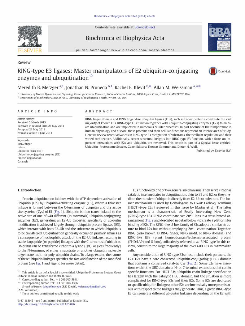
Protein ubiquitination initiates with the ATP-dependent activation ofubiquitin (Ub) by ubiquitin-activating enzyme (E1), where a thioesterlinkage is formed between the C-terminus of ubiquitin and the activesite cysteine (Cys) of E1 (Fig. 1). Ubiquitin is then transthiolated to theactive site of one of ~40 different (in mammals) ubiquitin-conjugatingenzymes (E2), generating an E2~Ub thioester. Specificity of ubiquitinmodification is achieved largely through ubiquitin protein ligases (E3),which interact with both E2~Ub and the substrate to which ubiquitin isto be transferred. Ubiquitination generally occurs on primary amines asa consequence of nucleophilic attack on the E2~Ub linkage, resulting instable isopeptide (or peptide) linkages with the C-terminus of ubiquitin.Ubiquitin can be transferred either to a lysine (Lys), or (less frequently)to the N-terminus, of either a substrate or another ubiquitin moleculeto generate multi- or poly-ubiquitin chains. To a large extent, the natureof these ubiquitin linkages specifies the fate and function of themodifiedprotein (see Fig. 1 and legend for more detail).
tin–Proteasome System. Guest
.V.
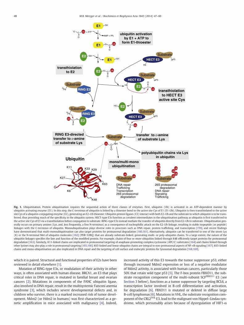
E3s function by one of two generalmechanisms. They serve either ascatalytic intermediates in ubiquitination, akin to E1 and E2, or they me-diate the transfer of ubiquitin directly from E2~Ub to substrate. The for-mer mechanism is used by Homologous to E6-AP Carboxy Terminus(HECT)-type E3s (reviewed in this issue by Martin et al). The lattermechanism is a characteristic of Really Interesting New Gene(RING)-type E3s. RINGs coordinate two Zn2+ ions in a cross-braced ar-rangement (Fig. 2 and described in detail below) to create a platform forbinding of E2s. The RING-like U-box family of E3s adopts a similar struc-ture to bind E2s but without employing Zn2+ coordination. Together,RING (also known as RING finger, RING motif, or RING domain) andRING-like E3s (plant homeodomain/leukemia-associated protein(PHD/LAP) and U-box), collectively referred to as ‘RING-type’ in this re-view, constitute the large majority of the over 600 E3s in mammaliancells.
Any consideration of RING-type E3smust include their partners, theE2s. E2s have a core conserved ubiquitin-conjugating (UBC) domainthat contains a conserved catalytic Cys (Fig. 2). Some E2s have inser-tions within the UBC domain or N- or C-terminal extensions that conferspecific functions. For HECT E3s, ubiquitin chain linkage specificationlies largely with the catalytic HECT domain, but the situation is morecomplicated for RING-type E3s and their E2s. Some E2s are dedicatedto specific ubiquitin linkages; other E2s are intrinsicallymore promiscu-ous with respect to the linkages they generate. Thus, a given RING-typeE3 can generate different ubiquitin linkages depending on the E2 with
E1
Ub + ATP
Substrate
RING E3
HECT E3
AMP + PPi
E1 S C
O
Ub
E2E1
S C
O
Ub
Ub
H2N
SH
SHSH
C
O
-O
E2S
C
O
Ub
C
O
Ub
NH2
HECT E3
C
O
Ub
NH2
HS
S
Substrate
Substrate
Ubiquitin
HECT E3 NH2
HS
Substrate
Substrate
RING E3H2N
Substrate
NH
Substrate
C
K48
K63
K29K33K11
K27
K6
ubiquitin activation by E1 + ATP to
form E1-thioester
RING E3-directed transfer to ε-amine
of substrate Lys
transthiolation to E2
transthiolation to HECT E3
active site Cys
N Ub
UbUb
Ub
Ub
UbUbUb
HS
polyubiquitin chains via Lys in ubiquitin
transfer to ε-amine of substrate Lys
SC
O
SubstrateUb
DNA repair Trafficking Transcription 26S proteasomal degradation
26S proteasomal degradationDNA repairSignalingTrafficking
E2
E2
E2
E2
mono/multi-monoubiquitination
Ub
Fig. 1. Ubiquitination. Protein ubiquitination requires the sequential action of three classes of enzymes. First, ubiquitin (Ub) is activated in an ATP-dependent manner byubiquitin-activating enzyme (E1). In this step, the C-terminus of ubiquitin is linked by a thioester bond to the active site Cys of E1 (E1~Ub). Ubiquitin is then transthiolated to the activesite Cys of a ubiquitin-conjugating enzyme (E2), generating an E2~Ub thioester. Ubiquitin protein ligases (E3) interactwith both E2~Ub and the substrate towhich ubiquitin is to be trans-ferred, thus providing much of the specificity in the ubiquitin system. HECT-type E3s function as covalent intermediates in the ubiquitination pathway as ubiquitin is first transferred tothe active site Cys of E3 via a transthiolation before conjugation to substrate. RING-type E3s insteadmediate the transfer of ubiquitin directly fromE2~Ub to substrate. Ubiquitination gen-erally occurs on primary amines (Lys, and, less frequently, a free N-terminus) as a consequence of nucleophillic attack on the E2~Ub linkage, resulting in stable isopeptide (or peptide)linkages with the C-terminus of ubiquitin. Monoubiquitination plays diverse roles in processes such as DNA repair, protein trafficking, and transcription [159], and recent findingshave demonstrated that multi-monoubiquitination can also target proteins for proteasomal degradation [160,161]. Alternatively, ubiquitin can be transferred to one of the seven Lys(K) or the N-terminal Met of ubiquitin molecules [162] (PDB 1UBQ) that are already substrate-linked, generating multi- or poly-ubiquitin chains. To a large extent, the nature of theubiquitin linkages specifies the fate and function of the modified protein. For example, chains of four or more ubiquitins linked through K48 efficiently target proteins for proteasomaldegradation [163]. Similarly, K11-linked chains are implicated in proteasomal targeting of anaphase-promoting complex/cyclosome (APC/C) substrates [164] and chains linked throughother lysinesmay also play a role in proteasomal targeting [165,166]. K63-linked and linear ubiquitin chains are integral to non-proteasomal aspects of NF-κB signaling [167]. K63-linkedchains and mono-ubiquitination are also implicated in DNA repair and the targeting of cell surface and endocytic proteins for lysosomal degradation [168,169].
48 M.B. Metzger et al. / Biochimica et Biophysica Acta 1843 (2014) 47–60
which it is paired. Structural and functional properties of E2s have beenreviewed in detail elsewhere [1].
Mutation of RING-type E3s, or modulation of their activity in otherways, is often associated with human disease. BRCA1, an E3 that playscritical roles in DNA repair, is mutated in familial breast and ovariancancers [2]. Mutations in components of the FANC ubiquitin ligase,also involved in DNA repair, result in themultisystemic Fanconi anemiasyndrome [3], which includes severe developmental defects and, inchildren who survive, there is a marked increased risk of tumor devel-opment. Mdm2 (or Hdm2 in humans) was first characterized as a ge-netic amplification in mice associated with malignancy [4]. Indeed,
increased activity of this E3 towards the tumor suppressor p53, eitherthrough increased Mdm2 expression or loss of a negative modulatorof Mdm2 activity, is associated with human cancers, particularly those50% that retain wild type p53 [5]. The F-box protein FBXO11, the sub-strate recognition component of the multi-subunit SCFFBXO11 E3 (seeSection 3 below), functions as a tumor suppressor by targeting BCL6, atranscription factor involved in B-cell differentiation and activation,for degradation [6]. FBXO11 is mutated or deleted in diffuse largeB-cell lymphomas [6]. Mutations in VHL, the substrate recognition com-ponent of the CRL2VHL E3, lead to themalignant vonHippel–Lindau syn-drome, which presumably arises because of dysregulation of HIF1-α
A
B
C
Fig. 2. RING domains coordinate Zn2+ in a crossbrace arrangement that serves as a plat-form for E2 binding. A) Representation of the crystal structure of the TRAF6 RING domain(blue) bound to the E2, Ubc13 (green) [89] (PDB3HCT) highlights a stereotypical RING:E2interaction. The catalytic Cys of Ubc13 is highlighted in yellow, while its RINGdomain-interacting regions are in purple. Yellow TRAF6 RING residues with sidechainsshown are those that coordinate Zn2+ (C3HC3D), forming the RING crossbrace structuremodeled in B). The two loops (Zn I, Zn II) and the intervening central α-helix formed bythis structure together serve as a conserved platform for E2 binding. B, C)Model of the in-terleaved RING crossbrace structure (B) and consensus sequence (C). The eightZn2+-coordinating residues are shown in yellow and X is any amino acid.
49M.B. Metzger et al. / Biochimica et Biophysica Acta 1843 (2014) 47–60
and/or HIF2-α [5]. Mutations in the RING–IBR (in between RING)–RINGE3 Parkin are associated with autosomal recessive juvenile Parkinson-ism (AR-JP) [7]. Additionally, a number of viruses, for example, herpessimplex virus type 1 (HSV-1), encode RING-type E3s as virulence factors[8,9]. In the case of HIV, the virus encodes an adaptor protein, Vpu, thatredirects SCFβTrCP to downregulate CD4 [10]. The importance ofRING-type E3s to human health and disease has contributed to their be-coming an intensively-studied family of proteins. This review will pro-vide an overview of their regulated function and structure and recentadvances in understanding how they mediate ubiquitination by E2s.
2. RING dimerization
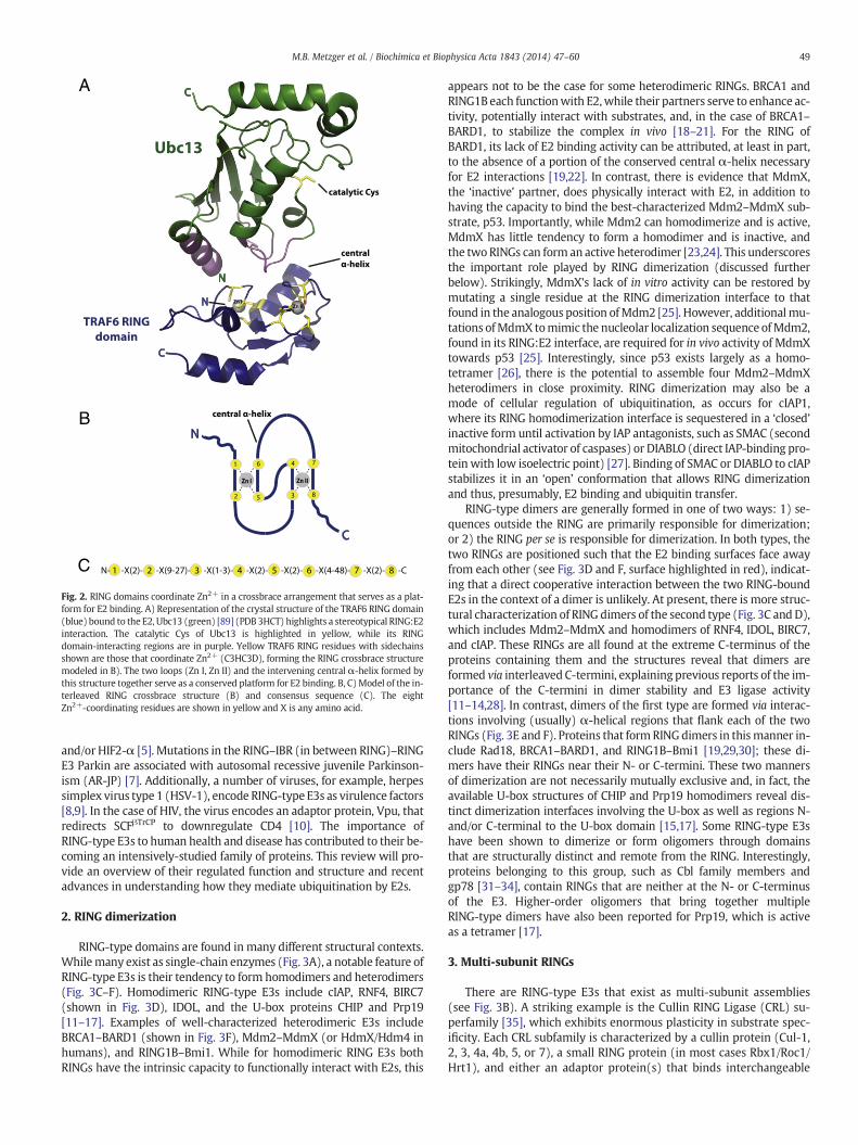
RING-type domains are found in many different structural contexts.Whilemany exist as single-chain enzymes (Fig. 3A), a notable feature ofRING-type E3s is their tendency to form homodimers and heterodimers(Fig. 3C–F). Homodimeric RING-type E3s include cIAP, RNF4, BIRC7(shown in Fig. 3D), IDOL, and the U-box proteins CHIP and Prp19[11–17]. Examples of well-characterized heterodimeric E3s includeBRCA1–BARD1 (shown in Fig. 3F), Mdm2–MdmX (or HdmX/Hdm4 inhumans), and RING1B–Bmi1. While for homodimeric RING E3s bothRINGs have the intrinsic capacity to functionally interact with E2s, this
appears not to be the case for some heterodimeric RINGs. BRCA1 andRING1B each functionwith E2,while their partners serve to enhance ac-tivity, potentially interact with substrates, and, in the case of BRCA1–BARD1, to stabilize the complex in vivo [18–21]. For the RING ofBARD1, its lack of E2 binding activity can be attributed, at least in part,to the absence of a portion of the conserved central α-helix necessaryfor E2 interactions [19,22]. In contrast, there is evidence that MdmX,the ‘inactive’ partner, does physically interact with E2, in addition tohaving the capacity to bind the best-characterized Mdm2–MdmX sub-strate, p53. Importantly, while Mdm2 can homodimerize and is active,MdmX has little tendency to form a homodimer and is inactive, andthe twoRINGs can form an active heterodimer [23,24]. This underscoresthe important role played by RING dimerization (discussed furtherbelow). Strikingly, MdmX's lack of in vitro activity can be restored bymutating a single residue at the RING dimerization interface to thatfound in the analogous position ofMdm2 [25]. However, additionalmu-tations ofMdmX tomimic the nucleolar localization sequence ofMdm2,found in its RING:E2 interface, are required for in vivo activity of MdmXtowards p53 [25]. Interestingly, since p53 exists largely as a homo-tetramer [26], there is the potential to assemble four Mdm2–MdmXheterodimers in close proximity. RING dimerization may also be amode of cellular regulation of ubiquitination, as occurs for cIAP1,where its RING homodimerization interface is sequestered in a ‘closed’inactive form until activation by IAP antagonists, such as SMAC (secondmitochondrial activator of caspases) or DIABLO (direct IAP-binding pro-tein with low isoelectric point) [27]. Binding of SMAC or DIABLO to cIAPstabilizes it in an ‘open’ conformation that allows RING dimerizationand thus, presumably, E2 binding and ubiquitin transfer.
RING-type dimers are generally formed in one of two ways: 1) se-quences outside the RING are primarily responsible for dimerization;or 2) the RING per se is responsible for dimerization. In both types, thetwo RINGs are positioned such that the E2 binding surfaces face awayfrom each other (see Fig. 3D and F, surface highlighted in red), indicat-ing that a direct cooperative interaction between the two RING-boundE2s in the context of a dimer is unlikely. At present, there is more struc-tural characterization of RING dimers of the second type (Fig. 3C andD),which includes Mdm2–MdmX and homodimers of RNF4, IDOL, BIRC7,and cIAP. These RINGs are all found at the extreme C-terminus of theproteins containing them and the structures reveal that dimers areformed via interleaved C-termini, explaining previous reports of the im-portance of the C-termini in dimer stability and E3 ligase activity[11–14,28]. In contrast, dimers of the first type are formed via interac-tions involving (usually) α-helical regions that flank each of the twoRINGs (Fig. 3E and F). Proteins that formRINGdimers in thismanner in-clude Rad18, BRCA1–BARD1, and RING1B–Bmi1 [19,29,30]; these di-mers have their RINGs near their N- or C-termini. These two mannersof dimerization are not necessarily mutually exclusive and, in fact, theavailable U-box structures of CHIP and Prp19 homodimers reveal dis-tinct dimerization interfaces involving the U-box as well as regions N-and/or C-terminal to the U-box domain [15,17]. Some RING-type E3shave been shown to dimerize or form oligomers through domainsthat are structurally distinct and remote from the RING. Interestingly,proteins belonging to this group, such as Cbl family members andgp78 [31–34], contain RINGs that are neither at the N- or C-terminusof the E3. Higher-order oligomers that bring together multipleRING-type dimers have also been reported for Prp19, which is activeas a tetramer [17].
3. Multi-subunit RINGs
There are RING-type E3s that exist as multi-subunit assemblies(see Fig. 3B). A striking example is the Cullin RING Ligase (CRL) su-perfamily [35], which exhibits enormous plasticity in substrate spec-ificity. Each CRL subfamily is characterized by a cullin protein (Cul-1,2, 3, 4a, 4b, 5, or 7), a small RING protein (in most cases Rbx1/Roc1/Hrt1), and either an adaptor protein(s) that binds interchangeable
RING
Skp
1
Cul1
F-box protein
A B
C C
C
C C
DN N
NN
C
N
Nedd8
Substrate Substrate
RING RING
RING RING
E
CC
N N
F
Rbx1
C
C
NN
Fig. 3. Architecture of RING-type E3s. A) Model of a monomeric RING-type E3, where its RING domain would mediate binding to E2 thioester-linked to ubiquitin. Binding to substrateoccurs generally through regions of the E3 other than the RING domain. B) Model of a multi-subunit RING E3 of the cullin RING ligase (CRL) superfamily, such as the well-studied SCF(CRL1) family, shown here. SCF consists of a cullin protein (Cul1) a small RING finger protein (Rbx1), and an adaptor protein (Skp1) that binds interchangeable substrate recognition el-ements (F-box proteins). The ubiquitin-like molecule, Nedd8, is reversibly conjugated to cullins and associated with activation of CRLs. C) Schematic of dimeric RING E3s, such as cIAP,RNF4, BIRC7, IDOL, Mdm2–MdmX, that dimerize through their RING domains and interleaved C-terminal tails. D) Ribbon diagram illustrating the homodimeric RING E3, BIRC7 [13](PDB 4AUQ) as a representative of the class of dimers schematized in C. The E2-interacting residues of one RING domain are highlighted in red. E) Model of dimeric RING E3s, such asBRCA1–BARD1 and RING1B–Bmi1, where α-helices both N- and C-terminal to the RING facilitate dimerization. In the case of BRCA1–BARD1 (illustrated), this occurs through a fourα-helix bundle (helices above RINGs in F). F) Ribbon diagram illustrating the heterodimeric RING dimer of BRCA1–BARD1 [19] (PDB 1JM7) modeled in E). The E2-interacting residuesof the RING domain of BRCA1 are highlighted in red.
50 M.B. Metzger et al. / Biochimica et Biophysica Acta 1843 (2014) 47–60
substrate recognition elements or, in the case of CRL3, proteins thatbind both to the cullin protein (Cul-3) and to substrate [36]. TheCRL superfamily is exemplified by the well-studied Skp1-Cul1-F-boxprotein (SCF) family (Fig. 3B), in which one of ~69 (in humans) inter-changeable F-box proteins can potentially recognize substrates [37](reviewed in this issue by Bassermann et al.). Exchange of F-box pro-teins within the SCF scaffold takes place through a complex cycle thatincludes dynamic attachment and removal of the ubiquitin-like mod-ifier, Nedd8 [38]. While the CRL superfamily overwhelmingly exhibitsthe greatest range of substrate recognition, other multi-subunit E3sexhibit even greater structural complexity. The anaphase promotingcomplex/cyclosome (APC/C) is a highly complex E3 that in humanscontains 13 core subunits including a cullin-like protein and a small
RING protein. It also has two interchangeable co-activator subunits,Cdc20 and Cdh1, which recognize distinct substrates and are activeduring different phases of the cell cycle [39] (reviewed in this issueby Bassermann et al.).
Another multi-subunit RING-containing ligase is the Fanconi anemia(FANC) E3. There are at least 13 complementation groups associatedwith this disease, and proteins corresponding to eight complementationgroups are components of the FANC ubiquitin ligase, including aRING-type protein (FancL). This E3 is recruited to sites of DNA damageto effect translesional repair. Despite its complexity, the role of theFANC E3, as we currently understand it, is limited tomonoubiquitinationof two associated proteins that are subsequently deubiquitinated as partof the DNA repair process. Degradation of a key FANC component,
51M.B. Metzger et al. / Biochimica et Biophysica Acta 1843 (2014) 47–60
FANCM, via SCFβTrCP is responsible for inactivating the function of theFANC E3 duringmitosis, thereby preventing chromosomal abnormalities[3,5,40].
Some multi-subunit RING-type E3s contain multiple RING proteins.The yeast GID (glucose-induced degradation deficient) complex,which targets fructose-1, 6 bisphosphatase for ubiquitination in re-sponse to glucose, consists of seven subunits including two interactingRINGs [41]. The yeast PEX ubiquitin ligase, which mono-ubiquitinatesthe peroxisome receptor, Pex5p, and possibly other substrates, includesthree distinct RING proteins as part of a multi-subunit complex [42,43].The specific function of each of the RINGs in such complexes is currentlyunknown.
Finally, there are single proteins that contain multiple RINGs.Mindbomb, involved in Notch signaling, has three RINGs in its C-terminal region, although to date only the most C-terminal of thesehas been studied and shown to be required for activity [44]. RING–IBR–RING (RBR) proteins are a class of ~13 proteins (in humans) that in-clude a RING consensus sequence (RING1) followed by a Cys-rich ‘in be-tween RING’ (IBR) region and a third domain, the RING2, that wasoriginally characterized as a second RING-like domain. Although RBRproteins were thought to function as canonical RING E3s, recent studieshave shown that they employ a RING–HECT hybrid mechanism[45–49]. The RING1 domain binds E2 (similar to the RING mechanism)but ubiquitin is transferred to a specific Cys within RING2 before beingtransferred to substrates (similar to the HECT mechanism). Well-known members of this family include Parkin, HHARI, HOIP, andHOIL-1L. The latter two are subunits of the Linear Ubiquitin Chain As-sembly Complex (LUBAC) E3 consisting of HOIP, HOIL-1L, and Sharpin(a non-RING-containing protein). This complex plays critical roles inNF-κB activation (reviewed in this issue by Kazuhiro et al).
4. RING-type E3s and their substrates
There is enormous diversity in substrate ubiquitination and its regu-lation, as the targets of RING-type E3s are incredibly varied. RING-typeE3s are implicated as tumor suppressors, oncogenes, and mediators ofendocytosis, and play critical roles in complex multi-step processessuch as DNA repair and activation of NF-κB signaling. A RING-type E3may have multiple substrates and several E3s can target the same sub-strate. Not surprisingly, the mechanisms of substrate recognition byRING-type E3s are highly varied, and occur in the context of networksof interactions that often also include HECT E3s and deubiquitinating en-zymes (DUBs). Substratesmay bind directly to a RING-type E3 ormay as-sociate indirectly. The capacity of RING-type E3s for self-ubiquitination,first utilized as a means of assessing their potential to function withE2s [50], frequently occurs in vivo, as does ubiquitination of RING E3sby heterologous RING or HECT-type E3s as part of regulatory networks[51].
E3-substrate interactions may be constitutive, and, in such cases,regulation can occur at the level of E3 transcription or degradation.The complexity of such trans-regulation is illustrated by the interplayof SCF and APC/C E3s during the cell cycle where, for example, APC/CCdh1 targets the F-boxprotein Skp2 for degradation in early G1, therebystabilizing p27 and preventing premature G1-S transition, and SCFβTrCP
targets the ‘pseudo-substrate’ and suppressor of APCCdc20, EMI1, fordegradation in late G2 [5] (reviewed in this issue by Bassermann etal.). Similarly, SCFFBXO11 ubiquitinates and targets Cdt2, the conservedsubstrate recognition subunit of CRL4Cdt2, for degradation, stabilizingits substrates, such as p21 and Set8, and allowing for cells to properlyexit the cell cycle [52,53].
An emerging theme is a role for metabolites in substrate recogni-tion and E3 activity. As above, the effect may be via a direct interac-tion between a metabolite and a RING-type E3, or may be indirect,for example via interaction with the substrate. In the latter category,sterols serve as feedback regulators of their own synthesis by regulat-ing the association of Insig-1 with the ER-resident RING E3 gp78 and
hence the stability of the former, which is critical to the regulation ofcholesterol biosynthesis [54]. The plant hormones auxin and jasmonicacid are examples of regulation by a direct metabolite:E3 interaction[55,56]. These hormones bind directly to SCF complexes and targettranscriptional repressors for ubiquitination and degradation. Thisstrategy provides a way to de-repress gene expression and alter thetranscriptional profile in response to environmental factors in plants.Another intriguing example of regulation by a metabolite is a reportthat the RING E3 TRAF-2 is inactive due to its RING structure beingunsuitable for E2 interactions, but is activated by its associationwith sphingosine-1-phosphate [57]. A structural understanding ofhow this occurs awaits further studies. Nevertheless, as nature rarelyuses a good idea just once, it seems likely that additional examples ofsmall molecule or metabolite activation (or inhibition) of E3s will beuncovered in the future.
The most common means of regulating substrate ubiquitination isby post-translational modifications that alter either ligase activity orsubstrate recognition by RING-type E3s. Examples of regulation via pro-tein phosphorylation are widespread. Regulated substrate phosphory-lation on either Ser or Thr residues allows for the recognition ofnumerous substrates by SCFβTrCP and SCFFbw7 [37]. Tyrosine phosphor-ylation of activated receptor tyrosine kinases (RTKs) facilitates theirrecognition by the Cbl family of RING E3s (Cbl, Cbl-b and Cbl-c) [58].Cbl-c phosphorylation also modulates the dynamic interaction of itsRING with E2~Ub (see below). The net result of activation of Cbl familymembers includes the ubiquitination of RTKs, leading to their lysosomaldegradation and an attenuation of signaling. Both phosphorylation ofthe core RING subunit and dephosphorylation of Cdc20 play roles inthe activation of the APC/CCdc20 [59]. The level of complexity that phos-phorylation offers is exemplified by p53 and Mdm2. Regulated phos-phorylation of specific residues on either p53 or Mdm2 can eitherinhibit or enhance their interaction. Also,Mdm2degradation as a conse-quence of self-ubiquitination is enhanced by phosphorylation, whichprevents interaction of Mdm2 with the DUB HAUSP/USP7. The failureof Mdm2 to be deubiquitinated by USP7 leads to degradation ofMdm2 and increased p53 activity under conditions of genotoxic stress[5] (reviewed in this issue by Vousden et al).
A growing number of other post-translational modifications areimplicated in regulation of the ubiquitin system, including substratemodification by hydroxylation, glycosylation, acetylation, methyla-tion, modification by poly(ADP-ribose) (PAR), and attachment ofubiquitin-like modifiers. The HIF1-α and HIF2-α transcription factorsundergo proline hydroxylation in response to increased oxygen levelsand become substrates for the multi-subunit RING E3, CRL2VHL. Thehydroxylation reaction is mediated by proline-hydroxylase domain(PHD) proteins, which are themselves targets for SIAH RING E3s.SIAH expression is positively regulated by HIF transcriptionally, creat-ing a positive feedback loop to increase HIF1-α levels [5]. A subfamilyof F-box proteins, denoted as Fbs1–5, has the potential to recognizeglycosylated proteins in the cytosol, which have presumably beentransported out of the endoplasmic reticulum (ER). This provides ameans to target these displaced glycoproteins for ubiquitination andproteasomal degradation [60].
Modification of proteins with the small ubiquitin-like modifier(SUMO) family of proteins can inhibit substrate degradation by com-peting for specific sites of ubiquitination or by altering the localizationof modified proteins. Perhaps a more general role for sumoylation isto target proteins for ubiquitination, particularly those modifiedwith chains of SUMO (in metazoans SUMO 2/3). This occurs bySUMO-targeted ubiquitin ligases (STUbLs), which contain multipleSUMO-interactingmotifs (SIMs) in addition to their E3 ligase domains[61].
The RING E3 RNF146 uses a WWE domain to recognize substratesmodified with poly(ADP-ribose) (PAR). In response to Wnt signaling,axin is PARsylated by tankyrase and subsequently ubiquitinated byRNF146, ultimately resulting in its proteasomal degradation. In vitro,
52 M.B. Metzger et al. / Biochimica et Biophysica Acta 1843 (2014) 47–60
PAR as been shown to stimulate the ligase activity of RNF146, thoughthe mechanism is presently unknown [62–64]. There are otherRING-type E3s that contain WWE domains, including the Deltex fam-ily, whose members play an important role in Notch signaling. Thus, itis likely that additional examples of regulation by PARsylation willemerge in the future.
There is a complex dynamic between ubiquitination, acetylation,and methylation of lysines as part of the histone code [65]. Addition-ally, there is data to suggest that specific lysines in p53 that are tar-gets of Mdm2–MdmX are also acetylated, thereby preventing theirubiquitination [66]. The acetylation of Mdm2 itself may decrease itsactivity towards p53 [67]. There is also evidence for interplay be-tween acetylation and ubiquitination of transcription factors such asestrogen receptor-α [68].
Finally, for E3s to target substrates theymust exist in the same cellu-lar compartment. Some RING-type E3s have nuclear localization signalsand many RING-type E3s are transmembrane proteins targeted to sitessuch as the ER, plasmamembrane, endosomes, peroxisomes, and mito-chondria [42,69–73].
5. RING-type domain structure
RING structure is conformed as a consequence of a cross-braced ar-rangement of eight Zn2+ coordinating residues, generally Cys and His,with conserved spacing between these residues (Fig. 2B and C). Canon-ical RINGs have either one or two His in the linear arrangement ofcoordinating residues, denoted C3H2C3 or C3HC4, however other varia-tions exist. The PHD/LAP finger found in the transcription factor NF-X1and theMARCH family of membrane-bound E3s is defined by its C4HC3consensus. RINGs having a C8 configuration (CNOT4) or an Asp residuein the final position (e.g. Rbx1 and TRAF6) have been shown to have li-gase activity [74–76]. Thus, it has become apparent that categorizingRINGs by the linear arrangement of coordinating residues has little todo with functional properties of the domain. Nevertheless, contextdoes matter, as swapping Zn2+ liganding residues in a C3H2C3 RINGto create a C3HC4 configuration resulted in loss of activity for AO7(RNF25), one of the first RING E3s studied [50]. NF-X1 contains a se-quence in which both a RING and a PHD/LAP motif are recognizable,but only the PHD/LAP consensus is functional [77]. Unlike RING domainE3s, U-box proteins do not coordinate Zn2+ but adopt a RING-like ter-tiary structure for binding E2, stabilized by non-covalent interactionsamong core amino acids [16]. Additionally, some pathogenic bacteriahave evolved proteins that show no sequence homology to eukaryoticRING or U-box domains, yet fold into highly similar structures and dis-play robust ubiquitin ligase activity [78,79].
Crystallographic and NMR-based analyses have revealed that RINGsandU-boxes have a commonmodeof interactionwith E2s (Fig. 2A). Thekey structural elements are two loop-like regions, which, in the case ofRINGs, coordinate Zn2+. The loops surround a shallow groove formedby the central α-helix. Together these elements serve as a platform forinteractions with the UBC domain of E2s (Fig. 2A). The E2 surface thatinteracts with the RING domain overlaps with the region that interactswith E1, leading to the notion that dissociation of E2s from RINGs is re-quired for an E2 to be ‘reloaded’with ubiquitin by E1 [80–82]. A charac-teristic of RING:E2 interactions is that they are generally of low affinity,typically with Kd values in the high micromolar range. Thus, eventhough a RING domain may function robustly with an E2, assessingphysical interactions between these proteins using standard ‘pulldown’approaches is often not fruitful. Exceptions to this feature include E3ssuch as gp78 [83], Rad18 [84], and AO7 [50] (S. Li, Y. Liang, X. Ji, &A.M.W., unpublished observations), which contain regions outside theRING motif that bind E2s through distinct interfaces, resulting in highaffinity interactions (see below).
RING:E2 interactions typically involve conserved, bulky hydrophobicside chains.Mutation of these side chainsmitigates E2 binding and causesdecreased levels of ubiquitination activity in vitro, as demonstrated for
c-Cbl Trp408Ala [85], CNOT4 Leu16Ala [74], and BRCA1 Ile26Ala [86].However, because a given RING can function with a cohort of E2s withvarying binding affinities [86,87], mutation of such RING domain:E2 resi-dues may yield unexpected results. For example, residual activity is ob-served in vitro for the BRCA1 Ile26Ala mutant with select E2 pairings(J.N.P., D.M. Wenzel, & R.E.K., unpublished observations), and the analo-gous mutation in other RING E3s does not consistently eliminate activity(J. Callis, UC Davis, personal communications). Thus, the relationshipbetween E2 binding and activity remains to be fully characterized andwill require mutants where, in the context of a correctly folded RING(i.e. retaining its Zn2+-coordinating residues), E2 binding is abolished.Conversely, in vivo analysis of RING function demands a truly ‘ligase-dead’mutant that retains E2~Ub binding. Identification of such mutants awaitsa more thorough definition of RING catalytic function.
6. RINGs as activators of E2~Ub
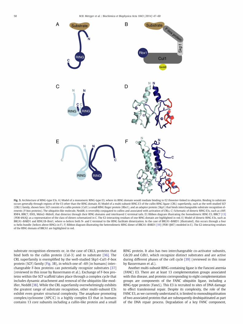
In contrast to HECT-type E3 ligases, RING-type E3s lack a bona fidecatalytic center. A lingering question in the field has been whetherRINGs serve solely to position E2~Ub relative to the substrate, or if theyalso serve as activators of E2~Ub. Clearly, unwanted ubiquitinationevents in the cell are detrimental, so it follows that the reactive E2~Ubspeciesmust be activated at the opportunemoment for transfer. Special-ized examples have indeed provided evidence in support of an activatingrole for RING E3s. One involves the E2 Ubc13 (Ube2N in humans), whichcatalyzes free, K63-linked polyubiquitin chains in the presence of its ac-cessory protein, Mms2. A crystal structure of the Mms2:Ubc13~Ub com-plex revealed thatMms2 binds an incoming substrate ubiquitin in a waythat orients the ubiquitin K63 directly towards the Ubc13~Ub thioester[88]. Thus, this heterodimeric E2 carries its own substrate-binding do-main and does not require an E3 to coordinate substrate. Nevertheless,ubiquitin chain formation by Mms2:Ubc13 is dramatically enhanced inthe presence of a minimal RING domain [89]. A second example, in-spired by work of the late Cecile Pickart, showing E2-catalyzed Ubtransfer onto free Lys, demonstrates an increased rate of ubiquitin dis-charge to small molecule nucleophiles in the presence of minimalRING domains [45,90]. Use of these substrate-independent assays hasallowed theobservation of a catalytic role for RING-type E3s in ubiquitintransfer reactions uncoupled fromproximity effects afforded by E3:sub-strate interactions.
In all available E2:E3 structures, the RING-type domain binds the E2on a surface that is remote from the active site Cys (and therefore fromthe ubiquitin thioester) (Fig. 2). The non-contiguous E3-binding and ac-tive sites on the E2 imply that the role played by a RING to facilitateubiquitin transfer may be indirect and, therefore, allosteric. However,apo- and E3-bound E2 structures are largely indistinguishable and failto suggest amechanism for the allostery. Recent structural studies char-acterizing the interactions of the more relevant E2~Ub conjugated spe-cies with RING-type domains have provided much needed insight.Notably, a solution-based study of E2~Ub conjugates established theirdynamic nature and that E2 and the thioester-linked ubiquitin adoptan array of ‘open’ and ‘closed’ conformations [91] (Fig. 4A). Three struc-tures of E2~Ub conjugates of the UbcH5 family (Ube2D1-3) in complexwith RING-type E3s (RNF4:UbcH5A~Ub, E4B:UbcH5C~Ub, and BIRC7:UbcH5B~Ub) provide the first glimpses at an E3:E2 complex poised totransfer ubiquitin [13,92,93]. A striking common feature is a ‘closed’conformation of the E2~Ub conjugate in which the Ile44 hydrophobicsurface of ubiquitin is positioned against the 310 helix, active site, andhelix 2 of E2 (Fig. 4A–C). In solution, where multiple species can existsimultaneously, E3 binding promotes a population shift in the highlyflexible E2~Ub towards closed conformations which, based on activitydata, primes the active site for transfer (Fig. 4A) [13,93,94]. The closedE2~Ub states are readily disrupted and even conservative mutations ofhydrophobic residues in the interface between E2 and ubiquitin can de-stabilize the closed state and greatly decrease E3-stimulated ubiquitintransfer [93]. It is interesting to note that E2~Ub conjugates that
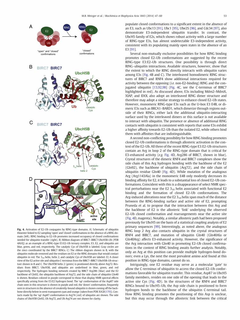
A
B
C
D
Fig. 4. Activation of E2~Ub conjugates by RING-type domains. A) Schematic of ubiquitinthioester linked to E2 sampling ‘open’ and ‘closed’ confirmations in the absence of a RINGdo-main (left). RING binding to E2~Ub promotes increased occupancy of closed confirmationsneeded for ubiquitin transfer (right). B) Ribbon diagram of BIRC7–BIRC7:UbcH5B~Ub (PDB4AUQ) as an example of a RING-type E3:E2~Ub ternary complex. E3, E2, and ubiquitin areblue, green, and red, respectively. The catalytic Cys of UbcH5B is labeled. Gray circles arethe zincs coordinated by the BIRC7 RINGs. C) The ribbon diagram shown in B, with theubiquitinmolecule removed and the residues on E2 or the RING domains that would contactubiquitin in red. The 310 helix, helix 2, and catalytic Cys of UbcH5B are labeled. D) A closerviewof the E2 active site and ubiquitin C-terminus from the BIRC7–BIRC7:UbcH5B~Ub struc-ture shown in B and C. The UbcH5B helix 2 (green) is positioned directly above Arg72. Res-idues from BIRC7, UbcH5B, and ubiquitin are underlined in blue, green, and redrespectively. The hydrogen bonding network created by BIRC7 Arg286 (blue) and the E2backbone of Gln92, the ubiquitin backbone of Arg72, and the side chain of ubiquitin Gln40is shown. Residues colored in purple correspond to those that display NMR spectral effectsspecifically arising from the E3:E2 hydrogen bond. The ‘up’ conformation of the Asp87 sidechain seen in this structure is shown in purple and red, the ‘down’ conformation, frequentlyseen in structures in the absence of covalently-boundubiquitin is showncomingoff the back-bonedirectly below in semi-transparent cyan andorange (taken fromPDB3UGB [170]). Con-tacts made by the ‘up’ Asp87 conformation to Arg74 (red) of ubiquitin are shown. The sidechains of UbcH5B Gln92, Ub Arg72, and Ub Arg74 are not shown for clarity.
53M.B. Metzger et al. / Biochimica et Biophysica Acta 1843 (2014) 47–60
populate closed conformations to a significant extent in the absence ofan E3, such as Ubc13 [91], Ubc1 [95], Ube2S [96], and Cdc34 [97], alsodemonstrate E3-independent ubiquitin transfer. In contrast, theUbcH5 family of E2s, which shows robust activity with a large numberof RING-type E3s, has almost undetectable E3-independent activity,consistent with its populating mainly open states in the absence of anE3 [91].
Several non-mutually exclusive possibilities for how RING bindingpromotes closed E2~Ub conformations are suggested by the recentRING-type E3:E2~Ub structures. One possibility is through directRING–ubiquitin interactions. Available structures, however, show thatthe extent to which the RING directly interacts with ubiquitin variesamong E3s (Fig. 4B and C). The interleaved homodimeric RING struc-tures of BIRC7 and RNF4 show additional interactions required foractivity between the opposing (i.e. non-E2-binding) RING and the con-jugated ubiquitin [13,92,98] (Fig. 4C, see the C-terminus of BIRC7highlighted in red). As discussed above, E3s including Mdm2–MdmX,XIAP, and IDOL also adopt an interleaved RING dimer structure andtherefore may adopt a similar strategy to enhance closed E2~Ub states.However, monomeric RING-type E3s such as the U-box E3 E4B, or di-meric E3s such as BRCA1–BARD1, which dimerize through regions out-side of their RINGs, either lack the additional ubiquitin-interactingsurface used by the interleaved dimers or this surface is not availableto interact with ubiquitin. The presence or absence of additional RINGcontacts with ubiquitin is consistent with reports that some E3s exhibita higher affinity towards E2~Ub than the isolated E2, while others bindthem with affinities that are indistinguishable.
A second non-conflicting possibility for howRING binding promotesclosed E2~Ub conformations is through allosteric activation in the con-text of the E2~Ub. All three of the recent RING-type E3:E2~Ub structurescontain an Arg in loop 2 of the RING-type domain that is critical forE3-enhanced activity (e.g. Fig. 4D, Arg286 of BIRC7, shown in blue).Crystal structures of the dimeric RNF4 and BIRC7 complexes show theside chain of this Arg hydrogen bonding with the backbone of the E2(Gln92), the backbone of ubiquitin (Arg72), and the side chain ofubiquitin residue Gln40 (Fig. 4D). While mutation of the analogousArg (Arg1143Ala) in the monomeric E4B only modestly decreases itsbinding affinity for E2, it leads to a substantial loss of closed E2~Ub con-formations. Coincident with this is a disappearance of select NMR spec-tral perturbations near the E2 310 helix associated with functional E2binding and the formation of closed E2~Ub conformations. TheArg-induced alterations near the E2 310 helix spanmuch of the distancebetween the RING-binding surface and active site of E2, promptingPruneda et al. to propose that the interaction between this Arg andthe backbone of E2 is the allosteric ‘link’ underlying the observedE2~Ub closed conformation and rearrangements near the active site(Fig. 4D, magenta). Notably, a similar allosteric path had been proposedpreviously for UbcH5 on the basis of a statistical coupling analysis of E2primary sequences [99]. Interestingly, as noted above, the analogousRING loop 2 Arg also contacts ubiquitin in the crystal structures ofRNF4 and BIRC7, and mutation of ubiquitin Gln40 (Gln40Ala orGln40Arg) affects E3-enhanced activity. However, the significance ofthe Arg interaction with Gln40 in promoting E2~Ub closed confirma-tions in the context of RING binding awaits further analysis. Notably,only an Arg at this position can provide multiple hydrogen-bond do-nors; even a Lys, the next the most prevalent amino acid found at thisposition in RING-type domains, cannot do so.
Intriguingly, one E2 residue may serve as a molecular ‘gate’ toallow the C-terminus of ubiquitin to access the closed E2~Ub confor-mations favorable for ubiquitin transfer. This residue, Asp87 in UbcH5family members, resides on one side of the opening that leads to theactive site Cys (Fig. 4D). In the structures of the RNF4 and BIRC7RINGs bound to UbcH5~Ub, the Asp side chain is positioned to formhydrogen bonds to the backbone of the ubiquitin C-terminal tail.How RING binding promotes the positioning of this Asp is unclear,but this may occur through the allosteric link between the critical
54 M.B. Metzger et al. / Biochimica et Biophysica Acta 1843 (2014) 47–60
Arg of the RING with Gln92, which neighbors Asp87. Given the lack ofobservable changes in structure in this region when RING-type E3:E2and RING-type E3:E2~Ub structures are compared, the effect is likelyto be a subtle one, probably involving small but important changesin electrostatics. The steric and chemical nature of this moleculargate is critical for activity, as even a Glu substitution severely impactsubiquitination [45]. The residue corresponding to Asp87 is conservedas Asp, Asn, or Ser in most E2s, with the exceptions of UbcH7 andUbcH8, neither of which has been shown to function with RING-typeE3s. Notably, the SUMO-specific E2, Ubc9, which encodes a Ser at theposition analogous to Asp87 in UbcH5C, makes a similar contact withthe C-terminus of SUMO in the SUMO–RanGAP1–Ubc9–Nup358 struc-ture, consistent with a general mechanistic feature of ubiquitin andubiquitin-like transfer [100].
In sum, it is now clear that RING-type E3s are more than mere mo-lecular scaffolds. By binding E2~Ub conjugates and promoting closedconformations, RING-type E3s activate their cognate E2s to stimulateubiquitin transfer. Several non-mutually exclusive mechanisms forhow RING domains promote the structural arrangements associatedwith increasedubiquitin transfer activity have been suggested by recentstudies, but all require further vetting with other RING-type domainsand E2s. Nevertheless, these studies suggest that it will be possible tocreate ‘catalytic’ mutations within RING-type E3s that can be used inplace of, or together with, E2-binding mutations (such as the widelyused BRCA1–Ile26Ala) to create more profoundly ligase-dead versionsof RING-type E3s. Such strategies will pave the way for the generationof new tools to be used in investigations of E3 cellular functions andprotein substrates.
7. E2-binding domains distinct from the RING domain
7.1. E2-binding domains found in RING proteins
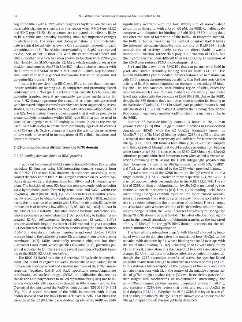
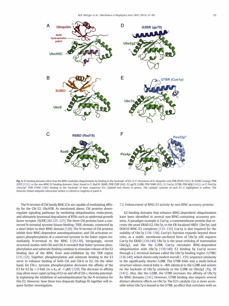
In addition to canonical RING:E2 interactions, RING-type E3s can alsomodulate E2 function using UBC-interacting domains separate fromtheir RINGs. Of the non-RING domains characterized structurally, mostcontact the ‘backside’ of the E2 UBC, a region centered on its β-sheet, op-posite its active site, and distinct from both RING- and E1-interacting re-gions. The backside of some E2s interacts non-covalently with ubiquitinvia a hydrophobic patch formed by Leu8, Ile44, and Val70 within theubiquitin β-sheet [84,101,102] (Fig. 5A). This surface of ubiquitin is com-monly recognized by ubiquitin-binding domains (UBDs) [103], and sim-ilar to the interaction of ubiquitin with UBDs, the ubiquitin:E2 backsideinteraction is of relatively low affinity (Kd of ~300 μM) [102]. At least inthe case of BRCA1–BARD1, the ubiquitin:E2 backside interaction en-hances processive polyubiquitination [102], potentially by facilitating in-creased E2~Ub self-assembly. Several ubiquitin E2-variant (UEV)proteins also bind ubiquitin via their backside site and the yeast ubiquitinE2 Ubc4 interacts with the Ubl-protein, Nedd8, using the same interface[104–106]. Arabidopsis thaliana membrane-anchored Ub-fold (MUB)proteins bind to the backside of some E2s and target them to the plasmamembrane [107]. MUBs structurally resemble ubiquitin, but theirC-terminal CAAX motif, which specifies lipidation [108], precludes po-tential activation by E1. There are also several examples of backside bind-ing to the SUMO E2, Ubc9 (see below).
The RING E3 Rad18 contains a C-terminal E2 backside-binding do-main. Rad18 and its cognate E2, Rad6 (Rad6a/Ube2A and Rad6b/Ube2Bin mammals), are conserved and essential members of the DNA damageresponse. Together, Rad18 and Rad6 specifically monoubiquitinateproliferating cell nuclear antigen (PCNA), a modification that recruitstranslesion DNA polymerases to stalled replication forks [109]. Rad18 in-teracts with Rad6 both canonically through its RING domain and via theC-terminal domain, called the Rad6-binding domain (R6BD) [110–113](Fig. 5C). A crystal structure of the R6BD in complex with humanRad6b revealed that the R6BD forms a ‘kinked α-helix’ that binds thebackside of the E2 [84]. The backside-binding site of the R6BD on Rad6
significantly overlaps with the low affinity site of non-covalentubiquitin binding and, with a Kd of ~60 μM, the R6BD can effectivelycompete with ubiquitin for binding to Rad6 [84]. R6BD binding doesnot alter the rate of formation of the Rad6~Ub thioester. Instead,the R6BD either in trans or in the context of intact Rad18 limitsthe inherent ubiquitin chain-forming activity of Rad6 [84]. Suchmodulation of activity likely serves to direct Rad6 towardsmonoubiquitination, rather than polyubiquitination of PCNA, butthis hypothesis has been difficult to assess directly as mutation ofthe R6BD also reduces PCNA monoubiquitination.
Bre1 and Ubr1, two other RING E3s that can partner with Rad6 inyeast, also contain secondary binding sites for Rad6 [114–116]. Thehuman RAD6/BRE1 pair monoubiquitinates histone H2B inmammaliancells [115], raising the interesting possibility that Bre1 also restricts theactivity of Rad6 to monoubiquitination through its secondary E2 bind-ing site. The non-canonical Rad6-binding region of Ubr1, called thebasic residues-rich (BRR) domain, mediates a low affinity (millimolarlevel) interaction with the backside of Rad6 [84,116]. Unlike the R6BD,though, the BRR domain does not outcompete ubiquitin for binding tothe backside of Rad6 [84]. The Ubr1/Rad6 pair polyubiquitinates N-endrule substrates [116–118], making it unlikely that binding through theBRR domain negatively regulates Rad6 function in a manner similar tothe R6BD.
Another E2 backside-binding domain is found in the humanpro-metastatic [119] RING E3 gp78, which functions in ER-associateddegradation (ERAD) with the E2 Ube2g2 (originally known asMmUbc7 [120]). The Ube2g2-binding region (G2BR) of gp78 is a discreteC-terminal domain that is necessary and sufficient for interaction withUbe2g2 [121]. The G2BR forms a high-affinity (Kd of ~20 nM) complexwith the backside of Ube2g2 that would preclude ubiquitin from bindingto this same surface [83]. In contrast to the R6BD,G2BR binding toUbe2g2stimulates polyubiquitin chain formation, evenwhen added in trans to re-actions containing gp78 lacking the G2BR. Intriguingly, polyubiquitinchain formation by two other Ube2g2-interacting RING E3s, hsHRD1and Trc8, can also be stimulated by the G2BR when provided in trans.
Crystal structures of the G2BR bound to Ube2g2 reveal it to be asingle α-helix (Fig. 5D). Relative to their respective E2s, the G2BR isoriented approximately perpendicular to the R6BD [32,83,84]. The ef-fect of G2BR binding on ubiquitination by Ube2g2 is mediated by twodistinct allosteric mechanisms [83]. First, G2BR binding ‘locks’ loopssurrounding Ube2g2's catalytic cysteine into more ‘closed’ orienta-tions and reorients the catalytic cysteine away from the accessible ac-tive site region defined by the orientation of the loops. These changesare associated with a decreased rate of E1-dependent ubiquitin load-ing of Ube2g2. Second, the G2BR increases the affinity of Ube2g2 forthe gp78 RING domain almost 50-fold. The latter effect is most signif-icant in the overall stimulation of ubiquitin transfer, as the increasedaffinity of Ube2g2 for the gp78 RING domain accounts for the ob-served stimulation of ubiquitination.
The high affinity interaction of gp78 with Ube2g2 afforded by bind-ing of two discrete domains raises the question of how Ube2g2 can bereloaded with ubiquitin by E1, whose binding site on E2 overlaps withthe site of RING binding [80–82]. Reloading of an E2 with ubiquitin byE1 (or at least, dissociation of a discharged E2 to allow association of acharged E2~Ub) must occur to achieve substrate polyubiquitination, al-though the G2BR-dependent transfer of active-site cysteine-linkedubiquitin chains from Ube2g2 to substrate has been reported [32,122].In this system, a full description of the dynamics of the G2BR and RINGdomain interactions with E2, in the context of the putative oligomeriza-tion of gp78 through a distinct region [32],will be needed to provide fur-ther insight into mechanisms of ubiquitination. Interestingly, thenon-RING-containing protein, ancient ubiquitous protein 1 (AUP1),also contains a G2BR-like region that binds and recruits Ube2g2 tolipid droplets [123,124]. Whether the AUP1 G2BR-like region has an ef-fect on ubiquitination by Ube2g2 is not yet known and a precise role forUbe2g2 in lipid droplets has not yet been described.
A
B
C
D
E
F
Fig. 5. E2 binding domains other than the RINGmodulate ubiquitination by binding to the ‘backside’ of E2s. A–F) Structures of A) ubiquitin (red, PDB 2FUH [102]); B) SUMO (orange, PDB2UYZ [151]); or the non-RING E2 binding domains (blue) found in C) Rad18 (R6BD, PDB 2YBF [84]); D) gp78 (G2BR, PDB 3H8K [83]); E) Cue1p (U7BR, PDB 4JQU [141]); or F) Pex22p(Pex22pS, PDB 2Y9M [148]) binding to the ‘backside’ of their respective E2s (labeled and shown in green). The catalytic cysteine of each E2 is highlighted in yellow. Thethioester-linked ubiquitin interaction surface is colored in magenta in panel A.
55M.B. Metzger et al. / Biochimica et Biophysica Acta 1843 (2014) 47–60
TheN-termini of Cbl family RING E3s are capable ofmodulating affin-ity for the Cbl E2, UbcH5B. As mentioned above, Cbl proteins down-regulate signaling pathways by mediating ubiquitination, endocytosis,and ultimately lysosomal degradation of RTKs such as epidermal growthfactor receptor (EGFR) [85,125–127]. The three Cbl proteins have a con-served N-terminal tyrosine kinase-binding (TKB) domain, connected bya short linker to their RING domain [128]. The N-termini of Cbl proteinsinhibit their RING-dependent autoubiquitination, and Cbl activation re-quires phosphorylation of a conserved tyrosine in the linker region im-mediately N-terminal to the RING [129,130]. Intriguingly, recentstructural studies with Cbl and Cbl-b revealed that linker tyrosine phos-phorylation and substrate binding combine to stimulate release of the E2binding face of the RING from auto-inhibition by the TKB region[131,132]. Together, phosphorylation and substrate binding to the E3serve to enhance binding of both Cbl and Cbl-b to E2. On the otherhand, for Cbl-c, tyrosine phosphorylation decreases the affinity of theE3 for E2 by >3-fold (to a Kd of ~1 μM) [129]. The decrease in affinitymay allowmore rapid cycling of E2 on and off of Cbl-c, thereby potential-ly explaining the inhibition of autoubiquitination by the N-terminus forthis E3. However, how these two disparate findings fit together will re-quire further investigation.
7.2. Enhancement of RING E3 activity by non-RING accessory proteins
E2-binding domains that enhance RING-dependent ubiquitinationhave been identified in several non-RING-containing accessory pro-teins. A paradigm example is Cue1p, a transmembrane protein that re-cruits the yeast ERAD E2, Ubc7p, to the ER-localized HRD1 (Der3p) andDOA10 RING E3 complexes [133–135]. Cue1p is also required for thestability of Ubc7p [136–138]. Cue1p's function expands beyond theseroles, as a stable, membrane-anchored form of Ubc7p still requiresCue1p for ERAD [139,140]. Ubc7p is the yeast ortholog of mammalianUbe2g2, and like the G2BR, Cue1p stimulates RING-dependentubiquitination with Ubc7p [139,140]. E2 binding by Cue1p occursthrough a C-terminal domain called the Ubc7p binding region (U7BR)[136,140], which shows onlymodest overall (~15%) sequence similarityto the significantly shorter G2BR. The U7BR folds into a multi-helicalstructure whose central helix is ~40% identical to the G2BR and orientson the backside of Ubc7p similarly to the G2BR on Ube2g2 (Fig. 5E[141]). Also, like the G2BR, the U7BR increases the affinity of Ubc7pfor RING domains [141]. However, U7BR binding also imparts severaldistinct allosteric effects on Ubc7p. The E2's catalytic Cys is more acces-siblewhenUbc7p is bound to the U7BR, an effect that correlateswith an
56 M.B. Metzger et al. / Biochimica et Biophysica Acta 1843 (2014) 47–60
enhancement in the rate of ubiquitin loading of Ubc7p by E1 and an in-crease in the rate of RING-independent ubiquitin transfer when U7BR isbound. Uniquely, Cue1p's ability to enhance ubiquitin loading indicatesthat Ubc7p could remain associated with the HRD1 or DOA10 E3 com-plexes via Cue1p while being reloaded with ubiquitin by E1, somethingthat RING binding precludes. In support of this, modeling of E1 ontothe Ubc7p:U7BR structure based on the recent crystal structure ofSchizosaccharomyces pombe ubiquitin E1 (Uba1) in complex with S.pombe Ubc4 [142] (PDB 4II2) indicates that E1 and U7BR could be mu-tually bound to Ubc7p. Interestingly, similar analysis predicts that si-multaneous binding of E1 and G2BR to Ube2G2 would be precluded;however, this is not the case for the R6BD or Pex22pS (see below). It isinteresting to consider how the U7BR, being part of a protein distinctfrom the RING itself, affords flexibility to the system and could allowUbc7p to simultaneously bind to E1 or pair with various RING E3s invivo.
Another example of activation of an E2 by a non-RING E3 protein isthe yeast E2 Pex4p, which is recruited to the peroxisome by the trans-membrane protein, Pex22p [143–145]. Pex4p functionswith a complexof RING E3s (Pex2p, Pex10p, Pex12p) [146] to monoubiquitinate Pex5pon a specific Cys residue, required for efficient peroxisomal protein im-port [147]. Pex22p binds to Pex4p with high affinity (Kd of ~2 nM) andits binding stimulates the formation of lysine-linked ubiquitin chains onPex4p in the absence of a RING domain. Pex22p:Pex4p binding is alsorequired for RING-dependent substrate ubiquitination in vivo [148].The enhancement of ubiquitin transfer activity does not appear to bemediated by an enhancement in the rate of E1 loading of Pex4p [148].A crystal structure of the soluble portion of Pex22p (Pex22pS) withPex4p reveals that Pex22pS adopts a novel mixed β-sheet and α-helixfold that contacts the C-terminal α3 and α4 helices of the E2, adjacentto the backside region [148] (Fig. 5F). The molecular mechanism ofPex4p stimulation by Pex22p remains unknown, and the binding tothis distinct region of the E2 by Pex22pmay reflect a novel mode of reg-ulation as well. Further investigation will reveal whether Pex22p andAUP1 (described above) have similar or distinct mechanistic effects ontheir respective E2s as the other E2 binding domains whose functionshave now been described.
7.3. Parallels in the SUMO system
FewE3 ligases have been identified and characterized for SUMOand,to date, no HECT-type SUMO E3s have been identified. Although theknown SUMO E3 ligases are not structurally similar to RINGs, likeRING-type E3s, they facilitate direct transfer of SUMO from the soleSUMO E2, Ubc9, to a substrate Lys. Thus, it is informative to compareand contrast features and strategies used by the SUMO system tothose of the ubiquitin system. Ubc9 interacts non-covalently withSUMO via the analogous E2 backside site and SUMO β-sheet[149–151] (Fig. 5B). Like ubiquitin binding, non-covalent SUMObindingto Ubc9 promotes SUMO chain formation on target proteins [149,151].Furthermore, the SUMO E3 Nup358/Ran-binding protein 2 (RanBP2),which is involved in nucleocytoplasmic trafficking [152–154], despitebeing structurally distinct from SUMO, contacts the Ubc9 backsideusing the same residues on Ubc9, making their bindingmutually exclu-sive [149]. How SUMO andNup358/RanBP2 interact in vivo tomodulatefunction remains to be determined.
SUMO accessory proteins also contain domains that, despite verylittle sequence identity, structurally mimic SUMO and bind the back-side of Ubc9. An example is Rad60/Esc2 and its human ortholog, nu-clear factor of activated T-cells (NFAT)-interacting protein of 45 kDa(Nip45), which contains a C-terminal SUMO-like domain (SLD) thatis a structural mimic for SUMO with respect to the Ubc9-interactingregion [155,156]. Notably, binding of SLD2 to the backside of Ubc9 in-hibits SUMO chain elongation in vitro and is important for survival ofgenotoxic stress in yeast [155,156].
Another putative SUMO accessory protein, the RWD domain-containing protein RSUME, enhances SUMO conjugation in severalways including: stimulating the loading of Ubc9 with SUMO, mediatingE3-independent SUMO transfer to substrates, such as IκB and HIF-1α,and stimulating sumoylation by the SUMO E3, PIAS [157]. RSUMEbinds to both SUMO and Ubc9 and enhances the non-covalent SUMObinding to Ubc9 [157]. Interestingly, like other RWD domains [158],the structure of RSUME closely resembles that of E2 Ub-conjugating en-zymes, despite limited sequence homology (PDB 2EBK).
8. Perspective and future directions
In the 14 years since RING finger function was discovered, ourknowledge of RING-type ubiquitin ligases has increased dramatically.Through their targeting of a diverse array of substrates, we are begin-ning to appreciate the range of roles played by this family of E3s indevelopment, in maintaining homeostasis, and in response to cellularsignals. Many challenges remain, however, as exemplified by the factthat substrates for most RING-type E3:E2 pairs are not yet known,
For some E3s, insights have emerged as to how their activity andsubstrate interactions can be regulated. We now know that a wide va-riety of protein–protein interactions are employed in substrate recog-nition and that post-translational protein modifications are, in manycases, critical to substrate binding. It is also evident that multiple sub-strates can be targeted by one RING-type E3, and that multiple E3scan target the same substrate. However, at both the cellular and or-ganismal levels, the overall significance of E3 redundancy in substrateubiquitination is, in general, poorly understood.
For substrates that are ubiquitinated on specific sites, with few ex-ceptions, the factors that specify these sites are unknown. Progresswill require a marked expansion of our understanding of the position-ing of both E2~Ub and the substrate in the context of the entireRING-type ligase. Related to this, an emerging concept is that someE2s function with RING-type ligases as ‘chain initiators,’ which putthe first ubiquitin on a substrate, while others are ‘chain builders’that add to an existing ubiquitin chain. Progress in parsing this issuehas been slowed by the fact that the most extensively studied E2s,the UbcH5 (Ube2D) family, can perform both functions. In consider-ing chain building, an important outstanding issue is whether differ-ent E2s provide specific local environments around their active sitesfavorable to particular lysines on acceptor ubiquitins, and therebyfavor certain ubiquitin chain linkages. If this is the case, understand-ing the nature of these local environmental factors, and how theyare influenced by RING binding, will be crucial to appreciating howthe myriad of ubiquitin signals is generated.
Until recently, the molecular basis by which RING-type domainsstimulate the transfer of ubiquitin from E2 was enigmatic. As reviewedherein, the pieces are falling into place for some E2:E3 pairs, as bothcrystal and solution structures of RING-type domains complexed withE2~Ub rather than E2 alone have recently been described and corrobo-rated with functional data. These findings are all consistent with criticalroles for both a RING-induced closed E2~Ub conformation and theRING's Zn II loop (or the equivalent in U-box proteins) in facilitating ac-tivation. While these paradigm-shifting observations provide insightsinto some aspects of the RING:E2 interface, they do not fully accountfor defects in ubiquitination seen with mutations in other regions ofthis interface. It will be necessary to expand upon the small number ofproductive E3:E2 pairs that have been studied in depth to fully under-stand this critical interface. Similarly, it is important to determine thedetails and significance of the RING:E2 binding interface for RING–IBR–RING E3s, as we now know these function as classic catalysts,akin to HECT-type E3s.
A common feature of RING-type domains is a tendency to form ac-tive homo- and/or heterodimers. For C-terminal interleaved dimers,the distal RING-type domain provides additional contacts to ubiquitin(E2~Ub) to facilitate the closed E2~Ub conformation and promote
57M.B. Metzger et al. / Biochimica et Biophysica Acta 1843 (2014) 47–60
catalysis. E3 dimerization/oligomerization, in instances where multi-ple RING-type domains can bind E2~Ub, could also enhance the prob-ability of successful ubiquitin chain formation by increasing the localconcentration of RING-type domains accessible to substrate. In thecases of BRCA1–BARD1 and RING1B–Bmi1, which dimerize throughnon-RING interactions, positioning of the second inactive (i.e.,non-E2-binding) RING finger is not predicted to play a direct role inthe binding of E2 ~ Ub. In these cases, the function of dimerizationin ubiquitination remains enigmatic.
A central remaining question in RING-type E3-mediatedubiquitination is what regulates the processivity of ubiquitination andthus the fate of the substrate. The answer is likely complicated and in-cludes E3 dimerization/oligomerization, the affinity for substrate, therelative affinities for E2 versus E2~Ub, and ubiquitin-binding domainsintrinsic to E3s or E3 complexes. All of these potentially positive factorsare, of course, countered by DUBs that are associated with E3s or sub-strates. Another factor that, in some cases, facilitates ubiquitination isthe non-covalent binding of ubiquitin to the backside of a subclass ofE2s. An emerging factor, reviewed herein, is the contribution ofnon-RING regions of E3s binding to E2s using surfaces distinct fromthe shared RING- and E1-interacting interface. In some cases, these in-teractions compete with non-covalent ubiquitin backside binding andlimit ubiquitination. In other cases, binding to a similar region of theE2 increases the affinity of the E2:E3 interaction and thereby enhancesprocessivity of ubiquitination. These secondary sites of E2 binding mayalso provide ameans to tether the E2 to the E3 complex,without contin-uous RING finger binding, and thereby provide a potential means to‘reload’ E2 with ubiquitin (E2~Ub) without dissociation from the E3complex. Whether such E2-specific binding is of general importancein vivo in determining combinatorial specificity in RING-type domain:E2 interactions and in the processivity of ubiquitination now becomeimportant questions.
Finally, RING-type E3s and their substrates are implicated in a widevariety of human diseases ranging from viral infections to neurodegen-erative disorders to cancer. Thus, they are attractive targets for thera-peutic development. However, the lack of a catalytic center inRING-type domains makes targeting strategies more difficult. As bio-chemical and biophysical/structural approaches converge on develop-ing an understanding of specific aspects of RING-type E3s and theirinteractions with E2s, the potential for generating therapeutics by in-corporation of structure-based design becomes increasingly promising.
Acknowledgements
The study of RING-type E3s continues to grow extremely rapidly.We regret that it was possible to only cite a fraction of the outstand-ing primary publications in this field. This work was supported by theNational Institute of General Medical Sciences grants R01 GM088055and R01 GM098503 (R.E.K.) and by the Intramural Research Programof the National Institutes of Health, National Cancer Institute, Centerfor Cancer Research (A.M.W.).
References
[1] D.M. Wenzel, K.E. Stoll, R.E. Klevit, E2s: structurally economical and functionallyreplete, Biochem. J. 433 (2011) 31–42.
[2] P.L. Welcsh, M.C. King, BRCA1 and BRCA2 and the genetics of breast and ovariancancer, Hum. Mol. Genet. 10 (2001) 705–713.
[3] G.L. Moldovan, A.D. D'Andrea, How the Fanconi anemia pathway guards the ge-nome, Annu. Rev. Genet. 43 (2009) 223–249.
[4] S.S. Fakharzadeh, S.P. Trusko, D.L. George, Tumorigenic potential associated withenhanced expression of a gene that is amplified in a mouse tumor cell line,EMBO J. 10 (1991) 1565–1569.
[5] S. Lipkowitz, A.M. Weissman, RINGs of good and evil: RING finger ubiquitin li-gases at the crossroads of tumour suppression and oncogenesis, Nat. Rev. Cancer11 (2011) 629–643.
[6] S. Duan, L. Cermak, J.K. Pagan, M. Rossi, C. Martinengo, P.F. di Celle, B. Chapuy, M.Shipp, R. Chiarle, M. Pagano, FBXO11 targets BCL6 for degradation and isinactivated in diffuse large B-cell lymphomas, Nature 481 (2011) 90–93.
[7] T. Kitada, S. Asakawa, N. Hattori, H. Matsumine, Y. Yamamura, S. Minoshima, M.Yokochi, Y. Mizuno, N. Shimizu, Mutations in the parkin gene cause autosomalrecessive juvenile parkinsonism, Nature 392 (1998) 605–608.
[8] C. Boutell, R.D. Everett, Regulation of alphaherpesvirus infections by the ICP0family of proteins, J. Gen. Virol. 94 (2012) 465–481.
[9] J.A. Nathan, P.J. Lehner, The trafficking and regulation of membrane receptors bythe RING-CH ubiquitin E3 ligases, Exp. Cell Res. 315 (2009) 1593–1600.
[10] F. Margottin, S.P. Bour, H. Durand, L. Selig, S. Benichou, V. Richard, D. Thomas, K.Strebel, R. Benarous, A novel human WD protein, h-beta TrCp, that interactswith HIV-1 Vpu connects CD4 to the ER degradation pathway through anF-box motif, Mol. Cell 1 (1998) 565–574.
[11] P.D. Mace, K. Linke, R. Feltham, F.R. Schumacher, C.A. Smith, D.L. Vaux, J. Silke,C.L. Day, Structures of the cIAP2 RING domain reveal conformational changes as-sociated with ubiquitin-conjugating enzyme (E2) recruitment, J. Biol. Chem. 283(2008) 31633–31640.
[12] C.W. Liew, H. Sun, T. Hunter, C.L. Day, RING domain dimerization is essential forRNF4 function, Biochem. J. 431 (2010) 23–29.
[13] H. Dou, L. Buetow, G.J. Sibbet, K. Cameron, D.T. Huang, BIRC7–E2 ubiquitin con-jugate structure reveals the mechanism of ubiquitin transfer by a RING dimer,Nat. Struct. Mol. Biol. 19 (2012) 876–883.
[14] L. Zhang, L. Fairall, B.T. Goult, A.C. Calkin, C. Hong, C.J. Millard, P. Tontonoz, J.W.Schwabe, The IDOL–UBE2D complex mediates sterol-dependent degradation ofthe LDL receptor, Genes Dev. 25 (2011) 1262–1274.
[15] Z. Xu, E. Kohli, K.I. Devlin, M. Bold, J.C. Nix, S. Misra, Interactions between thequality control ubiquitin ligase CHIP and ubiquitin conjugating enzymes, BMCStruct. Biol. 8 (2008) 26.
[16] M.D. Ohi, C.W. Vander Kooi, J.A. Rosenberg, W.J. Chazin, K.L. Gould, Structural in-sights into the U-box, a domain associated with multi-ubiquitination, Nat.Struct. Biol. 10 (2003) 250–255.
[17] C.W. Vander Kooi, M.D. Ohi, J.A. Rosenberg, M.L. Oldham, M.E. Newcomer, K.L.Gould, W.J. Chazin, The Prp19 U-box crystal structure suggests a common di-meric architecture for a class of oligomeric E3 ubiquitin ligases, Biochemistry45 (2006) 121–130.
[18] V. Joukov, J. Chen, E.A. Fox, J.B. Green, D.M. Livingston, Functional communica-tion between endogenous BRCA1 and its partner, BARD1, during Xenopus laevisdevelopment, Proc. Natl. Acad. Sci. U. S. A. 98 (2001) 12078–12083.
[19] P.S. Brzovic, P. Rajagopal, D.W. Hoyt, M.C. King, R.E. Klevit, Structure of a BRCA1–BARD1 heterodimeric RING–RING complex, Nat. Struct. Biol. 8 (2001) 833–837.
[20] R. Cao, Y. Tsukada, Y. Zhang, Role of Bmi-1 and Ring1A in H2A ubiquitylation andHox gene silencing, Mol. Cell 20 (2005) 845–854.
[21] H. Wang, L. Wang, H. Erdjument-Bromage, M. Vidal, P. Tempst, R.S. Jones, Y.Zhang, Role of histone H2A ubiquitination in Polycomb silencing, Nature 431(2004) 873–878.
[22] D.E. Christensen, P.S. Brzovic, R.E. Klevit, E2–BRCA1 RING interactions dictatesynthesis of mono- or specific polyubiquitin chain linkages, Nat. Struct. Mol.Biol. 14 (2007) 941–948.
[23] S. Uldrijan, W.J. Pannekoek, K.H. Vousden, An essential function of the ex-treme C-terminus of MDM2 can be provided by MDMX, EMBO J. 26 (2007)102–112.
[24] M.V. Poyurovsky, C. Priest, A. Kentsis, K.L. Borden, Z.Q. Pan, N. Pavletich, C.Prives, The Mdm2 RING domain C-terminus is required for supramolecular as-sembly and ubiquitin ligase activity, EMBO J. 26 (2007) 90–101.
[25] S. Iyappan, H.P. Wollscheid, A. Rojas-Fernandez, A. Marquardt, H.C. Tang, R.K.Singh, M. Scheffner, Turning the RING domain protein MdmX into an activeubiquitin–protein ligase, J. Biol. Chem. 285 (2010) 33065–33072.
[26] P.N. Friedman, X. Chen, J. Bargonetti, C. Prives, The p53 protein is an unusuallyshaped tetramer that binds directly to DNA, Proc. Natl. Acad. Sci. U. S. A. 90(1993) 3319–3323.
[27] E.C. Dueber, A.J. Schoeffler, A. Lingel, J.M. Elliott, A.V. Fedorova, A.M. Giannetti, K.Zobel, B. Maurer, E. Varfolomeev, P. Wu, H.J. Wallweber, S.G. Hymowitz, K.Deshayes, D. Vucic, W.J. Fairbrother, Antagonists induce a conformationalchange in cIAP1 that promotes autoubiquitination, Science 334 (2011) 376–380.
[28] K. Linke, P.D. Mace, C.A. Smith, D.L. Vaux, J. Silke, C.L. Day, Structure of theMDM2/MDMX RING domain heterodimer reveals dimerization is required fortheir ubiquitylation in trans, Cell Death Differ. 15 (2008) 841–848.
[29] Z. Li, R. Cao, M. Wang, M.P. Myers, Y. Zhang, R.M. Xu, Structure of a Bmi-1-Ring1Bpolycomb group ubiquitin ligase complex, J. Biol. Chem. 281 (2006) 20643–20649.
[30] A. Huang, R.G. Hibbert, R.N. de Jong, D. Das, T.K. Sixma, R. Boelens, Symmetry andasymmetry of the RING–RING dimer of Rad18, J. Mol. Biol. 410 (2011) 424–435.
[31] S. Fang, M. Ferrone, C. Yang, J.P. Jensen, S. Tiwari, A.M. Weissman, The tumor au-tocrine motility factor receptor, gp78, is a ubiquitin protein ligase implicated indegradation from the endoplasmic reticulum, Proc. Natl. Acad. Sci. U. S. A. 98(2001) 14422–14427.
[32] W. Li, D. Tu, L. Li, T. Wollert, R. Ghirlando, A.T. Brunger, Y. Ye, Mechanistic insightsinto active site-associated polyubiquitination by the ubiquitin-conjugating enzymeUbe2g2, Proc. Natl. Acad. Sci. U. S. A. 106 (2009) 3722–3727.
[33] G. Kozlov, P. Peschard, B. Zimmerman, T. Lin, T. Moldoveanu, N. Mansur-Azzam,K. Gehring, M. Park, Structural basis for UBA-mediated dimerization of c-Cblubiquitin ligase, J. Biol. Chem. 282 (2007) 27547–27555.
[34] M. Bartkiewicz, A. Houghton, R. Baron, Leucine zipper-mediated homodimerization ofthe adaptorprotein c-Cbl. A role in c-Cbl's tyrosinephosphorylation and its associationwith epidermal growth factor receptor, J. Biol. Chem. 274 (1999) 30887–30895.
[35] M.D. Petroski, R.J. Deshaies, Function and regulation of cullin–RING ubiquitin li-gases, Nat. Rev. Mol. Cell Biol. 6 (2005) 9–20.
[36] A. Sarikas, T. Hartmann, Z.Q. Pan, The cullin protein family, Genome Biol. 12(2011) 220.
58 M.B. Metzger et al. / Biochimica et Biophysica Acta 1843 (2014) 47–60
[37] D. Frescas, M. Pagano, Deregulated proteolysis by the F-box proteins SKP2 andbeta-TrCP: tipping the scales of cancer, Nat. Rev. Cancer 8 (2008) 438–449.
[38] D.M. Duda, D.C. Scott, M.F. Calabrese, E.S. Zimmerman, N. Zheng, B.A. Schulman,Structural regulation of cullin–RING ubiquitin ligase complexes, Curr. Opin.Struct. Biol. 21 (2011) 257–264.
[39] A. Schreiber, F. Stengel, Z. Zhang, R.I. Enchev, E.H. Kong, E.P. Morris, C.V.Robinson, P.C. da Fonseca, D. Barford, Structural basis for the subunit assemblyof the anaphase-promoting complex, Nature 470 (2011) 227–232.
[40] Y. Kee, J.M. Kim, A.D. D'Andrea, Regulated degradation of FANCM in the Fanconianemia pathway during mitosis, Genes Dev. 23 (2009) 555–560.
[41] R. Menssen, J. Schweiggert, J. Schreiner, D. Kusevic, J. Reuther, B. Braun, D.H.Wolf, Exploring the topology of the Gid complex, the E3 ubiquitin ligase in-volved in catabolite-induced degradation of gluconeogenic enzymes, J. Biol.Chem. 287 (2012) 25602–25614.
[42] H.W. Platta, F. El Magraoui, B.E. Baumer, D. Schlee, W. Girzalsky, R. Erdmann,Pex2 and pex12 function as protein–ubiquitin ligases in peroxisomal proteinimport, Mol. Cell. Biol. 29 (2009) 5505–5516.
[43] C. Williams, M. van den Berg, E. Geers, B. Distel, Pex10p functions as an E3 ligasefor the Ubc4p-dependent ubiquitination of Pex5p, Biochem. Biophys. Res.Commun. 374 (2008) 620–624.
[44] M. Itoh, C.H. Kim, G. Palardy, T. Oda, Y.J. Jiang, D. Maust, S.Y. Yeo, K. Lorick, G.J.Wright, L. Ariza-McNaughton, A.M. Weissman, J. Lewis, S.C. Chandrasekharappa,A.B. Chitnis, Mind bomb is a ubiquitin ligase that is essential for efficient activationof Notch signaling by Delta, Dev. Cell 4 (2003) 67–82.
[45] D.M. Wenzel, A. Lissounov, P.S. Brzovic, R.E. Klevit, UBCH7 reactivity profile re-veals parkin and HHARI to be RING/HECT hybrids, Nature 474 (2011) 105–108.
[46] D.M. Wenzel, R.E. Klevit, Following Ariadne's thread: a new perspective on RBRubiquitin ligases, BMC Biol. 10 (2012) 24.
[47] J.J. Smit, D. Monteferrario, S.M. Noordermeer, W.J. van Dijk, B.A. van der Reijden, T.K.Sixma, The E3 ligaseHOIP specifies linear ubiquitin chain assembly through its RING–IBR–RING domain and the unique LDD extension, EMBO J. 31 (2012) 3833–3844.
[48] B. Stieglitz, A.C. Morris-Davies, M.G. Koliopoulos, E. Christodoulou, K. Rittinger,LUBAC synthesizes linear ubiquitin chains via a thioester intermediate, EMBORep. 13 (2012) 840–846.
[49] M. Lazarou, D.P. Narendra, S.M. Jin, E. Tekle, S. Banerjee, R.J. Youle, PINK1 drivesParkin self-association and HECT-like E3 activity upstream of mitochondrialbinding, J. Cell Biol. 200 (2013) 163–172.
[50] K.L. Lorick, J.P. Jensen, S. Fang, A.M. Ong, S. Hatakeyama, A.M. Weissman, RINGfingers mediate ubiquitin-conjugating enzyme (E2)-dependent ubiquitination,Proc. Natl. Acad. Sci. U. S. A. 96 (1999) 11364–11369.
[51] A. Weissman, N. Shabek, A. Ciechanover, The predator becomes the prey: regu-lating the ubiquitin system by ubiquitylation and degradation, Nat. Rev. Mol.Cell Biol. 12 (2011) 605–620.
[52] T. Abbas, A.C. Mueller, E. Shibata, M. Keaton, M. Rossi, A. Dutta, CRL1–FBXO11 pro-motes Cdt2 ubiquitylation and degradation and regulates Pr-set7/set8-mediatedcellular migration, Mol. Cell 49 (2013) 1147–1158.
[53] M. Rossi, S. Duan, Y.T. Jeong, M. Horn, A. Saraf, L. Florens, M.P. Washburn, A.Antebi, M. Pagano, Regulation of the CRL4(Cdt2) ubiquitin ligase and cell-cycleexit by the SCF(Fbxo11) ubiquitin ligase, Mol. Cell 49 (2013) 1159–1166.
[54] J.N. Lee, B. Song, R.A. DeBose-Boyd, J. Ye, Sterol-regulated degradation of Insig-1mediated by the membrane-bound ubiquitin ligase gp78, J. Biol. Chem. 281(2006) 39308–39315.
[55] S.L. Stone, J. Callis, Ubiquitin ligases mediate growth and development by pro-moting protein death, Curr. Opin. Plant Biol. 10 (2007) 624–632.
[56] E.J. Chapman, M. Estelle, Mechanism of auxin-regulated gene expression inplants, Annu. Rev. Genet. 43 (2009) 265–285.
[57] S.E. Alvarez, K.B. Harikumar, N.C. Hait, J. Allegood, G.M. Strub, E.Y. Kim,M.Maceyka,H. Jiang, C. Luo, T. Kordula, S. Milstien, S. Spiegel, Sphingosine-1-phosphate is amissing cofactor for the E3 ubiquitin ligase TRAF2, Nature 465 (2010) 1084–1088.
[58] P.E. Ryan, G.C. Davies, M.M. Nau, S. Lipkowitz, Regulating the regulator: negativeregulation of Cbl ubiquitin ligases, Trends Biochem. Sci. 31 (2006) 79–88.
[59] H. Labit, K. Fujimitsu, N.S. Bayin, T. Takaki, J. Gannon, H. Yamano, Dephosphory-lation of Cdc20 is required for its C-box-dependent activation of the APC/C,EMBO J. 31 (2012) 3351–3362.
[60] Y. Yoshida, K. Tanaka, Lectin-like ERAD players in ER and cytosol, Biochim.Biophys. Acta 1800 (2009) 172–180.
[61] J.J. Perry, J.A. Tainer, M.N. Boddy, A SIM-ultaneous role for SUMO and ubiquitin,Trends Biochem. Sci. 33 (2008) 201–208.
[62] Y. Zhang, S. Liu, C. Mickanin, Y. Feng, O. Charlat, G.A. Michaud, M. Schirle, X. Shi,M. Hild, A. Bauer, V.E. Myer, P.M. Finan, J.A. Porter, S.M. Huang, F. Cong, RNF146is a poly(ADP-ribose)-directed E3 ligase that regulates axin degradation andWnt signalling, Nat Cell Biol 13 (2011) 623–629.
[63] H.C. Kang, Y.I. Lee, J.H. Shin, S.A. Andrabi, Z. Chi, J.P. Gagne, Y. Lee, H.S. Ko, B.D.Lee, G.G. Poirier, V.L. Dawson, T.M. Dawson, Iduna is a poly(ADP-ribose)(PAR)-dependent E3 ubiquitin ligase that regulates DNA damage, Proc. Natl.Acad. Sci. U. S. A. 108 (2011) 14103–14108.
[64] M.G. Callow, H. Tran, L. Phu, T. Lau, J. Lee, W.N. Sandoval, P.S. Liu, S. Bheddah, J.Tao, J.R. Lill, J.A. Hongo, D. Davis, D.S. Kirkpatrick, P. Polakis, M. Costa, Ubiquitinligase RNF146 regulates tankyrase and axin to promote Wnt signaling, PLoS One6 (2011) e22595.
[65] J. Cao, Q. Yan, Histone ubiquitination and deubiquitination in transcription, DNAdamage response, and cancer, Front. Oncol. 2 (2012) 26.
[66] C. Dai, W. Gu, p53 post-translational modification: deregulated in tumorigene-sis, Trends Mol. Med. 16 (2010) 528–536.
[67] X. Wang, J. Taplick, N. Geva, M. Oren, Inhibition of p53 degradation by Mdm2acetylation, FEBS Lett. 561 (2004) 195–201.
[68] Y. Ma, S. Fan, C. Hu, Q. Meng, S.A. Fuqua, R.G. Pestell, Y.A. Tomita, E.M. Rosen,BRCA1 regulates acetylation and ubiquitination of estrogen receptor-alpha,Mol. Endocrinol. 24 (2009) 76–90.
[69] E. Bartee, M. Mansouri, B.T. Hovey Nerenberg, K. Gouveia, K. Fruh, Downregulationof major histocompatibility complex class I by human ubiquitin ligases related toviral immune evasion proteins, J. Virol. 78 (2004) 1109–1120.
[70] F. El Magraoui, B.E. Baumer, H.W. Platta, J.S. Baumann, W. Girzalsky, R. Erdmann,The RING-type ubiquitin ligases Pex2p, Pex10p and Pex12p form a heteromericcomplex that displays enhanced activity in an ubiquitin conjugatingenzyme-selective manner, FEBS J. 279 (2012) 2060–2070.
[71] M. Karbowski, A. Neutzner, R.J. Youle, The mitochondrial E3 ubiquitin ligaseMARCH5 is required for Drp1 dependent mitochondrial division, J. Cell Biol.178 (2007) 71–84.
[72] N. Nakamura, Y. Kimura, M. Tokuda, S. Honda, S. Hirose, MARCH-V is a novelmitofusin 2- and Drp1-binding protein able to change mitochondrial morpholo-gy, EMBO Rep. 7 (2006) 1019–1022.
[73] Z. Kostova, Y.C. Tsai, A.M.Weissman, Ubiquitin ligases, critical mediators of endoplas-mic reticulum-associated degradation, Semin. Cell Dev. Biol. 18 (2007) 770–779.
[74] T.K. Albert, H. Hanzawa, Y.I. Legtenberg, M.J. de Ruwe, F.A. van den Heuvel, M.A.Collart, R. Boelens, H.T. Timmers, Identification of a ubiquitin–protein ligasesubunit within the CCR4–NOT transcription repressor complex, EMBO J. 21(2002) 355–364.
[75] T. Ohta, J.J. Michel, A.J. Schottelius, Y. Xiong, ROC1, a homolog of APC11, repre-sents a family of cullin partners with an associated ubiquitin ligase activity,Mol. Cell 3 (1999) 535–541.
[76] T. Kamura, D.M. Koepp, M.N. Conrad, D. Skowyra, R.J. Moreland, O. Iliopoulos,W.S. Lane, W.G. Kaelin Jr., S.J. Elledge, R.C. Conaway, J.W. Harper, J.W.Conaway, Rbx1, a component of the VHL tumor suppressor complex and SCFubiquitin ligase, Science 284 (1999) 657–661.
[77] S. Fang, K.L. Lorick, J.P. Jensen, A.M. Weissman, RING finger ubiquitin protein li-gases: implications for tumorigenesis, metastasis and for molecular targets incancer, Semin. Cancer Biol. 13 (2003) 5–14.
[78] R. Janjusevic, R.B. Abramovitch, G.B. Martin, C.E. Stebbins, A bacterial inhibitor ofhost programmed cell death defenses is an E3 ubiquitin ligase, Science 311(2006) 222–226.
[79] B. Wu, T. Skarina, A. Yee, M.C. Jobin, R. Dileo, A. Semesi, C. Fares, A. Lemak, B.K.Coombes, C.H. Arrowsmith, A.U. Singer, A. Savchenko, NleG Type 3 effectorsfrom enterohaemorrhagic Escherichia coli are U-Box E3 ubiquitin ligases, PLoSPathog. 6 (2010) e1000960.
[80] Z.M. Eletr, D.T. Huang, D.M. Duda, B.A. Schulman, B. Kuhlman, E2 conjugatingenzymes must disengage from their E1 enzymes before E3-dependent ubiquitinand ubiquitin-like transfer, Nat. Struct. Mol. Biol. 12 (2005) 933–934.
[81] D.T. Huang, A. Paydar, M. Zhuang, M.B. Waddell, J.M. Holton, B.A. Schulman,Structural basis for recruitment of Ubc12 by an E2 binding domain in NEDD8'sE1, Mol. Cell 17 (2005) 341–350.
[82] N. Zheng, P.Wang, P.D. Jeffrey, N.P. Pavletich, Structure of a c-Cbl–UbcH7 complex:RING domain function in ubiquitin–protein ligases, Cell 102 (2000) 533–539.
[83] R. Das, J. Mariano, Y.C. Tsai, R.C. Kalathur, Z. Kostova, J. Li, S.G. Tarasov, R.L.McFeeters, A.S. Altieri, X. Ji, R.A. Byrd, A.M. Weissman, Allosteric activation ofE2-RING finger-mediated ubiquitylation by a structurally defined specificE2-binding region of gp78, Mol. Cell 34 (2009) 674–685.
[84] R.G. Hibbert, A. Huang, R. Boelens, T.K. Sixma, E3 ligase Rad18 promotesmonoubiquitination rather than ubiquitin chain formation by E2 enzymeRad6, Proc. Natl. Acad. Sci. U. S. A. 108 (2011) 5590–5595.
[85] C.A. Joazeiro, S.S. Wing, H. Huang, J.D. Leverson, T. Hunter, Y.C. Liu, The tyrosinekinase negative regulator c-Cbl as a RING-type, E2-dependent ubiquitin–proteinligase, Science 286 (1999) 309–312.
[86] P.S. Brzovic, J.R. Keeffe, H. Nishikawa, K. Miyamoto, D. Fox III, M. Fukuda, T. Ohta,R. Klevit, Binding and recognition in the assembly of an active BRCA1/BARD1ubiquitin–ligase complex, Proc. Natl. Acad. Sci. U. S. A. 100 (2003) 5646–5651.
[87] D.E. Christensen, R.E. Klevit, Dynamic interactions of proteins in complex net-works: identifying the complete set of interacting E2s for functional investiga-tion of E3-dependent protein ubiquitination, FEBS J. 276 (2009) 5381–5389.
[88] M.J. Eddins, C.M. Carlile, K.M. Gomez, C.M. Pickart, C. Wolberger, Mms2–Ubc13covalently bound to ubiquitin reveals the structural basis of linkage-specificpolyubiquitin chain formation, Nat. Struct. Mol. Biol. 13 (2006) 915–920.
[89] Q. Yin, S.C. Lin, B. Lamothe, M. Lu, Y.C. Lo, G. Hura, L. Zheng, R.L. Rich, A.D. Campos,D.G. Myszka, M.J. Lenardo, B.G. Darnay, H. Wu, E2 interaction and dimerization inthe crystal structure of TRAF6, Nat. Struct. Mol. Biol. 16 (2009) 658–666.
[90] C.M. Pickart, I.A. Rose, Functional heterogeneity of ubiquitin carrier proteins, J.Biol. Chem. 260 (1985) 1573–1581.
[91] J.N. Pruneda, K.E. Stoll, L.J. Bolton, P.S. Brzovic, R.E. Klevit, Ubiquitin in motion:structural studies of the ubiquitin-conjugating enzyme~ubiquitin conjugate,Biochemistry 50 (2011) 1624–1633.
[92] A. Plechanovova, E.G. Jaffray, M.H. Tatham, J.H. Naismith, R.T. Hay, Structure of aRING E3 ligase and ubiquitin-loaded E2 primed for catalysis, Nature 489 (2012)115–120.
[93] J.N. Pruneda, P.J. Littlefield, S.E. Soss, K.A. Nordquist, W.J. Chazin, P.S. Brzovic, R.E.Klevit, Structure of an E3:E2~Ub complex reveals an allosteric mechanismshared among RING/U-box ligases, Mol. Cell 47 (2012) 933–942.
[94] S.E. Soss, R.E. Klevit, W.J. Chazin, Activation of UbcH5c~Ub is the result of a shiftin interdomain motions of the conjugate bound to U-box E3 ligase E4B, Bio-chemistry 52 (2013) 2991–2999.
[95] K.S. Hamilton, M.J. Ellison, K.R. Barber, R.S. Williams, J.T. Huzil, S. McKenna, C. Ptak,M. Glover, G.S. Shaw, Structure of a conjugating enzyme-ubiquitin thiolester inter-mediate reveals a novel role for the ubiquitin tail, Structure 9 (2001) 897–904.
59M.B. Metzger et al. / Biochimica et Biophysica Acta 1843 (2014) 47–60
[96] K.E. Wickliffe, S. Lorenz, D.E. Wemmer, J. Kuriyan, M. Rape, The mechanism oflinkage-specific ubiquitin chain elongation by a single-subunit E2, Cell 144(2011) 769–781.
[97] A. Saha, S. Lewis, G. Kleiger, B. Kuhlman, R.J. Deshaies, Essential role for ubiquitin–ubiquitin-conjugating enzyme interaction in ubiquitin discharge from Cdc34 tosubstrate, Mol. Cell 42 (2011) 75–83.
[98] Y. Nakatani, T. Kleffmann, K. Linke, S.M. Condon, M.G. Hinds, C.L. Day, Regulationof ubiquitin transfer by XIAP, a dimeric RING E3 ligase, Biochem. J. 450 (3)(2012) 629–638.
[99] E. Ozkan, H. Yu, J. Deisenhofer, Mechanistic insight into the allosteric activationof a ubiquitin-conjugating enzyme by RING-type ubiquitin ligases, Proc. Natl.Acad. Sci. U. S. A. 102 (2005) 18890–18895.
[100] D. Reverter, C.D. Lima, Insights into E3 ligase activity revealed by a SUMO–RanGAP1–Ubc9–Nup358 complex, Nature 435 (2005) 687–692.
[101] T. Miura, W. Klaus, B. Gsell, C. Miyamoto, H. Senn, Characterization of the bind-ing interface between ubiquitin and class I human ubiquitin-conjugating en-zyme 2b by multidimensional heteronuclear NMR spectroscopy in solution, J.Mol. Biol. 290 (1999) 213–228.
[102] P.S. Brzovic, A. Lissounov, D.E. Christensen, D.W. Hoyt, R.E. Klevit, A UbcH5/ubiquitinnoncovalent complex is required for processive BRCA1-directed ubiquitination, Mol.Cell 21 (2006) 873–880.
[103] L. Randles, K.J. Walters, Ubiquitin and its binding domains, Front. Biosci. 17(2012) 2140–2157.
[104] W.I. Sundquist, H.L. Schubert, B.N. Kelly, G.C. Hill, J.M. Holton, C.P. Hill, Ubiquitinrecognition by the human TSG101 protein, Mol. Cell 13 (2004) 783–789.
[105] H. Teo, D.B. Veprintsev, R.L. Williams, Structural insights into endosomal sortingcomplex required for transport (ESCRT-I) recognition of ubiquitinated proteins,J. Biol. Chem. 279 (2004) 28689–28696.
[106] M.J. Lewis, L.F. Saltibus, D.D. Hau, W. Xiao, L. Spyracopoulos, Structural basis fornon-covalent interaction between ubiquitin and the ubiquitin conjugating en-zyme variant human MMS2, J. Biomol. NMR 34 (2006) 89–100.
[107] R.T. Dowil, X. Lu, S.A. Saracco, R.D. Vierstra, B.P. Downes, Arabidopsismembrane-anchored ubiquitin-fold (MUB) proteins localize a specific subsetof ubiquitin-conjugating (E2) enzymes to the plasma membrane, J. Biol. Chem.286 (2011) 14913–14921.
[108] B.P. Downes, S.A. Saracco, S.S. Lee, D.N. Crowell, R.D. Vierstra, MUBs, a family ofubiquitin-fold proteins that are plasma membrane-anchored by prenylation, J.Biol. Chem. 281 (2006) 27145–27157.
[109] K.Y. Lee, K. Myung, PCNA modifications for regulation of post-replication repairpathways, Mol. Cells 26 (2008) 5–11.
[110] H.D. Ulrich, S. Jentsch, Two RING finger proteins mediate cooperation be-tween ubiquitin-conjugating enzymes in DNA repair, EMBO J. 19 (2000)3388–3397.
[111] V. Bailly, S. Prakash, L. Prakash, Domains required for dimerization of yeast Rad6ubiquitin-conjugating enzyme and Rad18 DNA binding protein, Mol. Cell. Biol.17 (1997) 4536–4543.
[112] J.W. Back, V. Notenboom, L.J. de Koning, A.O. Muijsers, T.K. Sixma, C.G. de Koster,L. de Jong, Identification of cross-linked peptides for protein interaction studiesusing mass spectrometry and 18O labeling, Anal. Chem. 74 (2002) 4417–4422.
[113] V. Notenboom, R.G. Hibbert, S.E. van Rossum-Fikkert, J.V. Olsen, M. Mann, T.K.Sixma, Functional characterization of Rad18 domains for Rad6, ubiquitin. DNAbinding and PCNA modification, Nucleic Acids Res. 35 (2007) 5819–5830.
[114] J. Kim, R.G. Roeder, Direct Bre1–Paf1 complex interactions and RINGfinger-independent Bre1–Rad6 interactions mediate histone H2B ubiquitylationin yeast, J. Biol. Chem. 284 (2009) 20582–20592.
[115] J. Kim, M. Guermah, R.K. McGinty, J.S. Lee, Z. Tang, T.A. Milne, A. Shilatifard, T.W.Muir, R.G. Roeder, RAD6-Mediated transcription-coupled H2B ubiquitylation di-rectly stimulates H3K4 methylation in human cells, Cell 137 (2009) 459–471.
[116] Y. Xie, A. Varshavsky, The E2–E3 interaction in the N-end rule pathway: theRING–H2 finger of E3 is required for the synthesis of multiubiquitin chain,EMBO J. 18 (1999) 6832–6844.
[117] K. Madura, R.J. Dohmen, A. Varshavsky, N-recognin/Ubc2 interactions in theN-end rule pathway, J. Biol. Chem. 268 (1993) 12046–12054.
[118] J.F. Watkins, P. Sung, S. Prakash, L. Prakash, The extremely conserved amino ter-minus of RAD6 ubiquitin-conjugating enzyme is essential for amino-endrule-dependent protein degradation, Genes Dev. 7 (1993) 250–261.
[119] Y.C. Tsai, A. Mendoza, J.M. Mariano, M. Zhou, Z. Kostova, B. Chen, T. Veenstra,S.M. Hewitt, L.J. Helman, C. Khanna, A.M. Weissman, The ubiquitin ligase gp78promotes sarcoma metastasis by targeting KAI1 for degradation, Nat. Med. 13(2007) 1504–1509.
[120] S. Tiwari, A.M. Weissman, Endoplasmic reticulum (ER)-associated degradationof T cell receptor subunits. Involvement of ER-associated ubiquitin-conjugatingenzymes (E2s), J. Biol. Chem. 276 (2001) 16193–16200.
[121] B. Chen, J. Mariano, Y.C. Tsai, A.H. Chan, M. Cohen, A.M. Weissman, The activityof a human endoplasmic reticulum-associated degradation E3, gp78, requiresits Cue domain, RING finger, and an E2-binding site, Proc. Natl. Acad. Sci. U. S.A. 103 (2006) 341–346.
[122] W. Li, D. Tu, A.T. Brunger, Y. Ye, A ubiquitin ligase transfers preformedpolyubiquitin chains from a conjugating enzyme to a substrate, Nature 446(2007) 333–337.
[123] J. Spandl, D. Lohmann, L. Kuerschner, C. Moessinger, C. Thiele, Ancient ubiquitousprotein 1 (AUP1) localizes to lipid droplets and binds the E2 ubiquitin conjugaseG2 (Ube2g2) via its G2 binding region, J. Biol. Chem. 286 (2011) 5599–5606.
[124] E.J. Klemm, E. Spooner, H.L. Ploegh, Dual role of ancient ubiquitous protein 1(AUP1) in lipid droplet accumulation and endoplasmic reticulum (ER) proteinquality control, J. Biol. Chem. 286 (2011) 37602–37614.
[125] G. Levkowitz, H. Waterman, E. Zamir, Z. Kam, S. Oved, W.Y. Langdon, L. Beguinot,B. Geiger, Y. Yarden, c-Cbl/Sli-1 regulates endocytic sorting and ubiquitination ofthe epidermal growth factor receptor, Genes Dev. 12 (1998) 3663–3674.
[126] G. Levkowitz, H. Waterman, S.A. Ettenberg, M. Katz, A.Y. Tsygankov, I. Alroy, S.Lavi, K. Iwai, Y. Reiss, A. Ciechanover, S. Lipkowitz, Y. Yarden, Ubiquitin ligase ac-tivity and tyrosine phosphorylation underlie suppression of growth factor sig-naling by c-Cbl/Sli-1, Mol. Cell 4 (1999) 1029–1040.
[127] M. Yokouchi, T. Kondo, A. Houghton, M. Bartkiewicz, W.C. Horne, H. Zhang, A.Yoshimura, R. Baron, Ligand-induced ubiquitination of the epidermal growthfactor receptor involves the interaction of the c-Cbl RING finger and UbcH7, J.Biol. Chem. 274 (1999) 31707–31712.
[128] W. Meng, S. Sawasdikosol, S.J. Burakoff, M.J. Eck, Structure of the amino-terminal do-main of Cbl complexed to its binding site on ZAP-70 kinase, Nature 398 (1999) 84–90.
[129] P.E. Ryan, N. Sivadasan-Nair, M.M. Nau, S. Nicholas, S. Lipkowitz, The N terminusof Cbl-c regulates ubiquitin ligase activity by modulating affinity for theubiquitin-conjugating enzyme, J. Biol. Chem. 285 (2010) 23687–23698.
[130] C.K. Kassenbrock, S.M. Anderson, Regulation of ubiquitin protein ligase activityin c-Cbl by phosphorylation-induced conformational change and constitutiveactivation by tyrosine to glutamate point mutations, J. Biol. Chem. 279 (2004)28017–28027.
[131] H. Dou, L. Buetow, A. Hock, G.J. Sibbet, K.H. Vousden, D.T. Huang, Structural basisfor autoinhibition and phosphorylation-dependent activation of c-Cbl, Nat.Struct. Mol. Biol. 19 (2012) 184–192.
[132] Y. Kobashigawa, A. Tomitaka, H. Kumeta, N.N. Noda, M. Yamaguchi, F. Inagaki,Autoinhibition and phosphorylation-induced activation mechanisms of humancancer and autoimmune disease-related E3 protein Cbl-b, Proc. Natl. Acad. Sci.U. S. A. 108 (2011) 20579–20584.
[133] N.W. Bays, R.G. Gardner, L.P. Seelig, C.A. Joazeiro, R.Y. Hampton, Hrd1p/Der3p isa membrane-anchored ubiquitin ligase required for ER-associated degradation,Nat Cell Biol 3 (2001) 24–29.
[134] P.M. Deak, D.H. Wolf, Membrane topology and function of Der3/Hrd1p as aubiquitin–protein ligase (E3) involved in endoplasmic reticulum degradation,J. Biol. Chem. 276 (2001) 10663–10669.
[135] R. Swanson, M. Locher, M. Hochstrasser, A conserved ubiquitin ligase of the nu-clear envelope/endoplasmic reticulum that functions in both ER-associated andMatalpha2 repressor degradation, Genes Dev. 15 (2001) 2660–2674.
[136] T. Biederer, C. Volkwein, T. Sommer, Role of Cue1p in ubiquitination and degra-dation at the ER surface, Science 278 (1997) 1806–1809.
[137] R.G. Gardner, A.G. Shearer, R.Y. Hampton, In vivo action of the HRD ubiquitin li-gase complex: mechanisms of endoplasmic reticulum quality control and sterolregulation, Mol. Cell. Biol. 21 (2001) 4276–4291.
[138] T. Ravid, M. Hochstrasser, Autoregulation of an E2 enzyme by ubiquitin-chainassembly on its catalytic residue, Nat Cell Biol 9 (2007) 422–427.
[139] O.A. Bazirgan, R.Y. Hampton, Cue1p is an activator of Ubc7p E2 activity in vitroand in vivo, J. Biol. Chem. 283 (2008) 12797–12810.
[140] Z. Kostova, J. Mariano, S. Scholz, C. Koenig, A.M. Weissman, A Ubc7p-binding do-main in Cue1p activates ER-associated protein degradation, J. Cell Sci. 122(2009) 1374–1381.
[141] M.B. Metzger, Y.-H. Liang, R. Das, J. Mariano, S. Li, J. Li, Z. Kostova, R.A. Byrd, X. Ji, A.M.Weissman, A structurally unique E2-binding domain activates ubiquitination by theERAD E2, Ubc7p, through multiple mechanisms, Mol. Cell 50 (2013) 516–527.
[142] S.K. Olsen, C.D. Lima, Structure of a ubiquitin E1–E2 complex: insights to E1–E2thioester transfer, Mol. Cell 49 (2013) 884–896.
[143] C.S. Collins, J.E. Kalish, J.C. Morrell, J.M. McCaffery, S.J. Gould, The peroxisomebiogenesis factors pex4p, pex22p, pex1p, and pex6p act in the terminal stepsof peroxisomal matrix protein import, Mol. Cell. Biol. 20 (2000) 7516–7526.
[144] A. Koller, W.B. Snyder, K.N. Faber, T.J. Wenzel, L. Rangell, G.A. Keller, S.Subramani, Pex22p of Pichia pastoris, essential for peroxisomal matrix proteinimport, anchors the ubiquitin-conjugating enzyme, Pex4p, on the peroxisomalmembrane, J. Cell Biol. 146 (1999) 99–112.
[145] B.K. Zolman, M. Monroe-Augustus, I.D. Silva, B. Bartel, Identification and func-tional characterization of Arabidopsis PEROXIN4 and the interacting proteinPEROXIN22, Plant Cell 17 (2005) 3422–3435.
[146] A. Kragt, T. Voorn-Brouwer, M. van den Berg, B. Distel, The Saccharomycescerevisiae peroxisomal import receptor Pex5p is monoubiquitinated in wildtype cells, J. Biol. Chem. 280 (2005) 7867–7874.
[147] R. Rucktaschel, W. Girzalsky, R. Erdmann, Protein import machineries of perox-isomes, Biochim. Biophys. Acta 1808 (2011) 892–900.
[148] C. Williams, M. van den Berg, S. Panjikar, W.A. Stanley, B. Distel, M. Wilmanns,Insights into ubiquitin-conjugating enzyme/co-activator interactions from thestructure of the Pex4p:Pex22p complex, EMBO J. 31 (2011) 391–402.
[149] A.D. Capili, C.D. Lima, Structure and analysis of a complex between SUMO and Ubc9illustrates features of a conservedE2–Ubl interaction, J.Mol. Biol. 369 (2007) 608–618.
[150] D.M. Duda, R.C. van Waardenburg, L.A. Borg, S. McGarity, A. Nourse, M.B.Waddell, M.A. Bjornsti, B.A. Schulman, Structure of a SUMO-binding-motifmimic bound to Smt3p–Ubc9p: conservation of a non-covalent ubiquitin-likeprotein-E2 complex as a platform for selective interactions within a SUMO path-way, J. Mol. Biol. 369 (2007) 619–630.
[151] P. Knipscheer, W.J. van Dijk, J.V. Olsen, M. Mann, T.K. Sixma, Noncovalent inter-action between Ubc9 and SUMO promotes SUMO chain formation, EMBO J. 26(2007) 2797–2807.
[152] H. Saitoh, R. Pu, M. Cavenagh, M. Dasso, RanBP2 associates with Ubc9p and amodified form of RanGAP1, Proc. Natl. Acad. Sci. U. S. A. 94 (1997) 3736–3741.
[153] M.J. Matunis, E. Coutavas, G. Blobel, A novel ubiquitin-like modification modu-lates the partitioning of the Ran-GTPase-activating protein RanGAP1 betweenthe cytosol and the nuclear pore complex, J. Cell Biol. 135 (1996) 1457–1470.
60 M.B. Metzger et al. / Biochimica et Biophysica Acta 1843 (2014) 47–60
[154] R. Mahajan, C. Delphin, T. Guan, L. Gerace, F. Melchior, A small ubiquitin-relatedpolypeptide involved in targeting RanGAP1 to nuclear pore complex proteinRanBP2, Cell 88 (1997) 97–107.
[155] J. Prudden, J.J. Perry, A.S. Arvai, J.A. Tainer, M.N. Boddy, Molecular mimicry ofSUMO promotes DNA repair, Nat. Struct. Mol. Biol. 16 (2009) 509–516.
[156] N. Sekiyama, K. Arita, Y. Ikeda, K. Hashiguchi, M. Ariyoshi, H. Tochio, H. Saitoh,M. Shirakawa, Structural basis for regulation of poly-SUMO chain by aSUMO-like domain of Nip45, Proteins 78 (2010) 1491–1502.
[157] A. Carbia-Nagashima, J. Gerez, C. Perez-Castro, M. Paez-Pereda, S. Silberstein,G.K. Stalla, F. Holsboer, E. Arzt, RSUME, a small RWD-containing protein, en-hances SUMO conjugation and stabilizes HIF-1alpha during hypoxia, Cell 131(2007) 309–323.
[158] N. Nameki, M. Yoneyama, S. Koshiba, N. Tochio, M. Inoue, E. Seki, T. Matsuda, Y.Tomo, T. Harada, K. Saito, N. Kobayashi, T. Yabuki, M. Aoki, E. Nunokawa, N.Matsuda, N. Sakagami, T. Terada, M. Shirouzu, M. Yoshida, H. Hirota, T. Osanai,A. Tanaka, T. Arakawa, P. Carninci, J. Kawai, Y. Hayashizaki, K. Kinoshita, P.Guntert, T. Kigawa, S. Yokoyama, Solution structure of the RWD domain of themouse GCN2 protein, Protein Sci. 13 (2004) 2089–2100.
[159] H.N. Ramanathan, Y. Ye, Cellular strategies for making monoubiquitin signals,Crit. Rev. Biochem. Mol. Biol. 47 (2012) 17–28.
[160] N.V. Dimova, N.A. Hathaway, B.H. Lee, D.S. Kirkpatrick, M.L. Berkowitz, S.P. Gygi,D. Finley, R.W. King, APC/C-mediated multiple monoubiquitylation provides analternative degradation signal for cyclin B1, Nat Cell Biol 14 (2012) 168–176.
[161] N. Shabek, Y. Herman-Bachinsky, S. Buchsbaum, O. Lewinson, M. Haj-Yahya, M.Hejjaoui, H.A. Lashuel, T. Sommer, A. Brik, A. Ciechanover, The size of the
proteasomal substrate determines whether its degradation will be mediatedby mono- or polyubiquitylation, Mol. Cell 48 (2012) 87–97.
[162] S. Vijay-Kumar, C.E. Bugg, W.J. Cook, Structure of ubiquitin refined at 1.8 A res-olution, J. Mol. Biol. 194 (1987) 531–544.
[163] C.M. Pickart, Targeting of substrates to the 26S proteasome, FASEB J. 11 (1997)1055–1066.
[164] K.E. Wickliffe, A. Williamson, H.J. Meyer, A. Kelly, M. Rape, K11-linked ubiquitinchains as novel regulators of cell division, Trends Cell Biol. 21 (2011) 656–663.
[165] W. Kim, E.J. Bennett, E.L. Huttlin, A. Guo, J. Li, A. Possemato, M.E. Sowa, R. Rad, J.Rush, M.J. Comb, J.W. Harper, S.P. Gygi, Systematic and quantitative assessmentof the ubiquitin-modified proteome, Mol. Cell 44 (2011) 325–340.
[166] P. Xu, D.M. Duong, N.T. Seyfried, D. Cheng, Y. Xie, J. Robert, J. Rush, M.Hochstrasser, D. Finley, J. Peng, Quantitative proteomics reveals the function ofunconventional ubiquitin chains in proteasomal degradation, Cell 137 (2009)133–145.
[167] A.C. Schmukle, H. Walczak, No one can whistle a symphony alone — how differ-ent ubiquitin linkages cooperate to orchestrate NF-kappaB activity, J. Cell Sci.125 (2012) 549–559.
[168] C.H. Ramaekers, B.G. Wouters, Regulatory functions of ubiquitin in diverse DNAdamage responses, Curr. Mol. Med. 11 (2011) 152–169.
[169] M.J. Clague, H. Liu, S. Urbe, Governance of endocytic trafficking and signaling byreversible ubiquitylation, Dev. Cell 23 (2012) 457–467.
[170] R.C. Page, J.N. Pruneda, J. Amick, R.E. Klevit, S. Misra, Structural insights into theconformation and oligomerization of E2 ubiquitin conjugates, Biochemistry 51(2012) 4175–4187.


![Competitive Retro-Cycloaddition Reaction in Fullerene Dimers …iqc.udg.es/articles/pdf/iqc679.pdf · 2010. 8. 9. · Competitive retro-cycloaddition in [60]- and [70]fullerene homodimers](https://img.pdfslide.us/doc/110x75/61498ad2080bfa626014ad59/competitive-retro-cycloaddition-reaction-in-fullerene-dimers-iqcudgesarticlespdf.jpg)