Embed Size (px)
Citation preview

1
SMALL NUCLEAR RING FINGER PROTEIN SNURF/RNF4 IN GONAD DEVELOPMENT AND TESTICULAR TUMORS
Sirpa J. Hirvonen-Santti
Institute of Biomedicine/Physiology Biomedicum Helsinki University of Helsinki
Finland
ACADEMIC DISSERTATION
Helsinki University Biomedical Dissertations No. 40
To be publicly discussed with the permission of the Medical Faculty of the University of Helsinki, in the lecture hall 3, Biomedicum Helsinki, Haartmaninkatu 8, on December 10th, 2003, at 12 a.m.
Helsinki 2003

2
Supervised by
Professor Olli A. Jänne, M.D., Ph.D. University of Helsinki
and
Docent Jorma J. Palvimo, Ph.D.
University of Helsinki
Reviewed by
Docent Oskari Heikinheimo, M.D., Ph.D. University of Helsinki
and
Docent Olli Ritvos, M.D., Ph.D.
University of Helsinki
ISBN 952-10-1488-1 (nid.) ISBN 952-10-1489-X (PDF)
ISSN 1457-8433 http://ethesis.helsinki.fi
Yliopistopaino Helsinki 2003

3
To Family
Contents
4
CONTENTS
CONTENTS .......................................................................................................................... 4 ABSTRACT .......................................................................................................................... 6 ORIGINAL PUBLICATIONS.............................................................................................. 7 ABBREVIATIONS............................................................................................................... 8 REVIEW OF THE LITERATURE....................................................................................... 9
Introduction ....................................................................................................................... 9 Sex Determination and Fetal Gonad Development ........................................................... 9 Postnatal Gonad Development ........................................................................................ 11
Ovarian folliculogenesis.............................................................................................. 11 Spermatogenesis .......................................................................................................... 13
Testicular Tumors and Male Infertility ........................................................................... 18 Testicular germ cell cancer (TGC) .............................................................................. 18 Male infertility............................................................................................................. 19
Nuclear Receptors ........................................................................................................... 20 Nuclear receptor superfamily ...................................................................................... 20 Androgen receptor (AR).............................................................................................. 20
Physiological roles of androgens............................................................................. 20 Functional domains of androgen receptor ............................................................... 21 Androgen receptor gene and disease ....................................................................... 22
Estrogen receptors (ER) .............................................................................................. 22 Transcriptional control by nuclear receptors............................................................... 24
Activating signals and ‘transcriptional cross-talk’ .................................................. 24 Targeting of chromatin remodeling complexes....................................................... 26
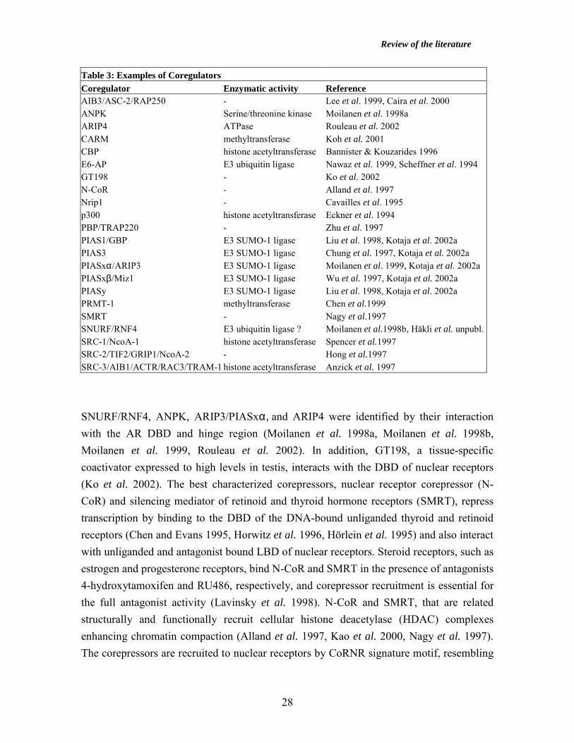
Coregulators .................................................................................................................... 27 Introduction ................................................................................................................. 27 CBP and p300.............................................................................................................. 29 p160 coactivators......................................................................................................... 30 Nrip1............................................................................................................................ 31 PPARγ –binding protein (PBP)/TRAP220.................................................................. 31 AIB3/RAP250/ASC-2 ................................................................................................. 32 PIAS protein family..................................................................................................... 32
Small nuclear RING finger protein SNURF/RNF4 ........................................................ 34 Structure of SNURF .................................................................................................... 34 Interaction partners of SNURF.................................................................................... 35
Contents
5
Expression of SNURF................................................................................................. 36 RING finger proteins in ubiquitylation ....................................................................... 37
AIMS OF THE STUDY...................................................................................................... 39 METHODS.......................................................................................................................... 40 RESULTS AND DISCUSSION ......................................................................................... 44
Expression of SNURF during murine gonad determination and prenatal differentiation (IV) .............................................................................................................................. 44 Expression of SNURF in spermatogenesis (II, III) ..................................................... 44 Expression of SNURF in murine postnatal ovary (IV) ............................................... 47 Transcriptional regulation of the murine SNURF gene (I) ......................................... 49 Hormonal regulation of SNURF gene expression in vivo in rodent gonads (II, IV)... 51 Expression of SNURF and estrogen receptor β in human testicular germ cells and testicular tumors (III, IV) ............................................................................................ 55
CONCLUDING REMARKS .............................................................................................. 59 ACKNOWLEDGEMENTS ................................................................................................ 60 REFERENCES.................................................................................................................... 61
Abstract
6
ABSTRACT
Steroid hormones, estrogens, androgens, progestins, glucocorticoids, and mineralocorticoids, regulate a variety of biological functions related to development, cell proliferation, differentiation, reproduction, and metabolism. The effects of the steroid hormones are mediated by steroid receptors, trans-acting transcription factors, which bind to regulatory sequences on target genes upon ligand binding, and either stimulate or repress gene expression. During recent years, a group of coregulatory proteins modulating steroid receptor responses in target gene regulation has been described. In gene inactivation experiments, some of the coregulatory proteins have been found to play essential roles in steroid receptor-mediated signaling, fertility, and fetal development. Small nuclear RING finger protein SNURF/RNF4, originally identified by its interaction with androgen receptor (AR), interacts with several transcription factors, such as TATA-binding protein (TBP) and promoter specificity protein 1 (Sp1). SNURF possesses a C-terminal RING finger domain, a motif typical of many E3 ubiquitin ligases and tumor suppressor proteins. Despite several in vitro interaction and expression studies reported during recent years, the biological function of SNURF has remained largely unknown. However, the expression of SNURF to high levels in testis suggests a role in reproduction and fertility. To gain insight into the role of SNURF in reproduction and testicular cancer, the expression of SNURF mRNA and protein was investigated during murine prenatal gonad development, in rodent postnatal folliculogenesis and spermatogenesis, and in human normal testis and testicular germ cell tumors. In murine fetal gonads, the expression of SNURF mRNA was detected from E10.5 dpc onwards in both sexes. SNURF mRNA was expressed to high levels in round and elongating spermatids in postnatal human and rat testis, and in oocytes of preantral follicles in postnatal murine ovary, respectively. Transcription from the murine SNURF promoter was governed by a single promoter, which showed very strong basal activity in reporter gene assays in mammalian cells. The proximal GC box at +9 nt of the SNURF promoter was identified as the main element governing transcription from the murine SNURF gene, and the Wilms’ tumor 1 (WT1) gene product was found as one of the potential activators of the murine SNURF promoter. Furthermore, the expression of SNURF mRNA was regulated by testosterone in vivo in rat testis and by gonadotropins and estrogen in vivo in rodent ovary. SNURF protein colocalized with estrogen receptor β (ERβ) protein in fetal and postnatal male germ cells. Down-regulated expression of SNURF and ERβ mRNA and protein was detected in human testicular germ cell tumors, suggesting a role for SNURF and estrogen signaling in the pathogenesis of testicular germ cell cancer.

Original Publications
7
ORIGINAL PUBLICATIONS This thesis is based on the following original articles, referred to in the text by their Roman numerals: I Hirvonen SJ, Santti HH, Jänne OA, and Palvimo JJ (2002) GC-rich elements
flanking the transcription start site govern strong activation on the SNURF gene. Biochem Biophys Res Commun 291: 897-902.
II Yan W, Hirvonen-Santti SJ, Toppari J, Palvimo JJ, and Jänne OA (2002)
Expression of nuclear RING finger protein SNURF/RNF4 during rat testis development suggests a role in spermatid maturation. Mech Dev 118: 247-253.
III Hirvonen-Santti SJ, Rannikko A, Santti H, Savolainen S, Nyberg M, Jänne OA,
and Palvimo JJ (2003) Down-regulation of estrogen receptor β and transcriptional coregulator SNURF/RNF4 in testicular germ cell cancer. Eur Urol, in press.
IV Hirvonen-Santti SJ, Sriraman V, Anttonen M, Savolainen S, Palvimo JJ,
Heikinheimo M, Richards JS, and Jänne OA (2003) Nuclear RING finger protein SNURF/RNF4 expression during gonad development: regulation by gonadotropins and estrogen in postnatal ovary. Endocrinology, submitted, in revision.
The original publications are reproduced with the permission of the copyright holders. In addition, some unpublished data are presented.

Abbreviations
8
ABBREVIATIONS AF activation function AP-1 activating protein-1 AR androgen receptor CBP CREB (cyclic AMP-responsive element -binding protein) -binding protein CREM cyclic AMP-responsive element modulator DBD DNA-binding domain DES diethylstilbestrol dpc days post coitum DRIP vitamin D receptor-interacting protein E2 estradiol EDS ethylene dimethane sulphonate EMSA electrophoretic mobility shift assay ER estrogen receptor FSH follicle-stimulating hormone GR glucocorticoid receptor HAT histone acetyltransferase hCG human chorionic gonadotropin HDAC histone deacetylase HRE hormone response element LBD ligand-binding domain LH luteinizing hormone MAA methoxyacetic acid N-CoR nuclear receptor corepressor NF-κB nuclear factor κB NLS nuclear localization signal Nrip1 Nuclear receptor -interacting protein 1 PGC primordial germ cell PIAS protein inhibitor of activated STAT PMSG gonadotropin from pregnant mare serum PPAR peroxisome proliferator-activated receptor PR progesterone receptor RNF4 ring finger protein 4 RT-PCR reverse transcription polymerase chain reaction SMRT silencing mediator for retinoic acid receptor and thyroid receptor SNURF small nuclear RING finger protein Sp1 promoter specificity protein 1 SRC steroid receptor coactivator SUMO small ubiquitin-like modifier T testosterone TBP TATA-binding protein TGC testicular germ cell cancer TRAP thyroid receptor-associated protein WT1 Wilms’ tumor 1 gene product
Review of the literature
9
REVIEW OF THE LITERATURE Introduction During fertilization, the fusion of sperm and egg, the specific combinations of genes encoded in the parental chromosomes are brought together to give life to an offspring in mammals. The female egg and male sperm originate from primordial germ cells (PGC) and differentiate into oocytes and gonocytes, respectively, prenatally. The germ cells gain full maturity during postnatal development in response to hormone signals and gonadal transcription factors. Disturbances during gonad development are likely to disrupt fertility and predispose to gonadal tumorigenesis, and thus better understanding of the mechanisms underlying gonadal development is mandatory for management of infertility and gonadal cancer. In this review, fetal and postnatal gonad development, testicular tumors, male infertility, steroid receptor signaling mechanisms, and coregulators are discussed. Sex Determination and Fetal Gonad Development Sex determination in mammals is a complex process, which is usually divided to determination of the gonad and its differentiation. During the first phase of gonad development, an indifferent bipotential primordium is established in both female and male mice from the thickening of coelomic epithelium on the ventrolateral surface of the mesonephros between embryonic day 10.5 and 11.5 (E10.5-11.5) dpc (for review, Capel 2000, Koopman et al. 2001, McLaren 2000, Swain and Lovell-Badge 1999, Veitia et al. 2001). The genital ridge is composed of the somatic cells derived from the mesonephros and proliferating primordial germ cells (PGC), which have migrated via the hindgut and mesonephros from the extraembryonic mesoderm at the base of the allantois. Expression of Wilms’ tumor 1 (WT1) and steroidogenic factor-1 (SF1) begins in the bipotential ridge by E10.0 dpc. SF1 is an orphan nuclear receptor that regulates genes involved in steroid synthesis in both the gonad and the adrenal gland. Impairment of WT1 and SF1 genes resulted in complete regression of the initially formed gonadal ridges by E14.5 dpc and E12.5 dpc, respectively (Kreidberg et al. 1993, Luo et al. 1994). The gonad initially develops in a non-sex-specific manner, being morphologically identical in XX and XY embryos up until E12.0 dpc. At E10.5-11.0 dpc, Sry, the sex determining gene on Y chromosome, begins to be expressed in the male genital ridge, and it guides the initial testis determination by

Review of the literature
10
triggering differentiation of pre-Sertoli cells (Clarkson and Harley 2002, Koopman et al. 2001). In addition, Sry-related Sox9 (Sry-like HMG-box protein 9) is expressed in the male gonad from E11.5 dpc onwards persisting there throughout the life, while it is not seen in the ovary (Clarkson and Harley 2002). SF1 and Sox9 activate the expression of the anti-müllerian hormone gene (AMH, also called Müllerian inhibiting substance, MIS), which induces the regression of the female urogenital tract forming Müllerian ducts in males (de Santa Barbara et al. 1998, Shen et al. 1994) (Fig. 1). Transgenic experiments where XX mice carrying the Sry and Sox9 genes develop as males have established the necessity of these factors in testis determination (Koopman et al. 1991, Vidal et al. 2001). By E12.5 dpc, the Sertoli cells have encircled germ cells and formed testis cords, surrounded by peritubular myoid cells. Other somatic cells in the developing testis are Leydig cells, which produce testosterone, critical for the development of Wolffian ducts to male urogenital tract and the formation of all male secondary sexual characteristics (Fig. 1, Table 1). At E13.5 dpc, oogonia in the murine ovary enter the meiotic prophase arresting at diplotene stage, while the majority of the germ cells within the testicular cords undergo mitotic arrest. Dax1, an orphan nuclear receptor, is expressed concomitantly with Sry in both sexes, but it is down-regulated in the testis and stays in the ovary during further prenatal gonad development (Swain et al. 1996). Dax1 has been thought to act antagonistically to Sry in a dosage-sensitive manner, since duplication of the Xp21 locus bearing the Dax1 gene resulted in XY sex reversal (Swain et al. 1998). However, murine Dax1 was essential for normal spermatogenesis in later life (Jeffs et al. 2001). In addition, Wnt-4 factor was involved in the suppression of Leydig cell formation in the ovary (Vainio et al. 1999). Wnt-4 null mutant female mice were masculinized (no Müllerian ducts and persistence of Wolffian structures), while the male phenotype was normal (Vainio et al. 1999). Table 1: Timeline for murine gonad development dpc E9.5-E11 PGCs migrate from yolk sac to posterior body wall, where they induce formation of genital
ridges. E11.0 Cells from the coelomic epithelium of mesonephros proliferate. E12.5 Sertoli cells encircle the germ cells and form testicular cords. E13.5 Testicular morphology recognizable. Female oogonia enter I meiosis, while male germ cells
enter mitotic arrest. Development of external genitalia begins. Modified from Rey R and Picard JY, Baillieres Clin Endocrinol Metab, 1998.

Review of the literature
11
Fig. 1 Development of the male and female internal genitalia from bipotential gonadal primordial.
Pre-Sertoli cells and Leydig cells in the fetal testis produce Müllerian inhibiting substance (MIS) and
testosterone (T), respectively. MIS induces the regression of Müllerian ducts in the male and T stimulates the
development of the male urogenital tract from the Wolffian ducts and the formation of external genitalia.
Modified from Rey and Picard, Baillieres Clin Endocrinol Metab, 1998.
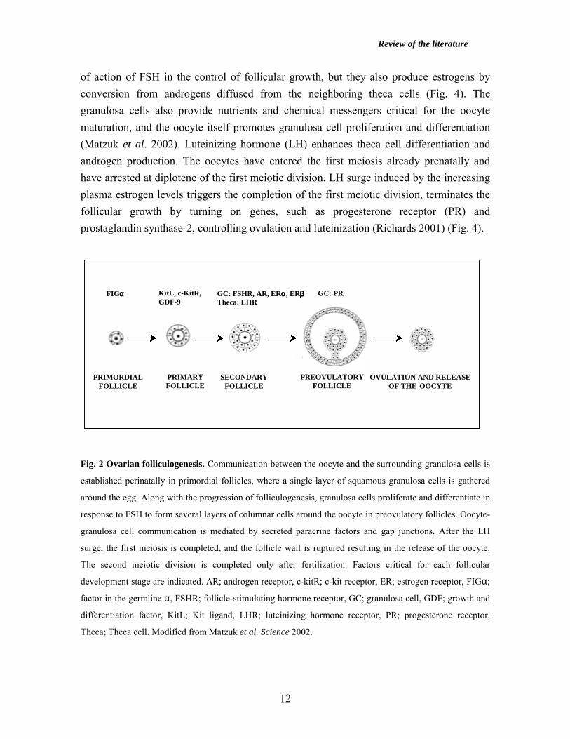
Postnatal Gonad Development Ovarian folliculogenesis During folliculogenesis, the primordial follicles consisting of an oocyte and a single layer of squamous granulosa cells develop into large preovulatory antral follicles, which contain several layers of columnar granulosa cells surrounding the oocytes (for review, Epifano and Dean 2002, Matzuk et al. 2002, Richards 2001) (Fig. 2). Growth of the primordial follicles to primary follicles requires the Kit ligand produced by the granulosa cells and c-kit receptor expressed in the egg as well as growth and differentiation factor 9 (GDF-9), an oocyte-specific member of TGFβ growth factor family (Elvin et al. 1999). GDF-9 null mice exhibited failed organization of theca cells, abnormal oocyte growth, and arrested follicle maturation to the primary follicle stage (Elvin et al. 1999, Richards 2001). The initiation of follicle growth occurs independently of pituitary gonadotropins, but follicle-stimulating hormone (FSH) is mandatory for the final growth of the preovulatory follicles. FSH promotes granulosa cell proliferation and stimulates expression of CYP450 aromatase in the granulosa cells (Richards 2001). The granulosa cells are not only the site
Indifferent gonads
mesonephros
Müllerian ductWolff ian duct
Male Testis Female Ovary
MIS, T
Wolffian duct:epididymis,vas deferens,seminal vesicle
Müllerian duct:fallopian tubes,uterus,upper vagina
Indifferent gonads
mesonephros
Müllerian ductWolff ian duct
Male Testis Female Ovary
MIS, T
Wolffian duct:epididymis,vas deferens,seminal vesicle
Müllerian duct:fallopian tubes,uterus,upper vagina
Review of the literature
12
of action of FSH in the control of follicular growth, but they also produce estrogens by conversion from androgens diffused from the neighboring theca cells (Fig. 4). The granulosa cells also provide nutrients and chemical messengers critical for the oocyte maturation, and the oocyte itself promotes granulosa cell proliferation and differentiation (Matzuk et al. 2002). Luteinizing hormone (LH) enhances theca cell differentiation and androgen production. The oocytes have entered the first meiosis already prenatally and have arrested at diplotene of the first meiotic division. LH surge induced by the increasing plasma estrogen levels triggers the completion of the first meiotic division, terminates the follicular growth by turning on genes, such as progesterone receptor (PR) and prostaglandin synthase-2, controlling ovulation and luteinization (Richards 2001) (Fig. 4). Fig. 2 Ovarian folliculogenesis. Communication between the oocyte and the surrounding granulosa cells is
established perinatally in primordial follicles, where a single layer of squamous granulosa cells is gathered
around the egg. Along with the progression of folliculogenesis, granulosa cells proliferate and differentiate in
response to FSH to form several layers of columnar cells around the oocyte in preovulatory follicles. Oocyte-
granulosa cell communication is mediated by secreted paracrine factors and gap junctions. After the LH
surge, the first meiosis is completed, and the follicle wall is ruptured resulting in the release of the oocyte.
The second meiotic division is completed only after fertilization. Factors critical for each follicular
development stage are indicated. AR; androgen receptor, c-kitR; c-kit receptor, ER; estrogen receptor, FIGα;
factor in the germline α, FSHR; follicle-stimulating hormone receptor, GC; granulosa cell, GDF; growth and
differentiation factor, KitL; Kit ligand, LHR; luteinizing hormone receptor, PR; progesterone receptor,
Theca; Theca cell. Modified from Matzuk et al. Science 2002.
FIGαααα KitL, c-KitR, GDF-9
GC: FSHR, AR, ERαααα, ERββββ Theca: LHR
GC: PR
PRIMORDIAL FOLLICLE
PRIMARY FOLLICLE
SECONDARY FOLLICLE
PREOVULATORY FOLLICLE
OVULATION AND RELEASE OF THE OOCYTE

Review of the literature
13
Spermatogenesis Spermatogenesis is a complex multi-step process, in which diploid spermatogonia differentiate into mature haploid spermatozoa in the epithelium of the seminiferous tubules during the postnatal testis development (for review, Grootegoed et al. 2000, Hecht 1998). Spermatogenesis is often divided into proliferation, meiotic, and spermatogenic phases (Russell et al. 1990). In the proliferative phase, most spermatogonia, which have arisen from the PGCs and gonocytes, located close to the basement membrane of the tubule, enter mitosis to give rise to a reserve of undifferentiated stem cells, and only a minority of spermatogonia become diploid preleptotene spermatocytes. During the meiotic phase, the diploid preleptotene cells pass through two meiotic divisions to produce haploid round spermatids. The long prophase of the first meiotic division lasting approximately three weeks in rodent testis, is initiated at leptotene spermatocytes and is subsequently followed by zygotene phase, in which homologous sister chromatids are paired and a synaptonemal complex is formed. In the ensuing pachytene spermatocytes, genetic material is recombined (crossing-over), and in diplotene spermatocytes it is segregated to sister chromatids. During the meiotic cell divisions, the germ cells remain connected by cytoplasmic bridges, and the syncytia move towards the tubular lumen. In differentiation or spermiogenic phase, the round spermatids undergo a series of molecular and morphological changes to generate mature spermatozoa. Changes in the composition of chromatin resulting in the formation of the spermatid head, acrosome development, and flagellum formation, take place during spermiogenesis to allow transmission of the paternal chromosomes to the oocyte at fertilization (Fig. 3). Germ cell development takes place in close association with the somatic Sertoli cells of the seminiferous epithelium (Fig. 3). Sertoli cell-germ cell interaction provides physical support, tight junctions, growth factors, and nutrients critical for germ cell development (for review, Griswold 1995, Griswold 1998). Importantly, the Sertoli cells form blood-testis barrier, which allows them to create a microenvironment in the tubular compartment, in which meiotic divisions and postmeiotic modifications take place. The associations between the Sertoli cells and the germ cells at different phases of maturation are referred to as stages and comprise the cycle of the seminiferous tubule (Leblond and Clermont 1952). Groups of spermatogonia enter the spermatogenic process at regular intervals, and the cycle is defined as a time interval between the appearance of germ cells at the same stage at the defined point of the tubule. In mice and men, spermatogenesis lasts for 35 and 70 days, respectively.

Review of the literature
14
Fig. 3 Top: The Flow of Spermatogenesis. During the fetal period, primordial germ cells (PGC)
differentiate into gonocytes, which give rise to spermatogonia right after birth. Spermatogonia proliferate to
maintain a pool of undifferentiated germ cells or, more rarely, give rise to preleptotene primary
spermatocytes. The primary spermatocytes proceed via the first and second meiotic divisions to become
secondary spermatocytes and round spermatids, respectively. During spermiogenesis, chromatin is
compacted, and acrosome and flagellum develop to generate mature haploid spermatozoa. Modified from
Sassone-Corsi, Cell, 1997.
Bottom: Organization of the developing germ cells and Sertoli cells in the seminiferous tubules in
testis. Spermatogonia, spermatocytes, and round spermatids are seen in successively higher levels within the
seminiferous epithelium. Sertoli cells and spermatogonia contact the basement membrane. Sertoli cells reach
from the base of the tubule to the lumen and encircle the developing germ cells. Tight junctions, gap
junctions, and desmosomes connect Sertoli cells to each other to create the blood-testis barrier and a
microenvironment for germ cell maturation. Modified from Russell et al., 1990.
PRIMORDIAL GERM CELL
GONOCYTE SPERMATOGONIA PRIMARY SPERMATOCYTE
SECONDARY SPERMATOCYTE
SPERMATID SPERMATOZOON
SPERMATOGONIA Fetal testis Postnatal testis
stem cell renewal pathway
mitosis 1st meiosis 2nd meiosis spermiogenesis
BASAL LAMINA
Tubule lumen
Interstitial compartment
ELONGATING SPERMATID
ROUND SPERMATID
SPERMATOCYTE
SPERMATOGONIA
MYOID CELL
TIGHT JUNCTION
SERTOLI CELL
PROLIFERATIVE MEIOTIC SPERMIOGENIC
Review of the literature
15
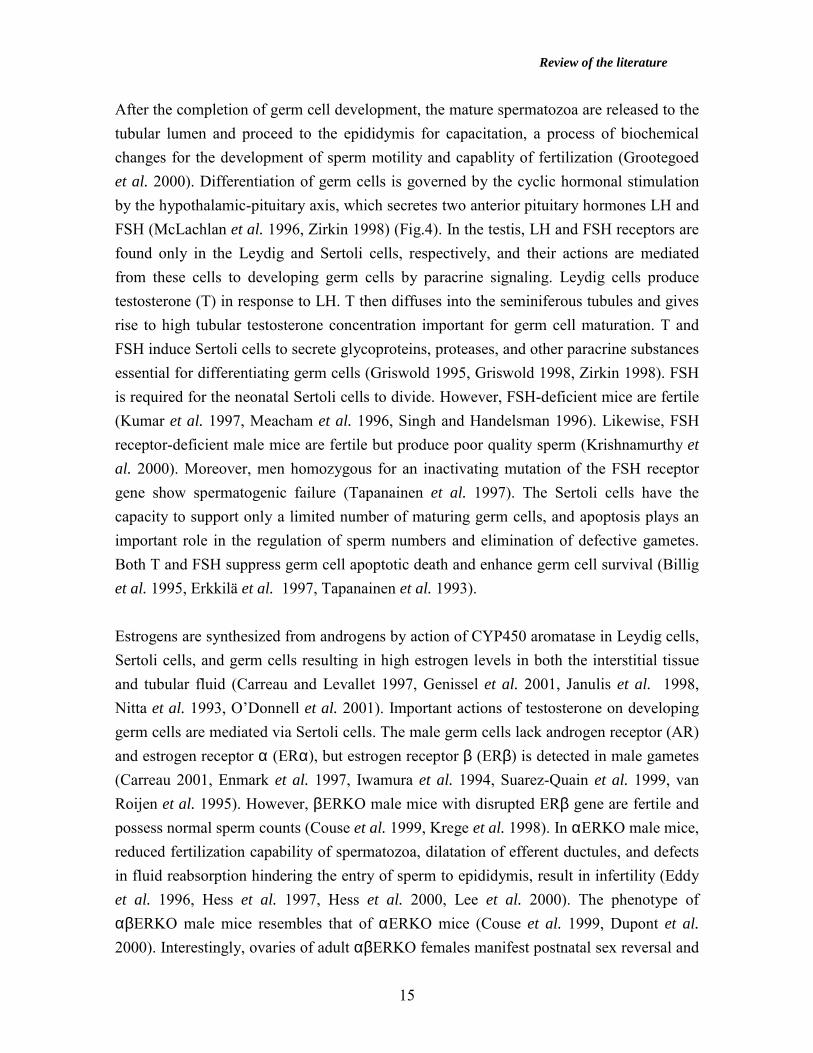
After the completion of germ cell development, the mature spermatozoa are released to the tubular lumen and proceed to the epididymis for capacitation, a process of biochemical changes for the development of sperm motility and capablity of fertilization (Grootegoed et al. 2000). Differentiation of germ cells is governed by the cyclic hormonal stimulation by the hypothalamic-pituitary axis, which secretes two anterior pituitary hormones LH and FSH (McLachlan et al. 1996, Zirkin 1998) (Fig.4). In the testis, LH and FSH receptors are found only in the Leydig and Sertoli cells, respectively, and their actions are mediated from these cells to developing germ cells by paracrine signaling. Leydig cells produce testosterone (T) in response to LH. T then diffuses into the seminiferous tubules and gives rise to high tubular testosterone concentration important for germ cell maturation. T and FSH induce Sertoli cells to secrete glycoproteins, proteases, and other paracrine substances essential for differentiating germ cells (Griswold 1995, Griswold 1998, Zirkin 1998). FSH is required for the neonatal Sertoli cells to divide. However, FSH-deficient mice are fertile (Kumar et al. 1997, Meacham et al. 1996, Singh and Handelsman 1996). Likewise, FSH receptor-deficient male mice are fertile but produce poor quality sperm (Krishnamurthy et al. 2000). Moreover, men homozygous for an inactivating mutation of the FSH receptor gene show spermatogenic failure (Tapanainen et al. 1997). The Sertoli cells have the capacity to support only a limited number of maturing germ cells, and apoptosis plays an important role in the regulation of sperm numbers and elimination of defective gametes. Both T and FSH suppress germ cell apoptotic death and enhance germ cell survival (Billig et al. 1995, Erkkilä et al. 1997, Tapanainen et al. 1993). Estrogens are synthesized from androgens by action of CYP450 aromatase in Leydig cells, Sertoli cells, and germ cells resulting in high estrogen levels in both the interstitial tissue and tubular fluid (Carreau and Levallet 1997, Genissel et al. 2001, Janulis et al. 1998, Nitta et al. 1993, O’Donnell et al. 2001). Important actions of testosterone on developing germ cells are mediated via Sertoli cells. The male germ cells lack androgen receptor (AR) and estrogen receptor α (ERα), but estrogen receptor β (ERβ) is detected in male gametes (Carreau 2001, Enmark et al. 1997, Iwamura et al. 1994, Suarez-Quain et al. 1999, van Roijen et al. 1995). However, βERKO male mice with disrupted ERβ gene are fertile and possess normal sperm counts (Couse et al. 1999, Krege et al. 1998). In αERKO male mice, reduced fertilization capability of spermatozoa, dilatation of efferent ductules, and defects in fluid reabsorption hindering the entry of sperm to epididymis, result in infertility (Eddy et al. 1996, Hess et al. 1997, Hess et al. 2000, Lee et al. 2000). The phenotype of αβERKO male mice resembles that of αERKO mice (Couse et al. 1999, Dupont et al. 2000). Interestingly, ovaries of adult αβERKO females manifest postnatal sex reversal and

Review of the literature
16
exhibit follicle transdifferentiation to structures resembling seminiferous tubules of the testis including Sertoli cell-like cells (Couse et al. 1999). Estradiol (E2) influences the negative feedback to the hypothalamus and the pituitary via conversion from androgens, and it controls the secretion of LH and FSH (Lindzey et al. 1998, O’Donnell et al. 2001). Thus, absence or inappropriate exposure to estrogens could disturb the balance of the hypothalamic-pituitary axis. In addition, estrogens play a role in testicular descent, the hormone-regulated migration of testes from the abdominal wall to scrotum, by regulating fetal Leydig cell gene expression (Hutson et al. 1997, Nef and Parada 2000, O’Donnell et al. 2001).
Fig. 4 Hypothalamic-pituitary-gonadal axis in the regulation of gonad function. Testis: Inhibin, a
protein hormone secreted by the Sertoli cells, feeds back to the anterior pituitary and mainly inhibits
secretion of FSH. Secretion of LH and gonadotropin-releasing hormone (GnRH) is inhibited mainly by
testosterone (T) secreted from the Leydig cells. T, acting locally, is essential for spermatogenesis. Ovary:
Estradiol (E2), in low plasma concentration during the early and middle follicular phase, feeds back
negatively to the hypothalamus and the anterior pituitary to decrease secretion of GnRH and LH,
respectively. Inhibin, secreted from the granulosa cells, acts on the pituitary to inhibit the secretion of FSH.
Estrogen, in high plasma levels preceding ovulation, triggers the secretion of LH surge from the pituitary
(positive feed-back). High plasma concentration of progesterone during the luteal phase and pregnancy
inhibits the hypothalamic neurons that secrete GnRH. Androgens produced in response to LH in theca cells
diffuse into granulosa cells, in which they are converted to estrogens by CYP450 aromatase. Modified from
Vander et al., Human Physiology, McGraw Hill, 2001.
+
+
+
+
HYPOTHALAMUS HYPOTHALAMUS
ANTERIOR PITUITARY ANTERIOR PITUITARY
TESTIS OVARY
Leydig cells: T Sertoli cells: inhibin, paracrine substances
Theca cells: Androstenedione Granulosa cells: inhibin, E2
T GnRH
T (inhibits only LH)
Inhibin (inhibits only FSH)
FSH, LH
E2GnRH
E2
+ (high E2 plasma levels)
Inhibin (inhibits only FSH)
FSH, LH

Review of the literature
17
Marked stage-specific gene regulation underlies the morphological changes of developing germ cells during spermatogenesis (for review, Sassone-Corsi 1997, Steger 1999). Various promoters display differential activity in male gametes due to the presence of testis-specific transcription factors (Foulkes et al. 1992). Some general transcription factors, such as TATA-binding protein (TBP), TFIIB, and RNA polymerase II are differentially regulated in germ cells and accumulate in postmeiotic round spermatids where high transcriptional activity takes place (Schmidt and Schibler 1995). In addition, many ubiquitously expressed genes generate testis-specific isoforms by the usage of alternative promoters, splicing, or polyadenylation (Daniel and Habener 1998, Moilanen et al. 1998b, Schmidt et al. 1997). The hypothalamic-pituitary axis controls the postmeiotic gene activation by enhancing accumulation of the CREM activator protein CREMτ in the male germ cells in response to FSH (Foulkes et al. 1993). CREMτ is a consensus cAMP response-element (CRE) binding protein, that is highly expressed from round spermatids onwards and regulates transcription from several postmeiotic genes, such as protamines, calspermin, and transition protein (Kistler et al. 1994, Sassone-Corsi 1998, Sun et al. 1995). In addition to CREMτ activator, the CREM gene encodes transcriptional repressors CREMα, β, and γ, which are expressed to low levels in premeiotic germ cells (Foulkes et al. 1992). Targeted disruption of the CREM gene results in complete arrest of the germ cell differentiation at the first step of spermiogenesis (Blendy et al. 1996, Nantel et al. 1996). Recently, male germ cell-specific transcriptional coregulators have also been identified, as exemplified by the LIM-only protein ACT, that enhances CREM-mediated transcription in the germ cells (Sassoni-Corsi 2002). Transcription ceases several days before the completion of spermatogenesis because of the condensation of the haploid genome to a volume of approximately 5% of that of somatic cell nuclei (Ward and Coffey 1991, Ward 1993). In round spermatids, the histones and non-histone proteins are replaced by transition proteins, and in elongating spermatids, the transition proteins are removed and replaced by protamines, the principal basic nuclear proteins in spermatozoa (Dadoune 1995, Oliva and Dixon 1991, Wouters-Tyrou et al. 1998). Translational regulation plays a significant role in maturing spermatids: a number of proteins appear in the late stages of spermiogenesis and must be translated from mRNA in postmeiotic germ cells (for review, Steger 1999). Translational regulation is exemplified by direct repression of mRNA by RNA-binding proteins, such as 70-kDa poly-A binding protein (PABP), and activation by deadenylation of long poly-A tails (Gu et al. 1995, Venables and Eperon 1999).
Review of the literature
18
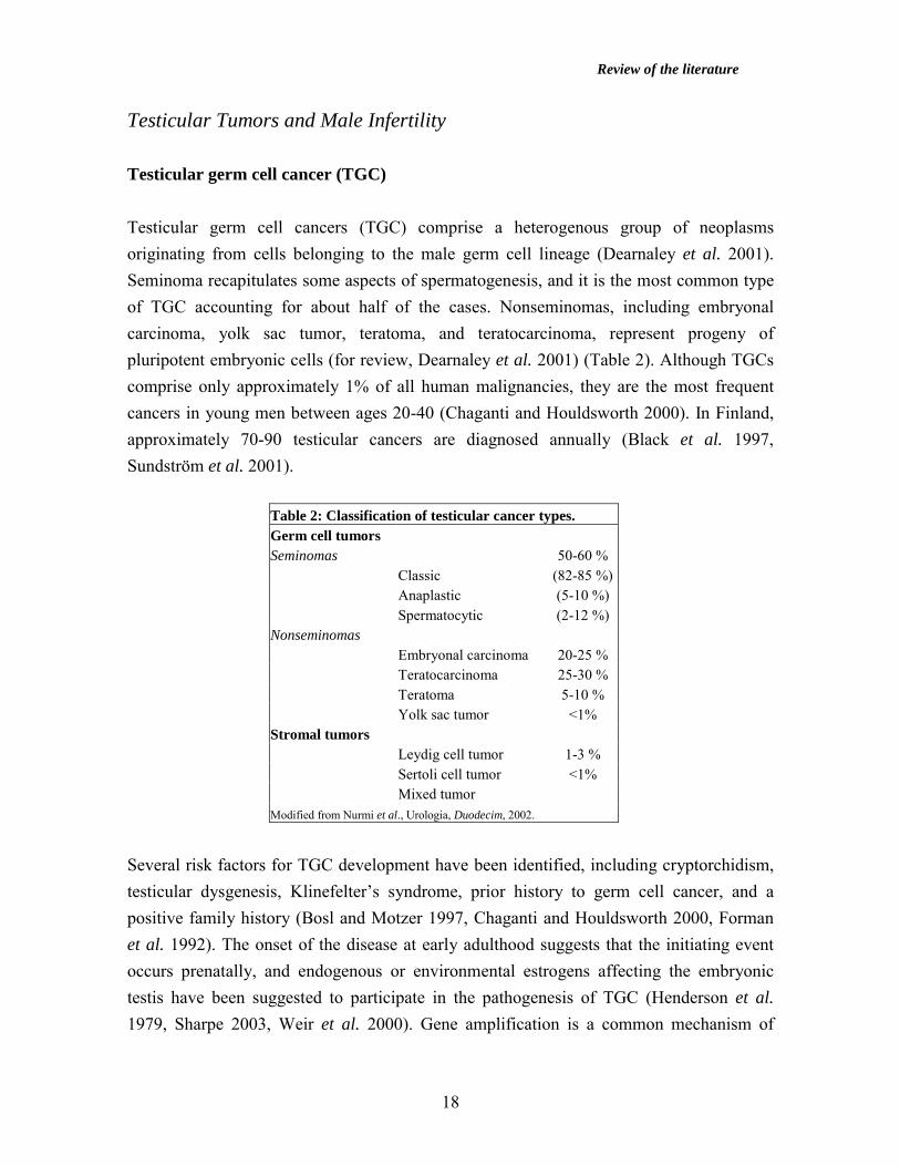
Testicular Tumors and Male Infertility Testicular germ cell cancer (TGC) Testicular germ cell cancers (TGC) comprise a heterogenous group of neoplasms originating from cells belonging to the male germ cell lineage (Dearnaley et al. 2001). Seminoma recapitulates some aspects of spermatogenesis, and it is the most common type of TGC accounting for about half of the cases. Nonseminomas, including embryonal carcinoma, yolk sac tumor, teratoma, and teratocarcinoma, represent progeny of pluripotent embryonic cells (for review, Dearnaley et al. 2001) (Table 2). Although TGCs comprise only approximately 1% of all human malignancies, they are the most frequent cancers in young men between ages 20-40 (Chaganti and Houldsworth 2000). In Finland, approximately 70-90 testicular cancers are diagnosed annually (Black et al. 1997, Sundström et al. 2001).
Table 2: Classification of testicular cancer types. Germ cell tumors Seminomas 50-60 % Classic (82-85 %) Anaplastic (5-10 %) Spermatocytic (2-12 %) Nonseminomas Embryonal carcinoma 20-25 % Teratocarcinoma 25-30 % Teratoma 5-10 % Yolk sac tumor <1% Stromal tumors Leydig cell tumor 1-3 % Sertoli cell tumor <1% Mixed tumor Modified from Nurmi et al., Urologia, Duodecim, 2002.
Several risk factors for TGC development have been identified, including cryptorchidism, testicular dysgenesis, Klinefelter’s syndrome, prior history to germ cell cancer, and a positive family history (Bosl and Motzer 1997, Chaganti and Houldsworth 2000, Forman et al. 1992). The onset of the disease at early adulthood suggests that the initiating event occurs prenatally, and endogenous or environmental estrogens affecting the embryonic testis have been suggested to participate in the pathogenesis of TGC (Henderson et al. 1979, Sharpe 2003, Weir et al. 2000). Gene amplification is a common mechanism of

Review of the literature
19
increased gene expression in human cancers. The most common cytogenetic aberration in TGC is the presence of an isochromosome (duplication) of the short arm of chromosome 12 in up to 80% of all TGC cases (Chaganti and Houldsworth 2000). In accordance with the overpresentation of 12p, mRNA encoded by the cyclin D2 gene, located at 12p13, was over-expressed in 69% of the TGCs by a mean factor of 7.9 as judged by semi-quantitative RT-PCR assay (Schmidt et al. 2001). Cyclin D2 along with the CDK4 protein regulates phosphorylation of the RB tumor suppressor protein and controls the G1-S cell cycle checkpoint (Schmidt et al. 2001). In addition, specific gains and losses from several other chromosomal regions, e.g. overpresentation of 17q, have been reported in TGC (Skotheim et al. 2002). Furthermore, the first mouse model of classical testicular seminoma has been identified in transgenic mice over-expressing glial cell-derived neurotrophic factor (GDNF), a member of TGFβ superfamily expressed also in Sertoli cells (Meng et al. 2001). The treatment of testicular tumors consists of radical orchiectomy (removal of the testis), and chemotherapy and/or radiotherapy depending on the tumor stage (Dearnaley et al. 2001, Nurmi et al. 2002). To date, cisplatin-containing chemotherapy has enabled cure rates >90-95% in lower-stage testicular malignancies (Dearnaley et al. 2001, Nurmi et al. 2002). However, liver, bone, or central nervous system metastases, high alpha-fetoprotein (>1000 ng/ml) or human chorionic gonadotropin (hCG>10 000 IU/l) marker levels, or the presence of primary mediastinal tumor indicate poorer prognosis with 70% 5-year survival rate among the Finnish testicular cancer patients (Aareleid et al. 1998, Mäenpää et al. 1996, Nurmi et al. 2002). Male infertility Approximately 10% of couples trying to conceive suffer from subfertility, with approximately one third of the cases deriving from male factor infertility. Potential causes of male infertility comprise varicocele, undescended testes, antibodies against spermatozoa, microdeletions in the Y-choromosomal AZF region, testicular and genital tract infections, chromosomal disorders such as Klinefelter’s syndrome (47XXY), systemic disease, external factors (such as drugs), radiotherapy, chemotherapy, and insufficient or excess reproductive hormone production. However, most often the reason for the poor quality of semen remains undetermined (for review, Cahill and Wardle 2002, Ford 2001, Hirsh 2003, Khorram et al. 2001). Patients suffering from hypogonadotropic hypogonadism as a major cause of infertility benefit from medication (Khorram et al.
Review of the literature
20
2001). Furthermore, introduction of intracytoplasmic sperm injection (ICSI) has dramatically improved the treatment of male factor infertility (Braude and Rowell 2003, Cahill and Wardle 2002, Ford 2001, Khorram et al. 2001). Nuclear Receptors Nuclear receptor superfamily The actions of lipophilic hormones, including steroids, retinoids, vitamin D, and thyroid hormone, are mediated through the conserved family of nuclear receptors which function as ligand-regulated, DNA-binding transcription factors (for review, Aranda and Pascual 2001, Beato et al. 1996, Mangelsdorf et al. 1995, McKenna et al. 1999). Fourty-eight human genes encoding members of the nuclear receptor superfamily sharing extensive homology have been identified to date (Mangelsdorf et al. 1995, McKenna and O’Malley 2002b). Identification of genes with structural features similar to those found in nuclear receptors has lead to the discovery of orphan receptor members, such as PPAR, ERR, Nurr1, LXR, and PXR, of the family without prior knowledge of their putative ligands (Giguère 1999). Recently, ligands have been found for many orphan receptors: LXR binds oxysterols, FXR bile acids, PXR pregnanes, and PPAR several fatty acids and prostaglandins (Giguère 1999). This chapter focuses mainly on the steroid receptors, especially androgen receptor (AR) and estrogen receptors (ER). Androgen receptor (AR) Physiological roles of androgens Androgen receptor (AR) mediates the responses of androgens that affect a male body in multiple ways. Androgens regulate the development of sex organs, the growth of facial, body, and pubic hair, the enlargement of vocal cords, the growth of the prostate, the production of sperm, and the development of muscle strength, and masculine behavior (Mooradian et al. 1987, Quigley et al. 1995). AR has two physiological ligands, testosterone and 5α-dihydrotestosterone (DHT), which is converted from testosterone by 5α-reductase in target cells (Mooradian et al. 1987). In certain cells, testosterone is converted to 17β-estradiol by CYP450 aromatase. After binding the androgen ligand, AR regulates the bodily functions by interacting with hormone response elements (HRE) located in the regulatory regions of the target genes. This leads to transcriptional activation

Review of the literature
21
or repression of the regulated genes (Beato et al. 1996). Among genes activated by AR are those encoding rat probasin, mouse sex-limited protein (slp), human glandular kallikrein (KLK2), and prostate-specific antigen (PSA) (Adler et al. 1992, Murtha et al. 1992, Rennie et al. 1993, Riegman et al. 1991). Functional domains of androgen receptor Androgen receptor consists of an amino-terminal region (region A/B), a DNA-binding domain (DBD, region C), a hinge region (region D), and a ligand-binding domain (LBD, region E)(for review, Gelmann 2002). The DBD and LBD of AR share 77-80% and 50-55% homology, respectively, with GR, PR, and MR. The amino-terminal modulatory A/B domain of AR is the most variable in both size and sequence among the steroid receptors. The A/B domain harbors a ligand-independent transactivation function (AF-1), communicates with other parts of the receptor and interacts with several coregulator proteins, such as SRC-1 and CBP (Alen et al. 1999, Ikonen et al. 1997). The DBD of the steroid receptors is the most conserved region, and it confers ability to recognize specific target sequences and activate genes. The DBD of AR is composed of two zinc finger modules comprising some 70 amino acid residues and a carboxyl-terminal extension that spans approximately 25 residues. The second zinc finger participates in receptor dimerization and, along with the carboxyl-terminal region, is involved in HRE recognition (Freedman 1992, Gelmann 2002, Schoenmakers et al. 1999). The D domain, which is less well conserved among the nuclear receptors, serves as a hinge between the DBD and the LBD, allowing rotation of the DBD. The hinge region contains the major part of the nuclear localization signal, which mediates the transfer of AR from the cell cytoplasm to its site of action in the nucleus (Jenster et al. 1993, Zhou et al. 1994). The LBD is the site of specific, high-affinity binding of the androgen ligand. Recently, the three-dimensional structure of the AR LBD has been determined (Matias et al. 2000, Sack et al. 2001). Like other steroid receptors, AR LBD is composed of 12 α-helices that form a ligand-binding pocket. After agonist binding, helix 12 is positioned over the pocket to enclose the ligand (Sack et al. 2001). The LBD of AR possesses the second activation function (AF-2) and interacts with coregulators (Ikonen et al. 1997). Deletion of AR LBD results in a constitutively active receptor, indicating strongly that AF-1 harbors the main transactivation activity of AR (Jenster et al. 1993, Gelmann 2002).

Review of the literature
22
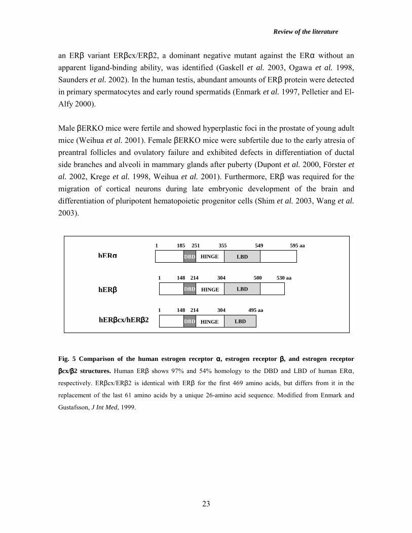
Androgen receptor gene and disease The human gene for AR encompasses approximately 80 kb and is mapped to a conserved region on chromosome Xq11-12 (Lubahn et al. 1988, Quigley et al. 1995). AR protein is approximately 110 kDa in size, and it is polymorphic due to the polymeric glutamine and proline repeats in the first exon of the AR gene. The glutamine (CAG) repeat length polymorphism contributes to the pathogenesis of Kennedy’s disease (also called spinal and bulbar muscular atrophy (SBMA)), that is an adult-onset, slowly progressing motoneuron disease (LaSpada et al. 1991, Yong et al. 2000). The number of CAG repeats increases with the severity of the Kennedy’s disease, and affected males show variable degrees of gynecomastia, testicular atrophy, and subfertility (LaSpada et al. 1991). In contrast, short CAG repeat length of AR was associated with more aggressive prostate cancer occurrence, earlier age of disease onset, and likelihood of prostate cancer recurrence (Bratt et al. 1999, Giovannucci et al. 1997, Hardy et al. 1996, Nam et al. 2000). The AR gene was amplified in 30% of hormone-refractory prostate cancers, and mutations in the LBD of AR were shown to broaden ligand specificity of the receptor in recurrent prostate cancer (Gregory et al. 2001). Loss of AR function in genetic (XY) males leads to complete androgen insensitivity syndrome with an external phenotype of a sterile woman, undescended testes, and lack of ovaries and uterus (Yong et al. 2000). In testicular feminized mice (Tfm) with a spontaneous single point mutation in the AR gene resulting in truncation and inactivation of AR, testicular migration and spermatogenesis are impaired (Charest et al. 1991, Lyon and Hawkes 1970, Lyon et al. 1975). Interestingly, AR knockout male mice (ARKO), generated by using the cre-lox conditional knockout strategy, showed female-like external genitalia, severely reduced testicular size, lowered testosterone concentration, Leydig cell hyperplasia, and arrested spermatogenesis at pachytene spermatocyte stage (Yeh et al. 2002). Estrogen receptors (ER) Estrogens have traditionally been connected to female reproduction, but during recent years they have been reported to play a role in the male reproduction, bone metabolism, and physiology of the cardiovascular and central nervous systems (Enmark and Gustafsson 1999). Estrogen signals are mediated by two estrogen receptors, estrogen receptor α (ERα) and estrogen receptor β (ERβ), of which only ERβ is expressed in the male germ cells (Fig. 5) (Enmark et al. 1997, Kuiper et al. 1996). The human ERβ gene is localized to a different chromosome than the ERα gene, to 14q22-24 (Enmark et al. 1997). Recently,
Review of the literature
23
an ERβ variant ERβcx/ERβ2, a dominant negative mutant against the ERα without an apparent ligand-binding ability, was identified (Gaskell et al. 2003, Ogawa et al. 1998, Saunders et al. 2002). In the human testis, abundant amounts of ERβ protein were detected in primary spermatocytes and early round spermatids (Enmark et al. 1997, Pelletier and El-Alfy 2000). Male βERKO mice were fertile and showed hyperplastic foci in the prostate of young adult mice (Weihua et al. 2001). Female βERKO mice were subfertile due to the early atresia of preantral follicles and ovulatory failure and exhibited defects in differentiation of ductal side branches and alveoli in mammary glands after puberty (Dupont et al. 2000, Förster et al. 2002, Krege et al. 1998, Weihua et al. 2001). Furthermore, ERβ was required for the migration of cortical neurons during late embryonic development of the brain and differentiation of pluripotent hematopoietic progenitor cells (Shim et al. 2003, Wang et al. 2003). Fig. 5 Comparison of the human estrogen receptor αααα, estrogen receptor ββββ, and estrogen receptor
ββββcx/ββββ2 structures. Human ERβ shows 97% and 54% homology to the DBD and LBD of human ERα,
respectively. ERβcx/ERβ2 is identical with ERβ for the first 469 amino acids, but differs from it in the
replacement of the last 61 amino acids by a unique 26-amino acid sequence. Modified from Enmark and
Gustafsson, J Int Med, 1999.
hERαααα
hERββββ
hERββββcx/hERββββ2
DBD LBD
DBD
DBD
LBD
LBD
HINGE
HINGE
HINGE
1 185 251 355 549 595 aa
1 148 214 304 500 530 aa
1 148 214 304 495 aa
Review of the literature
24
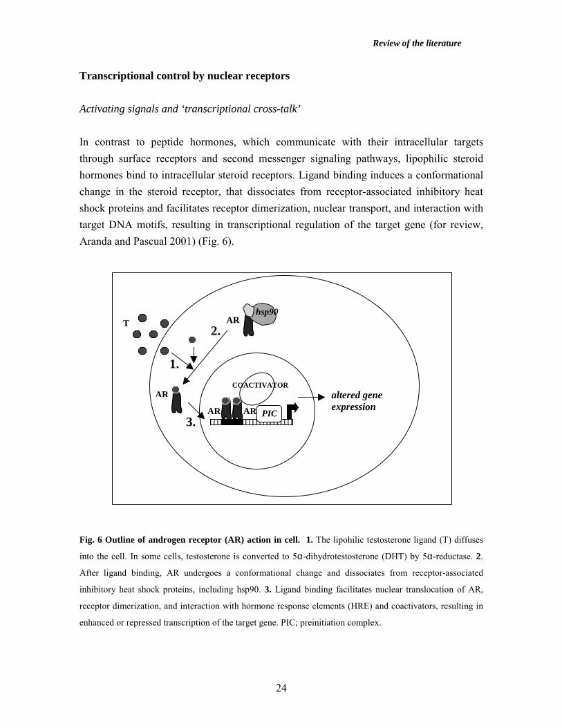
Transcriptional control by nuclear receptors Activating signals and ‘transcriptional cross-talk’ In contrast to peptide hormones, which communicate with their intracellular targets through surface receptors and second messenger signaling pathways, lipophilic steroid hormones bind to intracellular steroid receptors. Ligand binding induces a conformational change in the steroid receptor, that dissociates from receptor-associated inhibitory heat shock proteins and facilitates receptor dimerization, nuclear transport, and interaction with target DNA motifs, resulting in transcriptional regulation of the target gene (for review, Aranda and Pascual 2001) (Fig. 6). Fig. 6 Outline of androgen receptor (AR) action in cell. 1. The lipohilic testosterone ligand (T) diffuses
into the cell. In some cells, testosterone is converted to 5α-dihydrotestosterone (DHT) by 5α-reductase. 2.
After ligand binding, AR undergoes a conformational change and dissociates from receptor-associated
inhibitory heat shock proteins, including hsp90. 3. Ligand binding facilitates nuclear translocation of AR,
receptor dimerization, and interaction with hormone response elements (HRE) and coactivators, resulting in
enhanced or repressed transcription of the target gene. PIC; preinitiation complex.
T AR hsp90
AR
AR AR
COACTIVATOR
PIC
altered gene expression
1.
2.
3.

Review of the literature
25
A pair of palindromic 6-bp consensus AGAACA motifs, separated by three spacer nucleotides, is recognized by androgen, progesterone, glucocorticoid, and mineralocorticoid receptors, while the motif AGGTCA is preferentially recognized by estrogen receptors (Glass 1994). Steroid receptors usually bind to HRE as homodimers, while several orphan and non-steroidal nuclear receptors, such as thyroid and vitamin D receptors, bind to their HREs as heterodimers with retinoic acid receptor X (RXR) (Glass 1994). Interaction of ligand-bound steroid receptor with its HRE often results in transcriptional activation of the target gene. However, also negative HREs that bind liganded receptors resulting in hormone-mediated transrepression of the gene have been identified, such as for GR in the pro-opiomelanocortin and prolactin gene promoters (Drouin et al. 1989, Sakai et al. 1988). A subset of nuclear receptors, such as thyroid and retinoic acid receptors, repress basal transcription when bound to HRE in the absence of the ligand. The repressor activity is due to the binding of corepressors to the unliganded receptors. Binding of the hormone ligand to the receptor releases corepressors and leads to transactivation (Casanova et al. 1994, Tong et al. 1996). Steroid receptors interact with sequence-specific transcription factors through binding to nearby or overlapping binding sites. This is exemplified by the communication between GR and heterodimeric AP-1 transcription factors on a composite site containing both GRE and AP-1 elements at the proliferin promoter. The interaction results in GR-mediated transrepression or transactivation by AP-1 depending upon whether a Fos-Jun or Jun-Jun complex occupies the AP-1 site (Miner and Yamamoto 1992). Steroid receptors also affect transcriptional activity through positive or negative interference with other transcription factors without binding to the specific hormone response elements, referred to as ‘transcriptional cross-talk’ (for review, Beato et al. 1996, Göttlicher et al. 1998). For example, the collagenase type I gene is repressed by mutual antagonism between GR and AP-1 independent of DNA binding (Schüle et al. 1990, Jonat et al. 1990). In addition, AR was able to elicit both transactivation and transrepression of AP-1 factors without interacting directly with DNA-binding elements (Kallio et al. 1995). Moreover, NF-κB transcription factors participate in cross-talk with steroid receptors, as exemplified by mutual repression between AR and NF-κB factors by competition for a common cellular factor, the CBP coactivator (Aarnisalo et al. 1998, Dumont et al. 1998, Palvimo et al. 1996). Nuclear receptors are also activated or repressed by posttranslational covalent modifications, such as phosphorylation, acetylation, ubiquitination, and sumoylation.
Review of the literature
26
Steroid receptors are phosphorylated even in the absence of ligand, but ligand binding often increases the phosphorylation status of the receptors leading to enhancement of steroid receptor-mediated transcription (Kato et al. 1995, Kuiper et al. 1993, Nazareth and Weigel 1996, Orti et al. 1992, Shao and Lazar 1999, Weigel 1996). AR is phosphorylated on several sites, such as S81, S94, and S650, of which the phosphorylation of S650 seems to be necessary for full AR activity (Brinkmann et al. 1999, Zhou et al. 1995). Phosphorylation of AR is necessary for ubiquitination and degradation of AR by Mdm2 E3 ligase (Lee and Chang 2003). Mutation of the AR lysine residues to abrogate acetylation repressed transactivation of AR-regulated gene promoters, inhibited coactivation by several cofactors, increased corepressor N-CoR binding to the receptor, and inhibited MEKK1-induced apoptosis in prostate cancer cells (Fu et al. 2000, Fu et al. 2002). The AR is also covalently modified by small ubiquitin-like modifier 1 (SUMO-1) resulting in attenuation of AR-elicited transactivation (Poukka et al. 2000c). Targeting of chromatin remodeling complexes Two classes of chromatin remodeling factors play critical roles in transcriptional activation by nuclear receptors: ATP-driven nucleosome remodeling complexes and factors possessing histone acetyltransferase activity (for review, Narlikar et al. 2002, Workman and Kingston 1998, Wu 1997). Chromatin remodeling complexes, such as SWI/SNF/BRG1 and imitation of SWI (ISWI), alter the nucleosome structure in an ATP-dependent manner to facilitate promoter access, transcription factor binding, and transcription initiation (Narlikar et al. 2002, Owen-Hughes et al. 1996, Workman and Kingston 1998, Wu 1997). For example, progesterone receptor targeted ISWI-containing complexes to the mouse mammary tumor virus (MMTV) promoter and glucocorticoid receptor tethered the SWI/SNF complex to the chromatinized templates containing glucocorticoid response elements (DiCroce et al. 1999, Ostlund Farrants et al. 1997). Histone acetyltransferases covalently modify histones by decreasing their positive charge by acetylation, which is thought to result in weakened interaction with DNA and more accessible chromatin for transcription factor binding (Struhl 1998, Workman and Kingston 1998, Wu 1997). Histone acetylation usually increases transcriptional activity while hypoacetylation often correlates with transcriptional inactivity (Pazin and Kadonaga 1997, Workman and Kingston 1998). Factors with intrinsic histone acetyltransferase activity include members of the general transcription machinery, such as TAFII250, and coactivators of nuclear receptor action, including p/CAF (p300/CBP-associated factor),

Review of the literature
27
SRC family members, CREB -binding protein (CBP), and p300 (Glass and Rosenfeld 2000). Coregulators Introduction Steroid receptors interact directly with components of the basal transcription machinery in vitro (Lee et al. 2000, McEwan and Gustafsson 1997). In addition, coregulators, defined broadly as cellular factors recruited by nuclear receptors that complement receptor function mediating the cellular response to endocrine signals, have been identified during recent years. Most of the coregulators were initially identified by their stimulative or repressive effects on nuclear receptor action in transient transfection assays, and the proteins are called as coactivators and corepressors, respectively (for review, Beato et al. 1996, Freedman 1999, Glass and Rosenfeld 2000, Hermanson et al. 2002, McKenna et al. 1999, McKenna and O’Malley 2002a,b, Xu et al. 1999). Chromatin immunoprecipitation studies have shown that nuclear receptors and coregulators assemble on target promoters in a sequential and rapidly cycling dynamic fashion (Shang et al. 2000). Coactivators modulate nuclear receptor function in a variety of ways. Coactivators recruit additional cofactors to DNA-bound nuclear receptors, as exemplified by the recruitment of CBP by SRC family proteins (Demarest et al. 2002, Leo and Chen 2000, Yao et al. 1996). Several coactivators, such as SRC-1, SRC-3, CBP/p300, and p/CAF, acetylate nucleosomal histones at promoter regions to derepress chromatin structure (Chan and La Thangue 2001, Chen et al. 1997, Spencer et al. 1997). In addition, coactivator-associated arginine methyltransferase (CARM) and protein arginine methyltransferase (PRMT-1) function as histone methylases further relieving chromatin compaction (Chen et al. 1999, Koh et al. 2001). Some coactivators, such as the human TRAP/DRIP (TR-associated protein/VDR-interacting protein) complex, may recruit RNA polymerase II to nuclear receptor target promoters (Malik and Roeder 2000). Coregulators interact with various domains of nuclear receptors. SRC family, Nrip1, AIB3/RAP250/ASC-2, and PBP/TRAP220 proteins interact with the LBD of nuclear receptors via α-helical motifs related to Leu-X-X-Leu-Leu (LXXLL motif or NR box) (for review, Cavailles et al. 1995, Feng et al. 1998, Freedman 1999, Glass and Rosenfeld 2000, Heery et al. 1997, Lee et al. 1998, Leo and Chen 2000, McKenna et al. 1999).
Review of the literature
28
Table 3: Examples of Coregulators Coregulator Enzymatic activity Reference AIB3/ASC-2/RAP250 - Lee et al. 1999, Caira et al. 2000 ANPK Serine/threonine kinase Moilanen et al. 1998a ARIP4 ATPase Rouleau et al. 2002 CARM methyltransferase Koh et al. 2001 CBP histone acetyltransferase Bannister & Kouzarides 1996 E6-AP E3 ubiquitin ligase Nawaz et al. 1999, Scheffner et al. 1994 GT198 - Ko et al. 2002 N-CoR - Alland et al. 1997 Nrip1 - Cavailles et al. 1995 p300 histone acetyltransferase Eckner et al. 1994 PBP/TRAP220 - Zhu et al. 1997 PIAS1/GBP E3 SUMO-1 ligase Liu et al. 1998, Kotaja et al. 2002a PIAS3 E3 SUMO-1 ligase Chung et al. 1997, Kotaja et al. 2002a PIASxα/ARIP3 E3 SUMO-1 ligase Moilanen et al. 1999, Kotaja et al. 2002a PIASxβ/Miz1 E3 SUMO-1 ligase Wu et al. 1997, Kotaja et al. 2002a PIASy E3 SUMO-1 ligase Liu et al. 1998, Kotaja et al. 2002a PRMT-1 methyltransferase Chen et al.1999 SMRT - Nagy et al.1997 SNURF/RNF4 E3 ubiquitin ligase ? Moilanen et al.1998b, Häkli et al. unpubl.SRC-1/NcoA-1 histone acetyltransferase Spencer et al.1997 SRC-2/TIF2/GRIP1/NcoA-2 - Hong et al.1997 SRC-3/AIB1/ACTR/RAC3/TRAM-1 histone acetyltransferase Anzick et al. 1997
SNURF/RNF4, ANPK, ARIP3/PIASxα, and ARIP4 were identified by their interaction with the AR DBD and hinge region (Moilanen et al. 1998a, Moilanen et al. 1998b, Moilanen et al. 1999, Rouleau et al. 2002). In addition, GT198, a tissue-specific coactivator expressed to high levels in testis, interacts with the DBD of nuclear receptors (Ko et al. 2002). The best characterized corepressors, nuclear receptor corepressor (N-CoR) and silencing mediator of retinoid and thyroid hormone receptors (SMRT), repress transcription by binding to the DBD of the DNA-bound unliganded thyroid and retinoid receptors (Chen and Evans 1995, Horwitz et al. 1996, Hörlein et al. 1995) and also interact with unliganded and antagonist bound LBD of nuclear receptors. Steroid receptors, such as estrogen and progesterone receptors, bind N-CoR and SMRT in the presence of antagonists 4-hydroxytamoxifen and RU486, respectively, and corepressor recruitment is essential for the full antagonist activity (Lavinsky et al. 1998). N-CoR and SMRT, that are related structurally and functionally recruit cellular histone deacetylase (HDAC) complexes enhancing chromatin compaction (Alland et al. 1997, Kao et al. 2000, Nagy et al. 1997). The corepressors are recruited to nuclear receptors by CoRNR signature motif, resembling

Review of the literature
29
the LXXLL motif of coactivators (Hu and Lazar 1999, Nagy et al. 1999). In the following chapters, some of the most extensively studied coregulators are described in more detail. CBP and p300 CREB-binding protein and p300 are functionally conserved histone acetyltransferases, and coactivate nuclear receptor action (Bannister and Kouzarides 1996, Chakravarti et al. 1996, Chan and La Thangue 2001, Eckner et al. 1994, Lundblad et al. 1995, Ogryzko et al. 1996). CBP and p300 acetylate not only histones but also transcription factors, such as p53, AR, ERα, TFIIE, and TFIIF, and function as coactivators for several signaling pathways involving CREB, AP-1, p53, and STATs (Arias et al. 1994, Bhattacharya et al. 1996, Fu et al. 2000, Gu and Roeder 1997, Horvai et al. 1997, Imhof et al. 1997, Jancknecht and Hunter 1996, Zhang et al. 1996). Only limiting amounts of CBP and p300 reside in cells, and several signal transducers compete for their interaction, as exemplified by the inhibition of AP-1 activity through CBP-recruiting nuclear receptors (Kamei et al. 1996). CBP and p300 function as bridging proteins by contacting nuclear receptors and basal transcription factors, form a scaffold for the assembly of multiprotein complexes, and may increase the local concentration of additional histone acetyltransferases, such as SRC-1 and p/CAF (Chan and La Thangue 2001, Jenster et al. 1997, Kee et al. 1996, Spencer et al. 1997, Yang et al. 1996). In addition, CBP and p300 participate in the in vivo polyubiquitination, but not monoubiquitination, of p53 (Grossman et al. 2003). Gene-disrupted p300-/-, CBP-/-, and p300+/-CBP+/- mouse embryos died around embryonic day 10 due to the defects in neural tube closure and heart development (Yao et al. 1998). In addition, p300-/- murine embryonic fibroblasts showed retarded cell proliferation and were defective in retinoic acid-dependent transcription (Yao et al. 1998). CBP+/- mouse embryos presented abnormal skeletal patterning with similarities to patients with Rubinstein-Taubi syndrome, a disorder of mental retardation, craniofacial and skeletal defects, and increased incidence of neoplasia caused by mutations in one CBP gene locus in humans (Petrij et al. 1995). In addition, studies on CBP+/- mice suggest that CBP functions as a tumor suppressor in the hematopoietic system (Kung et al. 1999). Translocation between acetyltransferases MOZ and CBP resulting in MOZ-CBP fusion protein has been found in patients suffering from acute myeloid leukemia (Borrow et al. 1996). Moreover, biallelic mutations of p300 were found in patients with colorectal and gastric carcinomas (Gayther et al. 2000, Giles et al. 1998, Muraoka et al. 1996).

Review of the literature
30
p160 coactivators Three related genes, encoding approximately 160-kDa proteins, SRC1/NcoA-1, TIF2/GRIP1/NcoA-2, and p/CIP/AIB1/ACTR/SRC-3/TRAM-1/RAC3, constitute the p160 or SRC family of nuclear receptor coactivators (Anzick et al. 1997, Chen et al. 1997, Hong et al. 1997, Leo and Chen 2000, Li et al. 1997, Onate et al. 1995, Takeshita et al. 1997, Torchia et al. 1997). The p160 factors possess highly conserved amino-terminal basic helix-loop-helix (bHLH) and PAS domains, and the carboxyl-terminus of SRC-1 and SRC-3 harbor histone acetyltransferase (HAT) activity (Chen et al. 1997, Spencer et al. 1997). The carboxyl-terminus of the p160 factors acts as a platform for assembly of arginine methyltransferases CARM1 and PRMT1, that enhance nuclear receptor activity by methylating histones (Chen et al. 1999, Koh et al. 2001). Mice with the disrupted SRC-1 gene were viable and fertile, but responded only partially to thyroid and steroid hormones in the development and growth of target organs, such as testis, uterus, prostate, and mammary gland (Qi et al. 1999, Weiss et al. 1999, Xu et al. 1998). The relatively subtle defects seen in the SRC-1 knockout mice may be explained by the compensatory over-expression of transcriptional intermediary factor 2 (TIF2) (Xu et al. 1998, Wang et al. 2000). TIF2/GRIP1 null mutant mice showed impaired fertility in both sexes (Gehin et al. 2002). Male hypofertility was caused by teratozoospermia (i.e., abnormalities in morphology of the spermatozoa) and age-dependent testicular degeneration, while female subfertility derived from defects in placental development (Gehin et al. 2002). TIF2 knockout mice possessed normal thyroid function, but double heterozygous disruption of TIF2 and SRC-1 resulted in thyroid hyposensitivity (Takeuchi et al. 2002, Weiss et al. 2002). In addition, gene deletion experiments have revealed that SRC-1 and TIF2 modulate energy metabolism in the white and brown adipose tissue (Picard et al. 2003). Furthermore, the inv(8)(p11q13) resulted in a fusion between TIF2 and MOZ genes in human acute myeloid leukemia patients, and interaction of the MOZ-TIF2 fusion protein with CBP was essential for transformation (Carapeti et al. 1998, Deguchi et al. 2003). Inactivation of the gene encoding SRC-3, which is highly amplified and over-expressed in primary breast cancers, resulted in dwarfism, delayed puberty, abnormal female reproductive function, and mammary gland growth retardation (Xu et al. 2000, Yuan et al. 2002). These data underscore the biological significance of the SRC family proteins in the developmental and hormonal regulation.
Review of the literature
31
Nrip1 Nuclear receptor -interacting protein 1 (Nrip1, formerly RIP140), which was originally identified as an estrogen-dependent cofactor of mouse ERα, is widely expressed in tissues and cells (Cavailles et al. 1995, Lee et al. 1998). Nrip1 inhibits the activity of several nuclear receptors, and its expression was regulated by estradiol in breast cancer cells (Cavailles et al. 1995, Subramaniam et al. 1999, Thenot et al. 1999, Treuter et al. 1998). In the ovary, the highest expression level of Nrip1 was detected in granulosa cells, whereas lower levels were detected in the thecal and interstitial compartments (White et al. 2000). Furthermore, Nrip1 expression was temporally regulated in the corpora lutea at different stages of pregnancy (Leonardsson et al. 2002). Null mutation of Nrip1 led to female infertility due to a complete ovulatory failure (White et al. 2000). In addition, approximately 50% of the oocytes were entrapped in luteinizing follicles in superovulated heterozygous mice, underscoring the need of the absolute level of Nrip1 for ovulation (White et al. 2000). However, embryo and ovarian transplantation experiments demonstrated that by by-passing ovulation, Nrip1-/- mice were capable of establishing and maintaining pregnancy (Leonardsson et al. 2002). PPARγγγγ –binding protein (PBP)/TRAP220 The TRAP/DRIP complex that interacts with thyroid and vitamin D receptors is recruited to nuclear receptors by an LXXLL motif of PPARγ-binding protein (PBP)/ DRIP205/TRAP220 (Fondell et al. 1996, Fondell et al. 1999, Rachez et al. 2000). Several proteins of the TRAP/DRIP complex are components of mediator, a protein assembly associating with RNA polymerase II, and the TRAP/DRIP complex probably acts as a bridging protein recruiting the polymerase to nuclear receptor target promoters (Rachez et al. 1998, Rachez et al. 1999). Disruption of the PBP/TRAP220 coactivator gene resulted in embryonic lethality at E11.5 dpc due to defects in placental vascular network, heart failure, and impaired neurological development with extensive apoptosis (Ito et al. 2000, Zhu et al. 2000). In addition, deletion of the PBP gene lead to paucity of retinal pigment, defective lens formation, excessive systemic angiogenesis, a deficiency in the number of megakaryocytes, and an arrest in erythrocyte differentiation (Crawford et al. 2002). Moreover, heterozygous mice showed growth retardation, dysfunction of the pituitary-thyroid axis, and transcriptional impairment in the testis and in the brain (Ito et al. 2000). Murine embryonic fibroblasts
Review of the literature
32
derived from the TRAP220-/- embryos showed significant decrease in thyroid receptor function, and TRAP220 was also required for PPARγ2-mediated adipogenesis (Ge et al. 2002, Ito et al. 2000). Furthermore, PBP/TRAP220 was amplified and overexpressed in breast carcinomas (Zhu et al. 1997, Zhu et al. 1999). AIB3/RAP250/ASC-2 Amplified in breast cancer-3 (AIB3)/RAP250/ASC-2 is a coactivator of several nuclear receptors, including thyroid hormone receptors, retinoic acid receptor α, PPARα, PPARγ, ERα, and GR (Lee et al. 1999, Caira et al. 2000). The human AIB3 gene is located to a highly amplified chromosomal region 20q11-12, and AIB3 was amplified in 10% of breast cancers, 30% of colon cancers, and 13% of lung cancers studied (Lee et al. 1999). Since AIB3 interacts with several transcription factors, such as nuclear receptors, serum response factor (SRF), AP-1, and NF-κB, it could contribute to malignant transformation by affecting multiple signal transduction pathways governing cell proliferation and differentiation (Lee et al. 2000). Murine embryos with disrupted AIB3 gene died during E9.75-13.5 dpc due to the cardiac hypoplasia and defects in placental vascular network and nervous system development (Antonson et al. 2003, Kuang et al. 2002). The placental defects included failure of labyrinthine development, dilation of maternal blood sinuses, massive erythrophagocytosis by trophoblasts, and alteration of trophoblast populations (Antonson et al. 2003, Kuang et al. 2002). In microinjection studies, anti-ASC-2 antibody abrogated the ligand-dependent transactivation by retinoic acid receptor. Moreover, the repression was fully reversible by coinjection of ASC-2 expression plasmid, thus confirming the essential role of AIB3/ASC-2 in nuclear receptor-mediated signaling in mammalian cells (Lee et al. 1999). In addition, AIB3 was required for full PPARγ action in murine fibroblasts (Antonson et al. 2003, Kuang et al. 2002). PIAS protein family PIAS1/GBP, PIAS3, PIASy, PIASxβ/Miz1, and PIASxα/ARIP3, members of the protein inhibitor of activated STAT (signal transducer and activator of transcription) family, interact with steroid receptors (Chung et al. 1997, Kotaja et al. 2000, Kotaja et al. 2002a, Liu et al. 1998, Moilanen et al. 1999, Wu et al. 1997). STAT transcription factors are activated by several cytokines, that bind to their cell surface receptors resulting in tyrosine

Review of the literature
33
phosphorylation of STATs. After phosphorylation, STATs dimerize and translocate into the nucleus to induce transcription from their target genes (Levy and Darnell 2002). PIAS1 and PIAS3 inhibit DNA binding of STAT1 and STAT3 factors, respectively, whereas PIASy represses STAT1 and AR activity without affecting their DNA binding (Chung et al. 1997, Liu et al. 1998). PIAS proteins comprise a conserved group of putative zinc finger proteins with 60-80% homology in their amino acid sequences. They contain two LXXLL motifs and a N-terminal SAF box/SAP domain (SAF-A, Acinus, PIAS) (Moilanen et al. 1999, Tan et al. 2002). PIAS1, PIASy, and PIASxα function as E3 ligases for small ubiquitin-like modifier-1 (SUMO-1) in sumoylation, a covalent posttranslational modification, of p53, TIF2/GRIP1, Sp3, LEF1, c-Jun, and AR (Kahyo et al. 2001, Kotaja et al. 2002b, Nishida and Yasuda 2002, Ross et al. 2002, Sachdev et al. 2001, Sapetschnig et al. 2002, Schmidt and Müller 2002). In addition, STAT1 is a substrate for SUMO-1 modification but inhibition of STAT1 by PIAS1 did not require sumoylation of STAT1 itself (Rogers et al. 2003, Ungureanu et al. 2003). Sumoylation proceeds by a three-step enzyme pathway resembling ubiquitylation (for review, Hochstrasser 2001, Jackson 2001, Kim et al. 2002, Müller et al. 2001). SUMO-1 is likely to target proteins to subcellular and subnuclear compartments, influence the activity of transcription factors, facilitate protein-protein interactions, and inhibit ubiquitylation. PIAS3 is widely expressed in various tissues, whereas the PIAS1, PIASy, PIASxα/ARIP3, and PIASxβ/Miz1 are predominantly expressed in testis (Chung et al. 1997, Liu et al. 1998, Santti et al. 2003, Tan et al. 2000, Tan et al. 2002, Yan et al. 2003). Inactivation of Su(var)2-10, a Drosophila PIAS orthologue, disrupted mitotic chromosome condensation and organization of interphase chromatin (Hari et al. 2001). In addition, yeast Siz1 (PIAS homologue) interacted with the condensin complex, suggesting for a role in chromatin condensation (Strunnikov et al. 2001). Furthermore, PIAS1 was down-regulated in RAS-transformed fibroblasts, repression of PIASy was associated with stage-progression in chronic myeloid leukemia, and PIAS3 was down-regulated in anaplastic lymphoma (Ohmine et al. 2001, Zhang et al. 2002, Zuber et al. 2000).
Review of the literature
34
Table 4: Homozygous null mutations of coregulators Targeted gene Phenotype CBP and p300 Embryonic lethality at E10.5 dpc, defects in neural tube closure and heart development SRC-1 Partial resistence to thyroid and steroid hormones TIF2/GRIP1 Impaired fertility in both sexes, teratozoospermia, age-dependent testicular degeneration, defects in placental development SRC-3/AIB1/ACTR/ Female subfertility, dwarfism, delayed puberty, mammary gland growth RAC3/TRAM-1/p/CIP retardation Nrip1 Female infertility, complete ovulatory failure TRAP220/PBP Embryonic lethality at E11.5 dpc, defects in placental vascular network, cardiac failure and impaired neurological development, arrest in erythrocyte differentiation, excessive systemic angiogenesis ASC-2/RAP250/AIB3 Embryonic lethality at E9.75-E13.5 dpc, cardiac hypoplasia, defects in placental vascular network and nervous system development
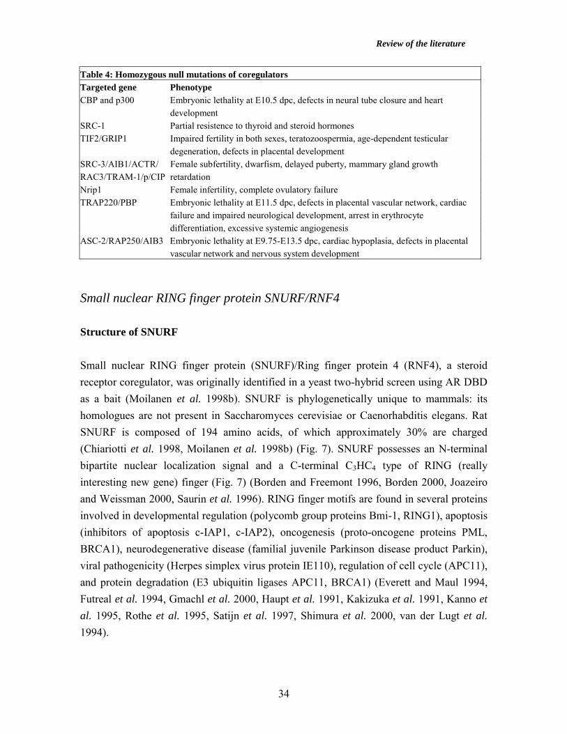
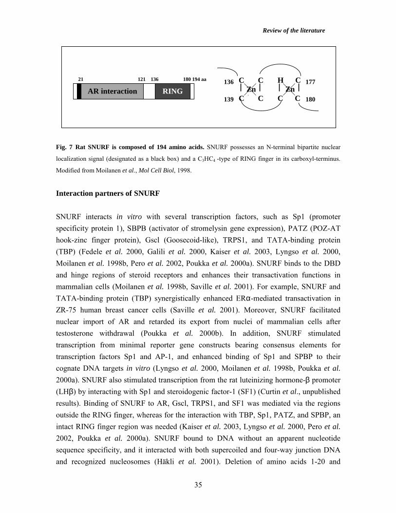
Small nuclear RING finger protein SNURF/RNF4 Structure of SNURF Small nuclear RING finger protein (SNURF)/Ring finger protein 4 (RNF4), a steroid receptor coregulator, was originally identified in a yeast two-hybrid screen using AR DBD as a bait (Moilanen et al. 1998b). SNURF is phylogenetically unique to mammals: its homologues are not present in Saccharomyces cerevisiae or Caenorhabditis elegans. Rat SNURF is composed of 194 amino acids, of which approximately 30% are charged (Chiariotti et al. 1998, Moilanen et al. 1998b) (Fig. 7). SNURF possesses an N-terminal bipartite nuclear localization signal and a C-terminal C3HC4 type of RING (really interesting new gene) finger (Fig. 7) (Borden and Freemont 1996, Borden 2000, Joazeiro and Weissman 2000, Saurin et al. 1996). RING finger motifs are found in several proteins involved in developmental regulation (polycomb group proteins Bmi-1, RING1), apoptosis (inhibitors of apoptosis c-IAP1, c-IAP2), oncogenesis (proto-oncogene proteins PML, BRCA1), neurodegenerative disease (familial juvenile Parkinson disease product Parkin), viral pathogenicity (Herpes simplex virus protein IE110), regulation of cell cycle (APC11), and protein degradation (E3 ubiquitin ligases APC11, BRCA1) (Everett and Maul 1994, Futreal et al. 1994, Gmachl et al. 2000, Haupt et al. 1991, Kakizuka et al. 1991, Kanno et al. 1995, Rothe et al. 1995, Satijn et al. 1997, Shimura et al. 2000, van der Lugt et al. 1994).
Review of the literature
35
Fig. 7 Rat SNURF is composed of 194 amino acids. SNURF possesses an N-terminal bipartite nuclear
localization signal (designated as a black box) and a C3HC4 -type of RING finger in its carboxyl-terminus.
Modified from Moilanen et al., Mol Cell Biol, 1998. Interaction partners of SNURF SNURF interacts in vitro with several transcription factors, such as Sp1 (promoter specificity protein 1), SBPB (activator of stromelysin gene expression), PATZ (POZ-AT hook-zinc finger protein), Gscl (Goosecoid-like), TRPS1, and TATA-binding protein (TBP) (Fedele et al. 2000, Galili et al. 2000, Kaiser et al. 2003, Lyngso et al. 2000, Moilanen et al. 1998b, Pero et al. 2002, Poukka et al. 2000a). SNURF binds to the DBD and hinge regions of steroid receptors and enhances their transactivation functions in mammalian cells (Moilanen et al. 1998b, Saville et al. 2001). For example, SNURF and TATA-binding protein (TBP) synergistically enhanced ERα-mediated transactivation in ZR-75 human breast cancer cells (Saville et al. 2001). Moreover, SNURF facilitated nuclear import of AR and retarded its export from nuclei of mammalian cells after testosterone withdrawal (Poukka et al. 2000b). In addition, SNURF stimulated transcription from minimal reporter gene constructs bearing consensus elements for transcription factors Sp1 and AP-1, and enhanced binding of Sp1 and SPBP to their cognate DNA targets in vitro (Lyngso et al. 2000, Moilanen et al. 1998b, Poukka et al. 2000a). SNURF also stimulated transcription from the rat luteinizing hormone-β promoter (LHβ) by interacting with Sp1 and steroidogenic factor-1 (SF1) (Curtin et al., unpublished results). Binding of SNURF to AR, Gscl, TRPS1, and SF1 was mediated via the regions outside the RING finger, whereas for the interaction with TBP, Sp1, PATZ, and SPBP, an intact RING finger region was needed (Kaiser et al. 2003, Lyngso et al. 2000, Pero et al. 2002, Poukka et al. 2000a). SNURF bound to DNA without an apparent nucleotide sequence specificity, and it interacted with both supercoiled and four-way junction DNA and recognized nucleosomes (Häkli et al. 2001). Deletion of amino acids 1-20 and
AR interaction RING21 121 136 180 194 aa C C
Zn C C
H C Zn C C
136
139
177
180
Review of the literature
36
mutations R8-11A/K9A and R8A of SNURF protein abolished DNA binding activity and severely decreased transactivation on the Sp1 minimal promoter (Häkli et al. 2001). Table 5: Interaction partners of SNURF Factor Interaction domain of SNURF Reference AR aa 20-121 Moilanen et al. 1998b ERα aa 31-65, RING finger Saville et al. 2001 TBP RING finger Moilanen et al. 1998b Sp1 RING finger Poukka et al. 2000a Gscl aa 39-125 Galili et al. 2000 PATZ RING finger Pero et al. 2002 HMGI(Y) full length protein Pero et al. 2000 SBPB RING finger Lyngso et al. 2000 TRPS1 aa 6-65 Kaiser et al. 2003 SF1 aa 31-65 Curtin et al., unpublished
Expression of SNURF SNURF is expressed as a single mRNA species of 3.0 kb in size in all human tissues analyzed. In addition to the ubiquitously expressed 3.0-kb transcript, another 1.0-1.6-kb testis-specific transcript is detected in rodents (Moilanen et al. 1998b). SNURF is expressed in murine embryonic tissues from E7.5 dpc on throughout the development, with high levels of expression in the developing gonads and nervous system (Galili et al. 2000). The gene coding for human SNURF/RNF4 maps at chromosomal region 4p16.3, which is associated with several genetic and neoplastic diseases (Chiariotti et al. 1998). The 47-kb spanning human RNF4 gene was located immeadiately proximal to the anonymous position D4S183, between the huntingtin (HD) and fibroblast growth factor receptor 3 (FGFR3) genes (Chiariotti et al. 1998). Deletion at 4p16.3 between the markers D4S43 and D4S127, the region bearing the RNF4 gene, was detected in breast carcinomas, and the loss of heterozygosity at 4p16.3 was common in bladder carcinomas (Elder et al. 1994, Shivapurkar et al. 1999). Interestingly, expression of SNURF was down-regulated in RAS-transformed cells (Zuber et al. 2000). Mutational activation of the RAS genes is important in the multi-step pathogenesis of several malignancies, such as pancreas, colon, lung, and myeloid cancers. Expression of SNURF was repressed in testicular germ cell tumors of yolk sac, embryonal carcinoma, and seminoma types (Pero et al. 2001). In addition, ectopically overexpressed SNURF

Review of the literature
37
inhibited the growth of cultured human embryonal kidney (293T) and teratocarcinoma (NTERA-2) cells (Pero et al. 2001). RING finger proteins in ubiquitylation Ubiquitin is a 76-amino acid protein. Its successive covalent attachments to target proteins result in their degradation, endocytosis, or conformational alteration (Hershko and Ciechanover 1998, Varhavsky 1997, Weissman 2001). Protein modification by the ubiquitin pathway proceeds in a three-step fashion (for review, Hershko and Ciechanover 1998, Varhavsky 1997, Weissman 2001) (Fig. 8). First, ubiquitin is activated by a ubiquitin-activating enzyme, E1. Secondly, the activated ubiquitin moiety is transferred to a cysteine residue of a ubiquitin-conjugating enzyme, E2, from which a ubiquitin ligase, E3, transfers it to the target protein. Several RING finger proteins and HECT (homology to the E6-AP carboxyl-terminus) domain ligases function as E3 ubiquitin ligases. E3 ligase facilitates the covalent attachment of ubiquitin to the substrate via the formation of an isopeptide bond between the C-terminal lysine of ubiquitin and ε-NH2 group of an internal lysine residue of the substrate. In subsequent reactions, several ubiquitin molecules are added to the substrate, and the polyubiquitin-tagged protein is degraded by the 26S proteasome. Many factors of the ubiquitylation pathway show remarkable evolutionary conservation, from yeast to mammalian species. The ubiquitin system participates in the regulation of several signal transduction processes, such as cell differentiation, cell cycle progression, endocytosis, immune response, programmed cell death, receptor down-regulation, and chromatin modification including DNA repair and gene silencing (Hershko and Ciechanover 1998, Weissman 2001). SNURF is auto-ubiquitylated in a RING domain-dependent fashion and may mediate ubiquitylation, suggesting its potential function as an E3 ubiquitin ligase (Häkli et al., unpublished results). The Angelman syndrome-associated protein E6-AP functions as an E3 ubiquitin ligase, targets p53 protein specifically to degradation, and coactivates nuclear receptors (Nawaz et al. 1999, Scheffner et al. 1994). Inactivation of the E6-AP gene in mice resulted in reduced fertility in both genders with defects in sperm production and in ovulation, and tissue-selective steroid hormone resistance (Smith et al. 2002).
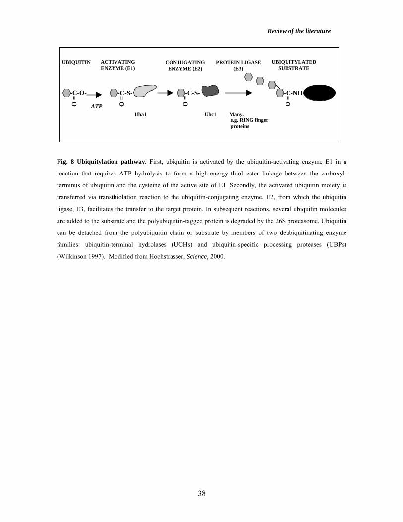
Review of the literature
38
Fig. 8 Ubiquitylation pathway. First, ubiquitin is activated by the ubiquitin-activating enzyme E1 in a
reaction that requires ATP hydrolysis to form a high-energy thiol ester linkage between the carboxyl-
terminus of ubiquitin and the cysteine of the active site of E1. Secondly, the activated ubiquitin moiety is
transferred via transthiolation reaction to the ubiquitin-conjugating enzyme, E2, from which the ubiquitin
ligase, E3, facilitates the transfer to the target protein. In subsequent reactions, several ubiquitin molecules
are added to the substrate and the polyubiquitin-tagged protein is degraded by the 26S proteasome. Ubiquitin
can be detached from the polyubiquitin chain or substrate by members of two deubiquitinating enzyme
families: ubiquitin-terminal hydrolases (UCHs) and ubiquitin-specific processing proteases (UBPs)
(Wilkinson 1997). Modified from Hochstrasser, Science, 2000.
UBIQUITIN ACTIVATING ENZYME (E1)
CONJUGATING ENZYME (E2)
PROTEIN LIGASE (E3)
UBIQUITYLATED SUBSTRATE
ATP Uba1 Ubc1 Many, e.g. RING finger proteins
-C-O-
O =
-C-S-
O =
-C-S-
O =
-C-NH-
O =
Aims of the Study
39
AIMS OF THE STUDY The physiological role of small nuclear RING finger protein SNURF (RNF4), a transcriptional coregulator, is largely unknown despite several in vitro interaction and expression studies reported. However, the expression of SNURF mRNA and protein to high levels in testis suggests a role for SNURF in reproduction. This thesis work aimed at studying the regulation of the SNURF gene expression during fetal gonad development, postnatal ovarian folliculogenesis and spermatogenesis, and in testicular germ cell tumors. The specific goals were as follows:
• To study the expression of SNURF mRNA and protein during gonad determination and prenatal differentiation, as well as in postnatal ovarian and testicular development.
• To characterize the main regulatory elements governing transcription from the
murine SNURF gene.
• To study the hormonal regulation of SNURF gene expression in vivo in rodent gonads.
• To analyze the expression of SNURF and steroid receptors in human testicular
germ cell cancer (TGC).

Methods
40
METHODS For detailed description of the materials and methods, the reader is referred to the original publications indicated in Table 6. In addition, some discussion on the methodology is included in the chapter ‘Results and Discussion’.
Table 6: Methods used in the study Described in publication no.
Cell culture and transfections I, IV
Electrophoretic mobility shift assay (EMSA) I
Immunohistochemistry II, III, IV
In situ hybridization II, IV
Northern blotting II
Primer extension I
Quantitative RT-PCR III, IV
Recombinant DNA techniques and plasmid I, II, IV
construction
Tissue samples All animal experiments were approved by the appropriate institutional review boards of the University of Turku (II), University of Helsinki (IV), and Baylor College of Medicine (Houston, TX, USA) (IV). For the description of the hormone-treated animal models the reader is referred to Table 8. The gonad samples from the hormone-treated and wild-type mice and rats (n=2-3 in each group) were fixed in 4% paraformaldehyde and embedded in paraffin for preparation of 5-µm tissue sections for in situ hybridization and immunohistochemistry. The human and rat testicular specimens were snap-frozen in liquid nitrogen and stored at –70oC until RNA extraction. Study plan of the testicular germ cell cancer project (III) was approved by the ethics committee of the Helsinki University Central Hospital. Paired specimens of normal testis tissue and adjacent tumor tissue were obtained from 12 patients with TGC (seminomas n=8, non-seminomas n=4), who undervent surgery at the Department of Urology, Helsinki University Central Hospital, Helsinki, Finland. Informed consent was received from each patient.

Methods
41
Plasmids The SNURF promoter fragments –617/+111, -202/+111, -38/+111, -8/+111, +32/+111, and –38/+36 nt (numbering refers to the transcription initiation site) were cloned into the luciferase encoding pGL3-basic vector (Promega, Madison, WI, USA). In reporter gene assays and cotransfections, 300-350 ng of the promoter constructs, 100 ng of pEVR-Sp1, pRc/CMV-Sp3, or pCB6-WT18A, and 50 ng of pCMVβ (Clontech, Palo Alto, CA, USA) were used per 3x10⁴ cells per well. Cell culture and transfections COS-1 (American Type Culture Collection, ATCC), HeLa (ATCC), and mSC-1 cells (a gift from Prof. Ilpo Huhtaniemi, University of Turku, Finland) were maintained in Dulbecco’s minimal essential medium containing penicillin (25 000 U/l), streptomycin (25 000 U/l), and 10% (vol/vol) fetal bovine serum. Twenty-four hours before transfection, the cells were seeded on 12-well plates (3x10⁴ cells per well) with indicated expression plasmids and 50 ng of β-galactosidase encoding pCMVβ using FuGENE 6 reagent (Roche Molecular Biochemicals, Indianapolis, IN, USA). The cells were harvested 48 hours after transfection. Luciferase activities were measured with reagents from Promega using a Luminoskan RT reader (Labsystems, Helsinki, Finland) and β-galactosidase activities were determined according to Rosenthal (1987). The luciferase values were normalized by the β-galactosidase values. Electrophoretic mobility shift assay (EMSA) EMSA experiments were carried out with 1 µg of HeLa cell nuclear extract (4C Biotech, Belgium), 1.6 µg of poly(dI-dC)(dI-dC) (Amersham Pharmacia Biotech, Aylesbury, UK), and 32P-labeled promoter fragments –8/+111 nt and –38/+111 nt in 20 mM HEPES (pH 7.9), 10% glycerol (vol/vol), 50 mM KCl, 0.5 mM EDTA, 1 mM DTT, 1 mM MgCl2, 0.5 mM phenylmethylsulfonyl fluoride (PMSF), and 1 µM leupeptin. Reaction products were separated on 4% polyacrylamide gels run in 22.5 mM Tris-borate, 0.5 mM EDTA for 2-3 h at 200V at 22oC. In oligomer competition assays, nuclear extracts were preincubated with 100-fold molar excess of non-radioactive double-stranded oligonucleotides encompassing indicated promoter regions or consensus binding sites for NF-κB, CREB, and Sp1.

Methods
42
Immunohistochemistry Paraffin-embedded sections of gonads were used for immunohistochemical staining with polyclonal anti-SNURF antibody raised against rat SNURF in rabbit (1:1000-1:2000 dilution in Tris-buffered saline (TBS) containing 1% bovine serum albumin (BSA), Moilanen 1998b), and the positive cells were visualized using Vectastain Elite ABC kit (Vector laboratories, Burlingame, CA, USA) according to the manufacturer’s instructions. As a control, 500 ng of GST-SNURF were first incubated with the antibody (1:2000 dilution) for 16 h at 4°C, and the resulting preadsorbed anti-SNURF antibody was used to monitor the specificity of staining. ERβ antigen was detected using anti-ERβ 503 antibody that was raised against human ERβ in chicken (a gift from Dr. Jan-Åke Gustafsson, Novum, Huddinge, Sweden, diluted to 1:1000 in phosphate-buffered saline (PBS) with 3% BSA and 0.05% Tween 20) and peroxidase-conjugated rabbit anti-chicken IgG (1:1000 dilution in PBS and 2% rat normal serum) (Sigma-Aldrich, St. Louis, MO, USA). Samples were incubated with the primary antibodies at 4oC overnight. Peroxidase activity was visualized with diaminobenzidine as the substrate (Vector laboratories, Burlingame, CA, USA), and the sections were stained with Mayer’s hematoxylin, dehydrated, and mounted. In situ hybridization The tissue sections of gonads were deparaffinized, permeabilized, dehydrated, and hybridized to antisense and sense SNURF RNA probes (corresponding to nt 156–536 of murine SNURF cDNA, or to nt 234-967 and nt 968-1644 of rat SNURF cDNA) synthesized in the presence of [α-35S]UTP (Amersham Pharmacia Biotech, Aylesbury, UK). The tissue sections were incubated with 5x10⁴ cpm of 35S-labeled probes in a total volume of 100 µl and incubated overnight at 55oC. Hybridization signals were detected after a 10-14-day exposure to NTB-2 emulsion (Eastman Kodak, Rochester, NY, USA) at 4oC. Northern blotting Ten micrograms of total rat testicular RNA were size-fractionated in 1% denaturing agarose gel and transferred onto Hybond-N+ nylon membrane (Amersham Pharmacia Biotech, Aylesbury, UK). After hybridization with 32P-labeled SNURF probes (corresponding to nt 234-967 and nt 968-1644 of rat SNURF cDNA) at 68oC overnight, the blots were stripped and subsequently hybridized with 28S rRNA cDNA probe labeled with

Methods
43
[α-32P]dCTP (Amersham Pharmacia Biotech, Aylesbury, UK) by the random priming method (Prime-a-gene kit, Promega, Madison, WI, USA). Real-time quantitative RT-PCR Real-time quantitative RT-PCR was performed using the Lightcycler system, FastStart DNA Master SYBR Green I mix (Roche Molecular systems, Indianapolis, IN, USA), and indicated primers according to the manufacturer’s instructions. For detailed protocol of the RT-PCR reactions the reader is referred to Fig. 11. Granulosa cell cultures Granulosa cells were harvested by needle puncture from the ovaries of E2-primed (1.5 mg s.c. daily for 3 days) immature rats (aged 26 days). The cells were cultured on serum-coated 12-well culture plates at a density of 0.5x10⁶ cells per 1.5 ml in Dulbecco’s minimal essential medium:F12 containing 100 IU/ml penicillin and streptomycin. On the following day, the cells were washed and cultured in fresh serum-free medium containing FSH (50 ng/ml, NIH FSH-16), testosterone (10 ng/ml), forskolin (10 µM), and/or PMA (20 nM) and harvested after selected time intervals for RNA extraction. Semi-quantitative RT-PCR Total RNA were extracted from whole ovaries of PMSG/hCG-primed immature rats, hypophysectomized (H) rats, and E2/FSH/hCG-primed H rats (from Harlan Sprague Dawley, Inc., Chicago, IL, USA), as well as from granulosa cells using Trizol reagent (Life Technologies, Inc., Grand Island, NY, USA). Semi-quantitative RT-PCR was performed using primers specific for SNURF and the ribosomal protein L19. Total RNA (500 ng) was reverse transcribed using oligo-d(T) primer and AMV-RT at 42oC for 75 min and 95oC for 5 min. DNA products were amplified in the presence of [α-32P]dCTP and Taq polymerase for 22 cycles at 94oC for 1 min, 60oC for 2 min, and 72oC for 2 min. The amplified DNA was resolved on a 5% polyacrylamide gel, which was dried, and exposed to X-ray film, and the signal intensities were quantified by using Storm 860 PhosphorImager software (Molecular Dynamics, Sunnyvale, CA, USA).
Results and Discussion
44
RESULTS AND DISCUSSION
Expression of SNURF during murine gonad determination and prenatal differentiation (IV) The murine gonad development begins at E10.5 dpc by migration of primordial germ cells to the urogenital ridges. The gonads first develop in a non-sex-specific manner, but by E11.0 dpc, the Sertoli cells express masculinizing transcription factors, and at E13.5 dpc, the testicular cords have been established (Capel 2000). Previously, Galili et al. (2000) reported that SNURF mRNA was detected at high levels in murine embryos from E7.5 dpc onwards. To gain insight into the role of SNURF in gonad determination and differentiation, we analyzed the expression of SNURF mRNA in developing murine gonads at E10.5-E17.5 dpc by in situ hybridization. The expression of SNURF mRNA was detected at moderate levels in fetal gonads of both sexes from E10.5 dpc onwards. SNURF mRNA accumulated to high levels in the fetal testis by E15.5-E17.5 dpc, with the most intense signal being localized to testicular cords, indicating that SNURF mRNA accumulation was developmentally regulated. Immunohistochemical staining using polyclonal anti-SNURF antibody (Moilanen et al. 1998b) confirmed the presence of SNURF protein in gonocytes, Leydig cells, and some Sertoli cells and in oogonia and supporting cells in the fetal testes and ovaries, respectively, at E17.5 dpc. SNURF interacts with SF1, an orphan nuclear receptor essential for gonadogenesis expressed in pre-Sertoli cells from E10.5 dpc onwards (Curtin et al., unpublished results, Luo et al. 1994). Moreover, SNURF is able to indirectly facilitate transcription elicited by GATA4, that is expressed to high levels in Leydig cells and Sertoli cells during fetal testis development (Kaiser et al. 2003, Ketola et al. 1999). Interestingly, GATA4 enhances transcription from the Müllerian inhibiting substance gene by interacting directly with SF1 (Tremblay and Viger 1999). Thus, SNURF might regulate gonad development by modulating transcriptional responses elicited by SF1 and GATA4. Expression of SNURF in spermatogenesis (II, III) A variety of widely expressed transcription factors exhibit alternatively spliced testis-specific transcript species. They may be differently regulated in gonads due to tissue-specific alternative promoters, or by signals from cell-specific transcription factors (Daniel and Habener 1998, Schmidt and Schibler 1995). The SNURF gene encodes a 1.0-1.6-kb alternative transcript in adult rodent testis, in addition to the ubiquitous 3.0-kb transcript
Results and Discussion
45
(Moilanen et al. 1998b, Lyngso et al. 2000). We isolated the rat testis-specific SNURF mRNA sequence from the same cDNA library, from which the ubiquitous SNURF transcript was originally identified (Moilanen et al. 1998b). Both rat transcripts encode a full-length SNURF protein of 194 amino acids. The shorter rat SNURF mRNA is generated by the use of an alternative poly(A)+ sequence. Similarly, both murine SNURF transcripts encode the full-length SNURF protein and only differ in their 3’untranslated regions (Pero et al. 2003). To analyze whether expression of the SNURF gene was developmentally regulated during postnatal rat testis development, we performed in situ hybridization experiments using a series of rat testicular samples representing different developmental phases. The 3.0-kb SNURF mRNA was expressed from newborn to mature testis, whereas the 1.6-kb mRNA appeared only after postnatal day 30, as detected using transcript-specific RNA probes in in situ hybridization experiments. The 1.6-kb transcript was abundantly expressed in postmeiotic haploid round and elongating spermatids (step 4-11 spermatids), while the 3.0-kb mRNA localized mainly to spermatogonia and somatic Leydig and Sertoli cells. In agreement with our data, Pero et al. (2003) detected the 3.0-kb SNURF transcript in murine spermatogonia and Sertoli cells, whereas the shorter transcript was seen in spermatogenic cell populations. We confirmed the cell type-specific expression of the SNURF transcripts by using methoxyacetic acid (MAA) treatment, which depletes spermatocytes at stages I-VI within 24 h of oral administration (Bartlett et al. 1988, Table 8). In MAA rat model, the stages are devoid of spermatocytes on day 4 after the treatment, and on days 18 and 30 the stages lack round and elongating spermatids, respectively (Bartlett et al. 1988). The 3.0-kb SNURF mRNA remained constant in testis on post-treatment days 4, 18, and 30. Interestingly, the level of the 1.6-kb SNURF transcript was markedly decreased on day 18, possibly due to the depletion of most of the SNURF-expressing spermatids at stages IV-VI. MAA treatment provided additional evidence that the 3.0-kb and 1.6-kb SNURF mRNAs accumulated mainly in somatic cells and spermatogonia, and spermatids, respectively. SNURF protein was detected in moderate amounts in postnatal rat testis from day 20 onwards (Fig. 9). It localized mainly to Sertoli cell nuclei before postnatal day 30, but from day 30 onwards nuclei of both Sertoli cells and spermatids were coated with polyclonal anti-SNURF antibody in agreement with the in situ hybridization data. Furthermore, the expression of SNURF protein was high at stages VII-VIII, IX-XII, and II-VI, and low at
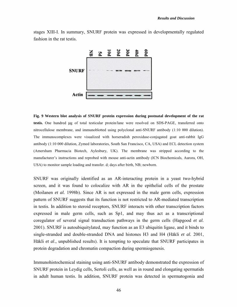
Results and Discussion
46
stages XIII-I. In summary, SNURF protein was expressed in developmentally regulated fashion in the rat testis.
Fig. 9 Western blot analysis of SNURF protein expression during postnatal development of the rat
testis. One hundred µg of total testicular protein/lane were resolved on SDS-PAGE, transferred onto
nitrocellulose membrane, and immunoblotted using polyclonal anti-SNURF antibody (1:10 000 dilution).
The immunocomplexes were visualized with horseradish peroxidase-conjugated goat anti-rabbit IgG
antibody (1:10 000 dilution, Zymed laboratories, South San Francisco, CA, USA) and ECL detection system
(Amersham Pharmacia Biotech, Aylesbury, UK). The membrane was stripped according to the
manufacturer’s instructions and reprobed with mouse anti-actin antibody (ICN Biochemicals, Aurora, OH,
USA) to monitor sample loading and transfer. d; days after birth, NB; newborn.
SNURF was originally identified as an AR-interacting protein in a yeast two-hybrid screen, and it was found to colocalize with AR in the epithelial cells of the prostate (Moilanen et al. 1998b). Since AR is not expressed in the male germ cells, expression pattern of SNURF suggests that its function is not restricted to AR-mediated transcription in testis. In addition to steroid receptors, SNURF interacts with other transcription factors expressed in male germ cells, such as Sp1, and may thus act as a transcriptional coregulator of several signal transduction pathways in the germ cells (Hapgood et al. 2001). SNURF is autoubiquitylated, may function as an E3 ubiquitin ligase, and it binds to single-stranded and double-stranded DNA and histones H3 and H4 (Häkli et al. 2001, Häkli et al., unpublished results). It is tempting to speculate that SNURF participates in protein degradation and chromatin compaction during spermiogenesis. Immunohistochemical staining using anti-SNURF antibody demonstrated the expression of SNURF protein in Leydig cells, Sertoli cells, as well as in round and elongating spermatids in adult human testis. In addition, SNURF protein was detected in spermatogonia and
Results and Discussion
47
spermatocytes at low levels in the human testis. However, on the contrary to the rodent Leydig cells showing slight immunoreactivity, human Leydig cell nuclei stained intensively with the anti-SNURF antibody (III). In testes, SNURF localized mainly to cell nuclei, although predominantly cytoplasmic expression was reported in developing murine neural structures, such as dorsal root ganglia and the neural tube, and in human primary fibroblasts (Galili et al. 2000, Kaiser et al. 2003). Since SNURF is phosphorylated by casein kinase II in vitro and is ubiquitylated in vitro (Moilanen et al. 1998b, Häkli et al., unpublished). Subcellular localization of SNURF may be regulated by protein-protein interactions elicited by covalent modifications in specific cell types. Interestingly, SNURF enhanced nuclear transport of AR and association of AR to nuclear matrix in COS-1 cells by an unknown mechanism (Poukka et al. 2000b). Other nuclear receptor coregulators expressed to high levels in male germ cells include PIASxα, PIAS1, and AIB3/RAP250 (Yan et al. 2003, Zhang et al. 2003). In addition, GRIP1/TIF2 was expressed in Sertoli cell-specific fashion in murine testis (Gehin et al. 2002). Expression of SNURF in murine postnatal ovary (IV) Several factors important for spermatogenesis, such as c-Kit receptor and Kit ligand and steroid receptors, play also important roles during folliculogenesis (Richards 2001). The abundant amounts of SNURF mRNA and protein in fetal gonads and in spermatogenesis prompted us to investigate the expression of SNURF mRNA during ovarian development. A series of ovarian specimens from newborn to mature mice were subjected to in situ hybridization using SNURF RNA probe. We detected SNURF mRNA throughout folliculogenesis appearing at high levels in the oocytes of preantral follicles from postnatal day 14 onwards. SNURF mRNA was detected at moderate levels in granulosa cells of preantral, antral, and Graafian follicles and in the luteal gland. Moreover, immunohistochemical staining using anti-SNURF antibody demonstrated SNURF protein in oocytes, in granulosa and luteal cells, as well as in theca and interstitial cells. Unfortunately, studies of SNURF expression in human ovary and ovarian tumors was out of reach in this work. SNURF protein colocalizes with AR, ERβ, and PR in the granulosa cells, ERα in the oocytes and in the corpus luteum, suggesting that SNURF facilitates steroid receptor-mediated signaling during follicle maturation and luteinization. Some other coregulator proteins have already been implicated in the regulation of female fertility, as exemplified by Nrip1, the null mutants of which are infertile due to ovulatory failure (White et al. 2000). Moreover, deletion of the gene encoding SRC-3 resulted in female subfertility (Xu et al. 2000). In addition, female mice lacking TAFII105, a TATA-

Results and Discussion
48
binding protein associated factor, are infertile because of a defect in folliculogenesis (Freiman et al. 2001).
Table 7. Expression of SNURF mRNA during gonad development Prenatal murine Male Female development Germ cells (pre)SC LC Germ cells GC CL E10.5 dpc + + - + + - E12.5 dpc + + - + + - E13.5 dpc + + + + + - E15.5 dpc ++ + + + + - E17.5 dpc ++ + + + + - Newborn mice + + + + + - Postnatal rat development NB + + - nd nd nd 5d + + - nd nd nd 10d + + - nd nd nd 20d + + + + + nd 30d ++ + + nd nd nd 40d ++ + + nd nd nd 60d ++ + + nd nd nd Postnatal murine development 7d nd nd nd + + - 14d nd nd nd ++ + - 25d nd nd nd ++ + - 59d nd nd nd ++ + + 11 wk nd nd nd ++ + + Adult human ++ + ++ nd nd nd nd; not determined, SC; Sertoli cell, LC; Leydig cell, GC; granulosa cell, CL; corpus luteum.
Posttranslational modifications, such as phosphorylation, acetylation, sumoylation, and ubiquitylation, modify steroid receptor-mediated signaling by affecting the transcriptional activity and/or subcellular localization of the receptors. In the periovulatory granulosa cells, ubiquitin-related modifier SUMO-1 and PR interacted in vivo, and inhibition of SUMO-1 expression by luteinizing hormone receptor stimulation was linked to periovulatory induction of PR levels (Shao et al. 2003). Recently, ret finger protein-like 4 (Rfpl4), exhibiting a RING finger-like structure with E3 ubiquitin ligase activity, was shown to regulate proteosome-mediated degradation of maturation-promoting factor (MPF), a heterodimer of p34Cdc2 and cyclin B1, during the progression of meiotic cell

Results and Discussion
49
cycle in murine oocytes (Suzumori et al. 2003). As SNURF possesses a RING finger domain present in many E3 ubiquitin ligases, the abundant expression of SNURF in the maturing oocytes might implicate in a role in regulation of oocyte meiosis. Although proteasomal degradation has been well established in the steroid receptor-dependent transactivation (Lonard et al. 2000), SNURF does not appear to mediate ubiquitylation of AR (Tian et al., unpublished results). Transcriptional regulation of the murine SNURF gene (I) Transcriptional regulation is mediated via promoter regions usually located upstream of transcription initiation sites of the target genes. The diversity of developmental and tissue-specific signals is conveyed by binding of a variety of cellular transcription factors to the specific target sequences on the promoter regions, as exemplified by binding of AR to AREs on the PEM homeodomain gene promoter (Rao et al. 2003). Along with the identification of the two alternative SNURF transcripts in rodent testis (Moilanen et al. 1998b), we became interested in the regulatory determinants governing transcription from the murine SNURF gene. To identify the regulatory regions responsible for the generation of murine SNURF transcripts, 0.7 kb of the SNURF promoter was isolated. Alternative transcripts may be generated using alternative transcription start sites or different tissue-specific promoters of the gene, as exemplified by the testis-specific promoter of TBP (Schmidt et al. 1997). To determine the transcription start sites of the SNURF gene, we performed primer extension assays using two different antisense oligomers and mouse total testicular RNA as a template. A single primer extension product was detected using both primers, suggesting that a single promoter governs the generation of both SNURF transcripts. In addition, Pero et al. (2003) published primer extension data that agree with our observations, in that the murine RNF4 transcripts also originate from the same promoter. There are several GC-rich elements within the SNURF promoter characteristic of housekeeping genes, but a consensus TATA box or a CCAAT box were not located in the vicinity of the transcription start site. To determine the elements critical for basal promoter activity, different fragments of the promoter were cloned in front of the luciferase gene and transfected into mammalian cells. The longest construct (–617/+111 nt) showed very strong basal activity in HeLa, COS-1, and mSC-1 cells. The activity of the region –38/+36 nt exhibited 29-43% of the maximal promoter activity in mammalian cells, indicating that the fragment harbored the elements critical for the basal activity of the SNURF promoter.
Results and Discussion
50
Detailed sequence analysis revealed a GC box at +9 nt and a NF-κB consensus site at -8 nt within the core promoter (Fig.10). The GC box was conserved in the human RNF4 gene, suggesting functional importance of the element (Chiariotti et al. 1998). Although SNURF is expressed at high levels in spermatids, consensus elements for the testis-specific transcription factor CREM were not found within the SNURF promoter region (data not shown). Binding of nuclear proteins to the SNURF promoter was examined by electrophoretic mobility shift assay using 32P-labeled probes corresponding to –8/+111 nt and –38/+111 nt. Using nuclear extract, we detected one major and few minor nuclear protein-DNA complexes, which were abolished by the addition of a 100-fold molar excess of unlabeled probes or double-stranded –8/+34 or –8/+24 oligomers. Interestingly, a non-radioactive Sp1 consensus oligomer reduced the formation of the major protein-DNA complex indicating that the Sp family members might bind to the promoter. NF-κB and CREB consensus oligomers did not challenge the probe markedly. Deletion of the proximal GC box from the probe –8/+111 abolished nuclear protein binding to the SNURF promoter and reduced promoter activity to 21% and 13% of the corresponding wild-type construct activity in COS-1 and HeLa cells, respectively. In search for factors regulating SNURF gene activity, we transfected plasmids encoding Sp1, Sp3, and WT1 gene products, factors binding to GC-rich elements, with the promoter constructs into mammalian cells (Hapgood et al. 2001). Ectopically expressed Sp1 marginally stimulated the SNURF promoter in HeLa cells, an effect not seen in the presence of a construct bearing the mutated proximal GC box. WT1, essential for urogenital development, activated the –8/+111 fragment >4-fold in HeLa cells. Interestingly, the WT1 gene product is expressed concomitantly with SNURF at E10.5 dpc in genital ridges of both sexes (Kreidberg et al. 1993). Colocalized expression of SNURF and WT1 during early gonad development supports the possibility that SNURF gene activation is dependent on WT1 signaling in fetal gonads in vivo. SNURF binds to Sp1, which interacts with components of the TFIID complex and several transcription factors, such as NF-κB and GATA-1 (Chiang and Roeder 1995, Emili et al. 1994, Gill et al. 1994). The critical GC element on the SNURF promoter may thus mediate activation by tissue-specific and developmental signals in addition to constitutive induction (Hirano et al. 1998, Merika et al. 1995). Interestingly, Sp1 and Sp3, present in the male germ cells, are among the factors responsible for the testis-specific expression of PIASx coregulators, and

Results and Discussion
51
they may thus also stimulate the SNURF promoter in the spermatogenic cells (Santti et al. 2003).
Fig. 10 Schematic presentation of the murine SNURF core promoter. The TATAless proximal promoter
contains GC boxes starting at +9 nt and –25 nt, and a putative NF-κB site at –8 nt. The GC element at +9 nt
was critical for the proximal SNURF promoter activity, and Sp1 and WT1 gene products were found as
potential activators of the murine SNURF gene in mammalian cells.
Hormonal regulation of SNURF gene expression in vivo in rodent gonads (II, IV) SNURF interacts with steroid receptors augmenting their function and is expressed to high levels in steroid hormone target tissues, the testis and the ovary. Thus, we hypothesized that SNURF mRNA might be regulated by steroids in vivo. Despite the presence of several steroid receptor response element half-sites in the murine SNURF promoter (at nt +58, -50, -238, -407, and –431 for AR, and +18 and –123 for ER, respectively), ectopically expressed AR or ER in the presence of testosterone or estradiol, respectively, did not affect the activity of SNURF promoter fragments in mammalian cells (data not shown). However, administration of ethylene dimethane sulfonate (EDS), which depletes testicular Leydig cells within 24 h, resulted in undetectable SNURF mRNA levels between post-treatment days 2 and 15 in rat testis concomitantly with the declining testicular testosterone (T) levels (Teerds 1996, Table 8). Twenty days after the EDS administration, by the time that the Leydig cells had recovered their function, SNURF transcript levels accumulated in concert with the raising T levels. Rats, that received EDS and T implants simultaneously exhibited higher circulating T levels and 3-5-fold increased testicular SNURF mRNA levels, respectively, compared to control rats. Steroid hormone response elements are often located up to 10 kb upstream of the transcription start site, and the cis-element responsible for androgen-regulation of testicular SNURF mRNA could be located further upstream of the SNURF promoter analyzed in the transient transfections. The effects of testosterone may also be mediated indirectly. Steroid receptors interact with other transcription factors,
+36 bp
GCGCGACTGGCGGCACTCTCGCGGGAAGTGGAAGCTCCCGCAAGCAGGCGGGGCGTGACCGGAAGAAGGTGTAT
-38 bp +1 bp
GC box NF-κκκκ B GC box
Sp1 WT1+36 bp+36 bp
GCGCGACTGGCGGCACTCTCGCGGGAAGTGGAAGCTCCCGCAAGCAGGCGGGGCGTGACCGGAAGAAGGTGTAT
-38 bp-38 bp +1 bp
GC box NF-κκκκ B GC box
Sp1 WT1
Results and Discussion
52
such as NF-κB, and influence their transcriptional activity sometimes even without binding to DNA elements, referred to as ‘transcriptional cross-talk’ (Göttlicher et al. 1998). Androgen action to germ cells is mediated through Sertoli cells and Leydig cells that express AR, and accumulation of SNURF in germ cells is likely to be regulated indirectly by paracrine signaling from the neighboring cells. However, ERβ is expressed in male germ cells and the effects of androgens could alternatively be conveyd to the germ cells by conversion to estrogens through the action of CYP450 aromatase present in the spermatogenic cells (Enmark et al. 1997). Interestingly, testosterone has been shown to stimulate the expression of aromatase mRNA in the purified rat germ cells (Bourguiba et al. 2003). In the ovary, estradiol is produced in the granulosa cells by aromatization of the androgen precursors diffusing from the neighboring theca cells (Richards 2001). Because testosterone increased SNURF mRNA level in rat testis either by an indirect mechanism or via conversion to estrogens, we sought to study whether estradiol regulates the accumulation of SNURF mRNA in vivo in the murine ovary. In the mouse model, PMSG, a hormone with both FSH and LH-like activity in the rodents, stimulates the growth of preovulatory follicles that synthesize increased amounts of estrogen (Sztein et al. 2000, Table 8). We exposed 3-week-old immature female mice to 2.5 IU of PMSG, that marginally attenuated the SNURF mRNA within 48 h after administration, whereas a subsequent injection of 2.5 IU of hCG, mimicking the preovulatory LH surge, slightly augmented ovarian SNURF mRNA content. However, as determined by semi-quantitave RT-PCR, SNURF mRNA level was not dramatically changed in whole ovaries of immature intact rats treated with PMSG and hCG. The ovaries from mice treated daily with 125 µg of diethylstilbestrol for 3 days increased SNURF mRNA levels in oocytes compared to control mice treated with vehicle, an effect that was blocked by the pure antiestrogen ICI 182,780, suggesting that expression of the SNURF gene was controlled by ER-dependent signaling in the oocytes (Jain et al. 1999). The levels of SNURF mRNA remained unchanged in the ovaries of female mice treated with ICI 182,780 alone compared to control mice, implying that estrogen-dependent signaling is not the only mechanism that regulates SNURF gene expression in oocytes. Moreover, ERα, that is expressed in the oocyte, did not stimulate SNURF gene activity in the presence of estradiol in mammalian cells of non-ovarian origin (I).

Results and Discussion
53
Table 8: Hormone treatments in animal models model hormone treatment Consequence MAA 650 mg/kg p.o. single dose Depletion of spermatocytes at (Bartlett et al. 1988) stages I-VI after 24 h, lack of round
and elongating spermatids 18 and 30 days after treatment,
respectively. EDS 75 mg/kg i.p. single dose Depletion of Leydig cells within (Teerds 1996) 24 h. Testosterone undetectable
between days 2 and 15. PMSG + hCG 2.5 IU PMSG i.p. + after 48 h Preovulatory follicles 48 h after (Sztein et al. 2000) 2.5 IU hCG i.p. PMSG and 5 h after hCG, post-
ovulatory follicles 20 h after hCG, luteinizing follicles 5 d after hCG
DES 125 µg s.c. daily for 3 days Stimulation of granulosa cell (Heikinheimo 1997) proliferation ICI 182,780 50 µg s.c. daily for 3 days Inhibition of estrogen-mediated (Jain et al. 1999) granulosa cell proliferation Hypophysectomized rats (Zhou et al. 1995)
1.5 mg E2 s.c. daily for 3 days + 1.0 µg FSH i.p. twice daily + 10 IU hCG i.p.
Preovulatory follicles 48 h after FSH, postovulatory follicles 16 h after hCG, luteinizing follicles 24 h after hCG
To confirm the hormone-dependent regulation of SNURF mRNA observed in the whole murine ovary in vivo, we studied regulation of SNURF mRNA in hypophysectomized (H) rats (Zhou et al. 1995). In the PMSG model, it is difficult to dissociate the effects of the steroid from those of the gonadotropin. In the H rat model, E2 is required to stimulate granulosa cell proliferation and formation of the preantral follicles, which are highly responsive to FSH (Zhou et al. 1995). After hypophysectomy, SNURF protein levels declined in granulosa cells in vivo as determined by immunohistochemical staining, whereas SNURF was constitutively expressed in rat oocytes. Treatment of H rats with E2 for 3 days restored SNURF protein expression in granulosa cells to some extent, but did not increase SNURF protein levels in the oocytes. This provided further evidence that basal activity of the SNURF gene in the oocytes was regulated in an estrogen-independent fashion. On the other hand, estradiol-mediated stimulation of SNURF gene expression might not be clearly detectable at the protein level by immunohistochemical analysis. Granulosa cells were isolated from ovaries of the H rats before and after hormonal stimulation and were used as a source of total RNA for semi-quantitative RT-PCR. SNURF mRNA levels were increased in E2-primed granulosa cells by 3-fold, as determined by semi-quantitative RT-PCR, and were increased further by 12 h after an additional treatment with FSH and hCG. To the best of our knowledge, the specific genes regulated by estradiol in the oocyte have remained speculative, but estrogen-dependent
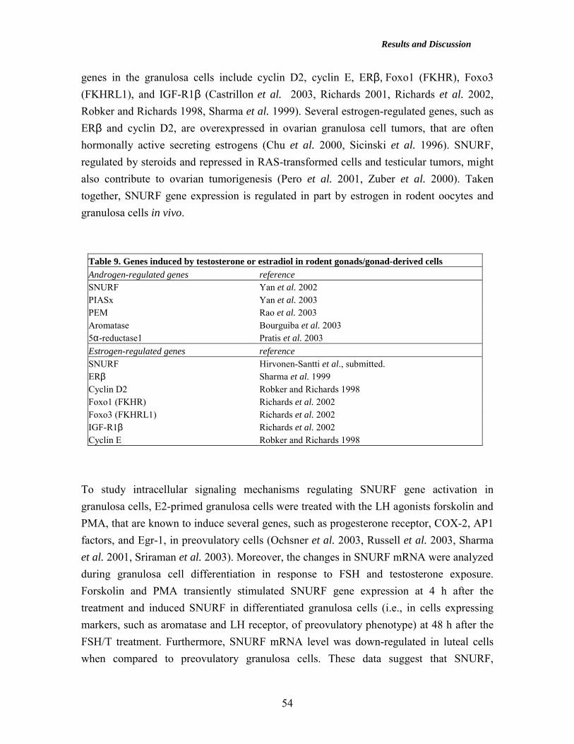
Results and Discussion
54
genes in the granulosa cells include cyclin D2, cyclin E, ERβ, Foxo1 (FKHR), Foxo3 (FKHRL1), and IGF-R1β (Castrillon et al. 2003, Richards 2001, Richards et al. 2002, Robker and Richards 1998, Sharma et al. 1999). Several estrogen-regulated genes, such as ERβ and cyclin D2, are overexpressed in ovarian granulosa cell tumors, that are often hormonally active secreting estrogens (Chu et al. 2000, Sicinski et al. 1996). SNURF, regulated by steroids and repressed in RAS-transformed cells and testicular tumors, might also contribute to ovarian tumorigenesis (Pero et al. 2001, Zuber et al. 2000). Taken together, SNURF gene expression is regulated in part by estrogen in rodent oocytes and granulosa cells in vivo.
Table 9. Genes induced by testosterone or estradiol in rodent gonads/gonad-derived cells Androgen-regulated genes reference SNURF Yan et al. 2002 PIASx Yan et al. 2003 PEM Rao et al. 2003 Aromatase Bourguiba et al. 2003 5α-reductase1 Pratis et al. 2003 Estrogen-regulated genes reference SNURF Hirvonen-Santti et al., submitted. ERβ Sharma et al. 1999 Cyclin D2 Robker and Richards 1998 Foxo1 (FKHR) Richards et al. 2002 Foxo3 (FKHRL1) Richards et al. 2002 IGF-R1β Richards et al. 2002 Cyclin E Robker and Richards 1998
To study intracellular signaling mechanisms regulating SNURF gene activation in granulosa cells, E2-primed granulosa cells were treated with the LH agonists forskolin and PMA, that are known to induce several genes, such as progesterone receptor, COX-2, AP1 factors, and Egr-1, in preovulatory cells (Ochsner et al. 2003, Russell et al. 2003, Sharma et al. 2001, Sriraman et al. 2003). Moreover, the changes in SNURF mRNA were analyzed during granulosa cell differentiation in response to FSH and testosterone exposure. Forskolin and PMA transiently stimulated SNURF gene expression at 4 h after the treatment and induced SNURF in differentiated granulosa cells (i.e., in cells expressing markers, such as aromatase and LH receptor, of preovulatory phenotype) at 48 h after the FSH/T treatment. Furthermore, SNURF mRNA level was down-regulated in luteal cells when compared to preovulatory granulosa cells. These data suggest that SNURF,
Results and Discussion
55
interacting with PR essential for ovulation (Moilanen et al. 1998b), may play a role in ovulation. Expression of SNURF and estrogen receptor ββββ
in human testicular germ cells and testicular tumors (III, IV) The onset of the testicular germ cell malignancies at adolescence implicates in a fetal origin of the disease, and endogenous or environmental estrogens have been suggested to contribute to tumorigenesis (Sharpe 2003). Fetal exposure to excess of estrogens could suppress testosterone production and AR expression in the fetal testis as well as inhibit fetal Leydig cell gene expression, e.g. insulin-like factor-3 (InsL3) production (Nef and Parada 2000, Sharpe 2003). The role of androgens, essential for the normal testicular development and spermatogenesis, is largely unknown in testicular cancer development. Since the testicular expression pattern of SNURF implicated a role in prenatal gonocyte differentiation and spermatid maturation, we determined SNURF mRNA levels in 12 paired normal testis-testicular germ cell cancer samples by real-time quantitative RT-PCR (Fig. 11). In addition to SNURF, we analyzed levels of mRNAs encoding ERβ, AR, PIAS1, and PIASx in the TGC specimen pairs. We found statistically significant repression of ERβ mRNA by 59% in seminomas (p=0.017, Wilcoxon signed ranks test) and down-regulation of SNURF/RNF4 and AR mRNA by 67% and 75%, respectively, in seminomas and teratocarcinomas (p=0.034 for both). In addition, the mRNA coding for ERβcx/β2 isoform was down-regulated in TGC samples (data not shown), whereas PIASx and PIAS1 mRNA levels were unaltered in TGC. We also examined the levels of cyclin D2 in our TGC material, and in agreement with previously reported observations, we found overexpression of cyclin D2 mRNA with an average increase of 6.8-fold. However, in our small patient group, overexpression of cyclin D2 remained statistically non-significant (p=0.136). No statistically significant association was observed between ERβ and SNURF mRNA in seminomas, or between AR and SNURF mRNA levels in all tumors, respectively (data not shown). ERβ and SNURF protein levels were also repressed in local pT1 seminomas as judged by immunohistochemical analysis. Our results confirm and complement the observations of Pero et al. (2001), who reported reduced expression of SNURF in TGC of yolk sac, embryonal carcinoma, and seminoma types. In normal human testis, ERβ protein localized to primary spermatocytes and round spermatids as previously described (Saunders et al. 2002). Recently, Pais et al. (2003) reported diminished expression of ERβ in seminomas,

Results and Discussion
56
embryonal cell carcinomas, and mixed germ cell tumors. Repressed ERβ mRNA and protein levels were reported in prostate cancer (Latil 2001). However, ERβ knockout male mice reproduced normally and showed prostatic hyperplasia, but did not exhibit testicular tumors (Weihua et al. 2001). The loss of AR mRNA in tumor samples probably reflects their cellular composition, i.e., the absence of AR-expressing Sertoli and Leydig cells. Accordingly, Tfm male mice bearing the mutated AR gene and AR knockout male mice showed Leydig cell hyperplasia, but no germ cell-derived cancer was reported (Lyon et al. 1975, Yeh et al. 2002). To our best knowledge, expression of other classical nuclear receptor coregulators has not been studied in testicular germ cell cancer. However, several studies indicate altered coregulator mRNA expression in hormone-dependent breast, ovarian, and prostate cancers (Anzick et al. 1997, Lee et al. 1999, Zhu et al. 1999), as exemplified by overexpression of SRC-1 and TIF2/GRIP1 proteins in hormone-refractory prostate cancer (Gregory et al. 2001). Examples of genes with altered expression in testicular germ cells tumors are presented in Table 10.
Table 10. Examples of genes with altered expression in TGC Gene Expression in TGC vs. normal Reference SNURF down-regulated Hirvonen-Santti et al. 2003 ERβ down-regulated Hirvonen-Santti et al. 2003 Cyclin D2 up-regulated Schmidt et al. 2001 CDK4 up-regulated Schmidt et al. 2001 Cyclin E down-regulated Schmidt et al. 2001 CDK2 down-regulated Schmidt et al. 2001 GRB7 up-regulated Skotheim et al. 2002 c-KIT down-regulated Rajpert-de Meyts et al. 1994 Ret finger protein down-regulated Tezel et al. 2002
Immunoreactivity of ERβ and SNURF proteins were detected in the gonocytes in murine fetal testis from E15.5 dpc and E10.5 dpc onwards, respectively, in agreement with the expression of ERβ in Sertoli cells and gonocytes at midgestation in fetal human testis (Gaskell et al. 2003). Therefore, already prenatal disturbances in the regulation of estrogen signaling and SNURF expression may play a role in the early stages of TGC development.

Results and Discussion
57
Fig. 11 Quantification of human SNURF gene expression using real-time RT-PCR with Lightcycler
system (Roche Molecular Systems, Indianapolis, IN, USA). Lightcycler measures fluorescent signals, that
are produced proportional to the concentrations of the PCR products. The higher the cDNA concentration of
the template, the earlier a significant accumulation of the PCR product occurs, and thereby the earlier the
fluorescent signal is measured. After the PCR run, logarithmic values of fluorescence are plotted against
cycle number. Top: A logarithmic plot of fluorescence above noise level during amplification. Serially
diluted (1:5) standard contained approximately 60 ng, 12 ng, 2.4 ng, and 0.48 ng of normal human testis
cDNA. Total RNA from testis samples was extracted using Trizol reagent (GIBCO BRL, Life Technologies)
and phenol-chloroform extraction according to the manufacturer’s recommendations. Three µg of total testis
RNA were annealed to 50 ng of oligo-d(T)12-18 primer (Amersham Pharmacia Biotech, Aylesbury, UK) at
70oC for 10 min, and cDNA synthesis was carried out with 5-10 U of avian myeloblastosis virus reverse
transcriptase (AMV-RT, Promega, Madison, WI, USA) at 50oC for 1 h and subsequently at 42oC for 1 h.
Thermocycling was carried out in a 20-µl reaction volume containing 0.5 µM primers, 3.5 mM MgCl2, 2 µl
of FastStart DNA Master SYBR Green I mix (Roche Molecular Systems, Indianapolis, IN, USA) and 2 µl of
template cDNA. To avoid amplification of genomic DNA, the forward and reverse primers were designed to
represent different exons. Human chromosome 1 contains an intronless SNURF pseudogene, but a number of
mismatches in the pseudogene enabled the design of primers specific for the bona fide SNURF gene. No
amplification of the product was seen in the absence of reverse transcriptase. Bottom: The crossing points
(cycle number), plotted against the logarithm of cDNA copy number to construct the standard curve, with the
help of which the concentrations of unknown patient samples were determined.
60 12 2.4 0.48

Results and Discussion
58
Moreover, the majority of seminomas (7/8) in this study were early stage pT1 tumors further indicating that dysregulation of SNURF and ERβ could be an early event in testicular tumorigenesis. It is believed that the germ cells pass through a carcinoma in situ stage before differentiating into a seminoma or a non-seminoma, and because SNURF is repressed in the major TGC types, it could be down-regulated already in the early stages of TGC (Fig. 12, Masters and Köberle 2003). In summary, our results suggest that ERβ and SNURF might contribute to the development of TGC. Further investigation with larger testicular germ cell cancer sample series are needed to elucidate the roles of SNURF and ERβ during the development and progression of TGC.
Fig. 12 Hypothetical model of SNURF gene expression in testicular germ cell cancer development.
SNURF mRNA, expressed to high levels in fetal testicular gonocytes, was down-regulated in both
seminomas and non-seminomas, suggesting that repression of the SNURF gene is an early event in testicular
tumorigenesis. Model modified from Masters and Köberle, Nature Reviews Cancer, 2003.
GERM CELL
CARCINOMA IN SITU
SEMINOMA EMBRYONAL CARCINOMA
EMBRYONICTISSUE
EXTRA-EMBRYONIC
TISSUE
YOLK SACTROPHOBLAST
TERATOMAYOLK SAC CARCINOMACHORIOCARCINOMA
SNURF?
NON-SEMINOMA
GERM CELL
CARCINOMA IN SITU
SEMINOMA EMBRYONAL CARCINOMA
EMBRYONICTISSUE
EXTRA-EMBRYONIC
TISSUE
YOLK SACTROPHOBLAST
TERATOMAYOLK SAC CARCINOMACHORIOCARCINOMA
SNURF?
GERM CELL
CARCINOMA IN SITU
SEMINOMA EMBRYONAL CARCINOMA
EMBRYONICTISSUE
EXTRA-EMBRYONIC
TISSUE
YOLK SACTROPHOBLAST
TERATOMAYOLK SAC CARCINOMACHORIOCARCINOMA
SNURF?
NON-SEMINOMA

Concluding Remarks
59
CONCLUDING REMARKS Gene expression data provide clues in definition of biological gene functions. To gain insight into the role of SNURF in reproduction, this thesis work characterized the temporal and spatial expression pattern of SNURF during rodent prenatal and postnatal gonad development as well as in human testicular germ cell tumors. Importantly, the results showed evidence for the regulation of SNURF gene expression in response to steroids and pituitary gonadotropins in vivo in the rodent gonads. This implies that SNURF may play a role in gonadal physiology, such as germ cell maturation and ovulation. The main results of the study are summarized below.
• SNURF mRNA is expressed to high levels in fetal and in postnatal germ cells as well as to lower levels in somatic cells in rodent gonads of both sexes.
• Transcription from the murine SNURF gene was governed by a single promoter,
which showed very strong basal activity in reporter gene assays in mammalian cells. A proximal GC box at +9 nt relative to the transcription start site was found to be critical for transcription from the murine SNURF gene. Sp1 and WT1 transcription factors were identified as potential activators of the SNURF gene.
• SNURF mRNA levels are regulated by testosterone in vivo in the rat testis and by
gonadotropins and estrogen in vivo in the rodent ovary.
• SNURF protein colocalizes with ERβ in fetal and postnatal male germ cells, and the expression of SNURF and ERβ mRNA and protein is down-regulated in human testicular germ cell tumors.
The study of the SNURF gene expression during gonad development and in testicular tumors offered an exciting area for basic research, the results of which are relevant to our understanding of steroid receptor-mediated signaling, reproduction, infertility, and testicular tumor development and progression. However, further studies using genetically modified mouse models are required for confirmation of the biological significance of the SNURF coregulator in reproduction and development. Furthermore, research with large tumor sample series is required for assessing the role of SNURF in hormone-dependent tumorigenesis.

Acknowledgements
60
ACKNOWLEDGEMENTS This thesis work was carried out in the laboratory of professor Olli A. Jänne and docent Jorma J. Palvimo at the Institute of Biomedicine, Department of Physiology, University of Helsinki, Finland, during years 1997-2003. I want to express my gratitude to Olli and Jorma for supervision, scientific expertise, and excellent facilities at the Biomedicum Helsinki. Docent Oskari Heikinheimo and docent Olli Ritvos are gratefully acknowledged for careful review of the thesis manuscript, and for valuable comments. I am grateful to the Helsinki Biomedical Graduate School (HBGS) for education and funding. In addition, docent Olli Ritvos and professor Heikki Rauvala are thanked for constructive comments and supervision in HBGS thesis committee meetings. The thesis work has been supported by grants from the Ahokas Foundation, Finnish Cultural Foundation, Helsinki University Research Funds, Lilly Foundation, and Research and Science Foundation of Farmos. All my co-authors, including Dr. Mikko Anttonen, professor Markku Heikinheimo, Dr. Marcus Nyberg, Dr. Antti Rannikko, professor JoAnne S. Richards, Dr. Henrikki Santti, M.Sc. Saija Savolainen, Dr. Venkataraman Sriraman, docent Jorma Toppari, and Dr. Wei Yan, are warmly thanked for collaboration. In addition, all past and present members of AR-lab and ‘orphan group’ in Helsinki are warmly thanked. I wish to express my special thanks to my husband Henrikki for sharing his life with me both at home and in the lab. My parents Anja and Pekka, and my sister Mervi, are thanked for continuos support, encouragement, and believing in me. Perception became reality. All my friends, especially Pia, Nina, Satu, Hannas, Daniela, and Leila, are thanked for encouragement, discussions, and friendship. In Helsinki, December 2003
Sirpa J. Hirvonen-Santti
References
61
REFERENCES
Aareleid T, Sant M, Hédelin G, and EUROCARE working group 1998 Improved survival for patients with testicular cancer in Europe since 1978. Eur J Cancer 34: 2236-2240. Aarnisalo P, Palvimo JJ, and Jänne OA 1998 CREB-binding protein in androgen-mediated signaling. Proc Natl Acad Sci USA 95: 2122-2127. Adler DJ, Danielsen M, and Robins DM 1992 Androgen-specific gene activation via a consensus glucocorticoid response element is determined by interaction with non-receptor factors. Proc Natl Acad Sci USA 89: 11660-11663. Alen P, Claessens F, Verhoeven G, Rombauts W, and Peeters B 1999 The androgen receptor amino-terminal domain plays a key role in p160 coactivator-stimulated gene transcription. Mol Cell Biol 19: 6085-6097. Alland L, Muhle R, Hou H Jr, Potes J, Chin L, Schreiber-Agus N, and DePinho RA 1997 Role of N-CoR and histone deacetylase in Sin3-mediated transcriptional repression. Nature 387: 49-55. Antonson P, Schuster GU, Wang L, Rozell B, Holter E, Flodby P, Treuter E, Holmgren L, and Gustafsson J-Å 2003 Inactivation of the nuclear receptor coactivator RAP250 in mice results in placental vascular dysfunction. Mol Cell Biol 23: 1260-1268. Anzick SL, Kononen J, Walker RL, Azorsa DO, Tanner MM, Guan XY, Sauter G, Kallioniemi OP, Trent JM, and Meltzer PS 1997 AIB1, a steroid receptor coactivator amplified in ovarian and breast cancers. Science 277: 965-968. Aranda A and Pascual A 2001 Nuclear hormone receptors and gene expression. Phys Rev 81: 1269-1304. Arias J, Alberts AS, Brindle P, Claret FX, Smeal T, Karin M, Feramisco J, and Montminy M 1994 Activation of cAMP and mitogen responsive genes relies on a common nuclear factor. Nature 370: 226-229. Bannister AJ and Kouzarides T 1996 The CBP coactivator is a histone acetyltransferase. Nature 284: 641-643. Bartlett JMS, Kerr JB, and Sharpe RM 1988 The selective removal of pachytene spermatocytes using methoxy acetic acid as an approach to study in vivo of paracrine interaction in the testis. J Androl 9: 31-40. Beato M, Chavez S, and Truss M 1996 Transcriptional regulation by steroid hormones. Steroids 61: 240-251. Bhattacharya S, Eckner R, Grossman S, Oldread E, Arany Z, D’Andrea A, and Livingstone DM 1996 Co-operation of Stat2 and p300/CBP in signalling induced by interferon. Nature 383: 344-347. Billig H, Furuta I, Rivier C, Tapanainen J, Parvinen M, and Hsueh AJW 1995 Apoptosis in testis germ cells: developmental changes in gonadotropin dependence and localization to selective tubule stages. Endocrinology 136: 5-12. Black RJ, Bray F, Ferlay J, and Parkin DM 1997 Cancer incidence and mortality in the European Union: cancer registry data and estimates of national incidence for 1990. Eur J Cancer 33: 1075-1107. Blendy JA, Kaestner KH, Weinbauer GF, Nieschlag E, and Schutz G 1996 Severe impairment of spermatogenesis in mice lacking the CREM gene. Nature 380: 162-165. Borden KLB and Freemont PS 1996 The RING Finger domain: a recent example of a sequence-structure family. Curr Opin Struct Biol 6: 395-401. Borden KLB 2000 RING domains: master builders of molecular scaffolds? J Mol Biol 295: 1103-1112. Borrow J, Stanton VP Jr, Andersen M, Becher R, Behm FG, Chaganti RSK, Civin CI, Disteche C, Dubé I, Frischauf AM, Horsman D, Mitleman F, Volinia S, Watmore AE, and Housman DE 1996 The translocation t(18;16)(p1;p13) of acute myeloid leukemia fuses a putative acetyltransferase to the CREB-binding protein. Nat Genet 14: 33-41. Bosl GJ, and Motzer RJ 1997 Testicular germ-cell cancer. N Eng J Med 337: 242-253.

References
62
Bourguiba S, Lambard S, and Carreau S 2003 Steroids control the aromatase gene expression in purified germ cells from the adult male rat. J Mol Endocrinol 31: 83-94. Bratt O, Borg A, Kristoffersson U, Lundgren R, Zhang QX, and Olsson H 1999 CAG repeat length in the androgen receptor gene is related to age at diagnosis of prostate cancer and response to endocrine therapy, but not to prostate cancer risk. Br J Cancer 81: 672-676. Braude P and Rowell P 2003 Assisted conception. II – in vitro fertilization and intracytoplasmic sperm injection. BMJ 327: 852-855. Brinkmann AO, Blok LJ, de Ruiter PE, Doesburg P, Steketee K, Berrevoets CA, and Trapman J 1999 Mechanisms of androgen receptor activation and function. J Steroid Biochem Mol Biol 69: 307-313. Byers M, Kuiper GGJM, Gustafsson J-Å, and Park-Sarge O-K 1997 Estrogen receptor-β mRNA expression in rat ovary: down-regulation by gonadotropins. Mol Endocrinol 11: 172-182. Cahill DJ, and Wardle PG 2002 Management of infertility. BMJ 325: 28-32. Caira F, Antonson P, Pelto-Huikko M, Treuter E, and Gustafsson J-Å 2000 Cloning and characterization of RAP250, a novel nuclear receptor coactivator. J Biol Chem 275: 5308-5317. Capel B 2000 The battle of sexes. Mech Dev 92: 89-103. Carapeti M, Aguiar RC, Goldman JM, and Cross NC 1998 A novel fusion between MOZ and the nuclear receptor coactivator TIF2 in acute myeloid leukemia. Blood 91: 3127-3133. Carreau S 2001 Germ cells: a new source of estrogens in male gonad. Mol Cell Endocrinol 178: 65-72. Carreau S and Levallet J 1997 Cytochrome P450 aromatase in male germ cells. Folia Histochem Cytobiol 35: 195-202. Casanova J, Helmer E, Selmi-Ruby S, Qi JS, Au-Fliegner M, Desai-Yajnik V, Koudinova N, Yarm F, Raaka BM, and Samuels HH 1994 Functional evidence for ligand-dependent dissociation of thyroid hormone and retinoic acid receptors from an inhibitory cellular factor. Mol Cell Biol 14: 5756-5765. Castrillon DH, Miao L, Kollipara R, Horner JW, and DePinho RA 2003 Suppression of ovarian follicle activation in mice by the transcription factor Foxo3a. Science 301: 215-218. Cavailles V, Dauvois S, L’Horset F, Lopez G, Hoare S, Kushner PJ, and Parker MG 1995 Nuclear factor RIP140 modulates transcriptional activity by the oestrogen receptor. EMBO J 14: 3741-3751. Chaganti RSK and Houldsworth J 2000 Genetics and biology of adult male germ cell tumors. Cancer res 60: 1475-1482. Chakravarti D, LaMorte VJ, Nelson MC, Nakajima T, Schulman IG, Juguilon H, Montminy M, and Evans RM 1996 Role of CBP/p300 in nuclear receptor signalling. Nature 383: 99-103. Chan HM and La Thangue NB 2001 p300/CBP proteins: HATs for transcriptional bridges and scaffolds. J Cell Science 114: 2363-2373. Charest NJ, Zhou ZX, Lubahn DB, Olsen KL, Wilson EM, and French FS 1991 A frameshift mutation destabilizes androgen receptor messenger RNA in the Tfm mouse. Mol Endo 5: 573-581. Chen JD and Evans RM 1995 A transcriptional corepressor that interacts with nuclear hormone receptors. Nature 377: 454-457. Chen H, Lin RJ, Schiltz RL, Chakravarti D, Nash A, Nagy L, Privalsky ML, Nakatani Y, and Evans RM 1997 Nuclear receptor coactivator ACTR is a novel histone acetyltransferase and forms a multimeric activation complex with p/CAF and CBP/p300. Cell 90: 569-580. Chen D, Ma H, Hong H, Koh SS, Huang S-M, Schurter BT, Aswad DW, and Stallcup MR 1999 Regulation of transcription by a protein methyltransferase. Science 284: 2174-2177.
References
63
Chiang CM and Roeder RG 1995 Cloning of an intrinsic human TFIID subunit that interacts with multiple transcriptional activators. Science 267: 531-536. Chiariotti L, Benvenuto G, Fedele M, Santoro M, Simeone A, Fusco A, and Bruno CB 1998 Identification and characterization of a novel RING-finger gene (RNF4) mapping at 4p16.3. Genomics 47: 258-265. Chu S, Mamers P, Burger HG, and Fuller PJ 2000 Estrogen receptor isoform gene expression in ovarian stromal and epithelial tumors. J Clin Endocrinol Metab 85: 1200-1205. Chung CD, Liao J, Liu B, Rao X, Jay P, Berta P, and Shuai K 1997 Specific inhibition of Stat3 signal transduction by PIAS3. Science 278: 1803-1805. Clarkson MJ and Harley VR 2002 Sex with two SOX on: SRY and SOX9 in testis development. Trends Endo Metab 13: 106-111. Couse JF, Hewitt SC, Bunch DO, Sar M, Walker VR, Davis BJ, and Korach KS 1999 Postnatal sex reversal of the ovaries in mice lacking estrogen receptors α and β. Science 286: 2328-2331. Crawford SE, Qi C, Misra P, Stellmach V, Rao MS, Engel JD, Zhu Y, and Reddy JK 2002 Defects of the heart, eye and megakaryocytes in peroxisome proliferator activator receptor-binding protein (PBP) null embryos implicate GATA family of transcription factors. J Biol Chem 277: 3585-3592. Dadoune JP 1995 The nuclear status of human sperm cells. Micron 26: 323-345. Daniel PB and Habener JF 1998 Cyclical alternative exon splicing of transcription factor cyclic adenosine monophosphate response element-binding protein (CREB) messenger ribonucleic acid during rat spermatogenesis. Endocrinology 139: 3721-3729. Dearnaley DP, Huddart DA, and Horwich A 2001 Managing testicular cancer. BMJ 322: 1583-1588. Deguchi K, Ayton PM, Carapeti M, Kutok JL, Snyder CS, Williams IR, Cross NCP, Glass CK, Cleary ML, and Gilliland DG 2003 MOZ-TIF2 induced acute myeloid leukemia requires the MOZ nucleosome binding motif and TIF2-mediated recruitement of CBP. Cancer Cell 3: 259-271. Demarest SJ, Martinez-Yamout M, Chung J, Chen H, Xu W, Dyson HJ, Evans RM, and Wright PE 2002 Mutual synergistic folding in recruitment of CBP/p300 by p160 nuclear receptor coactivators. Nature 415: 549-553. De Santa Barbara P, Bonneaud N, Boizet B, Desclozeaux M, Moniot B, Sudbeck P, Scherer G, Poulat F, and Berta P 1998 Direct interaction of SRY-related protein Sox9 and steroidogenic factor 1 regulates transcription of the human anti-müllerian hormone gene. Mol Cell Biol 18: 6653-6665. Di Croce L, Koop R, Venditti P, Westphal HM, Nightingale KP, Corona DF, Becker PB, and Beato M 1999 Two-step synergism between the progesterone receptor and the DNA-binding domain of nuclear factor 1 on MMTV mini-chromosomes. Mol Cell 4: 45-54. Drouin J, Trifiro MA, Plante RK, Nemer M, Eriksson P, and Wrange Ö 1989 Glucocorticoid receptor binding to a specific DNA sequence is required for hormone dependent repression of pro-opiomelanocortin gene transcription. Mol Cell Biol 9: 381-393. Dumont A, Hehner SP, Schmitz ML, Gustafsson J-Å, Lidén J, Okret S, van der Saag PT, Wissink S, van der Burg B, Herrlich P, Haegeman G, Bosscher K, and Fiers W 1998 Cross-talk between steroids and NF-κB: what language? Trends Biochem Sci 23: 233-235. Dupont S, Krust A, Gansmuller A, Dierich A, Chambon P, and Mark M 2000 Effect of single and compound knockouts of estrogen receptors α (ERα) and β (ERβ) on mouse reproductive phenotypes. Development 127: 4277-4291. Eckner R, Ewen ME, Newsome D, Gerdes M, DeCaprio JA, Lawrence JB, and Livingston DM 1994 Molecular cloning and functional analysis of the adenovirus E1A-associated 300kD protein (p300) reveals a protein with properties of a transcriptional adaptor. Genes Dev 8: 869-884.

References
64
Eddy EM, Washburn TF, Bunch DO, Goulding EH, Gladen BC, Lubahn DB, and Korach KS. 1996 Targeted disruption of the estrogen receptor gene in male mice causes alteration of spermatogenesis and infertility. Endocrinology 137: 4796-4805. Elder P, Bell SM, and Knowles MA 1994 Deletion of two regions on chromosome 4 on bladder carcinoma: definition of a critical 750 kb region at 4p16.3. Oncogene 9: 3433-3436. Elvin JA, Yan C, Wang P, Nishimori K, and Matzuk MM 1999 Molecular characterization of the follicle defects in the growth differentiation factor 9-deficient ovary. Mol Endocrinol 13: 1018-1034. Emili A, Greenblatt J, and Ingles CJ 1994 Species-specific interaction of the glutamine-rich activation domains of Sp1 with the TATA box-binding protein. Mol Cell Biol 14: 1582-1593. Enmark E, Pelto-Huikko M, Grandien K, Lagercrantz S, Lagercrantz J, Fried G, Nordenskjold M, and Gustafsson J-Å 1997 Human estrogen receptor beta - gene structure, chromosomal localization, and expression pattern. J Clin Endocrinol Metab 82: 4258-4265. Enmark E and Gustafsson J-Å 1999 Oestrogen receptors – an overview. J Int Med 246: 133-138. Epifano O and Dean J 2002 Genetic control of early folliculogenesis in mice. Trends Endo Metab 13: 169-173. Erkkilä K, Henriksén K, Hirvonen V, Rannikko S, Salo J, Parvinen M, and Dunkel L 1997 Testosterone regulates apoptosis in adult human seminiferous tubules in vitro. J Clin Endocrinol Metab 82: 2314-2321. Everett RD and Maul GG 1994 HSV-1 IE protein Vmw110 causes redistribution of PML. EMBO J 13: 5062-5069. Fedele M, Benvenuto G, Pero R, Majello B, Battista S, Lembo F, Vollono E, Day PM, Santoro M, Lania LL, Bruni CB, Fusco A, and Chiariotti L 2000 A novel member of the BTB/POZ family, PATZ, associates with the RNF4 RING Finger protein and acts as a transcriptional repressor. J Biol Chem 275: 7894-7901. Feng W, Ribeiro RCJ, Wagner RL, Nguyen H, Apriletti JW, Fletterick RJ, Baxter JD, Kushner PJ, and West BL 1998 Hormone-dependent coactivator binding to a hydrophobic cleft on nuclear receptors. Science 280: 1747-1749. Fondell JD, Ge H, and Roeder RG 1996 Ligand induction of a transcriptionally active thyroid hormone receptor coactivator complex. Proc Natl Acad Sci USA 93: 8329-8333. Fondell JD, Guermah M, Malik S, and Roeder RG 1999 Thyroid hormone receptor-associated proteins and general positive cofactors mediate thyroid hormone receptor function in the absence of the TATA box-binding protein-associated factors of TFIID. Proc Natl Acad Sci USA 96: 1959-1964. Ford WCL 2001 Biological mechanisms of male infertility. Lancet 357: 1223-1224. Forman D, Oliver DT, Brett AR, Marsh SG, Moses JH, Bodmer JG, Chilvers CE, and Pike MC 1992 Familial testicular cancer: a report of the UK family register, estimation of risk and an HLA class 1 sib-pair analysis. Br J Cancer 65: 255-262. Foulkes NS, Mellström B, Benusiglio E, and Sassone-Corsi P 1992 Developmental switch of CREM function during spermatogenesis: from antagonist to transcriptional activator. Nature 355: 80-84. Foulkes NS, Schlotter F, Pevet P, and Sassone-Corsi P 1993 Pituitary hormone FSH directs the CREM functional switch during spermatogenesis. Nature 362: 264-267. Freedman LP 1992 Anatomy of the steroid receptor zinc finger region. Endocr Rev 13: 129-145. Freedman LP 1999 Increasing the complexity of coactivation in nuclear receptor signalling. Cell 97: 5-8. Freiman RN, Albright SR, Zheng S, Sha WC, Hammer RE, and Tjian R 2001 Requirement of tissue-selective TBP-associated factor TAFII105 in ovarian development. Science 293: 2084-2087. Fu M, Wang C, Reutens AT, Wang J, Angeletti RH, Siconolfi-Baez L, Oqryzko V, Avantaggia M-L, and Pestell RG 2000 p300 and p300/cAMP responsive element-binding protein-associated factor acetylate the androgen receptor at sites governing hormone-dependent transactivation. J Biol Chem 275: 20853-20860.

References
65
Fu M, Wang C, Wang J, Zhang X, Sakamaki T, Yeung YG, Chang C, Hopp T, Fuqua SAW, Jaffray E, Hay RT, Palvimo JJ, Jänne OA, and Pestell RG 2002 Androgen receptor acetylation governs transactivation and MEKK1-induced apoptosis without affecting in vitro sumoylation and trans-repression function. Mol Cell Biol 22: 3373-3388. Futreal PA, Liu Q, Shattuck-Eidens D, Cochran C, Harshman K, Tavtigian S, Bennett LM, Haugen-Strano A, Swensen J, Miki Y, Eddington K, McClure M, Frye C, Weaver-Feldhaus J, Ding W, Gholami Z, Söderkvist P, Terry L, Jhanwar S, Berchuck A, Iglehart JD, Marks J, Ballinger DG, Barrett JC, Skolnick MH, Kamb A, and Wiseman R 1994 BRCA1 mutations in primary breast and ovarian carcinomas. Science 266: 120-122. Förster C, Mäkelä S, Wärri A, Kietz S, Becker D, Hultenby K, Warner M, and Gustafsson J-Å 2002 Involvement of estrogen receptor β in terminal differentiation of mammary gland development. Proc Natl Acad Sci 99: 15578-15583. Galili N, Nayak S, Epstein JA, and Buck CA 2000 Rnf4, a RING protein expressed in the developing nervous and reproductive system, interacts with Gsc1, a gene within the critical DiGeorge region. Dev Dyn 218: 102-111. Gaskell TL, Robinson LLL, Groome NP, Anderson RA, and Saunders PTK 2003 Differential expression of two estrogen receptor β isoforms in the human fetal testis during the second trimester of pregnancy. J Clin Endocrinol Metab 88: 424-432. Gayther SA, Batley SJ, Linger L, Bannister A, Thorpe K, Chin S, Daigo Y, Russell P, Wilson A, Sowter HM, Delhanty JD, Ponder BA, Kouzarides T, and Caldas C 2000 Mutations truncating the EP300 acetylase in human cancers. Nat Genet 24: 300-304. Ge K, Guermah M, Yuan C-X, Ito M, Wallberg AE, Spiegelmann BM, and Roeder RG 2002 Transcription coactivator TRAP220 is required for PPARγ2-stimulated adipogenesis. Nature 417: 563-567. Gehin M, Mark M, Dennefeld C, Dierich A, Gronemeyer H, and Chambon P 2002 The function of TIF2/GRIP1 in mouse reproduction is distinct from those of SRC-1 and p/CIP. Mol Cell Biol 22: 5923-5937. Gelmann EP 2002 Molecular biology of androgen receptor. J Clin Oncology 20: 3001-3015. Genissel C, Levallet J, and Carreau S 2001 Regulation of cytochrome P450 aromatase gene expression in adult rat Leydig cells: comparison with estradiol production. J Endocrinol 168: 95-105. Giguère V 1999 Orphan nuclear receptors: from gene to function. Endocr Rev 20: 689-725. Giles RH, Peters DJM, and Breuning MH 1998 Conjunction dysfunction: CBP/p300 in human disease. Trends Genet 14: 178-183. Gill G, Pascal E, Tseng ZH, and Tjian R 1994 A glutamine-rich hydrophobic patch in transcription factor Sp1 contacts the dTAFII110 component of the Drosophila TFIID complex and mediates transcriptional activation. Proc Natl Acad Sci USA 91: 192-196. Giovannucci E, Stamper MJ, Krithivas K, Brown M, Brufsky A, Talcott J, Hennekens CH, and Kantoff PW 1997 The CAG repeat within the androgen receptor gene and its relationship to prostate cancer. Proc Natl Acad Sci USA 94: 3320-3323. Glass CK 1994 Differential recognition of target genes by nuclear receptor monomers, dimers and heterodimers. Endocr Rev 15: 391-407. Glass CK and Rosenfeld MG 2000 The coregulator exchange in transcriptional functions of nuclear receptors. Genes Dev 14: 121-141. Gmachl M, Gieffers C, Podtelejnikov AV, Mann M, and Peters JM 2000 The RING-H2 finger protein APC11 and the E2 enzyme UBC4 are sufficient to ubiquitinate substrates of the anaphase-promoting complex. Proc Acad Natl Sci USA 97: 8973-8978. Gregory CW, He B, Johnson RT, Ford H, Mohler JL, French FS, and Wilson EM 2001 A mechanism for androgen receptor-mediated prostate cancer recurrence after androgen deprivation therapy. Cancer res 61: 4315-4319. Griswold MD 1995 Interactions between germ cells and Sertoli cells in the testis. Biol Reprod 52: 211-216.
References
66
Griswold MD 1998 The central role of Sertoli cells in spermatogenesis. Sem Cell Dev Biol 9: 411-416. Grootegoed JA, Siep M, and Baarends WM 2000 Molecular and cellular mechanisms in spermatogenesis. Best practice and research 14: 331-343. Grossman SR, Deato ME, Brignone C, Chan HM, Kung AL, Tagami H, Nakatani Y, and Livingston DM 2003 Polyubiquitination of p53 by a ubiquitin ligase activity of p300. Science 300: 342-344. Gu W, Kwon Y, Oko R, Hermo L, and Hecht NB 1995 Poly(A) binding-protein is bound to both stored and polysomal mRNAs in the mammalian testis. Mol Reprod Dev 40: 273-285. Gu W and Roeder RG 1997 Activation of p53 sequence specific DNA binding by acetylation of the p53 C-terminal domain. Cell 90: 595-606. Göttlicher M, Heck S, and Herrlich P 1998 Transcriptional cross-talk, the second mode of steroid hormone receptor action. J Mol Med 76: 480-489. Hapgood JP, Riedemann J, and Scherer SD 2001 Regulation of gene expression by GC-rich DNA cis-elements. Cell Biol Int 25: 17-31. Hardy DO, Scher HI, Bogen Reider T, Sabbatini P, Zhang ZF, Nanus DM, and Catterall JF 1996 Androgen receptor CAG repeat lengths in prostate cancer: correlation with age of onset. J Clin Endocrinol Metab 81: 4400-4405. Hari KL, Cook KR, and Karpen GH 2001 The Drosophila Su(var)2-10 locus regulates chromosome structure and function and encodes a member of the PIAS protein family. Genes Dev 15: 1334-1348. Haupt Y, Alexander WS, Barri G, Klinken SP, and Adams JM 1991 Novel zinc finger gene implicated as myc collaborator by retrovirally accelerated lymphomagenesis in Eµ-myc transgenic mice. Cell 65: 753-763. Hecht NB 1998 Molecular mechanisms of male germ cell differentiation. Bioessays 20: 555-561. Heery DM, Kalkhoven E, Hoare S, and Parker MG 1997 A signature motif in transcriptional coactivators mediates binding to nuclear receptors. Nature 387: 733-736. Heikinheimo M, Ermolaeva M, Bielinska M, Rahman NA, Narita N, Huhtaniemi I, Tapanainen JS, and Wilson DB 1997 Expression and hormonal regulation of transcription factors GATA-4 and GATA-6 in the mouse ovary. Endocrinology 138: 3505-3514. Henderson BE, Benton B, and Jing J 1979 Risk factors for cancer of the testis in young men. Int J Cancer 23: 598-602. Hermanson O, Glass CK, and Rosenfeld MG 2002 Nuclear receptor coregulators: multiple modes of modification. Trends Endoc Metab 13: 55-60. Hershko A and Ciechanover A 1998 The ubiquitin system. Annu Rev Biochem 67: 425-479. Hess RA, Bunick D, Lee KH, Bahr J, Taylor JA, Korach KS, and Lubahn DB 1997 A role for oestrogens in the male reproductive system. Nature 390: 509-512. Hess RA, Bunick D, Lubahn DB, Zhou Q, and Bouma J 2000 Morphologic changes in efferent ductules and epididymis in estrogen receptor-alpha knockout mice. J Androl 21: 107-121. Hirano F, Tanaka H, Hirano Y, Hiramoto M, Handa H, Makino I, and Scheidereit C 1998 Functional interference of Sp1 and NF-κB through the same DNA binding site. Mol Cell Biol 18: 1266-1274. Hirsh A 2003 ABC of subfertility. Male Subfertility. BMJ 327: 669-672. Hochstrasser M 2000 Biochemistry. All in the ubiquitin family. Science 289: 563-564. Hochstrasser M 2001 SP-RING for SUMO: new functions bloom for a ubiquitin-like protein. Cell 107: 5-8.

References
67
Hong H, Kohli K, Garabedian MJ, and Stallcup MR 1997 GRIP1, a transcriptional coactivator for the AF-2 transactivation domain of steroid, thyroid, retinoid and vitamin D receptors. Mol Cell Biol 17: 2735-2744. Horvai AE, Korzus E, Brard G, Kafalus D, Mullen T-M, Rose DW, Rosenfeld MG, and Glass CK 1997 Nuclear integration of JAK/STAT and Ras/AP-1 signalling by CBP and p300. Proc Natl Acad Sci USA 94: 1074-1079. Horwitz KB, Jackson TA, Bain DL, Richer JK, Takimoto GS, and Tung L 1996 Nuclear receptor coactivators and corepressors. Mol Endocrinol 10: 1167-1177. Hu X and Lazar MA 1999 The CoRNR motif controls the recruitement of corepressors by nuclear hormone receptors. Nature 402: 93-96. Hutson JM, Hasthorpe S, and Heyns CF 1997 Anatomical and functional aspects of testicular descent and cryptorchidism. Endocr Rev 18: 259-280. Häkli M, Karvonen U, Jänne OA, and Palvimo JJ 2001 The RING Finger protein SNURF is a bifunctional protein prosessing DNA binding activity. J Biol Chem 276: 23653-23660. Hörlein AJ, Näär AM, Heinzel T, Torchia J, Gloss B, Kurokawa R, Ryan A, Kamei Y, Söderström M, Glass CK, and Rosenfeld MG 1995 Ligand-independent repression by the thyroid hormone receptor determined by corepressor. Nature 377: 397-404. Ikonen T, Palvimo JJ, and Jänne OA 1997 Interaction between the amino- and carboxyl-terminal regions of the rat androgen receptor modulates transcriptional activity and is influenced by nuclear receptor coactivators. J Biol Chem 272: 29821-29828. Imhof A, Yang X-J, Ogryzko VV, Nakatani Y, Wolffe AP, and Ge H 1997 Acetylation of general transcription factors by histone acetyltransferases. Curr Biol 7: 689-692. Ito M, Yuan CX, Okano HJ, Darnell RB, and Roeder RG 2000 Involvement of the TRAP220 component of the TRAP/SMCC coactivator complex in embryonic development and thyroid hormone action. Mol Cell 5: 683-694. Iwamura M, Abrahamsson PA, Benning CM, Cockett AT, and di Sant'Agnese PA 1994 Androgen receptor immunostaining and its tissue distribution in formalin-fixed, paraffin-embedded sections after microwave treatment. J Histochem Cytochem 42: 783-788. Jackson PK 2001 A new RING for SUMO: wrestling transcriptional responses into nuclear bodies with PIAS family E3 SUMO ligases. Genes Dev 15: 3053-3058. Jain S, Saxena D, Kumar PG, Koide SS, and Laloraya M 1999 Effect of estradiol and selected antiestrogen on pro- and antioxidant pathways in mammalian uterus. Contraception 60: 111-118. Jancknecht R and Hunter T 1996 Transcriptional control: Versatile molecular glue. Current Biology 6: 951- 954. Janulis L, Bahr JM, Hess RA, Janssen S, Osawa Y, and Bunick D 1998 Rat testicular germ cells and epididymal sperm contain active P450 aromatase. J Androl 19: 65-71. Jeffs B, Meeks JJ, Ito M, Martinson FA, Matzuk MM, Jameson JL, and Russell LD 2001 Blockage of the rete testis and efferent ductules by ectopic Sertoli and Leydig cells causes infertility in Dax1-deficient male mice. Endocrinology 142: 4486-4495. Jenster G, Trapman J, and Brinkmann AO 1993 Nuclear import of the human androgen receptor. Biochem J 293: 761-768. Jenster G, Spencer TE, Burcin MM, Tsai SY, Tsai M-J, and O’Malley BW 1997 Steroid receptor induction of gene transcription: a two-step model. Proc Natl Acad Sci 94: 7879-7884. Joazeiro CAP and Weissman AM 2000 RING Finger proteins: mediators of ubiquitin ligase activity. Cell 102: 549-552. Jonat C, Rahmsdorf HJ, Park KK, Cato AC, Gebel S, Ponta H, and Herrlich P 1990 Antitumor promotion and anti-inflammation: down-regulation of AP-1 (Fos/Jun) activity by glucocorticoid hormone. Cell 62: 1189-2104.
References
68
Kahyo T, Nishida T, and Yasuda H 2001 Involvement of PIAS1 in the sumoylation of tumor suppressor p53. Mol Cell 8: 713-718. Kaiser FJ, Möröy T, Chang GTG, Horsthemke B, and Hermann-Josef L 2003 The RING finger protein RNF4, a co-regulator of transcription, interacts with the TRPS1 transcription factor. J Biol Chem 278: 38780-38785. Kakizuka A, Miller WH Jr, Umesono K, Warrell RP Jr, Frankel SR, Murty VV, Dmitrovsky E, and Evans RM 1991 Chromosomal translocation t(15;17) in human acute promyelocytic leukemia fuses RAR alpha with a novel putative transcription factor, PML. Cell 66: 663-674. Kallio PJ, Poukka H, Moilanen A, Jänne OA, and Palvimo JJ 1995 Androgen receptor-mediated transcriptional regulation in the absence of direct interaction with a specific element. Mol Endocrinol 9: 1017-1028. Kamei Y, Xu L, Heinzel T, Torchia J, Kurokawa R, Gloss B, Lin S-C, Heyman RA, Rose DW, Glass CK, and Rosenfeld MG 1996 A CBP integrator complex mediated transcriptional activation and AP-1 inhibition by nuclear receptors. Cell 85: 403-414. Kanno M, Hasegawa M, Ishida A, Isono K, and Taniguchi M 1995 mel-18, a Polycomb group-related mammalian gene, encodes a transcriptional negative regulator with tumor suppressor activity. Embo J 14: 5672-5678. Kao H-Y, Downes M, Ordentlich P, and Evans RM 2000 Isolation of a novel histone deacetyltransferase reveals that class I and class II deacetylases promote SMRT-mediated repression. Genes Dev 14: 55-66. Kato S, Endoh H, Masuhiro Y, Kitamoto T, Uchiyama S, Sasaki H, Masushige S, Gotoh Y, Nishida E, Kawashima H, Metzger D, and Chambon P 1995 Activation of estrogen receptor through phosphorylation by mitogen-activated protein kinase. Science 270: 1491-1494. Kee BL, Arias J, and Montminy MR 1996 Adaptor-mediated recruitment of RNA polymerase II to a signal-dependent activator. J Biol Chem 271: 2373-2375. Ketola I, Rahman N, Toppari J, Bielinska M, Porter-Tinge SB, Tapanainen JS, Huhtaniemi IT, Wilson DB, and Heikinheimo M 1999 Expression and regulation of transcription factors GATA-4 and GATA-6 in developing murine testis. Endocrinology 140: 1470-1480. Khorram O, Patrizio P, Wang C, and Swerdloff R 2001 Commentary: Reproductive technologies for male infertility. J Clin Endocrinol Metab 86: 2373-2379 Kim KI, Baek SH, and Chung CH 2002 Versatile protein tag, SUMO: Its enzymology and biological function. J Cell Physiology 191: 257-268. Kistler M, Sassone-Corsi P, and Kistler SW 1994 Identification of a functional cAMP response element in the 5’-flanking region of the gene for transition protein1 (TP1), a basic chromosomal protein of mammalian spermatids. Biol Reprod 51: 1322-1329. Ko L, Cardona GR, Henrion-Caude A, and Chin WW 2002 Identification and characterization of a tissue-spesific coactivator, GT198, that interacts with the DNA-binding domains of nuclear receptors. Mol Cell Biol 22: 357-369. Koh SS, Chen D, Lee Y-H, and Stallcup MR 2001 Synergistic enhancement of nuclear receptor function by p160 coactivators and two coactivators with protein methyltransferase activity. J Biol Chem 276: 1089-1098. Koopman P, Gubbay J, Vivian N, Goodfellow P, and Lovell-Badge R 1991 Male development of chromosomally female mice transgenic for Sry. Nature 351: 117-121. Koopman P 2001 The genetics and biology of vertebrate sex determination. Cell 105: 843-847. Kotaja N, Aittomäki S, Silvennoinen O, Palvimo JJ, and Jänne OA 2000 ARIP3 (Androgen Receptor Interacting Protein 3) and other PIAS (Protein Inhibitor of Activated STAT) proteins differ in their ability to modulate steroid receptor-dependent transcriptional activation. Mol Endocrinol 14: 1986-2000. Kotaja N, Vihinen M, Palvimo JJ, and Jänne OA 2002a Androgen-receptor interacting protein 3 and other PIAS proteins cooperate with glucocorticoid receptor-interacting protein 1 in steroid-dependent signaling. J Biol Chem 20: 17781-17788.
References
69
Kotaja N, Karvonen U, Jänne OA, and Palvimo JJ 2002b PIAS proteins modulate transcription factors by functioning as SUMO-1 ligases. Mol Cell Biol 22: 5222-5234. Krege JH, Hodgin JB, Couse JF, Enmark E, Warner M, Mahler JF, Sar M, Korach KS, Gustafsson J-Å, and Smithies O 1998 Generation and reproductive phenotypes of mice lacking estrogen receptor β. Proc Natl Acad Sci USA 95: 15677-15682. Kreidberg JA, Sariola H, Loring JM, Maeda M, Pelletier J, Housman D, and Jaenisch R 1993 WT-1 is required for early kidney development. Cell 74: 679-691. Krishnamurthy H, Danilovich N, Morales CR, and Sairam MR 2000 Qualitative and quantitative decline in spermatogenesis of the follicle-stimulating hormone receptor knockout (FORKO) mouse. Biol Reprod 62: 1146-1159. Kuang SQ, Liao L, Zhang H, Pereira FA, Yuan Y, DeMayo FJ, Ko L, and Xu J 2002 Deletion of the cancer-amplified coactivator AIB3 results in defective placentation and embryonic lethality. J Biol Chem 277: 45356-45360. Kuiper GGJM, DeRuiter PE, Trapman J, Boersma WJA, Grootegoed JA, and Brinkmann AO 1993 Localization and hormonal stimulation of phosphorylation sites in the LnCap-cell androgen receptor. Biochem J 291: 95-101. Kuiper G, Enmark E, Pelto-Huikko M, Nilsson S, and Gustafsson J-Å 1996 Cloning of a novel estrogen receptor expressed in rat prostate and ovary. Proc Natl Acad Sci USA 93: 5925-5930. Kumar TR, Wang Y, Lu N, and Matzuk M 1997 Follicle-stimulating hormone is required for ovarian follicle maturation but not male fertility. Nat Genet 15: 201-204. Kung AL, Rebel VI, Bronson RT, Chang L, Sieff CA, Livingston DM, and Yao T 1999 Gene dosage-dependent control of hematopoiesis and hematologic tumor suppression by CBP. Genes Dev 14: 272-277. LaSpada AR, Wilson EM, Lubahn DB, Harding AE, and Fischbeck KH 1991 Androgen receptor gene mutations in X-linked spinal and bulbar muscular atrophy. Nature 352: 77-79. Lavinsky RM, Jepsen K, Heinzel T, Torchia J, Mullen TM, Schiff R, DelRio AL, Ricote M, Ngo S, and Gemsch J 1998 Diverse signalling pathways modulate nuclear receptor recruitment of N-CoR and SMRT complexes. Proc Natl Acad Sci USA 95: 2920-2925. Leblond CP and Clermont Y 1952 Spermiogenesis of rat, mouse and guinea pig as revealed by the ‘periodic acid-fuchsin sulfurus acid’ technique. Am J Anat 90: 167-206. Lee CH, Chinpaisal C, and Wei L-N 1998 Cloning and characterization of mouse RIP140, a corepressor for nuclear orphan receptor TR2. Mol Cell Biol 18: 6745-6755. Lee DK, Duan HO, and Chang C 2000 From androgen receptor to the general transcription factor TFIIH. J Biol Chem 273: 16651-16654. Lee DK and Chang C 2003 Endocrine mechanisms of disease: expression and degradation of androgen receptor: mechanisms and clinical implication. J Clin Endocrinol Metab 88: 4043-4054. Lee KH, Hess RA, Bahr JM, Lubahn DB, Taylor J, and Bunick D 2000 Estrogen receptor alpha has a functional role in the mouse rete testis and efferent ductules. Biol Reprod 63: 1873-80. Lee S-K, Anzick SL, Choi J-E, Bubendorf L, Guan X-Y, Jung Y-K, Kallioniemi OP, Kononen J, Trent JM, Azorsa D, Jhun B-H, Cheong JH, Lee YC, Meltzer PS, and Lee JW 1999 A nuclear factor, ASC-2, as a cancer-amplified transcriptional coactivator essential for ligand-dependent transactivation by nuclear receptors in vivo. J Biol Chem 274: 34283-34293. Lee S-K, Na S-Y, Jung S-Y, Choi J-E, Jhun BH, Cheong JH, Meltzer PS, Lee YC, and Lee JW 2000 Activating protein-1, nuclear factor-κB, and serum response factor as novel target molecules of the cancer-amplified transcription coactivator ASC-2. Mol Endocrinol 14: 915-925. Leo C and Chen JD 2000 The SRC family of nuclear receptor coactivators. Gene 245: 1-11.
References
70
Leonardsson G, Jacobs MA, White R, Jeffery R, Poulsom R, Milligan S, and Parker M 2002 Embryo transfer experiments and ovarian transplantation identify the ovary as the only site in which nuclear receptor interacting protein1/RIP140 action is crucial for female fertility. Endocrinology 143: 700-707. Levy DE and Darnell JE Jr 2002 STATs: transcriptional control and biological impact. Nat Rev Mol Cell Biol 3: 651-662. Li H, Gomes PJ, and Chen JD 1997 RAC3, a steroid/nuclear receptor-associated coactivator that is related to SRC-1 and TIF2. Proc Natl Acad Sci USA 94: 8479-8484. Lindzey J, Wetsel WC, Couse JF, Stoker T, Cooper R, and Korach KS 1998 Effects of castration and chronic steroid treatments on hypothalamic gonadotropin-releasing hormone content and pituitary gonadotropins in male wild-type and estrogen receptor-alpha knockout mice. Endocrinology 139: 4092-4101. Liu B, Liao J, Rao X, Kushner SA, Chung CD, Chang DD, and Shuai K 1998 Inhibition of Stat1-mediated gene activation by PIAS1. Proc Natl Acad Sci USA 95: 10626-10631. Lonard DM, Nawaz Z, Smith CL, and O’Malley BW 2000 The 26S proteasome is required for estrogen receptor-α and coactivator turnover and for efficient estrogen receptor α transactivation. Mol Cell 5: 939-948. Lubahn DB, Joseph DR, Sullivan PM, Willard HF, French FS, and Wilson EM 1988 Cloning of human androgen receptor complementary DNA and localization to the X chromosome. Science 240: 327-330. Lundblad JR, Kwok RPS, Laurance ME, Harter ML, and Goodman RH 1995 Adenoviral E1A-associated protein p300 as a functional homologue of the transcriptional coactivator CBP. Nature 374: 85-88. Luo X, Ikeda Y, and Parker KL 1994 A cell-specific nuclear receptor is essential for adrenal and gonadal development and sexual differentiation. Cell 77: 481-490. Lyngso C, Bouteiller G, Damgaard CK, Ryom D, Sanchez-Munoz S, Norby PL, Bonven BJ, and Jorgensen P 2000 Interaction between the transcription factor SPBP and the positive cofactor RNF4. J Biol Chem 275: 26144-26149. Lyon MF and Hawkes SG 1970 X-linked gene for testicular feminization in mice. Nature 227: 1217-1219. Lyon MF, Glenister PH, and Lynn Lamoreux M 1975 Normal spermatozoa from androgen-resistant germ cells of chimearic mice and the role of androgen in spermatogenesis. Nature 258: 620-622. Malik S and Roeder RG 2000 Transcriptional regulation through Mediator-like coactivators in yeast and metazoan cells. Trend Biochem Sci 25: 277-283. Mangelsdorf DJ, Thummel C, Beato M, Herrlich P, Schütz G, Umesono K, Blumberg B, Kastner P, Mark M, Chambon P, and Evans RM 1995 The nuclear receptor superfamily: the second decade. Cell 83: 835-839. Masters JRW and Köberle B 2003 Curing metastatic cancer: lessons from testicular germ cell tumors. Nature Reviews Cancer 3: 517-525. Matias PM, Donner P, Coelho R, Thomaz M, Peixoto C, Macedo S, Otto N, Joschko S, Scholz P, Wegg A, Bäsler S, Schäfer M, Egner U, and Carrondo MA. Structural evidence for ligand specificity in the binding domain of the human androgen receptor: Implications for pathogenic gene mutations. J Biol Chem 275: 26164-26171. Matzuk MM, Burns KH, Viveiros MM, and Eppig JJ 2002 Intercellular communication in the mammalian ovary: oocytes carry the conversation. Science 296: 2178-2180. McEwan IJ and Gustafsson J-Å 1997 Interaction of the human androgen receptor transactivation function with the general transcription factor TFIIF. Proc Natl Acad Sci USA 94: 8485-8490. McKenna NJ and O’Malley BW 2002a Combinatorial control of gene expression by nuclear receptors and coregulators. Cell 108: 465-474. McKenna NJ and O’Malley BW 2002b Minireview: Nuclear receptor coactivators – an update. Endocrinology 143: 2461- 2465.

References
71
McKenna NJ, Lanz RB, and O’Malley BW 1999 Nuclear receptor coregulators: cellular and molecular biology. Endocr Rev 20: 321-344. McLachlan RI, Wreford NG, O’Donnell L, deKretser DM, and Robertson DM 1996 The endocrine regulation of spermatogenesis: independent roles for testosterone and FSH. J Endocrinol 148: 1-9. McLaren A 2000 Germ and somatic cell lineages in the developing gonad. Mol Cell Endocrinol 163: 3-9. Meacham SJ, McLachlan R, DeKretser D, Robertson DM, and Wreford NG 1996 Neonatal exposure of rats to recombinant follicle stimulating hormone increases adult Sertoli cell and spermatogenic cell numbers. Biol Reprod 54: 36-44. Meng X, de Rooij DG, Westerdahl K, Saarma M, and Sariola H 2001 Promotion of seminomatous tumors by targeted overexpression of glial cell line-derived neurotrophic factor in mouse testis. Cancer res 61: 3267-3271. Merika M and Orkin SH 1995 Functional synergy and physical interactions of the erythroid transcription factor GATA-1 with the Krüppel family proteins Sp1 and EKLF. Mol Cell Biol 15: 2437-2447. Miner JN and Yamamoto KR 1992 The basic region of AP-1 specifies glucocorticoid receptor activity at a composite response element. Genes Dev 6: 2491-2501. Moilanen A-M, Karvonen U, Poukka H, Jänne OA, and Palvimo JJ 1998a Activation of androgen receptor function by a novel nuclear protein kinase. Mol Cell Biol 9: 2527-2543. Moilanen A-M, Poukka H, Karvonen U, Häkli M, Jänne OA, and Palvimo JJ 1998b Identification of a novel RING finger protein as a coregulator in steroid-receptor mediated gene transcription. Mol Cell Biol 18: 5128-5139. Moilanen A-M, Karvonen U, Poukka H, Yan W, Toppari J, Jänne OA, and Toppari JJ 1999 A testis-specific androgen receptor coregulator that belongs to a novel family of nuclear proteins. J Biol Chem 274: 3700-3704. Mooradian AD, Morley JE, and Korenman SG 1987 Biological actions of androgens. Endocr Rev 8: 1-28. Muraoka M, Konishi M, Kikuchi-Yanoshita R, Tanaka K, Shitara N, Chong J-M, Iwama T, and Miyaki M 1996 p300 gene alterations in colorectal and gastric carcinomas. Oncogene 12: 1565-1569. Murtha P, Tindall DJ, and Young CYF 1992 Androgen induction of a human prostate-specific kallikrein, hKLK2: characterization of an androgen response element in the 5’-promoter region of the gene. Biochemistry 32: 6459-6464. Müller S, Hoege C, Pyrowlakis G, and Jentsch S 2001 Sumo, Ubiquitin’s mysterious cousin. Nature 2: 202-210. Mäenpää H, Blomqvist C, Wiklund T, Lehtonen T, and Elomaa I 1996 Results of treatment in testicular nonseminoma. Ann Med 28: 311-314. Nagy L, Kao H-Y, Chakravarti D, Lin RJ, Hassig CA, Ayer DE, Schreiber SL, and Evans RM 1997 Nuclear receptor repression mediated by a complex containing SMRT, mSin3A and histone deacetylase. Cell 89: 373-380. Nagy L, Kao H-Y, Love JD, Li C, Banayo E, Gooch JT, Krishna V, Chatterjee K, Evans RM, and Schwabe JWR 1999 Mechanism of corepressor binding and release from nuclear hormone receptors. Genes Dev 13: 3209-3216. Nam RK, Elhaji Y, Krahn MD, Hakimi J, Ho M, Chu W, Sweet J, Trachtenberg J, Jewett MAS, and Narod SA 2000 Significance of the CAG repeat polymorphism of the androgen receptor gene in prostate cancer progression. J Urol 164: 567-572. Nantel F, Monaco L, Foulkes NS, Masquilier D, LeMeur M, Henriksen K, Dierich A, Parvinen M, and Sassone-Corsi P 1996 Spermiogenesis deficiency and germ cell apoptosis in CREM-mutant mice. Nature 380: 159-162. Narlikar GJ, Fan H-Y, and Kingston RE 2002 Cooperation between complexes that regulate chromatin structure and transcription. Cell 108: 475-487. Nazareth LV and Weigel NL 1996 Activation of the human androgen receptor through a protein kinase A signalling pathway. J Biol Chem 271: 19900-19907.
References
72
Nawaz Z, Lonard DM, Smith CL, Lev-Lehman E, Tsai SY, Tsai M-Y, and O’Malley BW 1999 The Angelman syndrome-associated protein, E6-AP, is a coactivator for the nuclear hormone receptor superfamily. Mol Cell Biol 19: 1182-1189. Nef S and Parada LF 2000 Hormones in male sexual development. Genes Dev 14: 3075-3086. Nishida T and Yasuda H 2002 PIAS1 and PIASxα function as SUMO-E3 ligases toward androgen receptor and repress androgen receptor-dependent transcription. J Biol Chem 277: 41311-41317. Nitta H, Bunick D, Hess RA, Janulis L, Newton SC, Millette CF, Osawa Y, Shizuta Y, Toda K, and Bahr JM 1993 Germ cells of the mouse testis express P450 aromatase. Endocrinology 132: 1396-1401. Nurmi M, Lukkarinen O, Ruutu M, Taari K, and Tammela T 2002 Urologia, Duodecim, Finland. Ochsner SA, Russell DL, Day AJ, Breyer RM, and Richards JS 2003 Decreased expression of tumor necrosis factor-alpha-stimulated gene 6 in cumulus cells of the cyclooxygenase-2 and EP2 null mice. Endocrinology 144: 1008-1019. O’Donnell L, Robertson KM, Jones ME, and Simpson ER 2001 Estrogen and spermatogenesis. Endocr rev 22: 289-318. Ogawa S, Inoue S, Watanabe T, Orimo A, Hosoi T, Ouchi Y, and Muramatsu M 1998 Molecular cloning and characterization of human estrogen receptor βcx: a potential inhibitor of estrogen action in human. Nucleic Acids Research 26, 3505-3512. Ogryzko VV, Schiltz L, Russanova V, Howard BH, and Nakatani Y 1996 The transcriptional coactivators p300 and CBP are histone acetyltransferases. Cell 87: 953-959. Ohmine K, Ota J, Ueda M, Ueno S, Yoshida K, Yamashita Y, Kirito K, Imagawa S, Nakamura Y, Saito K, Akutsu M, Mitani K, Kano Y, Komatsu N, Ozawa K, and Mano H 2001 Characterization of stage progression in chronic myeloid leukemia by DNA microarray with purified hematopoietic stem cells. Oncogene 20: 8249-8257. Oliva R and Dixon GH 1991 Vertebrate protamine genes and the histone-to-protamine replacement reaction. Prog Nucleic Acids Res Mol Biol 40: 35-94. Onate SA, Tsai SY, Tsai MJ, and O’Malley BW 1995 Sequence and characterization of a coactivator for the steroid receptor superfamily. Science 270: 1354-1357. Orti E, Bodwell JE, and Munck A 1992 Phosphorylation of steroid hormone receptors. Endocr Rev 13: 105-128. Ostlund Farrants AK, Blomqvist P, Kwon H, and Wrange Ö 1997 Glucocorticoid receptor- glucocorticoid response element binding stimulates nucleosome disruption by the SWI/SNF complex. Mol Cell Biol 17: 895-905. Owen-Hughes T, Utley RT, Cote J, Peterson CL, and Workman JL 1996 Persistent site-specific remodeling of a nucleosome array by transient action of the SWI/SNF complex. Science 273: 513-516. Pais V, Leav I, Lau K-M, Jiang Z, and Ho S-M 2003 Estrogen receptor-β expression in human testicular germ cell tumors. Clin Cancer Res 9: 4475-4482. Palvimo JJ, Reinikainen P, Ikonen T, Kallio PJ, Moilanen AM, and Jänne OA 1996 Mutual transcriptional interference between RelA and androgen receptor. J Biol Chem 271: 24151-24156. Pazin MJ and Kadonaga JT 1997 What’s up and down with histone deacetylation and transcription? Cell 89: 325-328. Pelletier G and El-Alfy M 2000 Immunocytochemical localization of estrogen receptors α and β in the human reproductive organs. J Clin Endocrinol Metab 85: 4835-4840. Pero R, Lembo F, Di Vizio D, Boccia A, Chieffi P, Fedele M, Pierantoni GM, Rossi P, Iuliano R, Santoro M, Viglietto G, Bruni CB, Fusco A, and Chiariotti L 2001 RNF4 is a growth inhibitor expressed in germ cells but not in human testicular tumors. Am J Pathol 159:1225-1230. Pero R, Lembo F, Palmieri EA, Vitiello C, Fedele M, Fusco A, Bruni CB, and Chiariotti L 2002 PATZ attenuates the RNF4-mediated enhancement of androgen receptor-dependent transcription. J Biol Chem 277: 3280-3285.
References
73
Pero R, Lembo F, Chieffi P, Del Pozzo G, Fedele M, Fusco A, Bruni C, and Chiariotti L 2003 Translational regulation of a novel testis-specific RNF4 transcript. Mol Reprod Dev 66: 1-7. Petrij F, Giles RH, Dauwerse HG, Saris JJ, Hennekam RC, Masuno M, Tommerup N, van Ommen GJ, Goodman RH, and Peters DJ 1995 Rubinstein-Taubi syndrome caused by mutations in the transcriptional coactivator CBP. Nature 378: 348-351. Picard F, Géhin M, Annicotte J-S, Rocchi S, Champy M-F, O’Malley BW, Chambon P, and Auwerx J 2002 SRC-1 and TIF2 control energy balance between white and brown adipose tissue. Cell 111: 931-941. Poukka H, Aarnisalo P, Santti H, Jänne OA, and Palvimo JJ 2000a Coregulator Small Nuclear RING Finger protein (SNURF) enhances Sp1- and steroid receptor-mediated transcription by different mechanisms. J Biol Chem 275: 571-579. Poukka H, Karvonen U, Yoshikawa N, Tanaka H, Palvimo JJ, and Jänne OA 2000b The RING finger protein SNURF modulates nuclear trafficking of the androgen receptor. J Cell Sci 113: 2991-3001. Poukka H, Karvonen U, Jänne OA, and Palvimo JJ 2000c Covalent modification of the androgen receptor by small ubiquitin-like modifier 1 (SUMO-1). Proc Natl Acad Sci USA 97: 14145-14150. Pratis K, O´Donnell L, Ooi GT, Stanton PG, McLachlan RI, and Robertson DM 2003 Differential regulation of rat testicular 5α-reductase type 1 and 2 isoforms by testosterone and FSH. J Endocrinol 176: 393-403. Qi C, Zhu Y, Pan J, Yeldandi AV, Rao MS, Maeda N, Subbarao V, Pulikuri S, Hashimoto T, and Reddy JK 1999 Mouse steroid receptor coactivator-1 is not essential for peroxisome proliferator-activator receptor alpha-regulated gene expresion. Proc Natl Acad Sci USA 96: 1585-1590. Quigley CA, de Bellis A, Marschke KB, El-Awady MK, Wilson EM, and French FS 1995 Androgen receptor defects: historical, clinical and molecular perspectives. Endocr Rev 16: 271-321. Rachez C, Suldan Z, Ward J, Chang CP, Burakow D, Erdjument-Bromage H, Tempst P, and Freedman LP 1998 A novel protein complex that interacts with the vitamin D3 receptor in a ligand-dependent manner and enhaces VDR transactivation in a cell-free system. Genes Dev 12: 1787-1800. Rachez C, Lemon BD, Suldan Z, Bromleigh V, Gamble M, Naar AM, Erdjument-Bromage H, Tempst P, and Freedman LP 1999 Ligand-dependent transcription activation by nuclear receptors requires the DRIP complex. Nature 398: 824-828. Rachez C, Gamble M, Chang CP, Atkins GB, Lazar MA, and Freedman LP 2000 The DRIP complex and SRC-1/p160 coactivators share similar nuclear receptor binding determinants but constitute functionally distinct complexes. Mol Cell Biol 20: 2718-2726. Rajpert-de Meyts E and Skakkebaek NE 1994 Expression of the c-kit protein product in carcinoma in situ and invasive testicular germ cell tumors. Int J Androl 17: 85-92. Rao MK, Wayne CM, Meistrich ML, and Wilkinson MF 2003 Pem homeobox gene promoter sequences that direct transcription in a Sertoli cell-specific, stage-specific, and androgen-dependent manner in the testis in vivo. Mol Endocrinol 17: 223-233. Rennie PS, Bruchovsky N, Leco KJ, Sheppard PC, McQueen SA, Cheng H, Snoek R, Hamel A, Bock ME, MacDonald BS, Nickel BE, Chang C, Liao S, Cattini PA, and Matusik RJ 1993 Characterization of two cis-acting DNA elements involved in the androgen regulation of the probasin gene. Mol Endocrinol 7: 23-26. Rey R and Picard JY 1998 Embryology and endocrinology of genital development. Baillieres Clin Endocrinol Metab 12: 17-33. Richards JS 2001 Perspective: The ovarian follicle – perspective in 2001. Endocrinology 142: 2184-2193. Richards JS, Sharma SC, Falender A, and Lo YH 2002 Expression of FKHR, FKHRL1, and AFX genes in the rodent ovary: evidence for regulation by IGF-1, estrogen, and the gonadotropins. Mol Endocrinol 16: 580-599.

References
74
Riegman PH, Vliestra RJ, van der Korput HA, Brinkmann AO, and Trapman J 1991 The promoter of prostate-specific antigen gene contains a functional androgen response element. Mol Endocrinol 5: 1921-1930. Robker RL and Richards JS 1998 Hormonal control of the cell cycle in ovarian cells: proliferation versus differentiation. Biol Reprod 59: 476-482. Rogers RS, Horvath CM, and Matunis MJ 2003 SUMO modification of STAT1 and its role in PIAS-mediated inhibition of gene activation. J Biol Chem 278: 30091-30097. Ross S, Best JL, Zon LI, and Gill G 2002 SUMO-1 modification represses Sp3 transcriptional activation and modulates its subnuclear localization. Mol Cell 10: 831-842. Rosenthal N 1987 Identification of regulatory elements of cloned genes with functional assays. Meth Enzymol 152: 704-720. Rothe M, Pan MG, Henzel WJ, Ayres TM, and Goeddel DV 1995 The TNFR2-TRAF signalling complex contains two novel proteins related to baculoviral inhibitor of apoptosis proteins. Cell 83: 1243-1252. Rouleau N, Domans’kyi A, Reeben M, Moilanen A-M, Havas K, Kang Z, Owen-Hughes T, Palvimo JJ, and Jänne OA 2002 Novel ATPase of SNF2-like protein family interacts with androgen receptor and modulates androgen-dependent transcription. Mol Biol Cell 13: 2106-2119. Russell LD, Ettlin RA, Sinha Hikim AP, and Clegg ED 1990 Histological and histopathological evaluation of the testis. Cache River press, USA. Russell DL, Doyle KM, Gonzales-Robayna I, Pipaon C, and Richards JS 2003 Egr-1 induction in rat granulosa cells by follicle-stimulating and luteinizing hormone: combinatorial regulation by transcription factors cyclic adenosine 3’-5’-monophosphate regulatory element binding, serum response factor, Sp1, and early growth response factor-1. Mol Endocrinol 17: 520-533. Sachdev S, Bruhn L, Sieber H, Pichler A, Melchior F, and Grosschedl R 2001 PIASy, a nuclear matrix-associated SUMO E3 ligase, represses Lef1 activity by sequestration into nuclear bodies. Genes Dev 15: 3088-3103. Sack JS, Kish KF, Wang C, Attar RM, Kiefer SE, An Y, Wu GY, Scheffler JE, Salvati ME, Krystek SR, Weinmann R, and Einspahr HM 2001 Crystallographic structures of the ligand-binding domains of the androgen receptor and its T877A mutant complexed with the natural agonist dihydrotestosterone. Proc Natl Acad Sci USA 98: 4904-4909. Sakai DD, Helms S, Carlstedt-Duke J, Gustafsson J-Å, Rottman FM, and Yamamoto KR 1988 Hormone-mediated repression: a negative glucocorticoid response element from the bovine prolactin gene. Genes Dev 2: 1144-1154. Santti H, Mikkonen L, Hirvonen-Santti S, Toppari J, Jänne OA, and Palvimo JJ 2003 Identification of a short PIASx gene promoter that directs male germ cell –specific transcription in vivo. Biochem Biophys Res Comm 308: 139-147. Sapetschnig A, Rischitor G, Braun H, Doll A, Schergaut M, Melchior F, and Suske G 2002 Transcription factor Sp3 is silenced through SUMO modification by PIAS1. EMBO J 21: 5206-5215. Sassone-Corsi P 1997 Transcriptional checkpoints determining the fate of male germ cells. Cell 88: 163-166. Sassone-Corsi P 1998 Regulating the balance between differentiation and apoptosis: role of CREM in the male germ cells. J Mol Med 76: 811-817. Sassone-Corsi P 2002 Unique chromatin remodeling and transcriptional regulation in spermatogenesis. Science 296: 2176-2178. Satjin DPE, Gunster MJ, van der Vlag J, Hamer KM, Schul W, Alkema MJ, Saurin AJ, Freemont PS, van Driel R, and Otte AP 1997 RING1 is associated with the polycomb group protein complex and acts as a transcriptional repressor. Mol Cell Biol 17: 4105-4113. Saunders PTK, Millar MR, MacPherson S, Irvine DS, Groome NP, Evans LR, Sharpe RM, and Scobie GA 2002 ERβ1 and the ERβ2 splice variant (ERβcx/β2) are expressed in distinct cell populations in the adult human testis. J Clin Endocrinol Metab 87: 2706-2715.
References
75
Saurin AJ, Borden KLB, Boddy MN, and Freemont PS 1996 Does this have a familiar RING? Trends Biochem Sci 21: 208-214. Saville B, Poukka H, Wormke M, Jänne OA, Palvimo JJ, Stoner M, Samudio I, and Safe S 2002 Cooperative coactivation of estrogen receptor alpha in ZR-75 human breast cancer cells by SNURF and TATA-binding protein. J Biol Chem 277: 2485-2497. Scheffner M, Huibregtse JM, and Howley PM 1994 Identification of a human ubiquitin-conjugating enzyme that mediates the E6-AP-dependent ubiquitination of p53. Proc Natl Acad Sci USA 91: 8797- 8801. Schmidt BA, Rose A, Steinhoff C, Strohmeyer T, Hartmann M, and Ackermann R 2001 Up-Regulation of Cyclin-dependent kinase 4/Cyclin D2 expression but down-regulation of Cyclin-dependent kinase 2/Cyclin E in testicular germ cell tumors. Cancer res 61: 4214-4221. Schmidt D and Müller S 2002 members of the PIAS family act as SUMO ligases for c-Jun and p53 and repress p53 activity. Proc Natl Acad Sci USA 99: 2872-2877. Schmidt EE and Schibler U 1995 High accumulation of components of the RNA polymerase II transcription machinery in rodent spermatids. Development 121: 2373-2383. Schmidt EE, Ohbayashi T, Makino Y, Tamura T, and Schibler U 1997 Spermatid-specific overexpression of the TATA-binding protein gene involves recruitment of two potent testis-specific promoters. J Biol Chem 272: 5326-5334. Schoenmakers E, Laen P, Verridjt G, Peeters B, Verhoeven G, Rombauts W, and Claessens F 1999 Differential DNA binding by the androgen and glucocorticoid receptors involves the second Zn-finger and a C-terminal extension of the DNA-binding domains. Biochem J 341: 515-521. Schüle R, Rangarajan P, Kliewer S, Ransone LJ, Bolado J, Yang N, Verma IM, and Evans RM 1990 Functional antagonism between oncoprotein c-Jun and the glucocorticoid receptor. Cell 62: 1217-1226. Shang Y, Hu X, DiRenzo J, Lazar MA, and Brown M 2000 Cofactor dynamics and sufficiency in estrogen receptor-regulated transcription. Cell 103: 843-852. Shao D and Lazar MA 1999 Modulating nuclear receptor function: may the phos be with you. J Clin Invest 103: 1617-1618. Shao R, Zhang FP, Markstrom E, Palvimo JJ, Huhtaniemi I, and Billig H 2003 Inhibition of SUMO-1 Expression by LH Receptor Stimulation is Linked to Induction of Progesterone Receptor During Ovulation in Mouse Granulosa Cells. Endocrinology, in press. Sharma SC, Clemes JW, Pisarska MD, and Richards JS 1999 Expression and function of estrogen receptor subtypes in granulosa cells: regulation by estradiol and forskolin. Endocrinology 140: 4320-4334. Sharma SC and Richards JS 2000 Regulation of AP1 (Jun/Fos) factor expression and activation in ovarian granulosa cells. Relation of JunD and Fra2 to terminal differentiation. J Biol Chem 275: 33718-33728. Sharpe RM 2003 The ‘oestrogen hypothesis’ – where do we stand now? Int J Androl 26: 2-15. Shen W-H, Moore CCD, Ikeda Y, Parker KL, and Ingraham HA 1994 Nuclear receptor steroidogenic factor 1 regulates the müllerian inhibiting substance gene: a link to the sex determination cascade. Cell 77: 651-661. Shim G-J, Wang L, Andersson S, Nagy N, Levente Kis L, Zhang Q, Mäkelä S, Warner M, and Gustafsson J-Å 2003 Disruption of the estrogen receptor β gene in mice causes myeloproliferative disease resembling chronic myeloid leukemia with lymphoid blast crisis. Proc Natl Acad Sci USA 100: 6694-6699. Shimura H, Hattori N, Kubo S, Mizuno Y, Asakawa S, Minoshima S, Shimizu N, Iwai K, Chiba T, Tanaka K, and Suzuki T 2000 Familial Parkinson disease gene product, parkin, is a ubiquitin-protein ligase. Nat Genet 25: 302-305. Shivapurkar N, Sood S, Wistuba II, Virmani AK, Maitra A, Milchgrub S, Minna JD, and Gadzar AF 1999 Multiple regions of chromosome 4 demonstrating allelic losses in breast carcinomas. Cancer Res 59: 3576-3580.

References
76
Sicinski P, Donaher JL, Geng Y, Parker SB, Gardner H, Park MY, Robker RL, Richards JS, McGinnis LK, Biggers JD, Eppig JJ, Bronson RT, Elledge SJ, and Weinberg RA 1996 Cyclin D2 is an FSH-responsive gene involved in gonadal cell proliferation and oncogenesis. Nature 384: 470-474. Singh J and Handelsman DJ 1996 Neonatal administration of FSH increases Sertoli cell numbers and spermatogenesis in gonadotropin-deficient (hpg) mice. J Endocrinol 151: 37-48. Skotheim RI, Monni O, Mousses S, Fosså SD, Kallioniemi O-P, Lothe RA, and Kallioniemi A 2002 New insights into testicular germ cell tumorigenesis from gene expression profiling. Cancer res 62: 2359-2364. Smith CL, DeVera DG, Lamb DJ, Nawaz Z, Jiang Y-H, Beaudet AL, and O’Malley BW 2002 Genetic ablation of the steroid receptor coactivator-ubiquitin ligae, E6-AP, reuslts in tissue-selective steroid hormone resistance and defects in reproduction. Mol Cell Biol 22: 525-535. Soyal SM, Amieh A, and Dean J 2000 Figα, a germ-cell specific transcription factor required for ovarian follicle formation. Development 127: 4645-4654. Spencer TE, Jenster G, Burcin MM, Allis CD, Zhou J, Mizzen CA, McKenna NJ, Onate SA, Tsai SY, Tsai M-J, and O’Malley BW 1997 Steroid receptor coactivator-1 is a histone acetyltransferase. Nature 389: 194-198. Sriraman V, Sharma SC, and Richards JS 2003 Transactivation of the progesterone receptor gene in granulosa cells: evidence that Sp1/Sp3 binding sites in the proximal promoter play a key role in luteinizing hormone inducibility. Mol Endocrinol 17: 436-449. Steger K 1999 Transcriptional and translational regulation of gene expression in haploid spermatids. Anat Embryol 199: 471-487. Struhl K 1998 Histone acetylation and transcriptional regulatory mechanisms. Genes Dev 12: 599-606. Strunnikov AV, Aravind L, and Koonin EV 2001 Saccharomyces cerevisiae SMT4 encodes an evolutionarily conserved protease with a role in chromosome condensation regulation. Genetics 158: 95-107. Suarez-Quian CA, Martinez-Garcia F, Nistal M, and Regadera J 1999 Androgen receptor distribution in adult human testis. J Clin Endocrinol Metab 84: 350-358. Subramaniam N, Treuter E, and Okret S 1999 Receptor interacting protein 140 inhibits both positive and negative gene regulation by glucocorticoids. J Biol Chem 274: 18121-18127. Sun Z, Sassone-Corsi P, and Means A 1995 Calspermin gene transcription is regulated by two cyclic AMP response elements contained in an alternative promoter in the calmodulin kinase IV gene. Mol Cell Biol 15: 561- 571. Sundström J, Salminen E, Nurmi M, Toppari J, Pöllänen P, Pelliniemi LJ, Huhtala S, Rajala P, and Laato M 2001 Management of testicular cancer. 16 years’ experience from Southwest Finland. Scand J Urol Nephrol 35: 21-25. Suzumori N, Burns KH, Yan W, and Matzuk MM 2003 RFPL4 interacts with oocyte proteins of the ubiquitin-proteasome degradation pathway. Proc Natl Acad Sci USA 100: 550-555. Swain A, Zanaria E, Hacker A, Lovell-Badge R, and Camerino G 1996 Mouse Dax1 expression is consistent with a role in sex determination as well as adrenal and hypothalamus function. Nat Genet 12: 404-409. Swain A, Narvaez V, Burgoyne P, Camerino G, and Lovell-Badge R 1998 Dax1 antagonizes SRY action in mammalian sex determination. Nature 391: 761-767. Swain A and Lovell-Badge R 1999 Mammalian sex determination: a molecular drama. Genes Dev 13: 755-767. Sztein JM, Farley JS, and Mobraaten LE 2000 In vitro fertilization with cryopreserved inbred mouse sperm. Biol Reprod 63: 1774-1780. Takeshita A, Cardona GR, Koibuchi N, Suen CS, and Chin WW 1997 TRAM-1, a novel 160 kDa thyroid hormone receptor activator molecule, exhibits distinct properties from steroid receptor coactivator-1. J Biol Chem 272: 27629-27634.
References
77
Takeuchi Y, Murata Y, Sadow P, Hayashi Y, Seo H, Xu J, O’Malley BW, Weiss RE, and Retetoff S 2002 Steroid receptor coactivator-1 deficiency causes variable alterations in the modulation of T3-regulated transcription of genes in vivo. Endocrinology 143: 1346-1352. Tan J-A, Hall SH, Hamil KG, Grossman G, Petrusz P, Liao J, Shuai K, and French FS 2000 Protein inhibitor of activated STAT-1 (Signal transducer and activator of transcription-1) is a nuclear receptor coregulator expressed in human testis. Mol Endocrinol 14: 14-26. Tan J-A, Hall SH, Hamil KG, Grossman G, Petrusz P, and French FS 2002 Protein inhibitors of activated STAT resemble scaffold attachment factors and function as interacting nuclear receptor coregulators. J Biol Chem 277: 16993-17001. Tapanainen JS, Tilly JL, Vihko KK, and Hsueh AJW 1993 Hormonal control of apoptotic cell death in the testis: gonadotropins and androgens as testicular germ cell survival factors. Mol Endocrinol 7: 643-650. Tapanainen JS, Aittomäki K, Min J, Vaskivuo T, and Huhtaniemi IT 1997 Men homozygous for an inactivating mutation of the follicle-stimulating hormone (FSH) receptor gene present variable suppression of spermatogenesis and fertility. Nat Genet 15:205-206. Teerds KJ 1996 Regeneration of Leydig cells after depletion by EDS: a model for post-natal Leydig cell renewal. In: Payne AH, Hardy MP, and Russell LD (Eds.). The Leydig Cell, Cache River Press, Vienna, IL, USA. pp 203-219. Tezel G, Nagasaka T, Shimono Y, and Takahashi M 2002 Differential expression of RET finger protein in testicular germ cell tumors. Pathology Int 52: 623-627. Thenot S, Charpin M, Bonnet S, and Cavailles V 1999 Estrogen receptor cofactors expression in breast and endometrial human cancer cells. Mol Cell Endocrinol 156: 85-93. Tong GX, Jeyakumar M, Tanen MR, and Bagchi MK 1996 Transcriptional silencing by unliganded thyroid receptor beta requires a soluble corepressor that interacts with the ligand-binding domain of the receptor. Mol Cell Biol 16: 1909-1920. Torchia J, Rose DW, Inostroza J, Kamei Y, Westin S, Glass CK, and Rosenfeld MG 1997 The transcriptional co-activator p/CIP binds CBP and mediates nuclear-receptor function. Nature 387: 677-684. Tremblay JJ and Viger RS 1999 Transcription factor GATA-4 enhances Mullerian inhibiting substance gene transcription through a direct interaction with the nuclear receptor SF-1. Endocrinology 13: 1388-1401. Treuter E, Albrektsen T, Johansson L, Leers J, and Gustafsson J-Å 1998 A regulatory role for RIP140 in nuclear receptor activation. Mol Endocrinol 12: 841-881. Ungureanu D, Vanhatupa S, Kotaja N, Yang J, Aittomäki S, Jänne OA, Palvimo JJ, and Silvennoinen O 2003 PIAS proteins promote SUMO-1 conjugation to STAT1. Blood 102: 3311-3313. Vainio S, Heikkila M, Kispert A, Chin N, and McMahon AP 1999 Female development in mammals is regulated by Wnt-4. Nature 397: 405-409. Vander A, Sherman J, and Luciano D 2001 Human physiology, McGraw Hill, USA. Van der Lugt NMT, Domen J, Linders K, van Roon M, Robanus-Maandag E, te Riele H, van der Valk M, Deschamps J, Sofroniew M, van Lohuizen M, and Berns A 1994 Posterior transformation, neurological abnormalities, and severe hematopoietic defects in mice with a targeted deletion to the bmi-1 proto-oncogene. Genes Dev 8: 757-769. Van Roijen JH, Van Assen S, Van Der Kwast TH, De Rooij DG, Boersma WJ, Vreeburg JT, and Weber RF 1995 Androgen receptor immunoexpression in the testes of subfertile men. J Androl 16: 510-516. Varhavsky A 1997 The N-end rule pathway of protein degradation. Genes Cells 2: 13-28. Veitia RA, Salas-Cortés L, Ottolenghi C, Pailhoux E, Cotinot C, and Fellous M 2001 Testis determination in mammals: more questions than answers. Mol Cell Endocrinol 179: 3-16. Venables JP and Eperon IC 1999 The roles of RNA-binding proteins in spermatogenesis and male infertility. Curr Opin Genet Dev 9: 346-354.
References
78
Vidal VPI, Chaboissier M-C, de Rooij DG, and Schedl A 2001 Sox9 induces testis development in XX transgenic mice. Nat genet 28: 216-217. Wang Z-Y, Qiu Q-Q, Enger KT, and Deuel TF 1993 A second transcriptionally active DNA-binding site for the Wilms’ tumor gene product, WT1. Proc Natl Acad Sci USA 90: 8896-8900. Wang Z, Rose DW, Hermanson O, Liu F, Herman T, Wu W, Szeto D, Gleiberman A, Krones A, Pratt K, Rosenfeld R, Glass CK, and Rosenfeld MG 2000 Regulation of somatic growth by the p160 coactivator p/CIP. Proc Natl Acad Sci USA 97: 13549-13554. Wang L, Andersson S, Warner M, and Gustafsson J-Å 2003 Estrogen receptor (ER) β knockout mice reveal a role for ERβ in migration of cortical neurons in the developing brain. Proc Natl Acad Sci USA 100: 703-708. Ward WS 1993 Deoxyribonucleic acid loop domain tertiary structure in mammalian spermatozoa. Biol Reprod 48: 1193-1203. Ward WS and Coffey DS 1991 DNA packaging and organization in mammalian spermatozoa: comparison with somatic cell. Biol Reprod 44: 569-574. Weigel NL 1996 Steroid hormone receptors and their regulation by phosphorylation. Biochem J 319: 657-667. Weihua Z, Mäkelä S, Andersson LC, Salmi S, Saji S, Webster JI, Jensen EV, Nilsson S, Warner M, and Gustafsson J-Å 2001 A role for estrogen receptor β in the regulation of growth of the ventral prostate. Proc Natl Acad Sci USA 98: 6330-6335. Weir HK, Marrett LD, Kreiger N, Darlington GA, and Sugar L 2000 Pre-natal and peri-natal exposures and risk of testicular germ cell cancer. Int J Cancer 87: 438-443. Weiss RE, Xu J, Ning G, Pohlenz J, O’Malley BW, and Retetoff S 1999 Mice deficient in the steroid receptor co-activator 1 are resistant to thyroid hormone. EMBO J 18: 1900-1904. Weissman AM 2001 Themes and variations on ubiquitylation. Nat Rev Mol Cell Biol 2: 169-178. White R, Leonardsson G, Rosewell I, Jacobs MA, Milligan S, and Parker M 2000 The nuclear receptor co-repressor Nrip1 (RIP140) is essential for female fertility. Nature Med 6: 1368-1374. Wilkinson KD 1997 Regulation of ubiquitin-dependent processes by deubiquitinating enzymes. Faseb J 11: 1245-1256. Workman JL and Kingston RE 1998 Alteration of nucleosome structure as a mechanism of transcriptional regulation. Annu Rev Biochem 67: 545-579. Wouters-Tyrou D, Martinage A, Chevaillier P, and Sautiere P 1998 Nuclear basic proteins in spermiogenesis. Biochim Biophys Acta 80: 117-128. Wu C 1997 Chromatin remodeling and the control of gene expression. J Biol Chem 272: 28171-28174. Wu L, Wu H, Sangiorgi F, Wu N, Bell JR, Lyons GE, and Maxson R 1997 Miz1, a novel zinc finger transcription factor that interacts with Msx2 and enhances its affinity for DNA. Mech Dev 65: 3-17. Xu J, Qiu Y, DeMayo FJ, Tsai SY, Tsai M-J, and O’Malley BW 1998 Partial hormone resistance in mice with disruption of the steroid receptor coactivator-1 (SRC-1) gene. Science 279: 1922-1925. Xu J, Liao L, Ning G, Yoshida-Komiya H, Deng C, and O’Malley BW 2000 The steroid receptor coactivator SRC-3 (p/CIP/RAC3/AIB1/ACTR/TRAM-1) is required for normal growth, puberty, female reproductive function and mammary gland development. Proc Natl Acad Sci USA 97: 6379-6384. Xu L, Glass CK, and Rosenfeld MG 1999 Coactivator and corepressor complexes in nuclear receptor function. Curr Opin Genet Dev 9: 140-147. Yan W, Santti H, Jänne OA, Palvimo JJ, and Toppari J 2003 Expression of the E3 SUMO-1 ligases PIASx and PIAS1 during spermatogenesis in rat. Gene Expr Patterns 3: 301-308.

References
79
Yang X-J, Ogryzko VV, Nishikawa J, Howard BH, and Nakatani Y 1996 A p300/CBP associated factor that competes with the adenoviral oncoprotein E1a. Nature 382: 319-324. Yao T-P, Ku G, Zhou N, Scully R, and Livingston DM 1996 The nuclear receptor hormone coactivator SRC-1 is a specific target of p300. Proc Natl Acad Sci USA 93: 10626-10631. Yao T-P, Oh SP, Fuch M, Zhou N-D, Chang L-E, Newsome D, Bronson RT, Li E, Livingston DM, and Eckner R 1998 Gene-dosage dependent embryonic development and proliferation defects in mice lacking the transcriptional integrator p300. Cell 93: 361-372. Yeh S, Tsai M-Y, Xu Q, Mu X-M, Lardy H, Huang K-E, Lin H, Yeh S-D, Altuwaijri S, Zhou X, Xing L, Boyce BF, Hung M-C, Zhang S, Gan L, and Chang C 2002 Generation and characterization of androgen receptor knockout (ARKO) mice: an in vivo model for the study of androgen functions in selective tissues. Proc Natl Acad Sci USA 99: 13498-13503. Yong EL, Lim J, Qi W, Ong V, and Mifsud A 2000 Molecular basis of androgen receptor diseases. Ann Med 32: 15-22. Yuan Y, Liao L, Tulis DA, and Xu J 2002 Steroid receptor coactivator-3 is required for inhibition of neointima formation by estrogen. Circulation 105: 2653-2659. Zhang H, Liao L, Kuang S-Q, and Xu J 2003 Spatial distribution of the messenger ribonucleic acid and protein of the nuclear receptor coactivator, Amplified in breast cancer-3, in mice. Endocrinology 144: 1435-1443. Zhang JJ, Vinkemeier U, Gu W, Chakravarti D, Horvath CM, and Darnell E Jr 1996 Two contact regions between Stat1 and CBP/p300 in interferon gamma signaling. Proc Natl Acad Sci USA 93: 15092-15096. Zhang Q, Raghunath PN, Xue L, Majewski M, Carpentieri DF, Odum N, Morris S, Skorski T, and Wasik MA 2002 Multilevel dysregulation of STAT3 activation in anaplastic lymphoma kinase-positive T/null cell lymphoma. J Immunol 168: 466-474. Zhou J, Refuerzo J, and Bondy C 1995 Granulosa cell DNA synthesis is strictly correlated with the presence of insulin-like growth factor I and absence of c-fos/c-jun expression. Mol Endocrinol 9: 924-931. Zhou ZX, Sar M, Simental JA, Lane MV, and Wilson EM 1994 A ligand-dependent bipartite nuclear targeting signal in the human androgen receptor. J Biol Chem 269: 13115-13123. Zhou ZX, Kemppainen JA, and Wilson EM 1995 Identification of three proline-directed phosphorylation sites in the human androgen receptor. Mol Endocrinol 9: 605-615. Zhu Y, Qi C, Jain S, Rao MS, and Reddy JK 1997 Isolation and characterization of PBP, a protein that interacts with peroxisome proliferator-activated receptor. J Biol Chem 272: 25500-25506. Zhu Y, Qi C, Jain S, Le Beau MM, Espinosa R III, Atkins GB, Lazar MA, Yeldandi AV, Rao MS, and Reddy JK 1999 Amplification and overexpression of peroxisome proliferator – activated receptor binding protein (PBP/PPARBP) gene is breast cancer. Proc Natl Acad Sci USA 96: 10848-10853. Zhu Y, Qi C, Yuzhi J, Nye JS, Rao MS, and Reddy JK 2000 Deletion of PBP/PPARBP, the gene for nuclear receptor coactivator peroxisome proliferator-activated receptor-binding protein, results in embryonic lethality. J Biol Chem 275: 14779-14782. Zirkin BR 1998 Spermatogenesis: its regulation by testosterone and FSH. Sem Cell Dev Biol 9: 417-421. Zuber J, Tchernitsa OI, Hinzmann B, Schmitz A-C, Grips M, Hellriegel M, Sers C, Rosenthal A, and Schäfer R 2000 A genome-wide survey of RAS transformation targets. Nature genet 24: 144-152.


![Unit 10 Text I “Keep Class 2 Under Your Thumb”. fingers 1. thumb: 2.first finger/ index finger 3.the middle [long] finger 4.the ring finger 5.the little](https://img.pdfslide.us/doc/110x75/56649d565503460f94a3394a/unit-10-text-i-keep-class-2-under-your-thumb-fingers-1-thumb-2first.jpg)