Embed Size (px)
Citation preview
Biochemistry of Medicinals I – Nucleic Acids
Instructor: Natalia Tretyakova, Ph.D. 760E CCRB (Cancer Center)Tel. 6-3432e-mail [email protected]
Lecture: MWF 2:30-3:20 7-135 WDH
Web page: see “Web enhanced courses”

Chapter 1. DNA Structure.
Required reading: Stryer 5th Edition p. 117-125, 144-146, 152, 745-750, 754-762, 875-877) (or Stryer’s Biochemistry 4th edition p. 75-77,80-88, 119-122, 126-128, 787-799, 975-980)
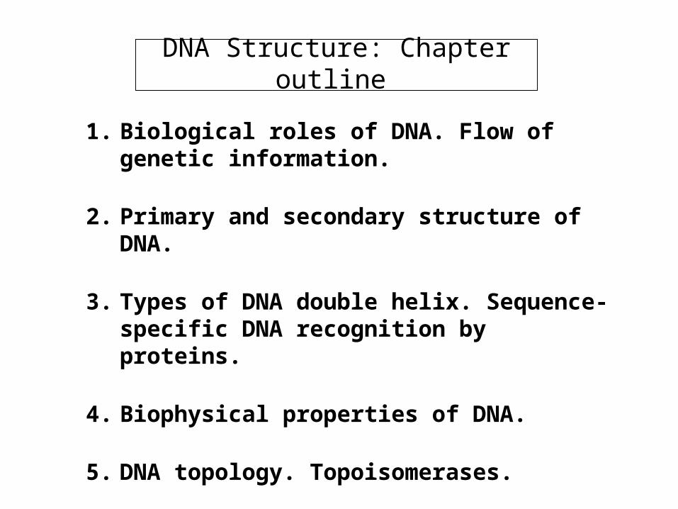
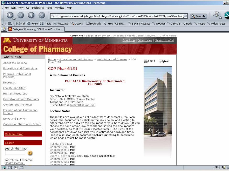
DNA Structure: Chapter outline
1. Biological roles of DNA. Flow of genetic information.
2. Primary and secondary structure of DNA.
3. Types of DNA double helix. Sequence-specific DNA recognition by proteins.
4. Biophysical properties of DNA.
5. DNA topology. Topoisomerases.
6. Restriction Endonucleases. Molecular Cloning

Nucleic Acids
DNA RNA
Central Dogma of Biology
DNA RNA Proteins Cellular Action
transcriptiontranslationDNA
rep
licati
on
(deoxyribonucleic acids) (ribonucleic acids)
Why ?
Questions?
•How is genetic information transmitted to progeny cells?•How is DNA synthesis initiated?•What causes DNA defects and what are their biological an physiological consequences?•What causes the differences between cells containing the same genetic information?
Relevance:
•Cancer: ex. Xeroderma pigmentosum•Genetic diseases: ex., cystic fibrosis, sickle cell anemia,
inborn errors of metabolism•Genetic typing: ex., drug metabolism•Rational drug design: ex., antitumor and antimicrobial
drugs•Biotechnology: ex., growth hormones

The Building Blocks of DNA
-OO
H(OH)
HH
HHO
OP
O
O-
Purine orPyrimidineBase
Phosphate
Pentose sugar
Nucleoside
Nucleotide
1'
2'3'
4'
5'-N-glycosidic bond
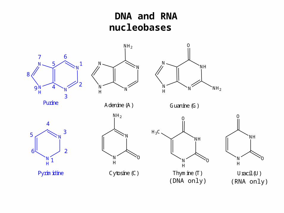
DNA and RNA nucleobases
NN
NNH
NH2
NNH
NNH
O
NH2
N
NH
NH2
O
H3C
NH
NH
O
O
Guanine (G)Adenine (A)
Thymine (T)Cytosine (C)
NH
NH
O
O
Uracil (U)
NN
NNH
Purine
N
NH
Pyrimidine
1
2
3
4
5
6
3
2
16
4
57
8
9
(DNA only) (RNA only)
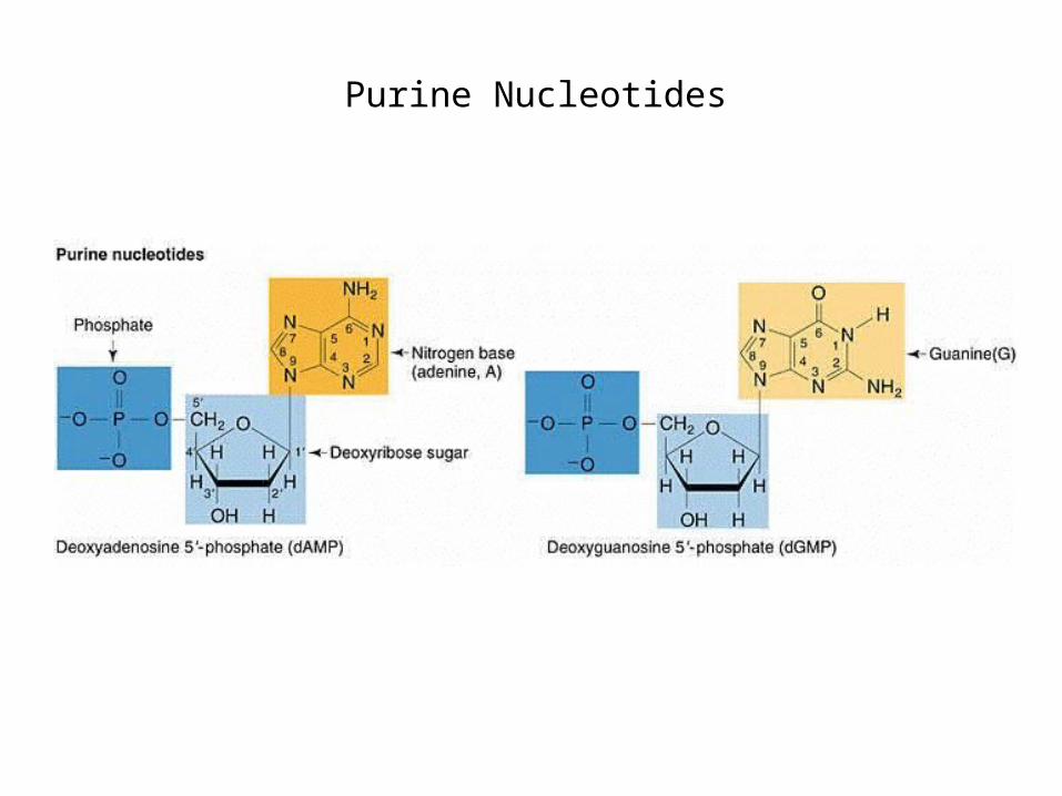
Purine Nucleotides
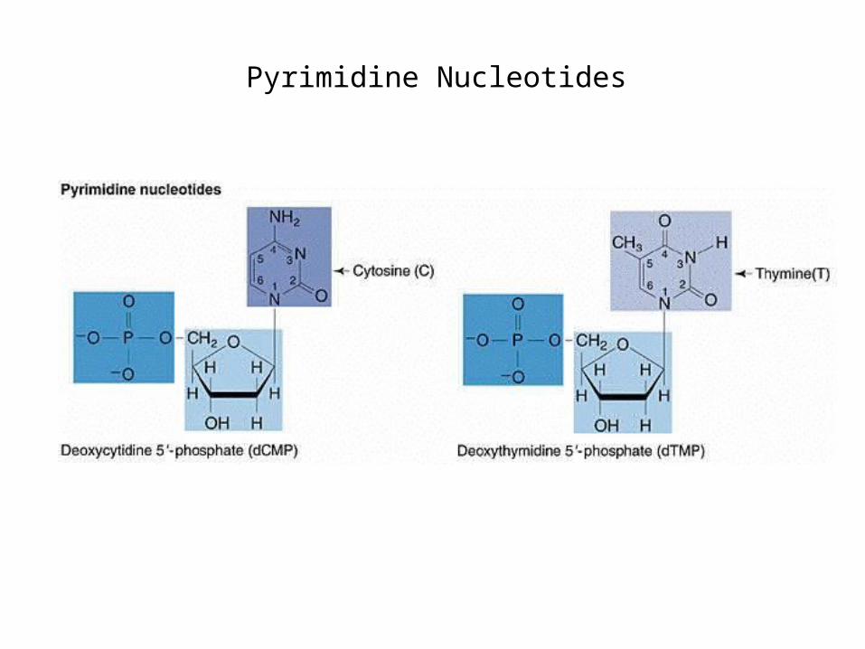
Pyrimidine Nucleotides
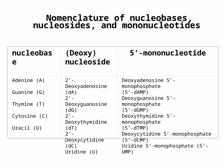
nucleobase (Deoxy)nucleoside
5’-mononucleotide
Adenine (A)
Guanine (G)
Thymine (T)
Cytosine (C)
Uracil (U)
2’-Deoxyadenosine (dA)2’- Deoxyguanosine (dG)2’- Deoxythymidine (dT)2’- Deoxycytidine (dC)Uridine (U)
Deoxyadenosine 5’-monophosphate (5’-dAMP)Deoxyguanosine 5’-monophosphate (5’-dGMP)Deoxythymidine 5’-monophosphate (5’-dTMP)Deoxycytidine 5’-monophosphate (5’-dCMP)Uridine 5’-monophosphate (5’-UMP)
Nomenclature of nucleobases, nucleosides,
and mononucleotides
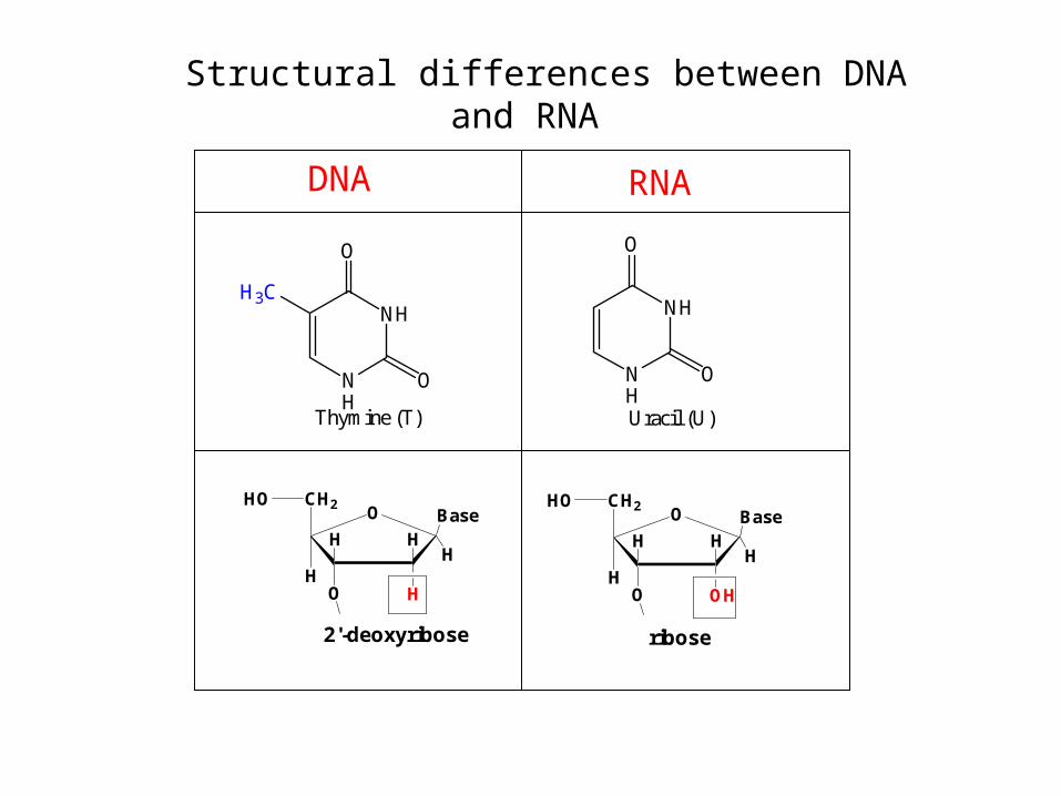
Structural differences between DNA and RNA
H3CNH
NH
O
O
Thymine (T)
NH
NH
O
O
Uracil (U)
DNA RNA
O
H
HHH
CH2
HO
HOBase
2'-deoxyribose
O
OH
HH
CH2
HO
HOBase
ribose
H
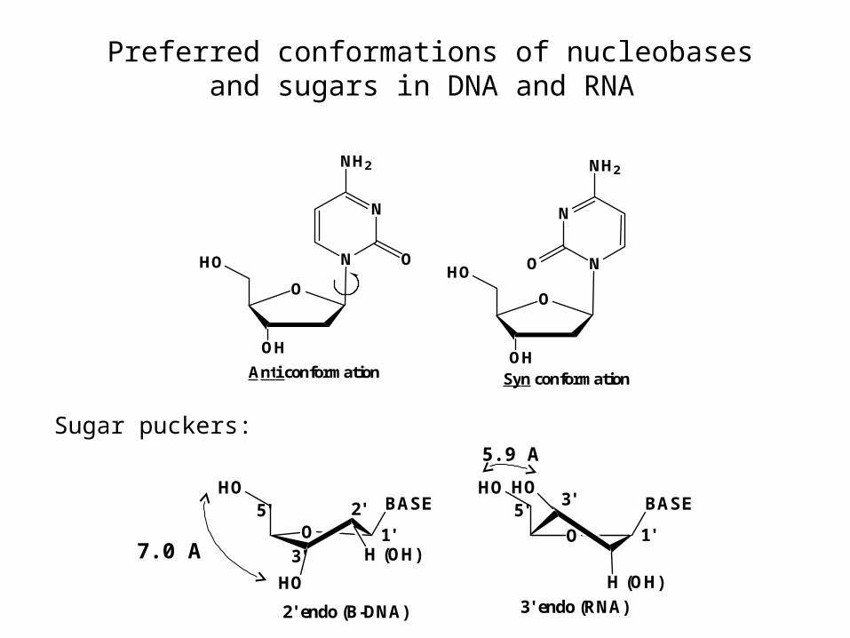
Preferred conformations of nucleobases and sugars in DNA and RNA
HO
O
OH
N
N
NH2
O
Anti conformation
HO
O
OH
N
N
NH2
O
Syn conformation
HO
OH (OH)
HO
BASEHO
O
H (OH)
HOBASE
2' endo (B-DNA)
1'
3' endo (RNA)
3'
1'3'
2' 5'5'
7.0 A
5.9 A
Sugar puckers:
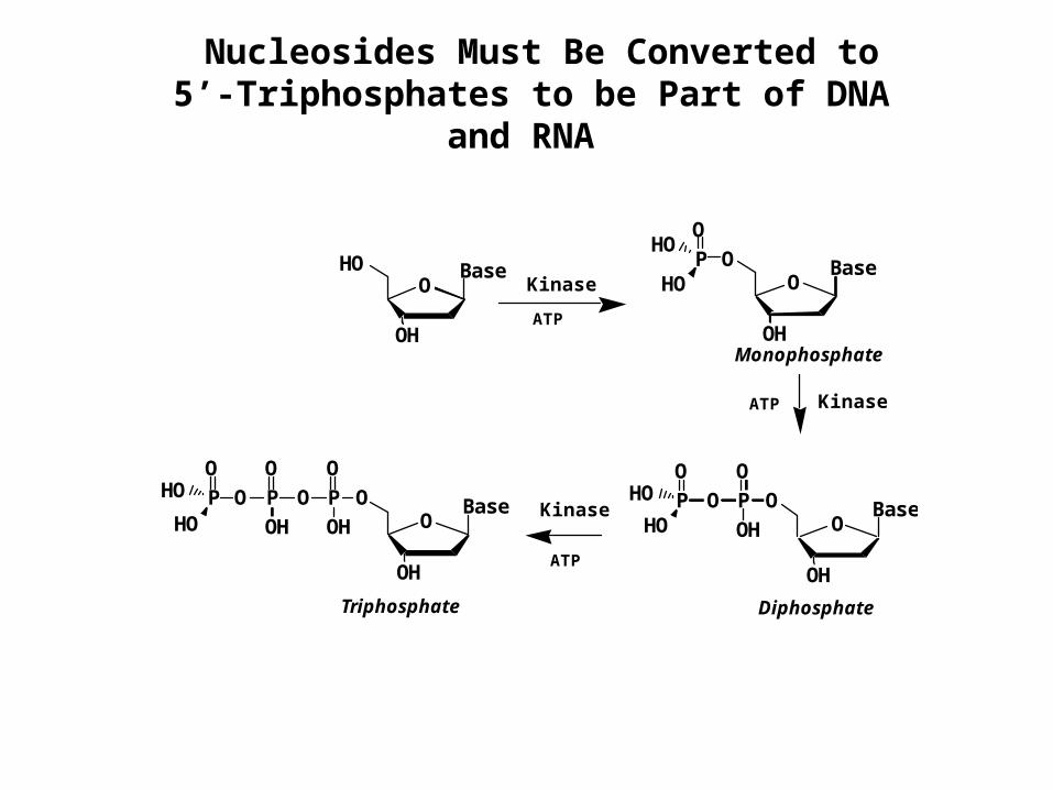
Nucleosides Must Be Converted to5’-Triphosphates to be Part of DNA and
RNA
HOO
OH
OO
OH
PHO
HO
O
OO
OH
P
O
P
OHO
HOO
OH
Base Base
BaseO
O
OH
P
O
P
O
O
OHBase
OH
OP
OHO
HO
Kinase
Kinase
Kinase
Monophosphate
DiphosphateTriphosphate
ATP
ATP
ATP

DNA isArranged5’ to 3’
Connected by
Phosphates
Linking in DNA biopolymer: DNA primary structure

DNA secondary structure – double helix
James Watson and Francis Crick, 1953- proposed a model for DNA structure
•DNA is the molecule of heredity (O.Avery, 1944)
•X-ray diffraction (R.Franklin and M. Wilkins)
•E. Chargaff (1940s) G = C and A = T in DNA
Francis Crick Jim Watson

Watson-Crick model of DNA was based on X-ray diffraction picture of DNA fibres
(Rosalind Franklin and Maurice Wilkins)
Rosalind Franklin

Watson-Crick model of DNA was consistent with Chargaff’s base composition rules
Erwin Chargaff (Columbia University)
G = C and A = T in DNA

DNA is Composed of Complementary Strands
NH
N
N O
NH2
NN
N
H2N
O
HN
N
O
O
NN
N
N NH2
G•C
A•T
DNA
NH
N
N O
NH2
NN
N
H2N
O
HN
N
O
O
NN
N
N NH2
G•C
A•U
RNA
A ::
G :::
T ::
T
C
A
3'
3' 5'
5'
Anti-parallel Strands of DNA
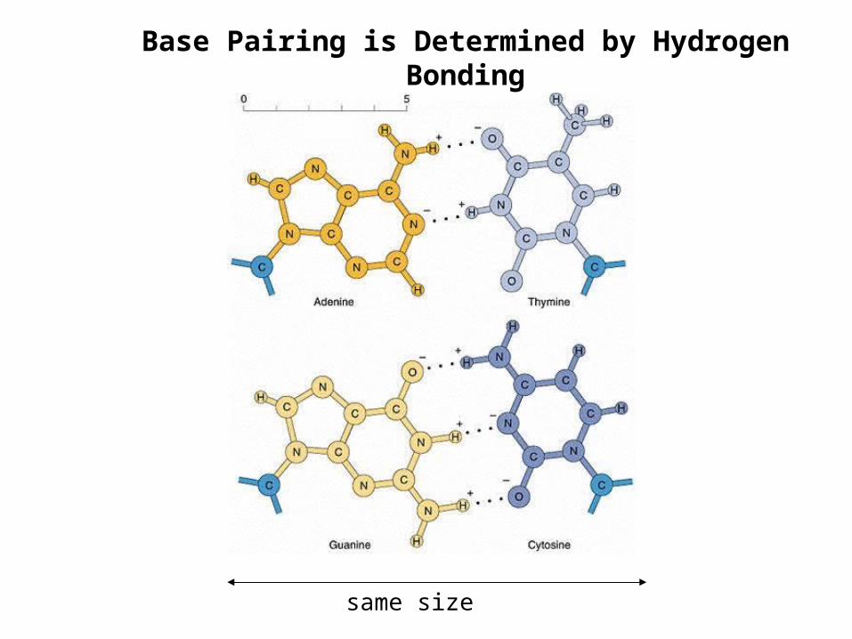
Base Pairing is Determined by Hydrogen Bonding
same size

Base stacking: an axial view of B-DNA
Forces stabilizing DNA double helix
1. Hydrogen bonding (2-3 kcal/mol per base pair)
2. Stacking (hydrophobic) interactions (4-15 kcal/mol per base pair)
3. Electrostatic forces.

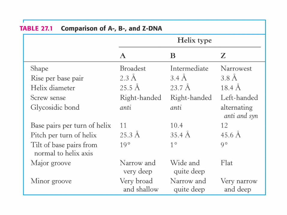
right handed helix
• planes of bases are nearlyperpendicular to the helix axis.
•Sugars are in the 2’ endo conformation.
•Bases are the anti conformation.
•Bases have a helical twist of 36º (10.4 bases per helix turn)
• Helical pitch = 34 A
B-DNA
• 3.4 A rise between base pairs
Wide and deep
Narrow and deep
HO
O
OH
N
N
NH2
O
HO
OH (OH)
HO
BASE
1'3'
2'5'
7.0 A
• helical axis passes through
base pairs
23.7 A
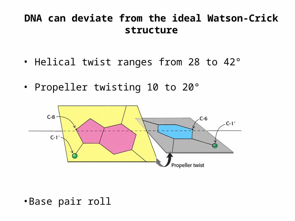
DNA can deviate from the ideal Watson-Crick structure
• Helical twist ranges from 28 to 42°
• Propeller twisting 10 to 20°
•Base pair roll
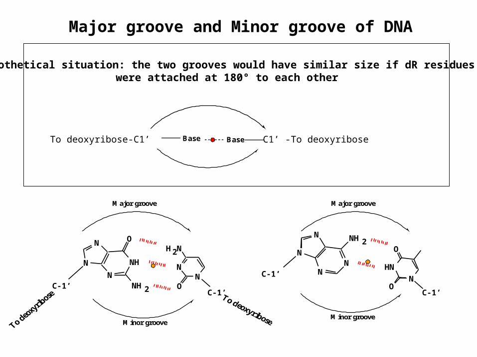
Major groove and Minor groove of DNA
Major groove
Minor groove
NH
N
N O
NH 2
N NN
H 2N
OC-1’C-1’
HN
N
O
O
NN
N
N NH 2
C-1’
C-1’
Major groove
Minor groove
Base BaseTo deoxyribose-C1’ C1’ -To deoxyribose
Hypothetical situation: the two grooves would have similar size if dR residues were attached at 180° to each other

Major and minor groove of the double helix
Wide and deep
Narrow and deep
Major groove
NH
N
N O
NH2
N NN
H2N
O
Minor grooveTo deo
xyrib
ose C-1’
C-1’
HN
N
O
O
NN
N
N NH2
C-1’
C-1’

B-type duplex is not possible for RNA
steric “clash”
O
OH
HH
CH2
HO
HOBase
ribose
H
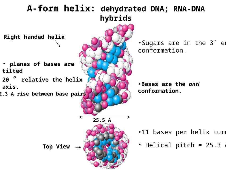
A-form helix: dehydrated DNA; RNA-DNA hybrids
Top View
Right handed helix
• planes of bases are tilted
20 ° relative the helix axis.
• 2.3 A rise between base pairs
•Sugars are in the 3’ endo conformation.
•Bases are the anti conformation.
•11 bases per helix turn
• Helical pitch = 25.3 A
25.5 A

The sugar puckering in A-DNA is 3’-endo
O
OH (OH)
O
BASEO
O
H (OH)
OBASE
2' endo (3' exo) B-DNA
1'
3' endo (A-DNA)
3'
1'3'
2'
5'
5'
2'
7.0 A
5.9 A
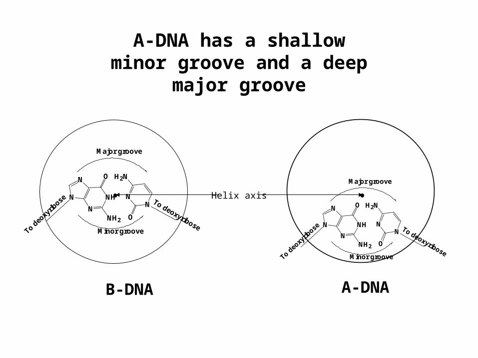
A-DNA has a shallow minor groove and a deep
major groove
N
NH
N
N
O
NH2
NN
H2N
O
To deo
xyrib
ose To deoxyribose
Major groove
Minor groove
B-DNA
N
NH
N
N
O
NH2
NN
H2N
O
To deo
xyrib
ose To deoxyribose
Major groove
Minor groove
Helix axis
A-DNA
• •

Z-form double helix: polynucleotides of alternating purines and pyrimidines (GCGCGCGC) at
high salt
Left handed helix
• Backbone zig-zags because sugar puckers alternate between 2’ endo pyrimidines and 3’ endo (purines)
• Bases alternate between anti (pyrimidines) and syn conformation (purines).
•12 bases per helix turn
• Helical pitch = 45.6 A
• planes of the bases are tilted 9° relative the helix axis.
• Flat major groove• Narrow and deep minor groove
18.4 A
• 3.8 A rise between base pairs

Sugar and base conformations in Z-DNA alternate:
N
N
NH2
ON
HN
NN
O
H2NHO
OH
HO
HO
O
H
HO1' 3'
1'3'
2'
5'
5'
GC
5’-GCGCGCGCGCGCG3’-CGCGCGCGCGCGC
C: sugar is 2’-endo, base is antiG: sugar is 3’-endo, base is syn

Biological relevance of the minor types of DNA secondary structure
•Although the majority of chromosomal DNA is in B-form, some regions assume A- or Z-like structure
• Runs of multiple Gs are A-like
•The upstream sequences of some genes contain 5-methylcytosine = Z-like duplex
N
NH
NH2
O
5-methylcytosine (5-Me-C)
H3C
• RNA-DNA hybrids and ds RNA have an A-type structure
• Structural variations play a role in DNA-protein interactions