Embed Size (px)
Citation preview

Biochemistry 1994, 33, 11493-1 1500 11493
Binding of Actinomycin D to the T(G)nT Motif of Double-Stranded DNA: Determination of the Guanine Requirement in Nonclassical, Non-GpC Binding
Sites+ Susan A. Bailey,* David E. Graves,*’$ and Randolph Rills
Department of Chemistry, University of Mississippi, University, Mississippi 38677, and Department of Chemistry and Institute of Molecular Biophysics, Florida State University, Tallahassee, Florida 32306-3006
Received February 24, 1994; Revised Manuscript Received July 15, 19948
ABSTRACT: Strong binding of the antitumor antibiotic actinomycin D to the sequence 5’-TGGGT-3’ in double-stranded D N A was recently established by equilibrium binding studies (Bailey et a l . , 1993). Actinomycin D binding to this -TGGGT- containing sequence was shown to be comparable to that of an -XGCY- containing oligonucleotide (K, - lo6 M-l). Investigation of -TGGGT- as a high-affinity binding site for actinomycin D follows from our 1989 sequencing study (Rill et al., 1989) in which the photoaffinity analog of actinomycin D (7-azidoactinomycin D) was used to determine D N A base sequence specificities and neighboring base effects. The studies presented here examine the guanine requirements for actinomycin D binding to such nonclassical (non-dGpC) sites by varying the number of central guanine residues in a series of selected duplex oligonucleotides. The central -T(G),T- motif varies from n equals 1 to 4. Actinomycin D binding to each of these undecamers is characterized and correlated with binding to oligonucleotides of identical length and similar sequences that contain classical dGpC binding sites. Binding affinities of actinomycin D to this series of oligonucleotide duplexes (10 “C) can be summarized as -TGGGT- > -TGGT- > -TGGGGT- > -TGT. The kinetics of SDS-induced dissociation of actinomycin D from these oligonucleotides reveal single-exponential decays with duration dependent on the sequence at the binding site. With the exception of the -TGGGT-containing oligomer, dissociation times for the T(G),T duplexes were drastically different and much shorter than times obtained for the dissociation of actinomycin D from oligonucleotides having classical dGpC sites. Dissociation constants ranged from 1463 s for -TGGGT- to 67 s for -TGGT-. The binding energetics for these drug-DNA interactions are quantitated and are presented as a function of the number of central guanines. W e observe an unusual (positive) binding enthalpy for the interaction of actinomycin D with the -TGGT- sequence, whereas binding to all of the other sequences exhibits negative enthalpies. Thus, addition or deletion of one G to or from the original -TGGGT- site causes significant changes in both binding affinities and energetics, even though the number of binding sites per duplex remains constant. These changes and how they correlate with changes in the base sequence are presented as a full thermodynamic profile.
Actinomycin D is a DNA intercalator proposed to exert its cytotoxic effect by inhibiting RNA polymerase through its interaction with double-stranded DNA (Goldberg et al . , 1962; Hurwitz et al . , 1962). The interaction of actinomycin D with DNA is highly sequence dependent, with an apparent requirement for guanine and preference for dGpC steps along double-stranded DNA (Muller & Crothers, 1968; Wells et al . , 1970). Sequence-specific binding of actinomycin D to dGpC steps has been confirmed by numerous studies using a wide variety of methods including NMR (Patel, 1974; Krugh & Neely, 1973; Brown et al., 1984) and DNA footprinting (Lane et al . , 1983; Fox & Waring, 1984; Goodisman et al . , 1992). The dGpC-selective model was first proposed from the X-ray diffraction pattern of a deoxyguanosine-actinomycin D complex by Sobell and Jain (1971) and more recently supported by Kamitori and Takusagawa (1992) in their description of an actinomycin D-d(GAAGCTTC)* cocrys- talline complex.
t This research was supported in part by US . Public Health Service Research Grants CA-41474 (D.E.G.), the Elsa U. Pardee Foundation (D.E.G), and Department of Energy Grant ER60588 (R.R.).
* Address correspondence to this author at the University of Mississippi. Email address: graves ~graves.chem.olemiss.edu; Tel: (601) 232-7732; FAX: (601) 232-7300.
University of Mississippi. 9 Florida State University. @ Abstract published in Advance ACS Abstracts, September 1, 1994.
0006-2960/94/0433-11493%04.50/0
<According to the model of Sobell and Jain the planar phenoxazone ring is described as being fully inserted (inter- calated) between the two adjacent GC base pairs of a dGpC step, with the two side chains residing in the minor groove. Their examination of the crystal structure of actinomycin D complexed with deoxyguanosine identified specific hydrogen bonds between the threonine carbonyl oxygen of the cyclic pentapeptide side chains of the drug and the guanine 2-amino group of the DNA. This binding geometry was recently confirmed by Kamitori and Takusagawa (1992), who used a longer DNA lattice for their structural determination of an actinomycin D-DNA complex. On the basis of the structural details derived from this complex, certain predictions were made concerning the effects that flanking bases might have on the binding of actinomycin D. From their study, Kamitori and Takusagawa postulated that the adjacent base on the 5’ side of the dGpC step must be either A or T due to steric limitations created by insertion of the pentapeptide chains within the DNA’s minor groove.
Recently, our laboratory examined the sequence-selective binding of actinomycin D by demonstrating the influences that flanking DNA base sequences (those bases which flank the intercalation site on both the 5’ and 3’ sides) exert on actinomycin D binding affinities and energetics. Comparisons of oligonucleotides containing central -TGCA- and -CGCA- sequences, both high-affinity actinomycin D binding sites,
0 1994 American Chemical Society

11494 Biochemistry, VoZ. 33, No. 38, 1994
revealed that changes to the 5’ flanking base alter both the binding affinities and enthalpy and entropy contributions toward binding energetics. The oligonucleotide containing -TGCA- bound actinomycin D with an affinity of 4.5 X lo6 M-l, whereas the -CGCA- containing oligomer bound acti- nomycin D with a slightly lower affinity of 2.5 X lo6 M-l. However, binding enthalpies varied significantly for these two DNA sequences. Both enthalpies were negative, but the AHo for -CGCA- was much larger in magnitude (-6.1 kcal/mol) than the AHo for -TGCA- (-2.7 kcal/mol). These studies revealed that subtle changes in base sequence of these oligonucleotides result in markedly different thermodynamic contributions to the interactions of this chemotherapeutic drug with its cellular target, indicating that the high degree of DNA base sequence specificity may be based on the ther- modynamic mechanism associated with complex formation.
In addition to identifying the influences of flanking bases on the thermodynamic binding properties of actinomycin D, our studies also revealed that a nonclassical DNA sequence (5’-TGGGT-3’) is a high-affinity binding site for actinomycin D. This sequence, as the central sequence of an undecamer duplex, is characterized by a high actinomycin D binding affinity (ICa = 1.5 X lo6 M-l), comparable to the well- documented -TGCA- and -CGCA- sites. The -TGGGT- sequence was first identified through our 1989 photoaffinity labeling study which utilized 7-azidoactinomycin D as a photoaffinity probe to monitor sites of covalent attachment as a measure of the sequence selectivity of actinomycin D binding. Highest reactivity occurred at the second G in this -TGGGT- sequence, which had not been previously identified as a strong actinomycin D binding site.
Binding of actinomycin D to -TGGGT-, now established as a high-affinity, non-dGpC binding site, would seemingly require a different binding mechanism than that first pro- posed by Sobell and Jain in 197 1. The existence of -TGGGT- as a high-affinity actinomycin D binding site is consistent with other observations in which actinomycin D was shown to bind non-dGpC sites (Wells & Larson, 1970; Scamrov & Beabealashvilli, 1983; Snyder et al., 1988; Waterloh & Fox, 1992). Scamrov and Beabealashvilli (1983) determined that the minimum requirement for actinomycin D binding to DNA tetranucleotide sequences was the presence of either a dGpC or dGpG step. In 1988 Breslauer and co-workers (Snyder et al., 1989) demonstrated that the self-complemen- tary octanucleotide d(CGTCGACG)Z binds two actino- mycin D molecules per duplex in a cooperative manner. Using batch calorimetry, they observed very strong binding to this seqence (Ka = 1.4 X lo7 M-l) and an atypical binding en- thalpy of + 1.8 kcal/mol. Their systematic comparison of octamers containing the same dGpT and dGpA steps as d(CGTCGACG)* showed no binding, except to this single sequence. The DNase I footprinting study by Waterloh and Fox (1992) demonstrated that actinomycin D binds well to dGpG steps of duplex DNA and that binding is potentiated by flanking base sequence. Weak or no footprints were observed on fragments containing dGpT steps, indicating only weaker or secondary binding at these sites.
These studies are consistent with the extreme degree of base sequence selectivity exhibited by actinomycin D, both at the intercalation site and at neighboring bases. In this manuscript we present additional evidence for the interaction of actinomycin D with non-dGpC binding sites and probe the requirement for guanines within the central core of the intercalation site. In addition, we offer kinetic and thermo- dynamic evidence to demonstrate differences in binding which can be attributed to variations in base sequence. We employ
Bailey et al.
a series of oligonucleotides containing -T(G)nT- as the central sequence motif in order to determine the influence of guanine in mediating actinomycin D binding. The number of central guanines in these oligomers is varied from n equals 1 to 4, and binding to each of these undecamers is quantitated. The following sequences (duplexes) are examined:
5’-CTATGGGGTAC-3’/5’-GTACCCCATAG-3’ (referred to as -TGGGGT-)
5’-CTATGGGTTAC-3’/5’-GTAACCCATAG-3’ (referred to as -TGGGT-)
5’-CTATGGTTAAC-3‘/5’-GTTAACCATAG-3’ (referred to as -TGGT-)
5’-CTATGTTATAC-3’/5‘-GTATAACATAG-3‘ - (referred to as -TGT-)
This group of non-self-complementary oligonucleotides was designed to have identical length, terminal sequence, and similar thermal stability so that the base sequences could be compared and differences in binding quantitated without any additional influences. The sequences flanking the T(G),,T sites are identical for all undecamers examined. The results obtained with the DNA duplexescontaining a central T(G),,T motif are compared and correlated with those for the well- characterized -TGCA- site in the undecamer 5’-CTATTG- CATAC-3’/5’-GTATGCAATAG-3’.
EXPERIMENTAL PROCEDURES
Oligonucleotide Preparation. Non-self-complementary undecamers of specific sequence were purchased from Re- search Genetics, Inc. Oligonucleotides were desalted by reverse-phase HPLC using an acetonitrile gradient (0-30%) which revealed greater than 98% purity of the oligonucleotide. The lyophilized strands were dissolved in 0.01 M sodium phosphate (pH 7.0), 0.001 M disodium EDTA, and 0.1 M sodium chloride. All subsequent experiments were carried out in this buffer. Molar absorptivities for each oligomer were determined by the nearest-neighbor method (Fasman, 1975).
Oligonucleotide Characterization. Stock solutions of the single strands were mixed in 1:l stoichiometry with their complement to obtain duplexes. Duplex-to-single strand thermal denaturation profiles for each of the oligonucleotide duplexes were obtained using a Cary 2290 (Varian) spec- trophotometer equipped with thermostated cell holders, mixers, and a programmable temperature controller (Lauda) which incremented the temperature at a rate of 0.25 OC/min. Thermodynamic parameters associated with helix formation were derived from analysis of the change in absorbance at 260 nm (AA) versus temperature as described by Petersheim and Turner (1983) and Freier et al. (1983). Melting curves were described by a two-state model to obtain AHo and ASo using Enzfitter (Biosoft). Since the melting temperatures of the oligonucleotide duplexes are concentration dependent, all DNA denaturation experiments were performed at identical duplex concentrations, which were also maintained during the drug binding experiments.
Denaturation studies of the oligonucleotides in the presence of bound actinomycin D were also performed. The amount of actinomycin D to be added was calculated using the equation:

Actinomycin D Binding to the -T(G),,T- Motif Biochemistry, Vol. 33, No. 38, 1994 11495
1968), representing the average value for a composite of all potential binding sites on the native DNA. However, in the case of these selected oligonucleotide duplexes, a primary concern was that, with only one potential binding site within the duplex, the molar absorptivity of the bound drug species might differ for each duplex due to the varying influences of the physical and chemical environment which result from differences in the base sequence. Molar absorptivities for the bound drug species weredetermined for thescatchard analyses described above. Interestingly, for each of the oligonucleotide duplexes, the eb values were found to be comparable (within 10%) to the Eb obtained for native calf thymus DNA.
Thermodynamic Analysis of Binding Data. The fixed ratio method outlined by Chaires (1985) and Shimer et al. (1988) was used to determinevan't Hoff enthalpies for the interactions of actinomycin D with each duplex. Data were obtained by adding a known concentration of drug to the DNA solution at 10 "C and determining the concentration of free and bound drug using the equations for cb and Cf as described earlier. At an approximatervalue of 0.5 (per duplex), the temperature was increased to 30 "C and allowed to equilibrate for 3 h. After thermal equilibrium had been achieved, the absorbance was determined and the temperature was decreased in 5-deg increments to 0 "C. The sample was allowed to equilibrate for 3 h at each new temperature, and the amounts of free and bound drug redetermined by monitoring the absorbance change. Due to the limited thermal stability of the oligo- nucleotide duplexes, the accessible temperature range for van't Hoff analyses for examining actinomycin D binding to duplex DNA was restricted to between 0 and 30 "C. The Scatchard equation was used to calculate K, from the measured r and r/Cf values at each temperature. A linear least-squares analysis of a plot of the ln(K,) versus l/[T(K)] was used to estimate the apparent enthalpy of binding from the van't Hoff relationship:
where fb is equal to the fraction of bound DNA (set to be greater than 0.95) and [D] is the drug concentration required to obtain 95% bound complex. The duplex oligonucleotide concentrations, binding constants, K,, and binding densities were previously determined for each of the sequences examined. Thermodynamic parameters for the melting transition in the presence of actinomycin D were obtained in the same manner as described above for the thermal transition of the DNA duplexes alone.
Actinomycin D-DNA Interactions. Actinomycin D binding to selected oligonucleotides containing the T(G)nT motif was monitored by UV-visible spectroscopy. Complex formation with DNA results in marked changes in the visible region of the drug's absorption spectrum, and these changes can be utilized to monitor the equilibrium binding of the drug to DNA. Known concentrations of duplex oligonucleotide were pipetted into 10 cm path length cylindrical cells maintained at 10 "C (f0.1 "C); the temperature was directly monitored by insertion of a Thermistor microprobe into the sample cuvette. Greater than 95% of the oligomer is in duplex conformation at this temperature, ionic strength, and DNA concentration, as determined from our helix-to-coil transition studies. Small aliquots of a known concentration of stock actinomycin D solution were titrated into the sample cell. After each addition, the solution was mixed and allowed to re-equilibrate to 10 "C. The absorbance was then determined directly at selected wavelengths of maximal absorbance change using a statistical algorithm which signal averages 300 readings and provides the mean and standard deviation of the absor- bance value. Total actinomycin D concentration (C,) was maintained at less than 5 pM to minimize aggregation of the actinomycin D in aqueous solution, which could lead to error in determining the correct drug concentration. The absorbance changes at 440 nm of the actinomycin D, resulting from complex formation with the oligonucleotides, were used to quantitate the intrinsic equilibrium binding constant, K,, and the site exclusion parameter, n. Bound and free drug concentrations (cb and Cf, respectively) were determined by the equations:
c b = A A / A t and cf = c, - cb where AA is the difference in the absorption between the free and bound drug species and Ae is the difference between the molar absorptivities for the free and bound drug species.
The Scatchard equation was used to determine the binding constant, K,, and site exclusion size, n (number of drugs bound per duplex), for each of the data sets by plotting r/Cf versus r, where r is equal to the bound drug concentration per DNA duplex. The Scatchard equation (Scatchard, 1949) adequately describes the binding of actinomycin D to these duplexes because of the presence of only one binding site, eliminating the need for neighbor exclusion and/or cooperative binding terms. K, and n were estimated from an iterative nonlinear least-squares fitting routine (Simplex):
r/Cf = K,(1 - nr)
The change in absorbance of the drug as a function of complex formation with DNA allows experimental determi- nation of €b (molar absorptivity of the fully bound drug species). In the case of actinomycin D-calf thymus DNA, this value was determined to be 12 000 M-I cm-I (Muller & Crothers,
where R is the gas constant. This method of van't Hoff analysis of ligand binding to
DNA has been shown to provide results comparable to those obtained by calorimetricmethods (Sauer et al., 1984; Chaires, 1985) which often require prohibitively high concentrations of oligonucleotides. Determination of the enthalpy of binding by this method provides sufficient data for a full thermody- namic profile which can be estimated by the following relationships:
AGO = -RT In K,
hs" = -(AGO - A H " ) / T
SDS- Induced Dissociation Kinetics. Time-dependent ab- sorbance changes at 440 nm were monitored at 20 "C using a CARY 2290 spectrophotometer equipped with a stirrer accessory. Data were recorded and displayed on the CARY 2290 at 10 s intervals immediately following addition of a 20% stock SDS solution sufficient to make a final SDS concentration of 1% in the sample cell. The SDS solution was added to an equilibratedmixtureof 1: 1 oligonucleotide duplex and actinomycin D.
The increase in absorbance, due to the dissociation of actinomycin D from the DNA, was monitored as a function of time. The resulting dissociation curves (A,4440nm versus time) were analyzed using the non-linear least-squares first-

11496 Biochemistry, Vol. 33, No. 38, 1994 Bailey et al.
Table 1 : Thermal Stability of Oligonucleotides Containing the T(G).T Motifa ~~~
free duplex duplex + drug obsd -AHo predicted -AHo As0 sequenceb '7% GC T m ("Cl A T m ("C) (kcal/mol) (kcal/moll lcal /(mol.K) 1
~~
5'-CTA TGGGGTAC-3' 55 41 9 62 80 175 GAT ACCCCA TG
5'-CTA TGGGT TAC-3' 45 38 7
5'-CTA m T A A C - 3 ' 36 35 3 GAT ACCCA ATG
GAT ACCA A T G
74
60
78
76
213
173
5'-CTA TATAC-3' 27 30 3 33 68 88 GAT ACA ATATG
a T,, AHo, and aSo determined by a nonlinear least-squares algorithm in ENZFITTER (Biosoft). * Oligonucleotide concentrations are as follows: -TGGGGT-, 2.0 pM; -TGGGT-, 1.7 pM; -TGGT-, 1.7 pM; -TGT-, 1.7 fiM (duplex). All melting profiles determined at 0.01 M sodium phosphate buffer (pH 7.0), 0.001 M disodium EDTA, 0.1 M sodium chloride. Predicted AHo values for the duplex denaturation were determined by the nearest-neighbor analysis of Breslauer and co-workers (Breslauer et al., 1986).
order rate equation of the Enzfitter fitting program (Biosoft). Rate constants were determined using a single exponential to analyze the observed absorbance change as a function of time.
RESULTS
Thermal Stability of Oligonucleotides Containing Central T(G),T Motifs. The oligomers used in this study were examined for any unusual thermal properties. Duplexes were denatured (see Experimental Procedures), and all melting profiles exhibited sigmoidal curves, characteristic of a two- state melting transition. As expected, we observed a linear relationship between the number of GC base pairs and duplex melting temperature for these oligomers (Table 1 ) . The melting temperatures (Tm) we observed ranged from 30 to 41 "C as the number of central guanines increases from 1 to 4.
Enthalpy values for duplex formation varied from 33 kcal/ mol for the -TGT-containing oligomer to 74 kcal/mol for the -TGGGT- containing oligomer. Interestingly, the most exothermic duplex formation was observed for the -TGGGT- sequence (45% GC) and not the -TGGGGT- (55% GC). The enthalpy and entropy contributions due to duplex formation for all of the sequences are provided in Table 1 . With the exception of the duplex 5'-CTATGTTATAC-3'/5'-GTATAA- CATAG-3', there is good agreement between observed and predicted melting enthalpies. Predicted values are based on the nearest-neighbor method of Breslauer and co-workers (Breslauer et al . , 1986).
Duplex denaturation experiments were carried out in the presence of a 1 : 1 ratio of bound actinomycin D per duplex to determine the degree of helix stabilization conferred upon the helix by drug binding. Typically, actinomycin D can be classified as a helix stabilizer (McGhee, 1976; Chen, 1988a,b; Snyder et al., 1989). For all of the selected T(G),T-containing oligomers, helix stabilization was observed in the presence of actinomycin D. However the degree of stabilization varied, ranging from 3 to 9 OC, and was highly dependent on the DNA base sequence. This ATm represents contributions made by both the DNA sequence and actinomycin D binding affinity. Interestingly, the stability of both -TGT- and -TGGT- containing duplexes increased by only 3 OC in the presence of actinomycin D; however, their respective actinomycin D binding affinities (at 10 "C) differ considerably (for -TGT-, Ka = 4.4 X lo5 M-l; and Ka = 7.7 X lo5 M-l for -TGGT-). Duplexes containing the -TGGGGT- and -TGGGT- binding sites showed increases in stabilization of 7 and 9 OC in the presence of actinomycin D.
Guanine Dependence of Actinomycin D Binding. The T(G),T motif was used as a means to determine the absolute guanine requirement for actinomycin D binding to nonclassical, non-dGpC sites in double-stranded DNA. Optical titrations
were used to characterize actinomycin D binding to the sequences listed in the introduction. By monitoring the decrease in the drug's absorbance at 440 nm, we obtained accurate actinomycin D binding isotherms for each of the selected oligonucleotide duplexes. These studies were carried out at 10 OC to ensure the DNA was double-stranded prior to the addition of actinomycin D. At this temperature and DNA concentration, thermal denaturation studies reveal the oligonucleotides in duplex conformation to be greater than 96% thus providing a suitable model for examining sequence dependence of actinomycin D binding to double-strand DNA.
The -TGGGT- site was previously shown to bind one actinomycin D molecule with an affinity of 1.5 X 10 M-l, which is comparable in magnitude to actinomycin D binding to classical dGpC sites (Bailey & Graves, 1993). Binding of actinomycin D to dGpC steps has been well-documented, with intercalation occurring between the adjacent GC base pairs. Our photoaffinity labeling studies revealed highest reactivity for 7-azidoactinomycin D at the second guanine of the five- base sequence -TGGGT-. We were surprised by the high affinity for -TGGGT- and wished to determine the influence of base sequences which flank this binding site. We have systematically changed this sequence to create a series of oligonucleotides, allowing us to identify the minimum guanine requirement for actinomycin D binding. We have already shown that the flanking bases surrounding a dGpC step play a major role in influencing actinomycin D binding.
Binding data are presented in the form of Scatchard plots as shown in Figure 1. Our results demonstrate the significant influence presented by guanine (both presence and number) on actinomycin D binding properties. In going from n equals 1 to 3, a linear relationship between the number of central guanines and binding affinity for actinomycin D is observed as illustrated in Figure 2. The -TGGGT- containing sequence exhibits the highest affinity, followed next by the -TGGT- site. Interestingly, both the -TGGGGT- and -TGT- containing duplexes demonstrated low affinities for actinomycin D (Figures 1 and 2 and Table 2). This was expected for the -TGT- sequence, but was surprising for the -TGGGGT- sequence. These oligonucleotides were designed so that a single target binding site was positioned in the center of the sequence in order to avoid drug binding at the ends of the duplex or having multiple binding sites (Le., site-exclusion effects). The number of binding sites per duplex was approximately one for each of the sequences used in this study.
Thermodynamic Parameters for Actinomycin D Binding. Using the fixed-ratio method of determining van't Hoff enthalpies, we were able to obtain a thermodynamic profile for actinomycin D binding to each of the oligonucleotides containing the T(G),T motif (Figure 3). Actinomycin D

Actinomycin D Binding to the -T(G)nT- Motif Biochemistry, Vol. 33, No. 38, 1994 1 1497
c
2 u!
X
is 2
0.0
6.0
4.0
2.0
8.0
6.0
4.0
2.0
0.2 0.4 0.6 0.8 0.2 0.4 0.6 0.0
r (duplex) FIGURE 1 : Scatchard plots comparing the interactions of actinomycin D with -TGGGGT- , -TGGGT- , -TGGT- , and -TGT-. Binding isotherms were obtained by serial titration of actinomycin D into a known concentration of oligonucleotideand monitoring the absorbance change at 440 nm usinga CARY 2290 UV-visiblespectrophotometer. Solid lines represent linear fits to the Scatchard equation. Temperature was maintained throughout the experiment at 10 "C to ensure the DNA was in the duplex form. All binding experiments were carried out in 0.01 M sodium phosphate buffer, pH 7.0,O.OOl M disodium EDTA, and 0.1 M sodium chloride. Drug concentrations were maintained at concentrations below 5 pM to eliminate aggregation effects.
2000000
4-
1500000 w v) c 0 0
1000000 3 L
i3 500000
0 1 2 3 4 Number of Central Guanines
FIGURE 2: Bar graph illustrating the effect of the number of central guanines in the T(G),,T motif on actinomycin D binding at 10 OC. Binding constants were obtained from Scatchard analysis (described in Experimental Procedures) of spectral titration data (Figure 1).
binding to the -TGGGT- sequence resulted in a favorable binding enthalpy of -3.7 kcal/mol. Again, in terms of binding energetics, the interaction of actinomycin D with thissequence is comparable to that of dGpC-containing sequence (Rill et al., 1989; Bailey et al., 1993).
An extremely interesting result of this study was the observation of a positive enthalpy for the interaction of actinomycin D with the -TGGT- containing oligonucleotide. Following -TGGGT- and having the second highest binding affinity, the -TGGT- site is characterized by a positive binding enthalpyof +4.7 kcal/mol. Snyder et al. (1 989) also reported an atypical positive binding enthalpy for the actinomycin D-d(CGTCGACG)* interaction. Their results indicated that the oligomer bound two actinomycin D molecules in a cooperative manner, in contrast to our sequence which binds only one drug. Thus, their rationale for the observation of the positive enthalpy (drug4rug interactions) can be ruled out in the case of the -TGGT- duplex.
Marky et al. (1983) also reported a positive enthalpy for the interaction of actinomycin D with salmon testes DNA.
Although small in magnitude, this endothermic binding enthalpy was taken to indicate an unusual mode(s) of binding, perhaps even the pseudo-intercalated mode described by Berman and co-workers (1982). This different mode of interaction, possibly indicated by the positive enthalpy values, is clearly different from the typical intercalation model accepted for actinomycin D with dGpC binding sites and other intercalators such as ethidium with DNA which give negative binding enthalpies.
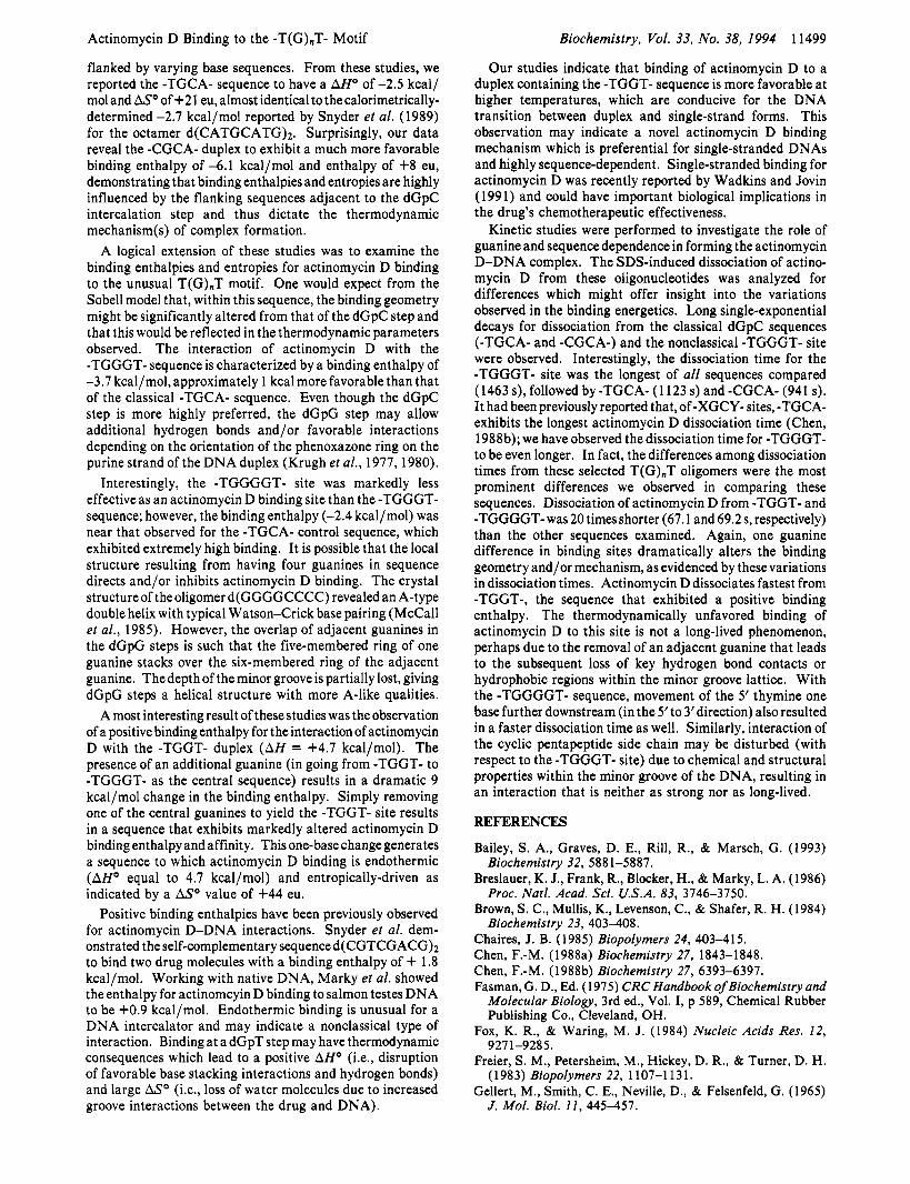
Dissociation Kinetics for Actinomycin D (Table 3). Ac- tinomycin D dissociation from native DNA has been char- acterized as slow, requiring threeexponentials to fit thedecay. The long residence time is thought to be important in the drug's pharmacological activity and represents the complexity of the interaction between the drug (both the chromophore and the side chains) and the DNA. With native DNA or longer lattices of synthetic oligonucleotides, dissociation is multiexponential, and several discrete time constants are observed. These differences may be attributed to nearest- neighbor effects and binding site heterogeneity (Miiller & Crothers, 1968). With these selected undecamers, actino- mycin D binding site heterogeneity is avoided and the dissociation curves can be completely described by a single- exponential rate equation.
With the exception of the -TGGGT- site, the dissociation times observed for the T(G)nT series were all drastically shorter in duration than for the classical -TGCA- site. Actinomycin D dissociation from the oligomer 5'-CTAT TGCATAC-3'/5'-GTATGCAATAG-3' is characterized by a rate constant of 1 123 s, similar to the long residence times reported by Chen (1988a) in his comparison of -XGCY- containing oligomers. Interestingly, the dissociation of ac- tinomycin D from the -TGGGT- sequence exhibits the the longest time observed at 1463 s, much longer than the 69 s observed for -TGGGGT- and the 67 s observed for -TGGT-.
As an added control, we determined the dissociation rate for actinomycin D from a second dGpC-containing oligomer,
CA-), which was earlier reported to be a high-affinity actinomycin D binding site (Bailey et al., 1993). We observed the dissociation rate of 941 s which is lower than the rates observed for -TGCA- and -TGGGT- sequences but much greater than the rates determined for the -TGGGGT- and -TGGT- sites.
DISCUSSION This study provides unique insight into the influences of
DNA base sequenceon the binding energetics and mechanistic properties which describe the interactions of actinomycin D with nucleic acids. This study specifically probes the binding of actinomycin D to atypical (non-dGpC) intercalation sites containing the sequence motif -T(G)IIT- where n varies from 1 to 4 guanine residues. In an effort to probe the role of guanine in the binding of actinomycin D to these non-dGpC sites in duplex DNA, we designed a series of non-self- complementary undecamers (and corresponding complemen- tary strands) whose 5' and 3' ends are identical but whose central regions contain the -TGT-, -TGGT-, -TGGGT-, and -TGGGGT- sequences, respectively. Our earlier DNA sequencing studies revealed the second G of the -TGGGT- sequence to be a highly-preferred site for binding actinomycin D; hence, these duplexes provide a direct model for determining the effects of the flanking base sequence and the influence of guanineon the formation of actinomycin D-DNA complexes.
The binding of actinomycin D to the four T(G)nT undecamer duplexes was compared to similar undecamers containing
5'-CTACCGCATAC-3'/5'-GTATGCGGTAG-3' (-CG-

11498 Biochemistry, Vol. 33, No. 38, 1994 Bailey et al.
Table 2: Thermodynamic Data for the Interactions of Actinomycin D with Selected Oligonucleotides Containing the T(G).T Motif at 10 O C
sequence Ka (M-l)" n (duplex)" AGO (kcal/mol)b AHo (kcal/mol)e ASo [cal/(moLK)ld 5'-CTATGGGGTAC-3' (0.60 f 0.04) X 106 0.9 f 0.2 -7.5 f 0.2 -2.4 f 0.1 +18 f 2
GAT ACCCCA TG
GAT ACCCA ATG
GAT ACCA ATTG
GAT ACA ATATG
5'-CTA TGGGT TAC-3' (1.50 f 0.20) X lo6 1.0 f 0.1 -8.0 f 0.1 -3.7 * 0.1 +15* 1
5'-CTA m T A A C - 3 ' (0.77 f 0.04) X lo6 1.0 f 0.1 -7.6 f 0.2 +4.7 f 0.2 +44 f 3
5'-CTA E L TATAC-3' (0.44 f 0.01) X lo6 1.2 f 0.2 -7.3 f 0.3
Binding parameters derived from Scatchard analysis. Calculated from Ka from the relation AGO = -RT In Ka. Values obtained from the slope of the van't Hoff plots. Entropy estimated from the relation ASo = -(AGO - A H o ) / T .
14
* 13.6 c -
13.2
1
3.35 3.45 3.55 I/T (K) x i o 3
FIGURE 3: van't Hoff plots showing the interaction of actinomycin D with oligonucleotides containing the T(G),,T motif. Solid lines represent linear least-squares fits to the data. Equilibrium binding constants (K.) were obtained from both complete titrations as well as by changing the temperature of the sample containing a fixed ratio of drug and DNA as described in the Experimental Procedures. The -TGGGGT- sequence is represented by open triangles (A); -TGGGT- is represented by open circles (0); and -TGGT- is represented by open squares (0).
Table 3: SDS-Induced Dissociation Times for Actinomycin D O
sequenceb dissociation time (s)
5'-CTA TGGGGtA c-3 ' GAT ACCCCA TG
5'-CTA m T A C - 3 ' 1463 f 190 GAT ACCCA ATG
69 f 7
5'-CTA m T A A C - 3 ' 67 f 4
5'-CTAT TAC-3' 1123 f 46 GAT ACCA ATTG
GTA TGCAATAG 5'-CTAC TAC-3'
GTAT GCGG TAG 941 f 35
0 Dissociation time constants determined by nonlinear least-squares analysis of absorbance change using a first-order rate equation. Oli- gonucleotide concentrations were kept the same for all experiments, 2.0 pM. Actinomycin D:duplex ratios were approximately 1:l (ratio determined from binding constant). All experiments were performed at 20 "C.
-TGCA- and -CGCA- as central actinomycin D binding sites. As shown in Figure 1 and Table 2, at 10 "C, the binding affinities of actinomycin D to the dGpC-containing duplexes are on the order of 2-3 times larger than those observed for the -TGGT- and -TGGGT- duplexes. However, the binding affinities to the -TGGT- and -TGGGT- sites are on the same order of magnitude as the dGpC-containing sequences. The dependence of actinomycin D binding on the number of central guanines was determined by comparing the binding properties of the selected T(G).T sequences. As shown in Table 2 and Figure 2, at 10 "C, there is a nearly linear relationship between actinomycin D binding and the number of guanines present with a binding order of -TGGGT- > -TGGT- > -TGGGGT-. As expected, actinomycin D binds relatively weakly to the
-TGT- site with a K, of 4 X lo5 M-l. Addition of a second and third guanine to form the -TGGT- and -TGGGT- sequences results in doubling and tripling of the actinomycin D binding affinities (8 X lo5 M-I and 1.5 X lo6 M-l), respectively. In contrast, the incorporation of four guanines to give the -TGGGGT- sequence results in a marked decrease in the binding affinity of actinomycin D, dropping it nearly to the level of the -TGT- sequence (the lowest binding we observed). This was surprising since the -TGGGT- site had demonstrated the highest level of binding of all of the T(G).T duplexes; it might be expected that an additional guanine would simply increase binding affinity in a linear fashion. These results suggest that not only is a dGpG step required for optimal binding, but the flanking bases are important in facilitating the binding of actinomycin D. In going from -TGGGT- to -TGGGGT-, substitution of the 3'-T to G dramatically alters the binding affinity, reducing it by more than 50%, from 1.5 X lo6 M-l to 6 X lo5 M-l.
Examination of Figure 3 at temperatures higher than 23 "C reveals a change in the binding order with actinomycin D binding the -TGGT- sequence better than -TGGGT-. The complex formed between actinomycin D and the -TGGT- containing oligonucleotide appears to be atypical based on our observations of the lack of helix stabilization, a positive binding enthalpy, and short dissociation time. Thermal denaturation studies indicate that, at 25 "C, greater than 80% of the -TGGT- undecamer remains in duplex form; however, the partially melted or single-strand DNA compo- nents present at 25 OC (and higher) may result in a different mode of actinomycin D binding.
Waterloh and Fox (1992) showed that actinomycin D binds better to dGpG steps than dGpT steps and that this binding is influenced by flanking base sequence. They attributed this discrimination to differences in local DNA structure and the presence of additional hydrogen bonds. Our spectroscopic binding data are consistent with their DNase I footprinting results. We also show actinomycin D to bind better to the second G of the -TGGGT- sequence (a dGpG step) than to the -TGGT- site (the second G is part of a dGpT step). The binding enthalpy for the interaction of actinomycin D with the -TGGGT- site is approximately 1.5 kcal/mol more exothermic, which is also indicative of enhanced binding.
The DNA binding energies of actinomycin D to these specific duplexes can be dissected into enthalpy and entropy compo- nents by analyzing the equilibrium binding properties as a function of temperature. Historically, the binding enthalpy of actinomycin D to DNA has been determined to be near zero (Gellert e? al., 1965; Snyder et al., 1989). Studies using DNAs with discrete dGpC binding sites have shown binding enthalpies to range from -1 to -2 kcal/mol. We recently reported the influence of flanking base sequences on actino- mycin D energetics by comparing non-self-complementary undecamer duplexes containing central dGpC sequences
Actinomycin D Binding to the -T(G)nT- Motif
flanked by varying base sequences. From these studies, we reported the -TGCA- sequence to have a AHo of -2.5 kcal/ mol and ASo of +21 eu, almost identical to thecalorimetrically- determined -2.7 kcal/mol reported by Snyder et al. (1989) for the octamer d(CATGCATG)z. Surprisingly, our data reveal the -CGCA- duplex to exhibit a much more favorable binding enthalpy of -6.1 kcal/mol and enthalpy of +8 eu, demonstrating that binding enthalpies and entropies are highly influenced by the flanking sequences adjacent to the dGpC intercalation step and thus dictate the thermodynamic mechanism(s) of complex formation.
A logical extension of these studies was to examine the binding enthalpies and entropies for actinomycin D binding to the unusual T(G)nT motif. One would expect from the Sobell model that, within this sequence, the binding geometry might be significantly altered from that of the dGpC step and that this would be reflected in the thermodynamic parameters observed. The interaction of actinomycin D with the -TGGGT- sequence is characterized by a binding enthalpy of -3.7 kcal/mol, approximately 1 kcal more favorable than that of the classical -TGCA- sequence. Even though the dGpC step is more highly preferred, the dGpG step may allow additional hydrogen bonds and/or favorable interactions depending on the orientation of the phenoxazone ring on the purine strand of the DNA duplex (Krugh et al., 1977,1980).
Interestingly, the -TGGGGT- site was markedly less effective as an actinomycin D binding site than the -TGGGT- sequence; however, the binding enthalpy (-2.4 kcal/mol) was near that observed for the -TGCA- control sequence, which exhibited extremely high binding. It is possible that the local structure resulting from having four guanines in sequence directs and/or inhibits actinomycin D binding. The crystal structure of the oligomer d(GGGGCCCC) revealed an A-type double helix with typical Watson-Crick base pairing (McCall et al., 1985). However, the overlap of adjacent guanines in the dGpG steps is such that the five-membered ring of one guanine stacks over the six-membered ring of the adjacent guanine. Thedepth of the minor groove is partially lost, giving dGpG steps a helical structure with more A-like qualities.
A most interesting result of these studies was the observation of a positive binding enthalpy for the interaction of actinomycin D with the -TGGT- duplex (AH = +4.7 kcal/mol). The presence of an additional guanine (in going from -TGGT- to -TGGGT- as the central sequence) results in a dramatic 9 kcal/mol change in the binding enthalpy. Simply removing one of the central guanines to yield the -TGGT- site results in a sequence that exhibits markedly altered actinomycin D binding enthalpy and affinity. This one-base change generates a sequence to which actinomycin D binding is endothermic (AHo equal to 4.7 kcal/mol) and entropically-driven as indicated by a ASo value of +44 eu.
Positive binding enthalpies have been previously observed for actinomycin D-DNA interactions. Snyder et al. dem- onstrated the self-complementary sequence d(CGTCGACG)Z to bind two drug molecules with a binding enthalpy of + 1.8 kcal/mol. Working with native DNA, Marky et al. showed the enthalpy for actinomcyin D binding to salmon testes DNA to be +0.9 kcal/mol. Endothermic binding is unusual for a DNA intercalator and may indicate a nonclassical type of interaction. Binding at a dGpT step may have thermodynamic consequences which lead to a positive AHo (Le., disruption of favorable base stacking interactions and hydrogen bonds) and large ASo (i.e., loss of water molecules due to increased groove interactions between the drug and DNA).
Biochemistry, Vol. 33, No. 38, 1994 11499
Our studies indicate that binding of actinomycin D to a duplex containing the -TGGT- sequence is more favorable at higher temperatures, which are conducive for the DNA transition between duplex and single-strand forms. This observation may indicate a novel actinomycin D binding mechanism which is preferential for single-stranded DNAs and highly sequence-dependent. Single-stranded binding for actinomycin D was recently reported by Wadkins and Jovin (1991) and could have important biological implications in the drug’s chemotherapeutic effectiveness.
Kinetic studies were performed to investigate the role of guanine and sequence dependence in forming the actinomycin D-DNA complex. The SDS-induced dissociation of actino- mycin D from these oligonucleotides was analyzed for differences which might offer insight into the variations observed in the binding energetics. Long single-exponential decays for dissociation from the classical dGpC sequences (-TGCA- and -CGCA-) and the nonclassical -TGGGT- site were observed. Interestingly, the dissociation time for the -TGGGT- site was the longest of all sequences compared (1463 s), followed by -TGCA- (1 123 s) and -CGCA- (941 s). It had been previously reported that, of -XGCY- sites, -TGCA- exhibits the longest actinomycin D dissociation time (Chen, 1988b); we have observed the dissociation time for -TGGGT- to be even longer. In fact, the differences among dissociation times from these selected T(G)nT oligomers were the most prominent differences we observed in comparing these sequences. Dissociation of actinomycin D from -TGGT- and -TGGGGT- was 20 times shorter (67.1 and 69.2 s, respectively) than the other sequences examined. Again, one guanine difference in binding sites dramatically alters the binding geometry and/or mechanism, as evidenced by these variations in dissociation times. Actinomycin D dissociates fastest from -TGGT-, the sequence that exhibited a positive binding enthalpy. The thermodynamically unfavored binding of actinomycin D to this site is not a long-lived phenomenon, perhaps due to the removal of an adjacent guanine that leads to the subsequent loss of key hydrogen bond contacts or hydrophobic regions within the minor groove lattice. With the -TGGGGT- sequence, movement of the 5’ thymine one base further downstream (in the 5’to 3’direction) also resulted in a faster dissociation time as well. Similarly, interaction of the cyclic pentapeptide side chain may be disturbed (with respect to the -TGGGT- site) due to chemical and structural properties within the minor groove of the DNA, resulting in an interaction that is neither as strong nor as long-lived.
REFERENCES
Bailey, S. A., Graves, D. E., Rill, R., & Marsch, G.
Breslauer, K. J., Frank, R., Blocker, H., & Marky, L. A.
Brown, S. C., Mullis, K., Levenson, C., & Shafer, R. H.
Chaires, J. B. (1985) Biopolymers 24, 403-415. Chen, F.-M. (1988a) Biochemistry 27, 1843-1848. Chen, F.-M. (1988b) Biochemistry 27, 6393-6397.
Biochemistry 32, 5881-5887.
Proc. Natl. Acad. Sci. U.S.A. 83, 3746-3750.
Biochemistry 23, 403-408.
1993)
1986)
1984)
Fasman, G. D., Ed. (1975) CRC Handbook of Biochemistry and Molecular Biology, 3rd ed., Vol. I, p 589, Chemical Rubber Publishing Co., Cleveland, OH.
Fox, K. R., & Waring, M. J. (1984) Nucleic Acids Res. 12,
Freier, S . M., Petersheim, M., Hickey, D. R., & Turner, D. H.
Gellert, M., Smith, C. E., Neville, D., & Felsenfeld, G. (1965)
9271-9285.
(1 983) Biopolymers 22, 1 107-1 13 1.
J. Mol. Biol. 1 1 , 445-457.

11 500 Biochemistry, Vol. 33, No. 38, 1994
Goldberg, I . H., Rabinowitz, M., & Reich, E. (1962) Proc. Natl. Acad. Sci. U.S.A. 48, 1222-1230.
Goodisman, J., Rehfuss, R., Ward, B., & Dabrowiak, J. C. (1992) Biochemistry 31, 1046-1058.
Hurwitz, J., Furth, J. J., Malamy, M., & Alexander, M. (1962) Proc. Natl. Acad. Sci. U.S.A. 48, 2094-2101.
Kamitori, S . , & Takusagawa, F. (1992) J . Mol. Biol. 225,445- 456.
Krugh, T. R., & Neely, J. W. (1973) Biochemistry 12, 1775- 1782.
Krugh, T. R., Mooberry, E. S., & Chiao, Y.-C. C. (1977) Biochemistry 16, 740-748.
Krugh, T. R., Hook, J. W., Blakrishnan, M. S., & Chen, F.-M. (1980) in Nucleic Acids Geometry and Dynamics (Sarma, R. H., Ed.) pp 351-366, Pergamon Press, New York.
Lane, M. J., Dabrowiak, J. C., & Vournakis, J. N. (1983) Proc. Natl. Acad. Sci. U.S.A. 80, 3260-3264 (1983).
Marky, L. A., Snyder, J. G., Remeta, D. P., & Breslauer, K.3. (1983) J . Biomol. Struct. Dyn. I , 487-507.
McCall, M., Brown, T., & Kennard, 0. (1985) J. Mol. Biol. 183,
McGhee, J. D. (1974) Biopolymers 15, 1345-1375. Muller, W., & Crothers, D. M. (1968) J . Mol. Biol. 35,25 1-290. Patel, D. J. (1974) Biochemistry 13, 2396-2402. Petersheim, M., & Turner, D. H. (1983) Biochemistry 22,256-
3 8 5-396.
263.
Bailey et al.
Rill, R. L., Marsch, G. A., & Graves, D. E. (1989) J. Biomol.
Sauer, B. B., Flint, R. A., Justice, J. B., & Trowbridge, C. G.
Scamrov, A. V., & Beabealashvilli, R. Sh. (1983) FEBS Lett.
Scatchard, G . (1949) Ann. N.Y. Acad. Sci. 51, 660-672. Shimer, G. H., Jr., Wolfe, A. R., & Meehan, T. (1988)
Biochemistry 27, 7960-7966. Snyder, J. G., Hartman, N. G., D’Estantoit, B. L., Kennard, O.,
Remeta, D. P., & Breslauer, K. J. (1989) Proc. Natl. Acad. Sci. U.S.A. 86, 3968-3972.
Struct. Dyn. 7, 591-605.
(1984) Arch. Biochem. Biophys. 234, 580-584.
164, 97-101.
Sobell, H. M., & Jain, S. C. (1972) J. Mol. Biol. 68, 21-34. Sobell, H. M., Jain, S. C., Sakore, T. D., & Nordman, C. E.
Takusagawa, F., Dabrow, M., Neidle, S., & Berman, H. M.
Wadkins, R. M., & Jovin, T. M. (1991) Biochemistry 30,9469-
Waterloh,K., & Fox, K. R. (1992) Biochim. Biophys. Acta1131,
Wells, R. D., Larson, J. E., Grant, R. C., Shortle, B. E., & Cantor,
(1971) Nature, New Biol. 231, 200-205.
(1982) Nature 296, 466-469.
9478.
300-306.
C. E. (1970) J. Mol. Biol. 54, 465-497.