Embed Size (px)
Citation preview
JournalofEndocrinology
ReviewR SENESE, F CIOFFI and others Nonclassical thyroid hormones 221 :2 R1–R12
Thyroid: biological actions of‘nonclassical’ thyroid hormones
Rosalba Senese*, Federica Cioffi1,*, Pieter de Lange, Fernando Goglia1 and
Antonia Lanni
Dipartimento di Scienze e Tecnologie Ambientali, Biologiche e Farmaceutiche, Seconda Universita degli Studi di
Napoli, Via Vivaldi 43, 81100 Caserta, Italy1Dipartimento di Scienze e Tecnologie, Universita degli Studi del Sannio, Via Port’Arsa 11, 82100 Benevento, Italy
*(R Senese and F Cioffi contributed equally to this work)
http://joe.endocrinology-journals.org � 2014 Society for EndocrinologyDOI: 10.1530/JOE-13-0573 Printed in Great Britain
Published by Bioscientifica Ltd.
Correspondence
should be addressed
to A Lanni
Abstract
Thyroid hormones (THs) are produced by the thyroid gland and converted in peripheral
organs by deiodinases. THs regulate cell functions through two distinct mechanisms:
genomic (nuclear) and nongenomic (non-nuclear). Many TH effects are mediated by the
genomic pathway – a mechanism that requires TH activation of nuclear thyroid hormone
receptors. The overall nongenomic processes, emerging as important accessory mechanisms
in TH actions, have been observed at the plasma membrane, in the cytoplasm and
cytoskeleton, and in organelles. Some products of peripheral TH metabolism (besides
triiodo-L-thyronine), now termed ‘nonclassical THs’, were previously considered as inactive
breakdown products. However, several reports have recently shown that they may have
relevant biological effects. The recent accumulation of knowledge on how classical and
nonclassical THs modulate the activity of membrane receptors, components of the
mitochondrial respiratory chain, kinases and deacetylases, opened the door to the discovery
of new pathways through which they act. We reviewed the current state-of-the-art on the
actions of the nonclassical THs, discussing the role that these endogenous TH metabolites
may have in the modulation of thyroid-related effects in organisms with differing
complexity, ranging from nonmammals to humans.
Key Words
" thyroid hormone
" nongenomic effects
" thyroid hormone metabolism
" thyroid hormone receptors
Journal of Endocrinology
(2014) 221, R1–R12
Introduction
General notions
The thyroid gland produces two main iodothyronines:
tetraiodo-L-thyronine (T4) and triiodo-L-thyronine (T3). In
humans, T4 is synthesized entirely within the thyroid and
acts as a pro-hormone to generate T3. Only 20% of the T3 in
circulation is secreted directly by the gland itself. The
remaining T3 derives from the peripheral monodeiodi-
nation of T4. Deiodinase activity regulates the local and
systemic availability of T3 and other iodothyronines.
Thyroid hormone (TH) deiodination is mediated by three
selenoenzymes: type 1 deiodinase (D1), preferentially
expressed in the liver and also expressed in the kidney,
thyroid, and pituitary; D2, present in the CNS, anterior
pituitary, brown adipose tissue, and placenta; and D3 in the
CNS, placenta, skin, and fetal tissue. For further details on
deiodinases, the reader is referred to Bianco (2011), Maia
et al. (2011), Orozco et al. (2012) and Luongo et al. (2013).
Other biochemical pathways are involved in TH
metabolism in addition to deiodination. Conjugation of
phenolic hydroxyl groups with sulfate or glucuronic acid
JournalofEndocrinology
Review R SENESE, F CIOFFI and others Nonclassical thyroid hormones 221 :2 R2
increases water solubility of substrates, facilitating biliary
and/or urinary clearance (Visser 1990). Decarboxylation
and deamination of THs lead to the formation of the so-
called acetic acid-TH analogs such as triiodothyroacetic
(Triac) and tetraiodothyroacetic (Tetrac) acids (Siegrist-
Kaiser & Burger 1994). Several transporters contribute to
the uptake of TH into the peripheral tissue, including
organic anion-transporting polypeptides (OATPs), L-type
amino acid transporters, monocarboxylate transporters
(MCT), and bile acid transporters see for a recent review:
Visser (2013).
TH actions
THs regulate cell functions through two distinct
mechanisms: genomic (nuclear) and nongenomic (non-
nuclear). Most effects of TH are mediated by the genomic
pathway – a mechanism that requires thyroid hormone
activation of nuclear receptors (TRs). This leads to a
conformational change allowing interaction with specific
thyroid hormone responsive elements located on the
promoters of THs target genes (Bassett et al. 2003, Moeller
& Broecker-Preuss 2011, Tata 2013, Pascual & Aranda
2013), regulating transcription rate. TRs homodimerize
or interact with other nuclear receptors such as the retinoic
X receptor (Forman et al. 1992, Bogazzi et al. 1994).
TRs belong to a large family of ligand-dependent transcrip-
tion factors, which includes nuclear hormone receptors
for vitamins, xenobiotics, and sex steroids (Weitzel &
Alexander Iwen 2011). They are termed as TRa and TRb and
are encoded by two genes (a and b) located on two different
chromosomes (Cheng 2000) that express differently in
developing and adult tissues (Oetting & Yen 2007, Cheng
et al. 2010). Highest Tra1 (Thra) expression is in the brain,
with lower levels in the kidney, skeletal muscle, lungs,
heart, and liver, whereas Trb1 is expressed predominantly
in the kidneys and liver, and at lower levels in the brain,
heart, thyroid skeletal muscle, lungs, and spleen (Williams
2000). TRb isoforms are involved in lipid metabolism
(Pramfalk et al. 2011) by reducing serum lipids (Johansson
et al. 2005, Angelin & Rudling 2010, Shoemaker et al. 2012).
TRb disruption in mice impairs fatty acid (FA) oxidation
(Araki et al. 2009) even in the presence of TRa over-
expression (Gullberg et al. 2000, 2002). TRb agonists have
approximately tenfold greater affinity for TRb than TRa,
with a marked effect on the liver and efficacy in lowering of
cholesterol (Webb 2010, Ladenson 2011). Although T3
exerts many of its actions through canonical transcrip-
tional regulation, an increasing amount of evidence shows
that many of T3 effects are initiated outside the nucleus and
http://joe.endocrinology-journals.org � 2014 Society for EndocrinologyDOI: 10.1530/JOE-13-0573 Printed in Great Britain
involve different signaling transduction pathways. These
effects are mediated by nongenomic actions. The overall
nongenomic processes are poorly understood but emerge
as important accessory mechanisms in TH actions and have
been observed at the plasma membrane, in the cytoplasm
and cytoskeleton, and in organelles (Wrutniak-Cabello
et al. 2001, Cheng et al. 2010). Membrane receptors,
consisting of specific integrin alpha V beta 3 (aVb3)
receptors, have been identified (Bergh et al. 2005). THs on
the cell surface trigger the serine–threonine kinase
(MPK/ERK) pathway via the integrin receptor (Bergh et al.
2005, Cody et al. 2007), initiating complex cellular events
(Lin et al. 2009a,b). In the cytoplasm, THs activate PI3K and
thereby downstream gene transcription of specific genes.
T3 also activates PI3K from the integrin avb3 hormone
receptor site (Lin et al. 2009b, Moeller & Broecker-Preuss
2011). Calcium is a second messenger regulated by THs
through the modulation of a Ca2C-ATPase (Galo et al.
1981). Del Viscovo et al. (2012) showed that THs exert
short-term nongenomic effects on intracellular calcium by
modulating plasma membrane and mitochondrial
pathways in rat pituitary GH3 cells. Furthermore, cellular
actions involving Akt/protein kinase B (shown in human
fibroblasts; Moeller et al. 2005) and AMP-activated protein
kinase (AMPK) (in mice) (Irrcher et al. 2008) are well
known. De Lange et al. (2008) showed that in rat skeletal
muscle, T3 stimulates FA and glucose metabolism through
rapid activation of AMPK and Akt/protein kinase B signal
transduction.
THs regulates mitochondrial activity and thus it
may perhaps not be surprising that the mitochondria
themselves are important target for THs. THs modulate
mitochondrial activity through two ways: direct or
indirect. The first requires the presence inside the
organelles of specific binding sites for THs that play
important physiological roles in regulation of the mito-
chondrial transcription apparatus (see for review, Cioffi
et al. (2013)). One of these binding sites, termed p43, has
been identified as a bona fide TR that binds to the D-loop
region that contains the promoters of the mitochondrial
genome (Wrutniak et al. 1995). By contrast, the indirect
way acts through increased, nuclear TR-dependent tran-
scription of factors that modulate the expression of
mitochondrial genes (see for review, Cioffi et al. (2013)).
The nonclassical THs
Besides T3, nonclassical THs exist. In the present review,
we summarize the highlights of their biological actions.
Published by Bioscientifica Ltd.
JournalofEndocrinology
Review R SENESE, F CIOFFI and others Nonclassical thyroid hormones 221 :2 R3
Tetrac and Triac
In humans, the amount of Triac produced by the liver and
other tissues accounts for about 14% of T3 metabolism
(Siegrist-Kaiser & Burger 1994). Triac is weakly TRb-
selective, with a 1.5-fold affinity for TRb (Schueler et al.
1990). Triac has been used to suppress thyroid-stimulating
hormone (TSH) secretion in TH-resistant patients
(Kunitake et al. 1989) and to increase metabolic rate in
obese patients (Dumas et al. 1982). It has been shown to be
more potent than T3 as both a b-adrenergic stimulator of
uncoupling protein 1 and inducer of lipoprotein lipase
mRNA, D3 activity, and mRNA (Medina-Gomez et al. 2003).
Triac inhibits expression and secretion of leptin in rat
primary white and brown adipocytes with a potency similar
to that of T3 (Medina-Gomez et al. 2004). The use of Tetrac
as a potential substitute for T4 has been studied in the
treatment of myxedema and for its ameliorating effect on
peripheral lipid metabolism in humans. The effects are
similar to those of T4, but require higher dosing (Lerman
1956). Tetrac is currently used in the clinic for the
treatment of TH resistance (Anzai et al. 2012). Therapeutic
doses of Triac to treat pituitary and thyroid disorders
exceed those required for T4 and T3 (Sherman & Ladenson
1992, Bracco et al. 1993), a property attributed to its short
half-life in humans and rodents (Pittman et al. 1980,
Moreno et al. 1994). Classic THs are transported within the
cell by TH transporters (Visser 2013). Tetrac does not seem
to depend on active transport, at least by the most
abundant transporter MCT8. Tetrac can replace T3 to
restore normal fetal mouse brain development in MCT8-
null mice (Horn et al. 2013).
Thyronamines
The structures of the thyronamines (TAMs), a novel class
of endogenous thyroid-signaling molecules, differ from T4
and deiodinated TH derivatives by the absence of a
carboxylate group on the alanine side chain. 3-Iodothyro-
namine (T1AM) and T0AM have been detected in vivo
(Scanlan et al. 2004, DeBarber et al. 2008) in the serum of
rodents and humans (Saba et al. 2010, Hoefig et al. 2011),
in rat liver, brain, and heart (Chiellini et al. 2007, Saba et al.
2010). Data from Piehl et al. (2008) present a role for
deiodinases in TAM biosynthesis, defining biosynthetic
pathways for T1AM and T0AM with T4 as a pro-hormone.
Seemingly in contrast, a recently developed method to
detect T1AM and T0AM in tissues and plasma (Ackermans
et al. 2010) failed to reproduce the above data. Using rats
treated with (13)C-labeled T4, the authors could detect
http://joe.endocrinology-journals.org � 2014 Society for EndocrinologyDOI: 10.1530/JOE-13-0573 Printed in Great Britain
in vivo conversion of T4 to T3 but not to T1AM in plasma or
brain samples, neither any endogenous T1AM nor T0AM
was detected in the plasma from rats and plasma and in
thyroid tissue from humans. Indeed, iodothyronine
decarboxylation to iodothyronamines has not been
demonstrated directly, and the aromatic amino acid
decarboxylase was shown to be unable to catalyze
iodothyronine decarboxylation (Hoefig et al. 2012). In
line with this, data from Hackenmueller et al. (2012)
suggest that T1AM is not an extrathyroidal metabolite of
T4, yet is produced within the thyroid by a process that
requires a sodium–iodide symporter and thyroperoxidase,
the same biosynthetic factors necessary for T4 synthesis.
These data shed new light on the pathways potentially
involved in T1AM production and imply that the
enzymatic conversion of iodothyronine to iodothyro-
namine is not simple. Steady-state physiological T1AM
serum concentrations are similar to those of T3, and tissue
concentrations of its metabolite, T0AM, exceed T4 and T3
metabolites by two- and 20-fold respectively (Hart et al.
2006, Chiellini et al. 2007). Physiological receptor(s) of
TAMs remain to be identified. In TR receptor binding/gene
activation assays, T1AM showed no affinity for TRb and
TRa, and inability to modulate nuclear TR-mediated
transactivation (Chiellini et al. 1998). Studies surrounding
TAM association with other receptors concluded that
neither T0AM nor 3-T1AM activated Gas-coupled dopa-
mine D1 and b2 adrenergic receptors (Scanlan et al. 2004).
T1AM, however, was found to be a potent agonist of trace
amine-associated receptor 1 (TAAR1), an orphan
G protein-coupled receptor (Zucchi et al. 2006). Rat and
mouse TAAR1 are activated by T1AM, with EC50 values of
14 and 112 nM respectively. The T1AM ligand pharmaco-
phore that activates TAAR1 was later characterized
(Hart et al. 2006, Tan et al. 2007, 2008, Snead et al.
2008). T1AM reduces activation of the proto-oncogene
c-fos (Manni et al. 2012). Ianculescu et al. (2009) reported
that the cellular uptake of T1AM occurs via specific,
saturable, and inhibitable transport mechanisms that are
sodium and chloride independent, pH dependent, TAM
specific, and do not involve candidate transporters of
monoamines, organic cations, or THs. By a novel RNAi
screening method, eight transporters of interest were
identified. Knockdown resulted in T1AM transport in
HeLa cells, but the physiological role of these transporters
remains unknown. Studies using COS-1 cells transfected
with multispecific OATPs, 1A2, 1B3, and 1C1, and the
specific TH transporters, MCT8 and MCT10, proved that
T1AM differentially inhibits T3 and T4 cellular uptake by
these transporters (Ianculescu et al. 2010). Notably, T1AM
Published by Bioscientifica Ltd.
JournalofEndocrinology
Review R SENESE, F CIOFFI and others Nonclassical thyroid hormones 221 :2 R4
also inhibits both T3 and T4 uptake via MCT8, the most
specific TH transporter. T1AM has no effect on TH
transport by OATP1B3 and MCT10.
In mice, Scanlan et al. (2004) showed that a single i.p.
injection of T1AM rapidly induced an w10 8C drop in
body temperature that peaked 1 h after injection and dose
dependently disappeared after 4–6 h. The same authors
further showed that T1AM reduction on cardiac
performance was a direct effect and independent of
T1AM-induced hypothermia. In a rat working heart
preparation held at 37 8C, introduction of T1AM into the
perfusion buffer resulted in large and immediate decreases
in both heart rate and systolic aortic pressure. Additional
studies on the heart have further supported direct actions
of T1AM on this organ (Chiellini et al. 2007, Frascarelli
et al. 2008). A single i.p. dose of T1AM dramatically
switched fuel utilization away from carbohydrates and
toward lipids (Braulke et al. 2008). Siberian hamsters
(Phodopus sungorus), a hibernating rodent species, and
mice completely shifted their respiratory quotients (RQ)
from a normal, mixed carbohydrate and lipid value (0.90
for hamsters and 0.83 for mice) to a complete and
persistent lipid-related RQ value of w0.7 with elevated
urine ketone content. The RQ effect (4.5 h after injection)
lagged behind hypothermia, bradycardia, or hyper-
glycemia (1 h after injection). I.v. infusion with a low
T1AM dose (0.5 mg/kg) into nonfasted naive rats rapidly
increased endogenous glucose production and plasma
glucose, plasma glucagon, and corticosterone, but did not
affect plasma insulin (Klieverik et al. 2009). Contrastingly,
in i.c.v. injected (130 ng/100 g body weight (BW)) short-
term fasted male mice (Manni et al. 2012), T1AM failed to
ameliorate lipid profiles. It is known to possess a central
effect, namely hypophagia, as well as peripheral effects of
raised plasma glucose levels and reduced peripheral
insulin sensitivity (the latter being also seen after i.p.
injection (Braulke et al. 2008, Klieverik et al. 2009)),
accompanied by pancreatic insulin production. Plasma
free T3 (fT3) levels were also lowered. Nonfasted, drug-
naive rats (Klieverik et al. 2009) treated with T1AM
(100 mg/kg) acutely increased endogenous glucose
production and hyperglucagonemia, while (in contrast
to the effect in fasted mice (Manni et al. 2012)) plasma
insulin decreased. T0AM had a similar effect that was less
profound (Klieverik et al. 2009). Interestingly, T1AM
injection in mice resulted in 12% of the injected dose in
the plasma, highlighting its systemic bioavailability
(Manni et al. 2012). Inhibition of T1AM conversion by
pretreatment with a mitochondrial amine oxidase
inhibitor, clorgyline (250 mg/100 g BW), prominently
http://joe.endocrinology-journals.org � 2014 Society for EndocrinologyDOI: 10.1530/JOE-13-0573 Printed in Great Britain
increased T1AM serum levels, but prevented the hyper-
glycemia and reduction of fT3 levels. This led the authors
to indicate that a metabolite of T1AM causes these adverse
effects. Central effects of T1AM administration also
included amelioration of memory and reduction in pain
threshold (Manni et al. 2013). T1AM’s enhancing effect on
learning renders this compound useful in the treatment of
neurodegenerative diseases.
3,3 0,5 0-Triiodo-L-thyronine
3,3 0,5 0-T3 (reverse T3, abbreviated as rT3), a product of
5-deiodination of T4 by D1 and D3, is a potent initiator of
actin polymerization in astrocytes. It portrays similar
effectiveness to T4 and much more than T3 (Farwell et al.
2006). In hypothyroid rodents, neurons and astrocytes
develop poor actin cytoskeletons that T3 replacement
cannot rescue. However, rT3 initiates reappearance of
filamentous actin within minutes without altering total
actin mRNA or protein content (Farwell et al. 1990,
Siegrist-Kaiser et al. 1990). This rT3 property is attributed
to TRDa1, a native TR isoform that lacks a nuclear
localization signal and is present in the extranuclear
compartment of astrocytes and neurons. This isoform
has the ligand affinity and specificity required for of actin
polymerization by rT3. A study of the astrocytes of the
developing mouse cerebellum deprived of both TRs
showed that TRDa1 rescued the actin cytoskeleton’s
response to rT3 (Flamant & Samarut 2003). Thus, THs
may require TR regions that are not necessarily canonical
DNA-binding regions. rT3 also inhibits free FA levels in
chickens stimulated with dexamethasone or adrenaline
(Bobek et al. 2002).
3,5-Diiodo-L-thyronine
Several studies have indicated 3,5-diiodo-L-thyronine (T2),
an endogenous metabolite of T3, as a peripheral mediator of
several TH metabolic effects. Although conversion of T3 to
T2 has not been demonstrated in vitro, indirect evidence
indicates that T2 is indeed formed from T3 in vivo through
deiodination (Moreno et al. 2002). Serum concentrations of
T2 in humans are within the picomolar range (16 pM in
healthy individuals to 50 pM maximum in individuals with
nonthyroidal illness; Pinna et al. 1997). Rat intra-hepatic T2
concentrations are 1.5 fmol/100 mg (Moreno et al. 2002). To
date, results in hypothyroid rats suggest that T2 has specific
actions on resting metabolic rate (RMR) that are distinct
from those of T3: they are more rapid and not attenuated by
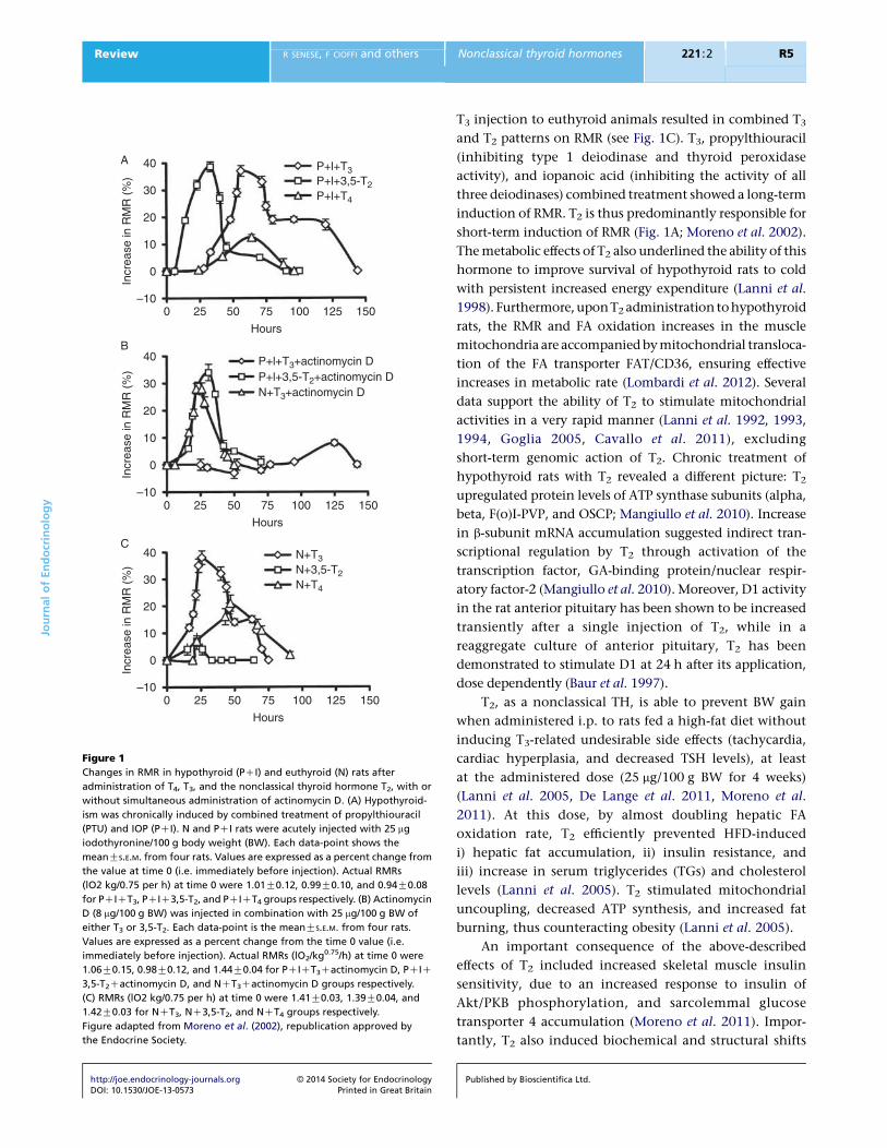
actinomycin D (see Fig. 1A and B; Lanni et al. 1996).
Published by Bioscientifica Ltd.
40A
B
C
30
20
10
0
–10
Incr
ease
in R
MR
(%
)
40
30
20
10
0
–10
Incr
ease
in R
MR
(%
)
40
30
20
10
0
–10
Incr
ease
in R
MR
(%
)
0 25 50 75 100 125 150
Hours
0 25 50 75 100 125 150
Hours
0 25 50 75 100 125 150
Hours
P+l+T3
P+l+T4
P+l+3,5-T2
P+l+T3+actinomycin DP+l+3,5-T2+actinomycin DN+T3+actinomycin D
N+T3
N+T4
N+3,5-T2
Figure 1
Changes in RMR in hypothyroid (PCI) and euthyroid (N) rats after
administration of T4, T3, and the nonclassical thyroid hormone T2, with or
without simultaneous administration of actinomycin D. (A) Hypothyroid-
ism was chronically induced by combined treatment of propylthiouracil
(PTU) and IOP (PCI). N and PCI rats were acutely injected with 25 mg
iodothyronine/100 g body weight (BW). Each data-point shows the
meanGS.E.M. from four rats. Values are expressed as a percent change from
the value at time 0 (i.e. immediately before injection). Actual RMRs
(lO2 kg/0.75 per h) at time 0 were 1.01G0.12, 0.99G0.10, and 0.94G0.08
for PCICT3, PCIC3,5-T2, and PCICT4 groups respectively. (B) Actinomycin
D (8 mg/100 g BW) was injected in combination with 25 mg/100 g BW of
either T3 or 3,5-T2. Each data-point is the meanGS.E.M. from four rats.
Values are expressed as a percent change from the time 0 value (i.e.
immediately before injection). Actual RMRs (lO2/kg0.75/h) at time 0 were
1.06G0.15, 0.98G0.12, and 1.44G0.04 for PCICT3Cactinomycin D, PCIC
3,5-T2Cactinomycin D, and NCT3Cactinomycin D groups respectively.
(C) RMRs (lO2 kg/0.75 per h) at time 0 were 1.41G0.03, 1.39G0.04, and
1.42G0.03 for NCT3, NC3,5-T2, and NCT4 groups respectively.
Figure adapted from Moreno et al. (2002), republication approved by
the Endocrine Society.
JournalofEndocrinology
Review R SENESE, F CIOFFI and others Nonclassical thyroid hormones 221 :2 R5
http://joe.endocrinology-journals.org � 2014 Society for EndocrinologyDOI: 10.1530/JOE-13-0573 Printed in Great Britain
T3 injection to euthyroid animals resulted in combined T3
and T2 patterns on RMR (see Fig. 1C). T3, propylthiouracil
(inhibiting type 1 deiodinase and thyroid peroxidase
activity), and iopanoic acid (inhibiting the activity of all
three deiodinases) combined treatment showed a long-term
induction of RMR. T2 is thus predominantly responsible for
short-term induction of RMR (Fig. 1A; Moreno et al. 2002).
The metabolic effects of T2 also underlined the ability of this
hormone to improve survival of hypothyroid rats to cold
with persistent increased energy expenditure (Lanni et al.
1998). Furthermore, uponT2 administration tohypothyroid
rats, the RMR and FA oxidation increases in the muscle
mitochondria are accompanied by mitochondrial transloca-
tion of the FA transporter FAT/CD36, ensuring effective
increases in metabolic rate (Lombardi et al. 2012). Several
data support the ability of T2 to stimulate mitochondrial
activities in a very rapid manner (Lanni et al. 1992, 1993,
1994, Goglia 2005, Cavallo et al. 2011), excluding
short-term genomic action of T2. Chronic treatment of
hypothyroid rats with T2 revealed a different picture: T2
upregulated protein levels of ATP synthase subunits (alpha,
beta, F(o)I-PVP, and OSCP; Mangiullo et al. 2010). Increase
in b-subunit mRNA accumulation suggested indirect tran-
scriptional regulation by T2 through activation of the
transcription factor, GA-binding protein/nuclear respir-
atory factor-2 (Mangiullo et al. 2010). Moreover, D1 activity
in the rat anterior pituitary has been shown to be increased
transiently after a single injection of T2, while in a
reaggregate culture of anterior pituitary, T2 has been
demonstrated to stimulate D1 at 24 h after its application,
dose dependently (Baur et al. 1997).
T2, as a nonclassical TH, is able to prevent BW gain
when administered i.p. to rats fed a high-fat diet without
inducing T3-related undesirable side effects (tachycardia,
cardiac hyperplasia, and decreased TSH levels), at least
at the administered dose (25 mg/100 g BW for 4 weeks)
(Lanni et al. 2005, De Lange et al. 2011, Moreno et al.
2011). At this dose, by almost doubling hepatic FA
oxidation rate, T2 efficiently prevented HFD-induced
i) hepatic fat accumulation, ii) insulin resistance, and
iii) increase in serum triglycerides (TGs) and cholesterol
levels (Lanni et al. 2005). T2 stimulated mitochondrial
uncoupling, decreased ATP synthesis, and increased fat
burning, thus counteracting obesity (Lanni et al. 2005).
An important consequence of the above-described
effects of T2 included increased skeletal muscle insulin
sensitivity, due to an increased response to insulin of
Akt/PKB phosphorylation, and sarcolemmal glucose
transporter 4 accumulation (Moreno et al. 2011). Impor-
tantly, T2 also induced biochemical and structural shifts
Published by Bioscientifica Ltd.
JournalofEndocrinology
Review R SENESE, F CIOFFI and others Nonclassical thyroid hormones 221 :2 R6
toward glycolytic myofibers (Moreno et al. 2011). For an
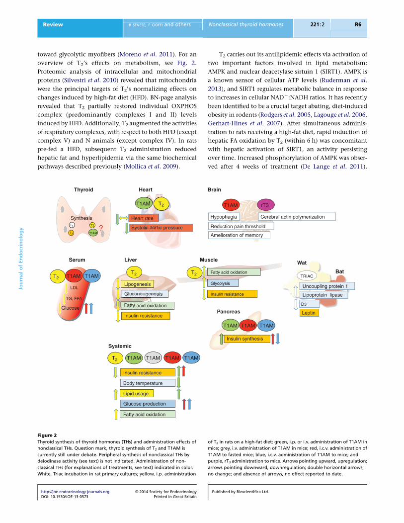
overview of T2’s effects on metabolism, see Fig. 2.
Proteomic analysis of intracellular and mitochondrial
proteins (Silvestri et al. 2010) revealed that mitochondria
were the principal targets of T2’s normalizing effects on
changes induced by high-fat diet (HFD). BN-page analysis
revealed that T2 partially restored individual OXPHOS
complex (predominantly complexes I and II) levels
induced by HFD. Additionally, T2 augmented the activities
of respiratory complexes, with respect to both HFD (except
complex V) and N animals (except complex IV). In rats
pre-fed a HFD, subsequent T2 administration reduced
hepatic fat and hyperlipidemia via the same biochemical
pathways described previously (Mollica et al. 2009).
Fatty acid oxidation
Insulin resistance
LDL Lipogenesis
GluconeogenesisTG, FFA
Glucose
T2T2
Synthesis T4
T3 T1AM
T2
T2
T1AM
Insulin resistance
T2
?
Body temperature
Lipid usage
T1AM
Glucose production
T1AM T1AM
T1AM T1AM
Heart rate
Systolic aortic pressure
T1AM
Fatty acid oxidation
T2
Thyroid
Serum Liver Mu
Systemic
Heart
Figure 2
Thyroid synthesis of thyroid hormones (THs) and administration effects of
nonclassical THs. Question mark, thyroid synthesis of T2 and T1AM is
currently still under debate. Peripheral synthesis of nonclassical THs by
deiodinase activity (see text) is not indicated. Administration of non-
classical THs (for explanations of treatments, see text) indicated in color.
White, Triac incubation in rat primary cultures; yellow, i.p. administration
http://joe.endocrinology-journals.org � 2014 Society for EndocrinologyDOI: 10.1530/JOE-13-0573 Printed in Great Britain
T2 carries out its antilipidemic effects via activation of
two important factors involved in lipid metabolism:
AMPK and nuclear deacetylase sirtuin 1 (SIRT1). AMPK is
a known sensor of cellular ATP levels (Ruderman et al.
2013), and SIRT1 regulates metabolic balance in response
to increases in cellular NADC:NADH ratios. It has recently
been identified to be a crucial target abating, diet-induced
obesity in rodents (Rodgers et al. 2005, Lagouge et al. 2006,
Gerhart-Hines et al. 2007). After simultaneous adminis-
tration to rats receiving a high-fat diet, rapid induction of
hepatic FA oxidation by T2 (within 6 h) was concomitant
with hepatic activation of SIRT1, an activity persisting
over time. Increased phosphorylation of AMPK was obser-
ved after 4 weeks of treatment (De Lange et al. 2011).
Hypophagia
Reduction pain threshold
Amelioration of memory
Fatty acid oxidation
Insulin resistance
Glycolysis
T1AM
Insulin synthesis
BatTRIAC
Leptin
T1AM T1AM
T1AM rT3
Cerebral actin polymerization
Uncoupling protein 1
Lipoprotein lipase
D3
scle Wat
Pancreas
Brain
of T2 in rats on a high-fat diet; green, i.p. or i.v. administration of T1AM in
mice; grey, i.v. administration of T1AM in mice; red, i.c.v. administration of
T1AM to fasted mice; blue, i.c.v. administration of T1AM to mice; and
purple, rT3 administration to mice. Arrows pointing upward, upregulation;
arrows pointing downward, downregulation; double horizontal arrows,
no change; and absence of arrows, no effect reported to date.
Published by Bioscientifica Ltd.
JournalofEndocrinology
Review R SENESE, F CIOFFI and others Nonclassical thyroid hormones 221 :2 R7
Induction of SIRT1 led to deacetylation of peroxisome
proliferator-activated receptor g coactivator-1a and sterol
receptor element binding protein-1c (SREBP-1c), associ-
ated with induction and reduction of expression of genes
involved in FA oxidation and lipogenesis respectively
(De Lange et al. 2011). These findings provide a clue to
explain T2’s effectiveness in lowering hepatic fat accu-
mulation and counteracting insulin resistance with
respect to T3. In a similar system, T3 has increased hepatic
lipogenesis (Cable et al. 2009).
A second clue indicating that T2 has contrasting effects
on hepatic lipogenesis involving SREBP-1c was found
in vitro. T3 increases an active precursor of SREBP-1c in
HepG2 cells without modulating SREBP-1c transcription
(Gnoni et al. 2012). T2, however, blocks proteolytic cleavage
and thus activation of SREBP-1c (Rochira et al. 2013)
independent of transcription. Consequently, FA synthase
expression reduced. The resulting inhibitory effect of T2 on
lipogenesis is concordant with in vivo findings (De Lange
et al. 2011) and mechanistically complementary to
SIRT1-dependent deacetylation of SREBP-1c (De Lange
et al. 2011). The liver is not the only organ in which T2
activates SIRT1: T2 has been shown to act through SIRT1
activation in the kidney (Shang et al. 2013). Treatment with
T2 prevented diabetic nephropathy (DN) in a diabetic
rat model via SIRT1-dependent deacetylation and p65,
a subunit of nuclear factor-kB, inactivation, thus inhibiting
the inflammatory process crucial to this pathology. In rat
mesangial cells, the DN phenotype was induced by
exposure to high glucose, and treatment with T2 under
these conditions counteracted the DN phenotype.
Co-treatment with T2 and sirtinol – a specific SIRT1
inhibitor (Shang et al. 2013) – abolished deacetylation of
p65. T2 leads to dephosphorylation of JNK independent
of SIRT1, and did not associate with abating the DN
phenotype (Shang et al. 2013). Thus, SIRT1 activation plays
a crucial role in T2’s relieving effect on DN.
As a first step in projecting rodent data to study T2’s
metabolic effects in humans, two healthy volunteers
were administered T2 (1–4 mg/kg BW) acutely. After 6 h, a
significant increase in RMR was detected in both patients.
Daily T2 administration for 28 days increased RMR byw15%
and decreased BW by about 4 kg. Ultrasonography
revealed that one subject showed reductions in steatosis.
Additionally, total serum cholesterol levels were lowered,
and no side effects were recorded (Antonelli et al. 2011).
A mechanism by which T2 reduces cholesterol in the serum
(Lanni et al. 2005, Antonelli et al. 2011, De Lange et al. 2011)
was recently called into question. T2’s LDL-lowering effects
are independent of the LDL receptor (Goldberg et al. 2012),
http://joe.endocrinology-journals.org � 2014 Society for EndocrinologyDOI: 10.1530/JOE-13-0573 Printed in Great Britain
as determined by feeding a western type diet to LDL
receptor-deficient mice (LdlrK/K) and treating with T2. The
diet was chosen because dietary absorption of cholesterol
and TG drives hepatic apoB production, especially in
LdlrK/K mice. These mice develop much higher levels of
cholesterol and atherosclerosis. T2 had no effect on TG
levels, probably due to increased lipolysis, but led to marked
reductions in liver apoB and circulating apoB48 and
apoB100 (Goldberg et al. 2012).
To study whether the lipid-lowering effects of T2 were
directly acting on the liver, or if they were secondary to
changes in endocrine or metabolic pathways, primary rat
hepatocytes, overloaded with lipids (to obtain ‘fatty
hepatocytes’) and treated with T2 (10K5 M) have been
employed (Grasselli et al. 2011a). This experimental setup
demonstrated that T2 reversed the effects induced by lipid
overload in these cells, thus supporting a direct effect of T2.
Moreover, rat hepatoma (FAO) cells defective for
functional TRs were used to answer whether T2-mediated
lowering of hepatic lipid profiles even requires TR action
(Grasselli et al. 2011b). Exposure to pharmacological doses
of T2 (10K5 M) for 24 h reversed the effects induced by FAs
and increased mitochondrial uncoupling, thus indicating
that the actions of T2 in these cells are independent of
transcriptionally functional TRs.
Intracellular action of T2 also is described in avian cells
during fetal development and cell differentiation (Incerpi
et al. 2002). T2 regulates DNA synthesis, cell-cycle proteins
(Alisi et al. 2004), and several membrane-associated
transport systems, whose activity is related to cell
proliferation (Incerpi et al. 2002, 2005). T2’s effect on the
NaC–HC exchanger was identified for 14-day- and 19-day-
old cells, whereas the effect on amino acid transport was
present at late stages of embryo development. Both
transport systems were activated through a signal trans-
duction pathway involving the PKC, MAPK, and PI3K
pathways (Incerpi et al. 2002). Moreover, T2 exerts a
short-term inhibitory effect on the NaC–KC-ATPase, the
magnitude of which strongly correlates to the develop-
mental age of the isolated cells (Scapin et al. 2009). The
NaC–KC-ATPase inhibition is mediated through the
activation of PKA, PKC, and PI3K (Scapin et al. 2009).
Signal transduction pathways contributing to the
modulation of the sodium pump by T2 are involved in
the control exerted on cell proliferation.
Recently, it has been shown that T2 exerts short-term
effects on intracellular calcium concentrations and NO
release by modulating plasma membrane and mito-
chondrial pathways in pituitary GH3 cells (Del Viscovo
et al. 2012). In particular, T2 facilitates physiological Ca2
Published by Bioscientifica Ltd.
Table 1 Involvement of TRs or alternative receptors in non-classical thyroid hormone action
Non-classical
thyroid hormone Affinity for/transactivation through TRa or TRb Affinity for other receptors
Triac High (Schueler et al. 1990) None yet identifiedT1AM Absent (Chiellini et al. 1998) High (trace amine-associated receptor 1
(TAAR1); Zucchi et al. 2006)rT3 Higha (TRDa1 (native TR isoform); Flamant & Samarut 2003) None yet identifiedT2 Weak (human TRa; Cioffi et al. 2010) None yet identified
Weak (human TRb; Ball et al. 1997, Cioffi et al. 2010,De Lange et al. 2011, Mendoza et al. 2013)
High (short TRb of tilapia fish; Mendoza et al. 2013,Navarrete-Ramirez et al. 2014)
aNongenomic action.
JournalofEndocrinology
Review R SENESE, F CIOFFI and others Nonclassical thyroid hormones 221 :2 R8
efflux from mitochondria through activation of mt-NCX
by interacting with different mitochondrial complexes
(Del Viscovo et al. 2012).
T2’s biological effects are not restricted to mammalian
species. Indeed, in the goldfish Carassius auratus, stimu-
lation of pyruvate-fueled liver and muscle mitochondrial
respiration was observed 5 min after 0.3 nM T2 incubation
(Leary et al. 1996). Garcıa et al. (2004) examined the effects
of short-term T4, T3, and T2 exposure (0.1 mM; 12 or 24 h)
on D1 and D2 activities and mRNA in killifish (tilapia) liver
and showed that these hormones decreased D2 activity,
the effect of T2 being relatively more rapid. T2 also
regulates thermal acclimation in zebra fish (Danio rerio)
with an efficiency comparable to T3 (Little et al. 2013). The
conserved role for T2 on regulating metabolic efficiency in
many species validates the motion to learn more about
biological actions of this nonclassical TH.
Mechanism of action of T2
Questions surrounding the cellular-molecular mechanism
of action of T2 remain. Both TR- and non TR-mediated
actions may be elicited by T2. Ball et al. (1997) reported
that T2 exerted selective thyromimetic effects. In the same
report, T2 showed a 60 times weaker affinity for TRb than
T3. Mendoza et al. (2013) reported that in teleosts, effects
of T2 may be mediated by an isoform of one of the two
known TRbs, namely TRb1 that contains a 9-amino-acid
insert in its ligand-binding domain (long TRb1), whereas
T3 binds preferentially to a short TRb1 isoform lacking this
insert. Moreover, the authors confirmed that T2 has a weak
affinity for human TRb (about 40-fold less than T3) and a
similarly weak transactivation capacity compared with T3
(Ball et al. 1997, Cioffi et al. 2010, De Lange et al. 2011,
Mendoza et al. 2013). In tilapia, both T3 and T2 are
http://joe.endocrinology-journals.org � 2014 Society for EndocrinologyDOI: 10.1530/JOE-13-0573 Printed in Great Britain
important in growth, a process, however, mediated by
different TRb1 isoforms (Navarrete-Ramirez et al. 2014). An
overview of TR or alternative receptor involvement in the
action of nonclassical THs is shown in Table 1.
Non TR-mediated effects of T2 are evident. It is known
that T2 specifically stimulates the activity of isolated
cytochrome c oxidase (COX) from bovine heart mito-
chondria. T3 barely stimulates COX. T2 binding to COX
induces conformational changes. Studies show specific
binding of labeled T2 to the subunit Va of COX from
bovine heart. T2, and to a small extent T3, but not
thyroxine and thyronine, abolished allosteric inhibition
of ascorbate respiration of reconstituted COX by ATP.
Inhibition is rescued by a monoclonal antibody to the
subunit Va. (Goglia et al. 1994, Arnold et al. 1998). T2
directly activates SIRT1 (De Lange et al. 2011), influencing
downstream pathways and inducing benefits. Shang et al.
(2013) showed that this interaction mitigates a DN by
using the Sirtuin inhibitor Sirtinol.
Conclusion and perspectives
It is clear that the so-called ‘nonclassical THs’ can induce
various biological actions. TH derivatives exert important
actions on metabolic parameters and on growth. At the
cellular–molecular level, several pathways are affected, the
most intriguing of which are related to lipid metabolism
and signaling pathways. Beneficial effects of these
molecules require more considerations due to their
potential to modulate human health.
Declaration of interest
The authors declare that there is no conflict of interest that could be
perceived as prejudicing the impartiality of the review.
Published by Bioscientifica Ltd.
JournalofEndocrinology
Review R SENESE, F CIOFFI and others Nonclassical thyroid hormones 221 :2 R9
Funding
This work has been supported, in part, by the following grants: MIUR
COFIN 2006 Prot. 2006051517 and MIUR COFIN 2008 Prot. 20089SRS2X.
References
Ackermans MT, Klieverik LP, Ringeling P, Endert E, Kalsbeek A & Fliers E
2010 An online solid-phase extraction-liquid chromatography–tandem
mass spectrometry method to study the presence of thyronamines in
plasma and tissue and their putative conversion from 13C6-thyroxine.
Journal of Endocrinology 206 327–334. (doi:10.1677/JOE-10-0060)
Alisi A, Spagnuolo S, Napoletano S, Spaziani A & Leoni S 2004 Thyroid
hormones regulate DNA-synthesis and cell-cycle proteins by activation
of PKCa and p42/44 MAPK in chick embryo hepatocytes. Journal of
Cellular Physiology 201 259–265. (doi:10.1002/jcp.20060)
Angelin B & Rudling M 2010 Lipid lowering with thyroid hormone and
thyromimetics. Current Opinion in Lipidology 21 499–506. (doi:10.1097/
MOL.0b013e3283402e9c)
Antonelli A, Fallahi P, Ferrari SM, Di Domenicantonio A, Moreno M, Lanni A
& Goglia F 2011 3,5-Diiodo-L-thyronine increases resting metabolic rate
and reduces body weight without undesirable side effects. Journal of
Biological Regulators and Homeostatic Agents 25 655–660.
Anzai R, Adachi M, Sho N, Muroya K, Asakura Y & Onigata K 2012
Long-term 3,5,3 0 triiodothyroacetic acid therapy in a child with
hyperthyroidism caused by thyroid hormone resistance: pharma-
cological study and therapeutic recommendations. Thyroid 22
1069–1075. (doi:10.1089/thy.2011.0450)
Araki O, Ying H, Zhu XG, Willingham MC & Cheng SY 2009 Distinct
dysregulation of lipid metabolism by unliganded thyroid hormone
receptor isoforms. Molecular Endocrinology 23 308–315. (doi:10.1210/
me.2008-0311)
Arnold S, Goglia F & Kadenbach B 1998 3,5-Diiodothyronine binds to
subunit Va of cytochrome-c oxidase and abolishes the allosteric
inhibition of respiration by ATP. European Journal of Biochemistry 252
325–323. (doi:10.1046/j.1432-1327.1998.2520325.x)
Ball SG, Sokolov J & Chin WW 1997 3,5-Diiodo-L-thyronine (T2) has
selective thyromimetic effects in vivo and in vitro. Journal of Molecular
Endocrinology 19 137–147. (doi:10.1677/jme.0.0190137)
Bassett JH, Harvey CB & Williams GR 2003 Mechanisms of thyroid
hormone receptor-specific nuclear and extra nuclear actions. Molecular
and Cellular Endocrinology 213 1–11. (doi:10.1016/j.mce.2003.10.033)
Baur A, Bauer K, Jarry H & Koehrle J 1997 3,5-Diiodo-L-thyronine stimulates
type 1 5 0deiodinase activity in rat anterior pituitaries in vivo and in
reaggregate cultures and GH3 cells in vitro. Endocrinology 138
3242–3248. (doi:10.1210/endo.138.8.5333)
Bergh JJ, Lin HY, Lansing L, Mohamed SN, Davis FB, Mousa S & Davis P
2005 Integrin aVb3 contains a cell surface receptor site for thyroid
hormone that is linked to activation of mitogen-activated protein
kinase and induction of angiogenesis. Endocrinology 146 2864–2871.
(doi:10.1210/en.2005-0102)
Bianco AC 2011 Minireview: cracking the metabolic code for thyroid
hormone signaling. Endocrinology 152 3306–3311. (doi:10.1210/
en.2011-1104)
Bobek S, Sechman A, Niezgoda J & Jacek T 2002 Reverse 3,3 0,5 0-
triiodothyronine suppresses increase in free fatty acids in chickens
elicited by dexamethasone or adrenaline. Journal of Veterinary Medicine.
A, Physiology, Pathology, Clinical Medicine 49 121–124. (doi:10.1046/
j.1439-0442.2002.00343.x)
Bogazzi F, Hudson LD & Nikodem VM 1994 A novel heterodimerization
partner for thyroid hormone receptor. Peroxisome proliferator
activated receptor. Journal of Biological Chemistry 269 11683–11686.
BraccoD,MorinO,SchutzY,LiangH, JequierE& BurgerAG1993Comparison
of the metabolic and endocrine effects of 3,5,30-triiodothyroacetic acid
http://joe.endocrinology-journals.org � 2014 Society for EndocrinologyDOI: 10.1530/JOE-13-0573 Printed in Great Britain
and thyroxine. Journal of Clinical Endocrinology and Metabolism 77
221–228. (doi:10.1210/jcem.77.1.8325946)
Braulke LJ, Klingenspor M, DeBarber A, Tobias SC, Grandy DK, Scanlan TS
& Heldmaier G 2008 3-Iodothyronamine: a novel hormone controlling
the balance between glucose and lipid utilisation. Journal of Comparative
Physiology. B, Biochemical, Systemic, and Environmental Physiology 178
167–177. (doi:10.1007/s00360-007-0208-x)
Cable EE, Finn PD, Stebbins JW, Hou J, Ito BR, van Poelje PD, Linemeyer DL
& Erion MD 2009 Reduction of hepatic steatosis in rats and mice after
treatment with a liver-targeted thyroid hormone receptor agonist.
Hepatology 49 407–417.
Cavallo A, Gnoni A, Conte E, Siculella L, Zanotti F, Papa S & Gnoni GV
2011 3,5-Diiodo-L-thyronine increases FoF1-ATP synthase activity and
cardiolipin level in liver mitochondria of hypothyroid rats.
Journal of Bioenergetics and Biomembranes 43 349–357. (doi:10.1007/
s10863-011-9366-3)
Cheng SY, Leonard JL & Davis PJ 2010 Molecular aspects of thyroid
hormone actions. Endocrine Reviews 31 139–170. (doi:10.1210/er.2009-
0007)
Cheng SY 2000 Multiple mechanisms for regulation of the transcriptional
activity of thyroid hormone receptors. Reviews in Endocrine and
Metabolic Disorders 1 9–18.
Chiellini G, Apriletti JW, Yoshihara HA, Baxter JD, Ribeiro RC & Scanlan TS
1998 A high-affinity subtype-selective agonist ligand for the thyroid
hormone receptor. Chemistry & Biology 5 299–306. (doi:10.1016/
S1074-5521(98)90168-5)
Chiellini G, Frascarelli S, Ghelardoni S, Carnicelli V, Tobias SC, DeBarber A,
BrogioniS,Ronca-TestoniS,CerbaiE,GrandyDK etal.2007Cardiaceffects
of 3-iodothyronamine: a new aminergic system modulating cardiac
function. FASEB Journal 21 1597–1608. (doi:10.1096/fj.06-7474com)
Cioffi F, Zambad SP, Chhipa L, Senese R, Busiello RA, Tuli D, Munshi S,
Moreno M, Lombardi A, Gupta RC et al. 2010 TRC150094, a novel
functional analog of iodothyronines, reduces adiposity by increasing
energy expenditure and fatty acid oxidation in rats receiving a high-fat
diet. FASEB Journal 24 3451–3461. (doi:10.1096/fj.10-157115)
Cioffi F, Senese R, Lanni A & Goglia F 2013 Thyroid hormones and
mitochondria: with a brief look at derivatives and analogues. Molecular
and Cellular Endocrinology 379 51–61. (doi:10.1016/j.mce.2013.06.006)
Cody V, Davis PJ & Davis FB 2007 Molecular modeling of the thyroid
hormone interactions with avb3 integrin. Steroids 72 165–170.
(doi:10.1016/j.steroids.2006.11.008)
DeBarber AE, Geraci T, Colasurdo VP, Hackenmueller SA & Scanlan TS 2008
Validation of a liquid chromatography–tandem mass spectrometry
method to enable quantification of 3-iodothyronamine from serum.
Journal of Chromatography. A 1210 55–59. (doi:10.1016/j.chroma.
2008.09.022)
De Lange P, Senese R, Cioffi F, Moreno M, Lombardi A, Silvestri E, Goglia F
& Lanni A 2008 Rapid activation by 3,5,3 0-L-triiodothyronine of
adenosine 5 0-monophosphate-activated protein kinase/acetyl-
coenzyme a carboxylase and akt/protein kinase B signaling pathways:
relation to changes in fuel metabolism and myosin heavy-chain
protein content in rat gastrocnemius muscle in vivo. Endocrinology 149
6462–6470. (doi:10.1210/en.2008-0202)
De Lange P, Cioffi F, Senese R, Moreno M, Lombardi A, Silvestri E,
De Matteis R, Lionetti L, Mollica MP, Goglia F et al. 2011 Nonthyrotoxic
prevention of diet-induced insulin resistance by 3,5-diiodo-L-thyronine
in rats. Diabetes 60 2730–2739. (doi:10.2337/db11-0207)
Del Viscovo A, Secondo A, Esposito A, Goglia F, Moreno M & Canzoniero LM
2012 Intracellular and plasma membrane-initiated pathways involved
in the [Ca2C]i elevations induced by iodothyronines (T3 and T2) in
pituitary GH3 cells. American Journal of Physiology. Endocrinology and
Metabolism 302 E1419–E1430. (doi:10.1152/ajpendo.00389.2011)
Dumas P, Autissier N, Loireau A & Michel R 1982 Effects of 3,5,3 0-
triiodothyroacetic acid (TRIAC) on protein metabolism of genetically
obese or non-obese Zucker rats. Comptes Rendus des Seances de la Societe
de Biologie et de Ses Filiales 176 178–183.
Published by Bioscientifica Ltd.
JournalofEndocrinology
Review R SENESE, F CIOFFI and others Nonclassical thyroid hormones 221 :2 R10
Farwell AP, Lynch RM, Okulicz WC, Comi AM & Leonard JL 1990 The actin
cytoskeleton mediates the hormonally regulated translocation of
type II iodothyronine 5 0-deiodinase in astrocytes. Journal of Biological
Chemistry 265 18546–18553.
Farwell AP, Dubord-Tomasetti SA, Pietrzykowski AZ & Leonard JL 2006
Dynamic nongenomic actions of thyroid hormone in the developing
rat brain. Endocrinology 147 2567–2574. (doi:10.1210/en.2005-1272)
Flamant F & Samarut J 2003 Thyroid hormone receptors: lessons from
knockout and knock-in mutant mice. Trends in Endocrinology and
Metabolism 14 85–90. (doi:10.1016/S1043-2760(02)00043-7)
Forman BM, Casanova J, Raaka BM, Ghysdael J & Samuels HH 1992 Half-
site spacing and orientation determines whether thyroid hormone and
retinoic acid receptors and related factors bind to DNA response
elements as monomers, homodimers, or heterodimers. Molecular
Endocrinology 6 429–442. (doi:10.1210/mend.6.3.1316541)
Frascarelli S, Ghelardoni S, Chiellini G, Vargiu R, Ronca-Testoni S,
Scanlan TS, Grandy DK & Zucchi R 2008 Cardiac effects of trace
amines: pharmacological characterization of trace amine associated
receptors. European Journal of Pharmacology 587 231–236.
(doi:10.1016/j.ejphar.2008.03.055)
Galo MG, Unates LE & Farıas RN 1981 Effect of membrane fatty acid
composition on the action of thyroid hormone on (Ca2CC Mg2C)-
adenosine triphosphatase from rat erythrocyte. Journal of Biological
Chemistry 256 7113–7114.
Garcıa GC, Jeziorski MC, Valverde RC & Orozco A 2004 Effects of
iodothyronines on the hepatic outer-ring deiodinating pathway in
killifish. General and Comparative Endocrinology 135 201–209.
(doi:10.1016/j.ygcen.2003.09.010)
Gerhart-Hines Z, Rodgers JT, Bare O, Lerin C, Kim SH, Mostoslavsky R,
Alt FW, Wu Z & Puigserver P 2007 Metabolic control of muscle
mitochondrial function and fatty acid oxidation through SIRT1/
PGC-1alpha. EMBO Journal 26 1913–1923.
Gnoni GV, Rochira A, Leone A, Damiano F, Marsigliante S & Siculella L
2012 3,5,3’triiodo-L-thyronine induces SREBP-1 expression by non-
genomic actions in human HEP G2 cells. Journal of Cellular Physiology
227 2388–2397.
Goglia F 2005 Biological effects of 3,5-diiodothyronine (T2). Biochemistry
70 164–172.
Goglia F, Lanni A, Barth J & Kadenbach B 1994 Interaction of
diiodothyronines with isolated cytochrome c oxidase. FEBS Letters
346 295–298. (doi:10.1016/0014-5793(94)00476-5)
Goldberg IJ, Huang LS, Huggins LA, Yu S, Nagareddy PR, Scanlan TS &
Ehrenkranz JR 2012 Thyroid hormone reduces cholesterol via a
non-LDL receptor-mediated pathway. Endocrinology 153 5143–5149.
(doi:10.1210/en.2012-1572)
Grasselli E, Voci A, Canesi L, De Matteis R, Goglia F, Cioffi F, Fugassa E,
Gallo G & Vergani L 2011a Direct effects of iodothyronines on excess
fat storage in rat hepatocytes. Journal of Hepatology 54 1230–1236.
(doi:10.1016/j.jhep.2010.09.027)
Grasselli E, Voci A, Canesi L, Goglia F, Ravera S, Panfoli I, Gallo G &
Vergani L 2011b Non-receptor-mediated actions are responsible for the
lipid-lowering effects of iodothyronines in FaO rat hepatoma cells.
Journal of Endocrinology 21 59–69. (doi:10.1530/JOE-11-0074)
Gullberg H, Rudling M, Forrest D, Angelin B & Vennstrom B 2000 Thyroid
hormone receptor b-deficient mice show complete loss of the normal
cholesterol 7a-hydroxylase (CYP7A) response to thyroid hormone but
display enhanced resistance to dietary cholesterol. Molecular
Endocrinology 14 1739–1749. (doi:10.1210/mend.14.11.0548)
Gullberg H, Rudling M, Salto C, Forrest D, Angelin B & Vennstrom B 2002
Requirement for thyroid hormone receptor b in T3 regulation of
cholesterol metabolism in mice. Molecular Endocrinology 16 1767–1777.
(doi:10.1210/me.2002-0009)
Hackenmueller SA, Marchini M, Saba A, Zucchi R & Scanlan TS 2012
Biosynthesis of 3 iodothyronamine (T1AM) is dependent on the
sodium–iodide symporter and thyroperoxidase but does not involve
http://joe.endocrinology-journals.org � 2014 Society for EndocrinologyDOI: 10.1530/JOE-13-0573 Printed in Great Britain
extrathyroidal metabolism of T4. Endocrinology 153 5659–5667.
(doi:10.1210/en.2012-1254)
Hart ME, Suchland KL, Miyakawa M, Bunzow JR, Grandy DK & Scanlan TS
2006 Trace amine-associated receptor agonists; synthesis and evalu-
ation of thyronamines and related analogues. Journal of Medicinal
Chemistry 49 1101–1112. (doi:10.1021/jm0505718)
Hoefig CS, Kohrle J, Brabant G, Dixit K, Yap J, Strasburger CJ & Wu Z 2011
Evidence for extrathyroidal formation of 3-iodothyronamine in
humans as provided by a novel monoclonal antibody-based chemi-
luminescent serum immunoassay. Journal of Clinical Endocrinology and
Metabolism 98 1864–1872. (doi:10.1210/jc.2010-2680)
Hoefig CS, Renko K, Piehl S, Scanlan TS, Bertoldi M, Opladen T,
Hoffmann GF, Klein J, Blankenstein O, Sc weizer U et al. 2012 Does
the aromatic L-amino acid decarboxylase contribute to thyronamine
biosynthesis? Molecular and Cellular Endocrinology 349 195–201.
(doi:10.1016/j.mce.2011.10.024)
Horn S, Kersseboom S, Mayerl S, Muller J, Groba C, Trajkovic-Arsic M,
Ackermann T, Visser TJ & Heuer H 2013 Tetrac can replace thyroid
hormone during brain development in mouse mutants deficient in
the thyroid hormone transporter mct8. Endocrinology 154 968–679.
(doi:10.1210/en.2012-1628)
Ianculescu AG, Giacomini KM & Scanlan TS 2009 Identification and
characterization of 3-iodothyronamine intracellular transport.
Endocrinology 150 1991–1999. (doi:10.1210/en.2008-1339)
Ianculescu AG, Friesema EC, Visser TJ, Giacomini KM & Scanlan TS 2010
Transport of thyroid hormones is selectively inhibited by 3-iodothyr-
onamine. Molecular BioSystems 6 1403–1410. (doi:10.1039/b926588k)
Incerpi S, De Vito P, Luly P, Spagnuolo S & Leoni S 2002 Short-term effects
of thyroid hormones and 3,5-diiodothyronine on membrane transport
systems in chick embryo hepatocytes. Endocrinology 143 1660–1668.
(doi:10.1210/endo.143.5.8767)
Incerpi S, Scapin S, D’Arezzo S, Spagnuolo S & Leoni S 2005 Short-term
effects of thyroid hormone in prenatal development and cell
differentiation. Steroids 70 434–443. (doi:10.1016/j.steroids.2005.
02.009)
Irrcher I, Walkinshaw DR, Sheehan TE & Hood DA 2008 Thyroid
hormone (T3) rapidly activates p38 and AMPK in skeletal muscle in vivo.
Journal of Applied Physiology 104 178–185. (doi:10.1152/japplphysiol.
00643.2007)
Johansson L, Rudling M, Scanlan TS, Lundasen T, Webb P, Baxter J,
Angelin B & Parini P 2005 Selective thyroid receptor modulation by
GC-1 reduces serum lipids and stimulates steps of reverse cholesterol
transport in euthyroid mice. PNAS 102 10297–10302. (doi:10.1073/
pnas.0504379102)
Klieverik LP, Foppen E, Ackermans MT, Serlie MJ, Sauerwein HP, Scanlan TS,
Grandy DK, Fliers E & Kalsbeek A 2009 Central effects of thyronamines
on glucose metabolism in rats. Journal of Endocrinology 201 377–386.
(doi:10.1677/JOE-09-0043)
Kunitake JM, Hartman N, Henson LC, Lieberman J, Williams DE, Wong M
& Hershman JM 1989 3,5,3 0-Triiodothyroacetic acid therapy for
thyroid hormone resistance. Journal of Clinical Endocrinology and
Metabolism 69 461–466. (doi:10.1210/jcem-69-2-461)
Ladenson PW 2011 Thyroid hormone analogues: ready for prime time.
Thyroid 21 101–102. (doi:10.1089/thy.2010.2100.com1)
Lagouge M, Argmann C, Gerhart-Hines Z, Meziane H, Lerin C, Daussin F,
Messadeq N, Milne J, Lambert P, Elliot P et al. 2006 Resveratrol improves
mitochondrial function and protects against metabolic disease by
activating SIRT1 and PGC-1alpha. Cell 127 1109–1122.
Lanni A, Moreno M, Cioffi M & Goglia F 1992 Effect of 3,3 0-diiodothyr-
onine and 3,5-diiodothyronine on rat liver oxidative capacity.
Molecular and Cellular Endocrinology 86 143–148. (doi:10.1016/
0303-7207(92)90138-V)
Lanni A, Moreno M, Cioffi M & Goglia F 1993 Effect of 3,3 0-di-
iodothyronine and 3,5-di-iodothyronine on rat liver mitochondria.
Journal of Endocrinology 136 59–64. (doi:10.1677/joe.0.1360059)
Published by Bioscientifica Ltd.
JournalofEndocrinology
Review R SENESE, F CIOFFI and others Nonclassical thyroid hormones 221 :2 R11
Lanni A, Moreno M, Horst C, Lombardi A & Goglia F 1994 Specific binding
sites for 3,3 0-diiodo-L-thyronine (3,3 0-T2) in rat liver mitochondria.
FEBS Letters 351 237–240. (doi:10.1016/0014-5793(94)00840-X)
Lanni A, Moreno M, Lombardi A & Goglia F 1996 Calorigenic effect of
diiodothyronines in the rat. Journal of Physiology 494 831–837.
Lanni A, Moreno M, Lombardi A & Goglia F 1998 3,5-Diiodo-L-thyronine
and 3,5,3 0-triiodo-L-thyronine both improve the cold tolerance of
hypothyroid rats, but possibly via different mechanisms. Pflugers Archiv:
European Journal of Physiology 436 407–414. (doi:10.1007/
s004240050650)
Lanni A, Moreno M, Lombardi A, de Lange P, Silvestri E, Ragni M,
Farina P, Chieffi Baccari G, Fallahi P, Antonelli A et al. 2005
3,5-Diiodo-L-thyronine powerfully reduces adiposity in rats by
increasing the burning of fats. FASEB Journal 19 1552–1554.
Leary SC, Barton KN & Ballantyne JS 1996 Direct effects of 3,5,3 0-
triiodothyronine and 3,5-diiodothyronine on mitochondrial
metabolism in the goldfish Carassius auratus. General and Comparative
Endocrinology 104 61–66. (doi:10.1006/gcen.1996.0141)
Lerman J 1956 The activity of dl-tribromthyronine. Journal of
Clinical Endocrinology and Metabolism 16 1395–1397. (doi:10.1210/
jcem-16-10-1395)
Lin HY, Sun M, Lin C, Tang HY, London D, Shih A, Davis FB & Davis PJ
2009a Androgen induced human breast cancer cell proliferation is
mediated by discrete mechanisms in estrogen receptor-a-positive and
-negative breast cancer cells. Journal of Steroid Biochemistry and Molecular
Biology 113 182–188. (doi:10.1016/j.jsbmb.2008.12.010)
LinHY, SunM,TangHY,LinC,Luidens MK,MousaSA, IncerpiS,DrusanoGL,
Davis FB & Davis PJ 2009b L-Thyroxine vs. 3,5,3-triiodo-L-thyronine and
cell proliferation: activation of mitogen-activated protein kinase and
phosphatidylinositol 3-kinase. American Journal of Physiology. Cell
Physiology 296 C980–C991. (doi:10.1152/ajpcell.00305.2008)
Little AG, Kunisue T, Kannan K & Seebacher F 2013 Thyroid hormone
actions are temperature-specific and regulate thermal acclimation
in zebrafish (Danio rerio). BMC Biology 26 11–26. (doi:10.1186/1741-
7007-11-26)
Lombardi A, De Matteis R, Moreno M, Napolitano L, Busiello RA, Senese R,
de Lange P, Lanni A & Goglia F 2012 Responses of skeletal muscle lipid
metabolism in rat gastrocnemius to hypothyroidism and iodothyr-
onine administration: a putative role for FAT/CD36. American
Journal of Physiology. Endocrinology and Metabolism 303 E1222–E1233.
(doi:10.1152/ajpendo.00037.2012)
Luongo C, Trivisano L, Alfano F & Salvatore D 2013 Type 3 deiodinase
and consumptive hypothyroidism: a common mechanism for a rare
disease. Frontiers in Endocrinology 115 1–7. (doi:10.3389/fendo.2013.
00115)
Maia AL, Goemann IM, Meyer EL & Wajner SM 2011 Deiodinases: the
balance of thyroid hormone: type 1 iodothyronine deiodinase in
human physiology and disease. Journal of Endocrinology 209 283–297.
(doi:10.1530/JOE-10-0481)
Mangiullo R, Gnoni A, Damiano F, Siculella L, Zanotti F, Papa S &
Gnoni GV 2010 3,5-Diiodo-L-thyronine upregulates rat-liver mito-
chondrial F(o)F(1)-ATP synthase by GA-binding protein/nuclear
respiratory factor-2. Biochimica et Biophysica Acta 1797 233–240.
(doi:10.1016/j.bbabio.2009.10.009)
Manni ME, De Siena G, Saba A, Marchini M, Dicembrini I, Bigagli E, Cinci L,
Lodovici M, Chiellini MG, Zucchi R et al. 2012 3-Iodothyronamine:
a modulator of the hypothalamus–pancreas–thyroid axes in mice.
British Journal of Pharmacology 166 650–658. (doi:10.1111/j.1476-5381.
2011.01823.x)
Manni ME, De Siena G, Saba A, Marchini M, Landucci E, Gerace E,
Zazzeri M, Mussili C, Pellegrini-Giampietro D, Matucci R et al. 2013
Pharmacological effects of 3-iodothyronamine (T1AM) in mice
include facilitation and memory acquisition and retention and
reduction of pain threshold. British Journal of Pharmacology 168
354–362. (doi:10.1111/j.1476-5381.2012.02137.x)
http://joe.endocrinology-journals.org � 2014 Society for EndocrinologyDOI: 10.1530/JOE-13-0573 Printed in Great Britain
Medina-Gomez G, Hernandez A, Calvo RM, Martin E & Obregon MJ 2003
Potent thermogenic action of triiodothyroacetic acid in brown
adipocytes. Cellular and Molecular Life Sciences 60 1957–1967.
(doi:10.1007/s00018-003-3158-9)
Medina-Gomez G, Calvo MR & Obregon MJ 2004 T3 and Triac inhibit leptin
secretion and expression in brown and white rat adipocytes. Biochimica
et Biophysica Acta 1682 38–47. (doi:10.1016/j.bbalip.2004.01.007)
Mendoza A, Navarrete-Ramırez P, Hernandez-Puga G, Villalobos P,
Holzer G, Renaud JP, Laudet V & Orozco A 2013 3,5-T2 is an
alternative ligand for the thyroid hormone receptor b1. Endocrinology
154 2948–2958. (doi:10.1210/en.2013-1030)
Moeller LC & Broecker-Preuss M 2011 Transcriptional regulation by
nonclassical action of thyroid hormone. Thyroid Research 4(Suppl 1) S6.
(doi:10.1186/1756-6614-4-S1-S6)
Moeller LC, Dumitrescu AM & Refetoff S 2005 Cytosolic action of thyroid
hormone leads to induction of hypoxia-inducible factor-1 and
glycolytic genes. Molecular Endocrinology 19 2955–2963. (doi:10.1210/
me.2004-0542)
Mollica MP, Lionetti L, Moreno M, Lombardi A, de Lange P, Antonelli A,
Lanni A, Cavaliere G, Barletta A & Goglia F 2009 3,5-Diiodo-L-
thyronine, by modulating mitochondrial functions, reverses hepatic
fat accumulation in rats fed a high-fat diet. Journal of Hepatology 51
363–370. (doi:10.1016/j.jhep.2009.03.023)
Moreno M, Kaptein E, Goglia F & Visser TJ 1994 Rapid glucuronidation of
tri- and tetraiodothyroacetic acid to ester glucuronides in human liver
and to ether glucuronides in rat liver. Endocrinology 135 1004–1009.
(doi:10.1210/endo.135.3.8070342)
Moreno M, Lombardi A, Beneduce L, Silvestri E, Pinna G, Goglia F &
Lanni A 2002 Are the effects of T3 on resting metabolic rate in
euthyroid rats entirely caused by T3 itself? Endocrinology 143 504–510.
(doi:10.1210/endo.143.2.8613)
Moreno M, Silvestri E, De Matteis R, de Lange P, Lombardi A, Glinni D,
Senese R, Cioffi F, Salzano AM, Scaloni A et al. 2011 3,5-Diiodo-L-
thyronine prevents high-fat-diet-induced insulin resistance in rat
skeletal muscle through metabolic and structural adaptations. FASEB
Journal 25 3312–3324. (doi:10.1096/fj.11-181982)
Navarrete-Ramirez P, Luna M, Valverde RC & Orozco A 2014 3,5-T2
stimulates tilapia growth through an alternate isoform of thyroid
hormone receptor b1. Journal of Molecular Endocrinology 52 1–9.
(doi:10.1530/JME-13-0145)
Oetting A & Yen PM 2007 New insights into thyroid hormone action.
Best Practice & Research. Clinical Endocrinology & Metabolism 21 193–208.
(doi:10.1016/j.beem.2007.04.004)
Orozco A, Valverde RC, Olvera A & Garcıa GC 2012 Iodothyronine
deiodinases: a functional and evolutionary perspective. Journal of
Endocrinology 215 207–219. (doi:10.1530/JOE-12-0258)
Pascual A & Aranda A 2013 Thyroid hormone receptors, cell growth
and differentiation. Biochimica et Biophysica Acta 1830 3908–3916.
(doi:10.1016/j.bbagen.2012.03.012)
Piehl S, Heberer T, Balizs G, Scanlan TS, Smits R, Koksch B & Kohrle J 2008
Thyronamines are isozyme-specific substrates of deiodinases.
Endocrinology 149 3037–3045. (doi:10.1210/en.2007-1678)
Pinna G, Meinhold H, Hiedra L, Thoma R, Hoell T, Graf KJ, Stoltenburg-
Didinger G, Eravci M, Prengel H, Brodel O et al. 1997 Elevated
3,5-diiodothyronine concentrations in the sera of patients with
nonthyroidal illnesses and brain tumors. Journal of Clinical Endo-
crinology and Metabolism 82 1535–1542. (doi:10.1210/jcem.82.5.3939)
Pittman CS, Shimizu T, Burger A & Chambers JB Jr 1980 The nondeiodi-
native pathways of thyroxine metabolism: 3,5,3 0,5-tetraiodothyroace-
tic acid turnover in normal and fasting human subjects. Journal of
Clinical Endocrinology and Metabolism 50 712–716. (doi:10.1210/
jcem-50-4-712)
Pramfalk C, Pedrelli M & Parini P 2011 Role of thyroid receptor b in lipid
metabolism. Biochimica et Biophysica Acta 1812 929–937. (doi:10.1016/
j.bbadis.2010.12.019)
Published by Bioscientifica Ltd.
JournalofEndocrinology
Review R SENESE, F CIOFFI and others Nonclassical thyroid hormones 221 :2 R12
Rochira A, Damiano F, Marsigliante S, Gnoni GV & Siculella L 2013
3,5-Diiodo-l-thyronine induces SREBP-1 proteolytic cleavage block and
apoptosis in human hepatoma (Hepg2) cells. Biochimica et Biophysica
Acta 1831 1679–1688.
Rodgers JT, Lerin C, Haas W, Gygi SP, Spiegelman BM & Puigserver P 2005
Nutrient control of glucose homeostasis through a complex of
PGC-1alpha and SIRT1. Nature 434 113–118.
Ruderman NB, Carling D, Prentki M & Cacicedo 2013 AMPK, insulin
resistance, and the metabolic syndrome. Journal of Clinical Investigation
123 2764–2772.
Saba A, Chiellini G, Frascarelli S, Marchini M, Chelardoni S, Rafaelli A,
Tonacchera M, Vitti P, Scanlan TS & Zucchi R 2010 Tissue distribution
and cardiac metabolism of 3-iodothyronamine. Endocrinology 151
5063–5073. (doi:10.1210/en.2010-0491)
Scanlan TS, Suchland KL, Hart ME, Chiellini G, Huang Y, Kruzich PJ,
Frascarelli S, Crossley DA, Bunzow JR, Ronca-Testoni S et al. 2004
3-Iodothyronamine is an endogenous and rapid-acting derivative of
thyroid hormone. Nature Medicine 10 638–642. (doi:10.1038/nm1051)
Scapin S, Leoni S, Spagnuolo S, Fiore AM & Incerpi S 2009 Short-term effects
of thyroid hormones on NaC–KC-ATPase activity of chick embryo
hepatocytes during development: focus on signal transduction.
American Journal of Physiology. Cell Physiology 296 C4–C12.
(doi:10.1152/ajpcell.90604.2007)
Schueler PA, Schwartz HL, Strait KA, Mariash CN & Oppenheimer JH 1990
Binding of 3,5,3 0-triiodothyronine (T3) and its analogs to the in vitro
translational products of c-erbA protooncogenes: differences in the
affinity of the a- and b-forms for the acetic acid analog and failure
of the human testis and kidney a-2 products to bind T3. Molecular
Endocrinology 4 227–234. (doi:10.1210/mend-4-2-227)
Shang G, Gao P, Zhao Z, Chen Q, Jiang T, Zhang N & Li H 2013 3,5-Diiodo-
L-thyronine ameliorates diabetic nephropathy in streptozotocin-
induced diabetic rats. Biochimica et Biophysica Acta 1832 674–684.
Sherman SI & Ladenson PW 1992 Organ-specific effects of tiratricol:
a thyroid hormone analog with hepatic, not pituitary, superagonist
effects. Journal of Clinical Endocrinology and Metabolism 75 901–905.
(doi:10.1210/jcem.75.3.1517383)
Shoemaker TJ, Kono T, Mariash CN & Evans-Molina C 2012 Thyroid
hormone analogues for the treatment of metabolic disorders: new
potential for unmet clinical needs? Endocrine Practice 18 954–964.
(doi:10.4158/EP12086.RA)
Siegrist-Kaiser C & Burger AG 1994 Modification of the side chain of
thyroid hormones. In Thyroid Hormone Metabolism, pp 17–198.
Eds SY Wu & TJ Visser TJ. Boca Raton: CRC Press, Inc.
Siegrist-Kaiser CA, Juge-Aubry C, Tranter MP, Ekenbarger DM & Leonard JL
1990 Thyroxine-dependent modulation of actin polymerization in
http://joe.endocrinology-journals.org � 2014 Society for EndocrinologyDOI: 10.1530/JOE-13-0573 Printed in Great Britain
cultured astrocytes. A novel, extranuclear action of thyroid hormone.
Journal of Biological Chemistry 265 5296–5302.
Silvestri E, Cioffi F, Glinni D, Ceccarelli M, Lombardi A, de Lange P,
Chambery A, Severino V, Lanni A, Goglia F et al. 2010 Pathways
affected by 3,5-diiodo-L-thyronine in liver of high fat-fed rats: evidence
from two-dimensional electrophoresis, blue-native PAGE, and mass
spectrometry. Molecular BioSystems 6 2256–2271. (doi:10.1039/
c0mb00040j)
Snead AN, Miyakawa M, Tan ES & Scanlan TS 2008 Trace amine-associated
receptor 1 (TAAR1) is activated by amiodarone metabolites. Bioorganic
& Medicinal Chemistry Letters 18 5920–5922. (doi:10.1016/j.bmcl.2008.
08.013)
Tan ES, Miyakawa M, Bunzow JR, Grandy DK & Scanlan TS 2007 Exploring
the structure–activity relationship of the ethylamine portion of
3-iodothyronamine for rat and mouse trace amine-associated
receptor 1. Journal of Medicinal Chemistry 50 2787–2798. (doi:10.1021/
jm0700417)
Tan ES, Groban ES, Jacobson MP & Scanlan TS 2008 Toward deciphering
the code to aminergic G protein-coupled receptor drug design.
Chemistry & Biology 15 343–353. (doi:10.1016/j.chembiol.2008.03.004)
Tata JR 2013 The road to nuclear receptors of thyroid hormone. Biochimica
et Biophysica Acta 1830 3860–3866.
Visser TJ 1990 Importance of deiodination and conjugation in the hepatic
metabolism of thyroid hormone. In The Thyroid Gland, pp 255–283.
Ed. MA Greer. New York: Raven Press.
Visser TJ 2013 Thyroid hormone transporters and resistance. Endocrine
Development 24 1–10. (doi:10.1159/000343695)
Webb P 2010 Thyroid hormone receptor and lipid regulation. Current
Opinion in Investigational Drugs 11 1135–1142.
Weitzel JM & Alexander Iwen K 2011 Coordination of mitochondrial
biogenesis by thyroid hormone. Molecular and Cellular Endocrinology
342 1–7. (doi:10.1016/j.mce.2011.05.009)
Williams GR 2000 Cloning and characterization of two novel thyroid
hormone receptor b isoforms. Molecular and Cellular Biology 20
8329–8342. (doi:10.1128/MCB.20.22.8329-8342.2000)
Wrutniak C, Cassar-Malek I, Marchal S, Rascle A, Heusser S, Keller JM,
Flechon J, Dauca M, Samarut J, Ghysdael J et al. 1995 A 43-kDa protein
related to c-Erb Aa1 is located in the mitochondrial matrix of rat liver.
Journal of Biological Chemistry 270 16347–16354. (doi:10.1074/jbc.270.
27.16347)
Wrutniak-Cabello C, Casas F & Cabello G 2001 Thyroid hormone action
in mitochondria. Journal of Molecular Endocrinology 26 67–77.
(doi:10.1677/jme.0.0260067)
Zucchi R, Chiellini G, Scanlan TS & Grandy DK 2006 Trace amine-
associated receptors and their ligands. British Journal of Pharmacology
149 967–978. (doi:10.1038/sj.bjp.0706948)
Received in final form 13 January 2014Accepted 21 January 2014Accepted Preprint published online 24 January 2014
Published by Bioscientifica Ltd.