Embed Size (px)
Citation preview

Bimodal Imaging of Inflammation with SPECT/CT and MRI UsingIodine-125 Labeled VCAM‑1 Targeting Microparticle ConjugatesN. Patel,†,‡,# B. A. Duffy,†,# A. Badar,† M. F. Lythgoe,† and E. Årstad*,‡
†Centre for Advanced Biomedical Imaging (CABI) and ‡Division of Medicine and Department of Chemistry and Institute of NuclearMedicine, University College London, London NW1 2BU, United Kingdom
*S Supporting Information
ABSTRACT: Upregulation of cell adhesion molecules on endothelialcells is a hallmark of inflammation and an early feature of severalneurological conditions. Here, we describe bimodal in vivo imaging of thisinflammatory event in the brain using functionalized micron-sizedparticles of iron oxide. The particles were conjugated to anti-VCAM-1antibodies and subsequently labeled with iodine-125. Radiolabeling of theantibody-coated particles was straightforward and proceeded in highradiochemical yields using commercially available iodination tubes. The corresponding contrast agent was evaluated in a ratmodel of cerebral inflammation based on intracerebral injection of tumor necrosis factor alpha and a rat model of statusepilepticus. Biodistribution studies and phosphorimaging of cryosections were used to verify in vivo imaging data obtained withsingle photon emission computed tomography (SPECT) and magnetic resonance imaging (MRI). The contrast agent showedrapid and highly localized binding to the vasculature of inflamed brain tissue, and was effectively cleared from the blood poolwithin 2 min postinjection. Overall, the pattern of hypointensities observed with MRI was in good agreement with thedistribution of the contrast agent as determined with SPECT and phosphorimaging; however, conspicuous differences in thesignal intensities were observed. The results demonstrate that radiolabeled micron-sized particles of iron oxide enable multimodalin vivo imaging with MRI and nuclear techniques, and highlight the value of validating different imaging methods against oneanother.
■ INTRODUCTION
Cerebral inflammation occurs as a result of infection or injuryand is known to be a feature of several neurological conditionssuch as stroke,1 epilepsy,2 traumatic brain injury,3 braintumors,4,5 and Alzheimer’s disease.6 Molecular imaging is apowerful tool which could help to determine whichinflammatory processes are beneficial and which may bedetrimental to patient outcome. Furthermore, it could be usedfor anti-inflammatory treatment monitoring and to identifywhich patients might benefit from such therapies.Recently, imaging vascular cell adhesion molecule-1 (VCAM-
1) expression using micron-sized particles of iron oxide(MPIOs) coupled with magnetic resonance imaging (MRI)has shown to be a highly sensitive and specific method ofdetecting and locating inflammation.7−14 The low constitutiveexpression of VCAM-1 renders it an ideal target for molecularimaging. It is expressed on the surface of endothelial cells andtherefore can be used to monitor pathology or therapies in thebrain without the need for contrast agents to cross the blood-brain barrier (BBB). VCAM-1 mediates the rolling andextravasation of leukocytes across the vascular endothelium.15
Furthermore, it is thought to play a key role in severalpathological conditions and may therefore provide a meaningfulbiomarker of disease progression. For example, it has beensuggested that VCAM-1 can be hijacked by tumor cells to aidadhesion to the vascular endothelium4 and may exacerbate the
initiation of seizures (ictogenesis) or the development ofepilepsy (epileptogenesis).16
The development of anti-VCAM-1 antibody coated MPIOsas contrast agents (CAs) for MRI has been a majortechnological advance as it enables neuroinflammation to bedetected and localized with high sensitivity and specificity.7,10
This has challenged the notion that MRI has insufficientsensitivity for molecular imaging in the brain, a domaintraditionally restricted to nuclear imaging modalities. MPIOsprovide marked contrast on MRI images, which can beattributed to the sizable payload of iron which in turn resultsin hypointense regions up to 50 times the physical diameter ofthe particles.17 However, particle size also affects the cellularinteractions, distribution, and clearance of CAs.The exquisite performance of MPIOs as CAs for MRI is
intriguing as extensive efforts over the past decade to developparticle-derived nuclear imaging agents has met with limitedsuccess, despite the superior detection limits of positronemission tomography (PET) and single photon emissioncomputed tomography (SPECT).18−21 In order to gain furtherinsights into their properties as CAs, we have labeled VCAM-1antibody coated MPIOs with iodine-125 (half-life 60 days).The presence of radioactive iodine enables ex vivo biodis-
Received: March 31, 2015Revised: July 27, 2015Published: July 28, 2015
Article
pubs.acs.org/bc
© 2015 American Chemical Society 1542 DOI: 10.1021/acs.bioconjchem.5b00380Bioconjugate Chem. 2015, 26, 1542−1549
This is an open access article published under a Creative Commons Attribution (CC-BY)License, which permits unrestricted use, distribution and reproduction in any medium,provided the author and source are cited.
tribution studies, measurement of blood clearance, andvalidation of the observed MRI contrast with in vivo nuclearimaging and ex vivo phosphorimaging. A further motivation forthis work was to determine whether MPIOs can be exploited asradiotracers for biomodal imaging of molecular targets in thevasculature. Herein, we report SPECT and MR imaging ofinflammation in a TNF-α model, and in the lithium−pilocarpine model of Status Epilepticus (SE).
■ RESULTSRadiochemistry. The radiolabeled contrast agent was
obtained directly from antibody coated MPIOs by incubationwith [125I]NaI in precoated iodogen tubes, and was isolated bymagnetic immobilization to give [125I]VCAM-MPIO in 85 ±5% (n = 11) radiochemical yield. Iodine-125 was used due tothe ease of labeling, long half-life (60 days), and low energygamma emission (maximum energy 35 keV), which allowed invivo imaging data to be corroborated by ex vivo phosphorimag-ing of brain sections.Biodistribution Studies. In healthy rats, [125I]VCAM-
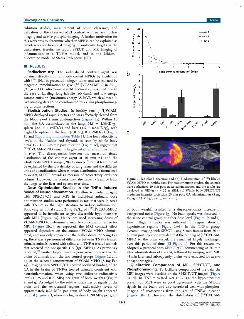
MPIO displayed rapid kinetics and was effectively cleared fromthe blood pool 2 min post-injection (Figure 1a). Within 10min, the CA accumulated in the lungs (4.8 ± 1.3%ID/g),spleen (3.4 ± 1.4%ID/g) and liver (1.5 ± 0.5%ID/g), withnegligible uptake in the brain (0.016 ± 0.005%ID/g) (Figure1b and Supporting Information Table 1). The low radioactivitylevels in the bladder and thyroid, as seen by whole bodySPECT/CT 20−25 min post-injection (Figure 1c), suggest that[125I]VCAM-MPIO remains largely intact after administrationin vivo. The discrepancies between the measured tissuedistribution of the contrast agent at 10 min p.i. and thewhole body SPECT image (20−25 min p.i.), can at least in partbe explained by the low density of lung tissue and the differentunits of quantification; whereas organ distribution is normalizedto weight, SPECT provides a measure of radioactivity levels pervolume. However, the results may also reflect clearance fromthe lungs to the liver and spleen over time.Dose Optimization Studies in the TNF-α Induced
Model of Neuroinflammation. To allow sequential imagingwith SPECT/CT and MRI in individual animals, doseoptimization studies were performed in rats that were injectedwith TNF-α in the right striatum to induce inflammation.Following an initial study, 2 mg Fe/kg of [125I]VCAM-MPIOappeared to be insufficient to give discernible hypointensitieswith MRI (Figure 2a). Hence, we used increasing doses ofVCAM-MPIO to determine a suitable concentration range forMRI (Figure 2b,c). As expected, the MRI contrast effectappeared dependent on the amount VCAM-MPIO adminis-tered, and was only apparent at the higher doses. At 5 mg Fe/kg, there was a pronounced difference between TNF-α treatedanimals, animals treated with saline, and TNF-α treated animalsthat received the nonspecific CA (IgG-MPIO). As previouslyreported,10 limited hypointense regions were observed in thebrains of animals from the two control groups (Figure 2d ande). At the selected concentration of VCAM-MPIO (5 mg Fe/kg), imaging with SPECT/CT showed localized binding of theCA in the brains of TNF-α treated animals, consistent withneuroinflammation, when using two different radioactivitylevels (0.25 and 0.90 MBq per gram of body weight) (Figure2f and g). As judged by the relative intensities of signals in thebrain and the extracranial regions, radioactivity levels ofapproximately 0.25 MBq per gram of body weight appearedoptimal (Figure 2f), whereas a higher dose (0.90 MBq per gram
of body weight) resulted in a disproportionate increase inbackground noise (Figure 2g). No brain uptake was observed inthe saline control group at either dose level (Figure 2h and i).Five milligrams Fe/kg was sufficient for segmentation ofhypointense regions (Figure 2j−l). In the TNF-α group,dynamic imaging with SPECT using 5 min frames from 20 to45 min post-injection revealed that the binding of [125I]VCAM-MPIO to the brain vasculature remained largely unchangedover this period of time (SI Figure 1). For this reason, weadopted a protocol with SPECT/CT commencing at 20 minafter administration of the CA, followed by imaging with MRI45 min later, and subsequently, brains were extracted for ex vivophosphorimaging.
Qualitative Comparison of MRI, SPECT/CT, andPhosphorimaging. To facilitate comparison of the data, theMRI images were overlaid on the SPECT/CT images (Figure3a−d). In TNF-α treated rats (n = 4), the hypointensitiespresent on MRI were in good agreement with the SPECTsignals in the brain, and also correlated well with phosphor-imaging of cryosections from the site of TNF-α injection(Figure 3f−h). However, the distribution of [125I]VCAM-
Figure 1. (a) Blood clearance and (b) biodistribution of 125I-labeledVCAM-MPIO in healthy rats. For biodistribution studies, the animalswere euthanized 10 min post tracer administration and the results aredisplayed as %ID/g (n = 3) ± SEM. (c) Whole body SPECT/CTmaximum intensity projection 20 min post CA administration (5 mgFe/kg, 0.25 MBq/g per gram, n = 1).
Bioconjugate Chemistry Article
DOI: 10.1021/acs.bioconjchem.5b00380Bioconjugate Chem. 2015, 26, 1542−1549
1543

MPIO in the cryosections appeared more widespread than wasevident from the in vivo imaging data obtained with MRI andSPECT. Interestingly, in the saline treated control group,phosphorimaging showed localized uptake of the CA at theinjection site, suggesting that the sham treatment also inducedlocal inflammation (Figure 3g). The low and homogeneousradioactivity levels observed with phosphorimaging in brainsfrom animals that received [125I]IgG-MPIO confirms that thenonspecific binding of the CA is negligible (Figure 3h).
Quantitative Comparison of SPECT and MRI Data. Toenable quantitative comparison of the data obtained withSPECT/CT and MRI, we determined the total signal from[125I]VCAM-MPIO in the whole brain, as well as in the twohemispheres. In the case of MRI, this was expressed as thepercentage contrast void across the brain volume. For SPECT,the background was defined as the average mean signalintensity across the extracranial field of view, and uptake of theCA was expressed as the signal-to-background ratio in order tonormalize counts for body weight and the amount ofradioactivity that was administered. Importantly, injection ofTNF-α into the right striatum induces widespread inflamma-tion that also affects the left hemisphere, as can be seen by 3Drepresentation of MRI images (Figure 4a,b). Nevertheless,comparison of the signal in the two brain hemispheres avoids
Figure 2. Representative coronal MRI and SPECT imagesdemonstrating binding of radiolabeled VCAM-MPIO at differentdoses of contrast agent (a−c) and levels of radioactivity (f−i). (a−c)TNF-α group following administration of VCAM-MPIO: (a) 2 mgFe/kg, (b) 5 mg Fe/kg, and (c) 6 mg Fe/kg. The optimum dose ofiron oxide (5 mg Fe/kg) was assessed in two control groups: (d) salineand (e) IgG. (f−i) SPECT/CT in the TNF-α and saline groupsadministered with a lower and higher dose of radioactivity: (f) TNF-α(0.25 MBq/g), (g) TNF-α (0.90 MBq/g), (h) Saline (0.25 MBq/g),(i) Saline (0.71 MBq/g). (j−l) Illustration of quantification procedure:(j) brain extraction, (k) hypointense regions are segmented using localthresholding, (l) segmented regions overlaid in red upon the originalMRI image, (m) blue crosshairs indicate the approximate location ofthe coronal slices for the displayed MRI and SPECT/CT images. Fordisplay purposes, SPECT images are thresholded at 40% of the 95thpercentile and linearly scaled between this value and the 95thpercentile.
Figure 3. Bimodal in vivo imaging and ex vivo phosphorimaging ofVCAM-1 expression. (a−d) Intracerebral TNF-α + [125I]VCAM-MPIO (5 mg Fe/kg): (a) coronal MRI image, (b) coronal SPECT/CT image, (c) co-registered MRI and CT image, (d) SPECT overlaidon MRI image. (e) Illustration of the approximate location for coronalsections. (f−h) Coronal phosphorimaging sections: (f) TNF-α, (g)saline control, (h) IgG control.
Figure 4. 3D representation of CA binding and in vivo quantitationusing SPECT and MRI. 3D representation of CA binding (VCAM-MPIO, 5 mg Fe/kg) based on in vivo MRI images for the (a) salinecontrol group and the (b) TNF-α treated group. Segmentedhypointense regions are shown in red, while the pink, green, andblue areas indicate the whole brain, left, and right regions of interest(respectively) used for quantification. Quantification of VCAM-MPIObinding as assessed by (a) in vivo MRI and (b) in vivo SPECT.Quantification was carried out over the whole brain (red) as well as theleft (green) and right (blue) cerebral hemispheres. The saline groupreceived intracerebral saline injections followed by radiolabeledVCAM-MPIO (5 mg Fe/kg, 0.25−0.9 MBq/g) (n = 3). The IgGgroup received intracerebral TNF-α injections followed by radio-labeled IgG-MPIO (5 mg Fe/kg, 0.25−0.9 MBq/g) (n = 3) and theTNF-α group received intracerebral TNF-α injections followed byradiolabeled VCAM-MPIO (5 mg Fe/kg, 0.25−0.9 MBq/g) (n = 4).Abbreviations: LLeft, WBWhole brain, RRight.
Bioconjugate Chemistry Article
DOI: 10.1021/acs.bioconjchem.5b00380Bioconjugate Chem. 2015, 26, 1542−1549
1544
bias from manually defining the regions of interest, and wasused in order to highlight differences in the data obtained withMRI and SPECT. Quantitation with both MRI and SPECTrevealed higher CA binding in TNF-α treated animals ascompared to the two control groups, globally as well as in eachof the brain hemispheres (Figure 4c,d and SI Figure 2). In theTNF-α treated animals, [125I]VCAM-MPIO localized predom-inantly to the affected (right) cerebral hemisphere (n = 4) asassessed by both SPECT (p = 0.03) and MRI (p = 0.002).While the SPECT and MRI data were in good overallagreement, there are important differences in the resultsobtained with the two modalities. In particular, MRI analysisindicated more pronounced difference in CA uptake betweenthe left and right hemispheres in the TNF-α group than wasevident with SPECT, and the statistical significance for thegroup comparisons with MRI was substantially higher than forSPECT. For instance, the difference in the group means whencomparing CA uptake in the right hemisphere reached asignificance of p = 1 × 10⁻⁶ with MRI, but only p = 0.045 withSPECT (Figure 4c,d). Unfortunately, the radioactivity levelsthat were needed for imaging with SPECT/CT were too highto allow quantification of the CA in brain sections withphosphorimaging.Distribution of [125I]VCAM-MPIO within the Brain
Vasculature. To determine the distribution of [125I]VCAM-MPIO within the brain vasculature, one TNF-α treated rat wasadministered a reduced dose of [125I]VCAM-MPIO (3 mg Fe/kg, 30 KBq/g). Phosphorimaging of brain cryosections (1 htime point, Figure 5) revealed the CA to be distributed in a
pattern consistent with the hypointensities observed with MRI(SI Figure 3) on slices both anterior and posterior to bregma.While the iron oxide particles appear to be localized in thevasculature, it is unclear whether they are on the arterial orvenous side of the circulation. Nevertheless, several large vesselsconsistent with major arteries were identified: these include theanterior striate arteries (astr), anterior choroidal artery (ach),transverse hippocampal arteries (trhi), the supracollicularnetwork (scol),22 as well as the choroidal artery. The prominentvessels which lie on the dorsal surface of both hemispheres alsocontained high levels of the CA.Imaging of Inflammation in the Lithium−Pilocarpine
Model of Status Epilepticus. We also assessed the potentialof [125I]VCAM-MPIO as a bimodal imaging agent to detectinflammation in the lithium−pilocarpine model of StatusEpilepticus (SE). All pilocarpine treated animals progressedto SE and displayed akinesia and facial automatizms. Tonic−clonic seizures and SE were induced 40−50 min afterpilocarpine administration. The seizures were terminated after90 min by administration of diazepam (10 mg/kg).Approximately 20 h after induction of SE, [125I]VCAM-
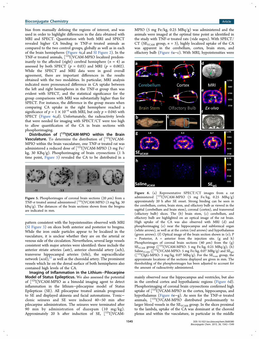
MPIO (5 mg Fe/kg, 0.25 MBq/g) was administered and theanimals were imaged at the optimal time point as identified inthe study with TNF-α treated rats (vide supra). With SPECT/CT (SEVCAM group, n = 3), highly localized uptake of the CAwas apparent in the cerebellum, cortex, brain stem, andolfactory bulb (Figure 6a−c). With MRI, hypointensities were
mainly observed near the hippocampus and ventricles, but alsoin the cerebral cortex and hypothalamic regions (Figure 6d).Phosphorimaging of coronal brain cryosections confirmed highuptake of [125I]VCAM-MPIO in the cortex, hippocampus, andhypothalamus (Figure 6e−g). As seen for the TNF-α treatedanimals, [125I]VCAM-MPIO distributed predominantly tolarger blood vessels in the SEVCAM group. In the slices proximalto the lambda, uptake of the CA was dominant at the choroidplexus and within the vasculature, in particular in the middle
Figure 5. Phosphorimages of coronal brain sections (20 μm) from aTNF-α treated animal administered [125I]VCAM-MPIO (3 mg/kg, 30kBq/g). The distances of the brain sections shown from the bregmaare indicated in mm.
Figure 6. (a) Representative SPECT/CT images from a ratadministered [125I]VCAM-MPIO (5 mg Fe/kg, 0.25 MBq/g)approximately 20 h after SE onset. Strong binding can be seen inthe cerebellum, cortex, brain stem, and olfactory bulb as viewed in thesagittal (cerebellum and brain stem), coronal (cortex), and transversal(olfactory bulb) slices. The (b) brain stem, (c) cerebellum, andolfactory bulb are highlighted on an optical image of the rat brain.High uptake of the CA was also observed with MRI (d) andphosphorimaging (e) near the hippocampus and subfornical organ(white arrows), as well as at the cortex (red arrows) and hypothalamus(green arrows). (f) Optical image of the brain section shown in (e); P= Posterior, A = anterior from the injection site. (g and h)Phosphorimages of coronal brain sections (40 μm) from the (g)SEVCAM group ([125I]VCAM-MPIO: 5 mg Fe/kg, 0.25 MBq/g); (h)SalineVCAM ([125I]VCAM-MPIO: 5 mg Fe/kg, 0.07 MBg/g) and SEIgG([125I]IgG-MPIO: 5 mg/kg, 0.07 MBq/g). For the SEvcam group, theapproximate locations of the sections displayed are given in mm. Thethresholding of the phosphorimages has been adjusted to account forthe amount of radioactivity administered.
Bioconjugate Chemistry Article
DOI: 10.1021/acs.bioconjchem.5b00380Bioconjugate Chem. 2015, 26, 1542−1549
1545

cerebral artery (Figure 6g). No uptake of the CA was evident inbrains from sham treated animals (SALINEVCAM, n = 3), nor incontrol animals that received [125I]IgG-MPIO (SEIgG group, n =3) (Figure 6h).
■ DISCUSSIONWe have used iodine-125 labeled, VCAM-1 antibody coated,micron-sized particles of iron oxide in two models ofneuroinflammation and have shown, for the first time, itspotential as a bimodal imaging agent; conspicuity of the CAwas not only evident by MRI, but also seen with SPECT/CTand phosphorimaging. Our kinetic studies revealed that[125I]VCAM-MPIO binds rapidly to vasculature in inflamedbrain tissue and is quickly cleared from the blood pool. Theblood clearance rate of [125I]VCAM-MPIO was in goodagreement with previously reported data for other microsizedparticles.23 This is significant, as the rapid binding and clearancemakes microsized particles suitable for labeling with short-livedpositron emitters, such as gallium-68 and fluorine-18, forimaging of molecular targets in the vasculature with PET. It isnoteworthy that previous efforts to develop particle-derivedradiotracers to date have focused almost exclusively onnanoparticles,18−21 despite the need for exotic long-livedradionuclides, such as copper-64 (half-life 12.7 h) and iodine-124 (half-life 4.2 days), to match the prolonged circulation timein the blood.24
Overall, there was good colocalization between the in vivoSPECT and MRI images, and significantly more contrast agentwas detected with both imaging techniques in rats treated withTNF-α compared to the two control groups. However,quantification of the imaging data yielded marked differencesbetween the two techniques. This may, at least in part, beexplained by attenuation of the low energy γ rays emitted byiodine-125 (35 keV) that was used for SPECT. In addition, thenonlinear relationship between the local CA concentration andcontrast volume as assessed by MRI25 may have furtherexacerbated the discrepancies in quantification.In the SE model, SPECT and MRI provided highly
complementary data with MRI hypointensities observed aroundthe hippocampus, the subfornical organ, and to a lesser extentin the cerebral cortex. In contrast, SPECT/CT failed to detectthe binding of [125I]VCAM-MPIO in the hippocampus (Figure6), likely due to attenuation, but revealed highly localizedsignals in the cortex, cerebellum, brain stem, and olfactory bulb(Figure 6a−d). Although the MRI data largely reflected thedistribution of [125I]VCAM-MPIO within the brain of SEanimals, comparison with SPECT and phosphorimaginghighlight the limitations of the MRI contrast effect for imagingin the outer regions of the brain. It is also evident that therelationship between the volume of hypointense pixels withinthe brain and the concentration of [125I]VCAM-MPIO isnonlinear, which should be taken into account when using thisagent.It is unclear to which degree the distribution of the CA is
dependent on cerebral blood flow. Phosphorimaging of brainsections showed that the CA localizes to large blood vessels,which may imply a flow component (Figure 5a). Although thedistribution to the larger vessels may be the result of asignificant first pass extraction, further studies are required todetermine how the interplay between blood flow, extraction,and VCAM-1 expression affects the binding of VCAM-MPIO.While it was outside the scope of this study to investigate thepathology of the disease models in detail, it is worth noting that
the distribution pattern of [125I]VCAM-MPIO in the brains ofanimals from the SE model was consistent with our previouslyreported results,10 and demonstrates that the uptake reflects theregions associated with inflammation.In conclusion, we have developed a highly efficient and
practical method to label VCAM-1 antibody coated iron oxidemicrosized particles with iodine-125, and characterized thecontrast agent in two models of inflammation using SPECT/CT, MRI, and phosphorimaging. Kinetic studies showed thatbinding of [125I]VCAM-MPIO to the vasculature of inflamedtissue remained relatively constant over a 20 min period andthat MPIOs are rapidly cleared from the blood pool. Thisensures high contrast, but is also a limitation in that the uptakeof the CA is likely to be influenced by regional blood flow. Thepattern of hypointensities observed with MRI was consistentwith the distribution of [125I]VCAM-MPIO as observed usingphosphorimaging. Overall, this study provides proof of conceptthat multimodal imaging of inflammation with both SPECTand MRI is feasible. In the pilocarpine model of SE, MRI andSPECT/CT proved highly complementary, with inflammationdetected in distinct brain regions by the two techniques,demonstrating the value of validating different imagingmethods against one another.
■ MATERIALS AND METHODSSynthesis of VCAM-MPIO. Monoclonal antibodies specific
to rat VCAM-1 (MR106) (ebioscience, USA) were conjugatedto micron-sized particles of iron oxide (MPIOs, 1 μm diameter,iron content: 26%, Invitrogen, Life Technologies, U.K.). Ingeneral, 40 μg of antibody was reacted per mg of iron/MPIO(Fe-MPIO) via a tosyl alkylation reaction following themanufacturers’ guidelines and as previously reported. First,MPIOs were washed. The beads in the original vial wereresuspended by vortexing for >30 s. The beads (5 mg of Fe perkg of animal weight, 19.2 mg/kg MPIO, 38 μL) were thenplaced in an eppendorf tube to which sodium borate buffersolution (1000 μL, 0.1 M, pH 9.5) was added and vortexed for>30 s. The eppendorf was placed next to a magnet (0.5 T,nickel plated, K&J Magnetics Inc., USA) to immobilize theparticles, and the supernatant was removed with a Gilsonpipette (Gilson Scientific Ltd., U.K.). After removing the tubefrom the magnet, to the washed beads was then added sodiumborate solution (415 μL, 0.1 M, pH 9.5) followed by theantibodies (154 μg, 154 μL). Finally, ammonium sulfatesolution (138 μL, 3 M dissolved in 0.1 M sodium borate buffer(100 mL, pH 9.4)) was added and the mixture was incubatedovernight at 37 °C with slow shaking to ensure the beads didnot settle during the incubation period. Following antibodyconjugation, a magnet was used to immobilize VCAM-MPIO.The supernatant containing unbound antibodies was removedand VCAM-MPIO was resuspended in heparinized PBS (0.1%,100 μL).
Synthesis of Iodine-125 Labeled VCAM-MPIO.[125I]NaI (>12.95 GBq/ml, 629 GBq/mg) (PerkinElmer,USA) was purchased as a non-carrier-added solution inreductant free 10−5 M aqueous sodium hydroxide solution(pH 8−11). All other chemicals were purchased from Sigma-Aldrich U.K. unless otherwise stated.VCAM-MPIO (19.2 mg MPIO, 40 μL) was suspended in
heparinized phosphate-buffered saline (PBS) (100 μL, pH 7.4),and then transferred into precoated iodogen tubes (ThermoFisher Scientific, USA). To this, [125I]NaI (30−120 MBq) wasadded and the mixture was left to incubate at room temperature
Bioconjugate Chemistry Article
DOI: 10.1021/acs.bioconjchem.5b00380Bioconjugate Chem. 2015, 26, 1542−1549
1546

for 30 min. After this time, the radioactive mixture wastransferred into an eppendorf tube and radiolabeled VCAM-MPIO ([125I]VCAM-MPIO) was purified using the magneticimmobilization technique described above. The particles werewashed with PBS (1 mL) prior to their administration into rats.Using this same protocol, [125I]IgG-MPIO was synthesizedusing nonspecific control antibodies (Southern Biotech, U.K).Animal Models. All animal procedures were carried out in
accordance with the UK Animals (Scientific Procedures) 1986Act and institutional ethics regulations. Adult male Sprague−Dawley rats (170−270 g) were obtained from the breedingcolony of the University College London (UCL) animal facility.Neuroinflammation Model. Rats were anaesthetized using a
combination of isoflurane (1% in pure oxygen) and urethane(1.5 g/kg, i.p.). TNF-α (300 ng, Life Technologies Ltd. U.K.)in saline (5 μL) was injected into the right striatum(coordinates from bregma: −0.5 mm anteroposterior, 3 mmmediolateral, 4 mm dorsoventral from dura) using a Hamiltonsyringe (Hamilton Company, USA) attached to a 31G needle.Saline (5 μL) was administered in place of TNF-α for thecontrol group.Lithium-Pilocarpine Model of Status Epilepticus (SE). Rats
were injected with lithium chloride (3 mg/kg, intraperitoneally(i.p.)) 3 h prior to methyl scopolamine nitrate (5 mg/kg, i.p.)administration. This was followed 30 min later by admin-istration of pilocarpine hydrochloride (30 mg/kg, i.p.) in orderto induce SE (SEVCAM). Animals were behaviorally assessed andthe onset of SE was defined as stage 3 on the Racine scale.Diazepam (10 mg/kg, i.p., Hameln Pharmaceuticals, U.K.) wasadministered 90 min after SE onset to terminate the seizure.Further injections of diazepam were administered as required.The control groups received saline in place of pilocarpinehydrochloride.Biological Evaluation of [125I]VCAM-MPIO. Unless
otherwise stated, rats were administrated [125I]VCAM-MPIOor [125I]IgG-MPIO (5 mg Fe/kg, 0.25−0.9 MBq/g) inheparinized phosphate-buffered saline (1 mL, pH 7.4) via acannula inserted into the right external jugular vein.Blood Clearance and Biodistribution. Blood samples (n =
3) were collected via a jugular vein cannula at predeterminedtime points (approximately: 85, 120 170, 230, 290 s) followingradiolabeled MPIO administration in rats anaesthetized by acombination of isoflurane (1% in pure oxygen) and urethane(1.5 g/kg). After each collection, the cannula was flushed withsaline (1 mL) to remove residual radioactivity. For biodis-tribution studies, radiolabeled MPIO (30−40 KBq/g) wasadministered via the lateral tail vein (n = 3). Ten minutes aftertracer administration, the rats were anaesthetized withisoflurane in pure oxygen. Blood was sampled through cardiacpuncture and the animals were sacrificed by cervical dislocation.The organs of interest were then removed for gamma counting(Wizard 2470, PerkinElmer, U.K.). The results are expressed as% injected dose per gram of tissue ± SD.In Vivo Imaging. Inflammation was induced by intrastriatal
injection of TNF-α as described above. Seven hours after theintracerebral injection (vide supra), [125I]VCAM-MPIO or[125I]IgG-MPIO was administered. Rats treated with Lithium-Pilocarpine received [125I]VCAM-MPIO or [125I]IgG-MPIO(SEVCAM and SEIgG) 20 h after the termination of seizures. Forthe sham control group saline was administered instead ofpilocarpine (SALINEVCAM). For the duration of the in vivoimaging experiments, isoflurane (1% in oxygen) was used tomaintain anesthesia in rats. A physiological monitoring system
(SA Instruments, USA) was used to monitor their respirationrate and rectal temperature. Temperature was maintained at 37± 0.5 °C using an air and water tubing warming system.In vivo SPECT/CT (computed tomography) was performed
using a nanoSPECT system (Bioscan Inc. USA) approximately20 min post contrast administration. A CT scan was conductedwith the following parameters: radial field of view (FOV) =40.5 mm2, axial FOV = 40.9 mm, exposure time per projection= 1 s, 360 projections, and 55 KvP tube voltage, which resultedin an acquisiton time of 6 min. Helical SPECT was performedacross the same FOV using 2.5 mm pinhole apertures with 20projections and an exposure time of 15 s per projection. Fordynamic imaging, 5 scans were performed in successionresulting in a total imaging time of 25 min. All SPECT/CTimages displayed are from the first frames captured 20 min postCA administration. To show the in vivo distribution of the CA,a scan was performed across the entire FOV of the animal with2.5 mm pinhole apertures and an acquisition time of 40 min.In vivo MRI was performed immediately following SPECT/
CT imaging using a 9.4T horizontal bore scanner (AgilentTechnologies, USA). MRI was performed between 1 and 2 hfollowing administration of [125I]VCAM-MPIO and [125I]IgG-MPIO. Iron oxide was detected using a 3D gradient echosequence (TR = 100 ms, TE = 11 ms, matrix = 192 × 192 ×160, FOV = 25 × 25 × 25 mm3, acquisition time ≈ 51 min).
Phosphorimaging. Following in vivo imaging, the animalswere euthanized, their brains were removed and fixed overnightin 4% paraformaldehyde (PFA) in PBS (pH 7.4). The brainswere then cryoprotected overnight in sucrose solution andsectioned coronally at 20 μm. Sectioning of the braincommenced close to the bregma. The sections were thenmounted on poly(L-lysine) coated glass slides (VWR interna-tional Ltd. U.K) which were subsequently exposed onunmounted GP 20 × 25 cm2 phosphor screens (VWRinternational LTD, U.K.) for 15 min. After this time, thescreens were scanned by a Typhoon 9410 Trio+ Phosphor-imager (GE Healthcare, U.K.), (25 μm resolution, acquisitiontime = 2 h 30 min) and the resulting images were processed byImageJ (NIH, USA).As the radioactivity levels in the brain sections were above
the optimal range for phosphorimaging, one animal in theTNF-α group was administered with a reduced dose of[125I]VCAM-MPIO (3 mg Fe/kg, 30 kBq/g). The animal wassacrificed 1 h post CA administration, and the brain wasremoved and treated with PFA and sucrose as previouslydescribed. Coronal sections (20 μm) of the brain were exposedon a phosphor screen for 1 week, which was then scanned usingthe parameters described above.
Optical Imaging. In order to correlate the observed signalsto particular brain regions, optical images of the sections usedfor phosphorimaging were captured on a bright field AxioSkop2 system (Gottingen, Germany) at 5× magnification. Theimages were processed using the tiling and stitching method(Lundin M, 2004) on the Zeiss Axio Vision 4.8 software(Imaging Associates, Germany).
Image Analysis. SPECT Reconstruction. SPECT imageswere reconstructed using the HiSPECT software package usingthe following parameters: smoothing = 45%, resolution = 67%,number of iterations = 10. CT reconstruction was performedusing InVivoScope (InviCRO, USA). For display purposes, thein vivo SPECT images are thresholded at 40% of the 95thpercentile and linearly scaled between this value and the 95thpercentile.
Bioconjugate Chemistry Article
DOI: 10.1021/acs.bioconjchem.5b00380Bioconjugate Chem. 2015, 26, 1542−1549
1547

MRI Quantification of Contrast Agent Binding. MRI datawere processed in MATLAB 2013a (MathWorks, Natick, MA).First, the data were zero-filled to 256 × 256 × 256 in k-spaceprior to reconstruction to give a final isotropic voxel size of 98μm. Brain extraction was performed using a multiatlasapproach.26−28 Hypointensities caused by the presence ofiron oxide were segmented using adaptive thresholding in orderto take into account local variations in signal intensity. This wasachieved by subtracting the MRI volume from the volumeconvolved with a three-dimensional Gaussian kernel of size 15× 15 × 15 voxels and a σ of 15, followed by thresholding withthe same empirically determined threshold used across all datasets. Regions of interest (ROIs) were defined over the left orright cerebral hemisphere surrounding the injection site. Theseconsisted of 30 consecutive slices (∼3 mm) anterior to and 30consecutive slices posterior to the injection site (as indicated inFigure 4a and b).Coregistration. Coregistration of MRI and CT images was
performed in AMIRA (Visualization Sciences Group, Burling-ton, MA) on the gradient images of the brain extracted MRImagnitude data and the CT intensity images using normalizedcorrelation as the cost function.SPECT Quantification of Contrast Agent Binding. ROIs as
defined on MRI images were propagated to the SPECT/CTnative space using the same transformation matrix. In this way,identical ROIs were used for both the MRI and SPECTanalyses. Background signal intensity was defined as the meansignal intensity across all nonbrain tissue across the entire 3Dfield of view and CA binding was expressed as the signal-to-background ratio to normalize counts for body weight and thelevel of radioactivity that was administered.Statistical Analysis. Statistical analysis was performed in
MATLAB. Group means were compared using one-wayANOVA. Group means between the left and right hemisphereswere compared using one-tailed paired t tests. All error bars areshown as mean ± standard error of the mean (SEM). Statisticalsignificance was assigned at p < 0.05.
■ ASSOCIATED CONTENT*S Supporting InformationThe Supporting Information is available free of charge on theACS Publications website at DOI: 10.1021/acs.bioconj-chem.5b00380.
Ex vivo biodistribution 125I labeled VCAM-MPIO inhealthy rats; dynamic SPECT following administration ofthe CA in TNF-α treated rats; MRI images showing thedistribution of VCAM-MPIO (PDF)
■ AUTHOR INFORMATIONCorresponding Author*E-mail: [email protected] Contributions#N. Patel and B. A. Duffy contributed equally.NotesThe authors declare no competing financial interest.
■ ACKNOWLEDGMENTSThis work was funded by the UK Medical Research Council,(B.D., N.P.) and was undertaken at UCLH/UCL who receiveda proportion of funding from the Department of Health’sNIHR Biomedical Research Centres funding scheme (E.A.).The research leading to these results has received funding from
the European Union’s Seventh Framework Programme (FP7/2007-2013) under grant agreement n°602102 (EPITARGET)(E.A.). We also acknowledge support from King’s CollegeLondon and UCL Comprehensive Cancer Imaging Centre CR-UK & EPSRC, in association with the MRC and DoH(England) (M.L.) M.L. receives funding from Medical ResearchCouncil (MR/J013110/1); the National Centre for theReplacement, Reduction and Refinement of Animal in Research(NC3Rs); UK Regenerative Medicine Platform Safety Hub(MRC: MR/K026739/1).
■ REFERENCES(1) del Zoppo, G., Ginis, I., Hallenbeck, J. M., Iadecola, C., Wang, X.,and Feuerstein, G. Z. (2000) Inflammation and stroke: putative rolefor cytokines, adhesion molecules and iNOS in brain response toischemia. Brain Pathol. 10, 95−112.(2) Vezzani, A., and Granata, T. (2005) Brain inflammation inepilepsy: Experimental and clinical evidence. Epilepsia 46, 1724−1739.(3) Morganti-Kossmann, M. C., Rancan, M., Stahel, P. F., andKossmann, T. (2002) Inflammatory response in acute traumatic braininjury: a double-edged sword. Curr. Opin. Crit. Care. 8, 101−105.(4) Serres, S., Soto, M. S., Hamilton, A., McAteer, M. A., Carbonell,W. S., Robson, M. D., Ansorge, O., Khrapitchev, A., Bristow, C.,Balathasan, L., et al. (2012) Molecular MRI enables early and sensitivedetection of brain metastases. Proc. Natl. Acad. Sci. U. S. A. 109, 6674−6679.(5) Soto, M. S., Serres, S., Anthony, D. C., and Sibson, N. R. (2014)Functional role of endothelial adhesion molecules in the early stages ofbrain metastasis. Neuro Oncol. 16, 540−551.(6) Tuppo, E. E., and Arias, H. R. (2005) The role of inflammation inAlzheimer’s disease. Int. J. Biochem. Cell Biol. 37, 289−305.(7) McAteer, M. A., Sibson, N. R., von zur Muhlen, C., Schneider, J.E., Lowe, A. S., Warrick, N., Channon, K. M., Anthony, D. C., andChoudhury, R. P. (2007) In vivo magnetic resonance imaging of acutebrain inflammation using microparticles of iron oxide. Nat. Med. 13,1253−1258.(8) Hoyte, L. C., Brooks, K. J., Nagel, S., Akhtar, A., Chen, R. L.,Mardiguian, S., McAteer, M. A., Anthony, D. C., Choudhury, R. P.,Buchan, A. M., et al. (2010) Molecular magnetic resonance imaging ofacute vascular cell adhesion molecule-1 expression in a mouse modelof cerebral ischemia. J. Cereb. Blood Flow Metab. 30, 1178−1187.(9) Serres, S., Mardiguian, S., Campbell, S. J., McAteer, M. A., Akhtar,A., Krapitchev, A., Choudhury, R. P., Anthony, D. C., and Sibson, N. R.(2011) VCAM-1-targeted magnetic resonance imaging revealssubclinical disease in a mouse model of multiple sclerosis. FASEB J.25, 4415−4522.(10) Duffy, B. A., Choy, M., Riegler, J., Wells, J. A., Anthony, D. C.,Scott, R. C., and Lythgoe, M. F. (2012) Imaging seizure-inducedinflammation using an antibody targeted iron oxide contrast agent.NeuroImage 60, 1149−1155.(11) Montagne, A., Gauberti, M., Macrez, R., Jullienne, A., Briens, A.,Raynaud, J.-S., Louin, G., Buisson, A., Haelewyn, B., Docagne, F., et al.(2012) Ultra-sensitive molecular MRI of cerebrovascular cell activationenables early detection of chronic central nervous system disorders.NeuroImage 63, 760−770.(12) Akhtar, A. M., Chen, Y., Schneider, J. E., Digby, J. E., McAteer,M. A., Wood, K., and Choudhury, R. P. (2008) Magnetic ResonanceImaging Of Renal Ischemia Reperfusion Injury Using MicroparticlesOf Iron Oxide Targeting VCAM-1. Circulation 118, S_555.(13) McAteer, M. A., Schneider, J. E., Ali, Z. A., Warrick, N., Bursill,C. A., von zur Muhlen, C., Greaves, D. R., Neubauer, S., Channon, K.M., and Choudhury, R. P. (2007) Magnetic resonance imaging ofendothelial adhesion molecules in mouse atherosclerosis using dual-targeted microparticles of iron oxide. Arterioscler., Thromb., Vasc. Biol.28, 77−83.(14) Akhtar, A. M., Schneider, J. E., Chapman, S. J., Jefferson, A.,Digby, J. E., Mankia, K., Chen, Y., McAteer, M. A., Wood, K. J., andChoudhury, R. P. (2010) In vivo quantification of VCAM-1 expression
Bioconjugate Chemistry Article
DOI: 10.1021/acs.bioconjchem.5b00380Bioconjugate Chem. 2015, 26, 1542−1549
1548

in renal ischemia reperfusion injury using non-invasive magneticresonance molecular imaging. PLoS One 5, e12800−e.(15) Elices, M. J., Osborn, L., Takada, Y., Crouse, C., Luhowskyj, S.,Hemler, M. E., and Lobb, R. R. (1990) VCAM-1 on ActivatedEndothelium Interacts with the Leukocyte Integrin VLA-4 at a SiteDistinct from the VLA-4 Fibronectin Binding-Site. Cell 60, 577−584.(16) Fabene, P. F., Mora, G. N., Martinello, M., Rossi, B., Merigo, F.,Ottoboni, L., Bach, S., Angiari, S., Benati, D., Chakir, A., et al. (2008) Arole for leukocyte-endothelial adhesion mechanisms in epilepsy. Nat.Med. 14, 1377−1383.(17) Shapiro, E. M., Skrtic, S., and Koretsky, A. P. (2005) Sizing itup: Cellular MRI using micron-sized iron oxide particles. Magn. Reson.Med. 53, 329−338.(18) Louie, A. (2010) MultimodalityImaging Probes: Design andChallenges. Chem. Rev. 110, 3146−3195.(19) Xie, J., Liu, G., Eden, H. S., Al, H., and Chen, X. (2011) Surface-engineered magnetic nanoparticle platforms for cancer imaging andtherapy. Acc. Chem. Res. 44, 883−892.(20) Sun, X., Cai, W., and Chen, X. (2015) Positron EmissionTopography Imaging Using Radiolabeled Inorganic Nanomaterials.Acc. Chem. Res. 48, 286−294.(21) Tamba, B. I., Dondas, A., Leon, M., Neagu, A. N., Dodi, G.,Stefanescu, C., and Tijani, A. (2015) Silica Nanoparticles: Prerpara-tion, Characterization, and In Vitro/In Vivo Biodistribution Studies.Eur. J. Pharm. Sci. 71, 46−55.(22) Scremin, O. U. (2004) Cerebral Vascular System. The RatNervous System, 3rd ed. (Paxinos, G.) pp 1167−1202, Chapter 33,Academic Press, Burlington.(23) Muro, S., Garnacho, C., Champion, J. A., Leferovich, J.,Gajewski, C., Schuchman, E. H., Mitragotri, S., and Muzykantov, V. R.(2008) Control of Endothelial Targeting and Intracellular Delivery ofTherapeutic Enzymes by Modulating the Size and Shape of ICAM-1targeted Carriers. Mol. Ther. 16, 1450−1458.(24) Benezra, M., Penate-Medina, O., Zanzonico, P. B., Schaer, D.,Ow, H., Burns, A., DeStanchina, E., Longo, V., Herz, E., Iyer, S., et al.(2011) Multimodal Silica Nanoparticles are Effectiver Cancer-Targeted Probes in a Model of Human Melanoma. J. Clin. Invest.121, 2768−2780.(25) Mendonca Dias, M. H., and Lauterbur, P. C. (1987) Contrastagents for magnetic resonance imaging. Biol. Trace Elem. Res. 13, 229−239.(26) Duffy, B. A., Chun, K. P., Ma, D., Lythgoe, M. F., and Scott, R.C. (2014) Dexamethasone exacerbates cerebral edema and brain injuryfollowing lithium-pilocarpine induced status epilepticus. Neurobiol. Dis.63, 229−36.(27) Ma, D., Cardoso, M. J., Modat, M., Powell, N., Wells, J., Holmes,H., Wiseman, S., Tybulewicz, V., Fisher, E., Lythgoe, M. F., et al.(2014) Automatic structural parcellation of mouse brain MRI usingmulti-atlas label fusion. PLoS One 9, e86576.(28) Jenks, K. R., Lucas, M. M., Duffy, B. A., Robbins, A. A., Gimi, B.,Barry, J. M., and Scott, R. C. (2013) Enrichment and training improvecognition in rats with cortical malformations. PLoS One 8, e84492.
Bioconjugate Chemistry Article
DOI: 10.1021/acs.bioconjchem.5b00380Bioconjugate Chem. 2015, 26, 1542−1549
1549