Embed Size (px)
Citation preview
1
1
BCMB 3100: Partial notesChapter 4 (Part 1)
• Diversity of proteins
• 3D structure of proteins
• Fibrous vs globular proteins
• Conformation vs configuration
• 1°, 2°, 3° and 4° structure
• Peptide groups in polypeptide
• vs angles, Ramachandran plot
• Xray crystallography & NMR
• helix vs -sheet2
Diversity of proteins
• ________________ - study of large sets of proteins, such as the entire complement of proteins produced by a cell
• E. coli has about _________ different polypeptides (average size 300 amino acids, Mr 33,000)
• Fruit fly (Drosophila melanogaster) about 16,000, humans/other mammals about ~20,000 - 40,000 different polypeptides
3
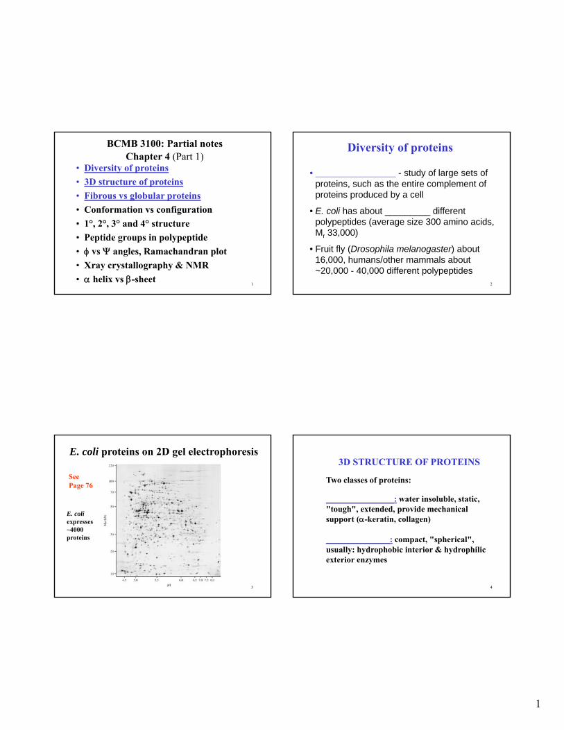
E. coli proteins on 2D gel electrophoresis
E. coliexpresses ~4000 proteins
See Page 76
4
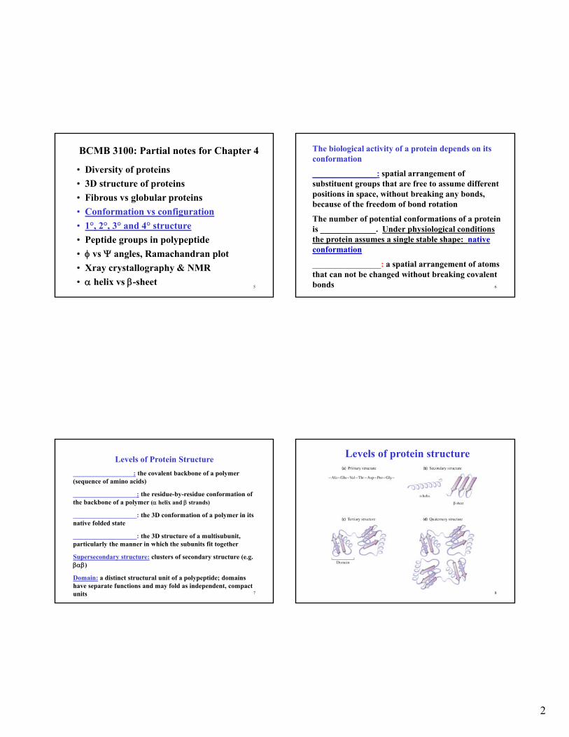
3D STRUCTURE OF PROTEINS
Two classes of proteins:
________________: water insoluble, static, "tough", extended, provide mechanical support (-keratin, collagen)
_______________: compact, "spherical", usually: hydrophobic interior & hydrophilic exterior enzymes
2
5
• Diversity of proteins
• 3D structure of proteins
• Fibrous vs globular proteins
• Conformation vs configuration
• 1°, 2°, 3° and 4° structure
• Peptide groups in polypeptide
• vs angles, Ramachandran plot
• Xray crystallography & NMR
• helix vs -sheet
BCMB 3100: Partial notes for Chapter 4
6
The biological activity of a protein depends on its conformation
_______________: spatial arrangement of substituent groups that are free to assume different positions in space, without breaking any bonds, because of the freedom of bond rotation
The number of potential conformations of a protein is _____________. Under physiological conditions the protein assumes a single stable shape: native conformation
________________: a spatial arrangement of atoms that can not be changed without breaking covalent bonds
7
Levels of Protein Structure
__________________: the covalent backbone of a polymer (sequence of amino acids)
___________________: the residue-by-residue conformation of the backbone of a polymer ( helix and strands)
___________________: the 3D conformation of a polymer in its native folded state
___________________: the 3D structure of a multisubunit, particularly the manner in which the subunits fit together
Supersecondary structure: clusters of secondary structure (e.g. )
Domain: a distinct structural unit of a polypeptide; domains have separate functions and may fold as independent, compact units 8
Levels of protein structure
3
9
• Diversity of proteins
• 3D structure of proteins
• Fibrous vs globular proteins
• Conformation vs configuration
• 1°, 2°, 3° and 4° structure
• Peptide groups in polypeptide
• vs angles, Ramachandran plot
• Xray crystallography & NMR
• helix vs -sheet
BCMB 3100: Partial notes for Chapter 4
10
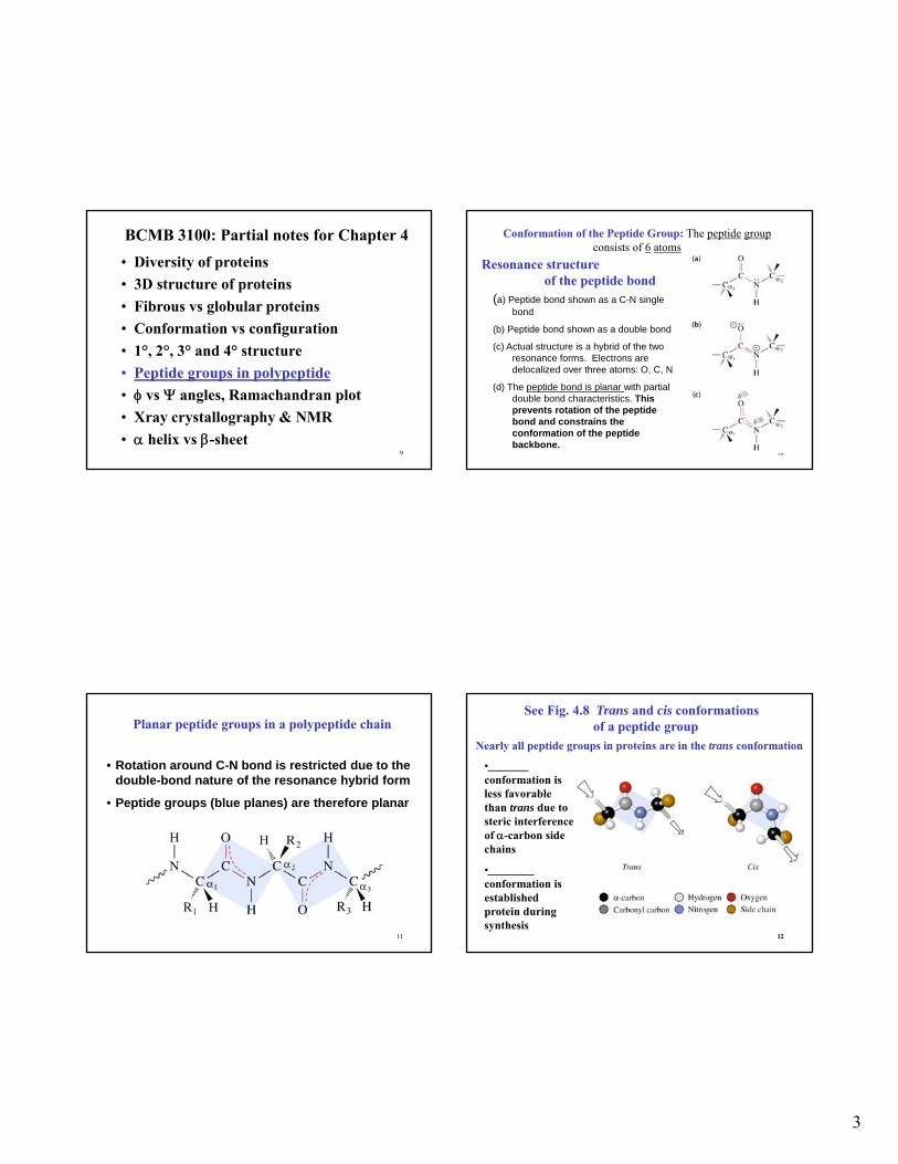
Resonance structure of the peptide bond
(a) Peptide bond shown as a C-N single bond
(b) Peptide bond shown as a double bond
(c) Actual structure is a hybrid of the two resonance forms. Electrons are delocalized over three atoms: O, C, N
(d) The peptide bond is planar with partial double bond characteristics. This prevents rotation of the peptide bond and constrains the conformation of the peptide backbone.
Conformation of the Peptide Group: The peptide groupconsists of 6 atoms
11
Planar peptide groups in a polypeptide chain
• Rotation around C-N bond is restricted due to the double-bond nature of the resonance hybrid form
• Peptide groups (blue planes) are therefore planar
1212
See Fig. 4.8 Trans and cis conformations of a peptide group
Nearly all peptide groups in proteins are in the trans conformation
•_______conformation is less favorable than trans due to steric interference of -carbon side chains
•________ conformation is established protein during synthesis
4
13
• Diversity of proteins
• 3D structure of proteins
• Fibrous vs globular proteins
• Conformation vs configuration
• 1°, 2°, 3° and 4° structure
• Peptide groups in polypeptide
• vs angles, Ramachandran plot
• Xray crystallography & NMR
• helix vs -sheet
BCMB 3100: Partial notes for Chapter 4
14
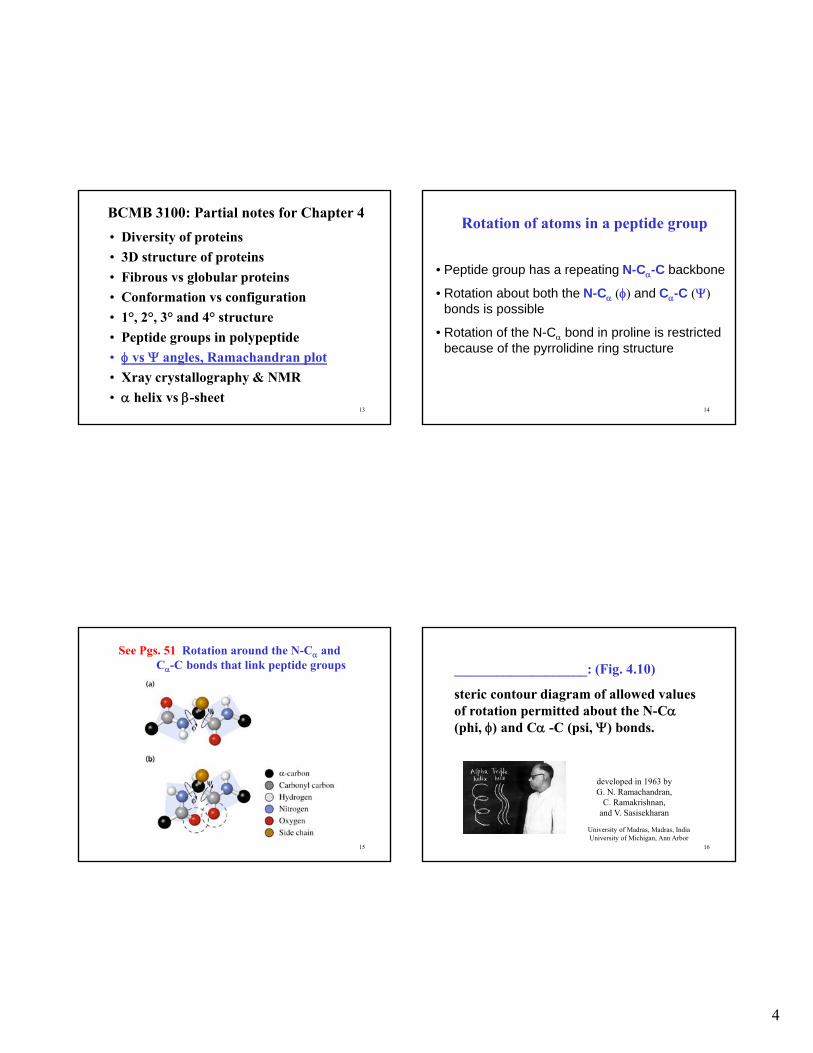
Rotation of atoms in a peptide group
• Peptide group has a repeating N-C-C backbone
• Rotation about both the N-C () and C-C () bonds is possible
• Rotation of the N-C bond in proline is restricted because of the pyrrolidine ring structure
15
See Pgs. 51 Rotation around the N-C and C-C bonds that link peptide groups
16
___________________: (Fig. 4.10)
steric contour diagram of allowed values of rotation permitted about the N-C(phi, ) and C -C (psi, ) bonds.
developed in 1963 by G. N. Ramachandran,
C. Ramakrishnan, and V. Sasisekharan
University of Madras, Madras, IndiaUniversity of Michigan, Ann Arbor

5
17
See Fig. 10 (a) Ramachandran Plot
(a) Solid lines: range of permissible and values
Dashed lines: outer limits for an alanine residue
Blue dots: values for known conformations
18
(b) Ramachandran Plot
• Observed and values in known structures. Crosses are typical values for a single protein.
• -Helix residues (red), -Strand residues (blue), others (green) .
19
• Diversity of proteins
• 3D structure of proteins
• Fibrous vs globular proteins
• Conformation vs configuration
• 1°, 2°, 3° and 4° structure
• Peptide groups in polypeptide
• vs angles, Ramachandran plot
• X-ray crystallography & NMR
• helix vs -sheet
BCMB 3100: Partial notes for Chapter 4
20
____________________: technique that reveals 3D position of atoms in a protein.
1. Need crystallized protein
2. Source of _________
X-ray strikes protein & & some are scattered by electrons in protein.
3.Scattering is detected by X-ray film or solid state electronic detector.
4. Pattern of recombinant waves depends upon atomic structure.
5. Pattern converted to atomic image by computer analysis.

6
21
Methods for Determining Protein Structure
• X-ray crystallography is used to determine the three-dimensional conformation of proteins
22
Ribonuclease A
(a) Space-filling model (bound substrate analog black)
(b) Cartoon ribbon model (shows secondary structure)
(c) Substrate-binding site view
23
NMR determination of protein structure
Ribonuclease Adetermined by NMR (polypeptide chain backbone)
•____________________________________ a method used to determine structure of small proteins in solution. Nuclear magnetic resonance (NMR) is a physical phenomenon in which nuclei in a magnetic field absorb and re-emit electromagnetic radiation.
Protein structures are deposited in the databases: e.g., Protein Data Bank (PDB)
24
• Diversity of proteins
• 3D structure of proteins
• Fibrous vs globular proteins
• Conformation vs configuration
• 1°, 2°, 3° and 4° structure
• Peptide groups in polypeptide
• vs angles, Ramachandran plot
• Xray crystallography & NMR
• helix vs -sheet
BCMB 3100: Partial notes for Chapter 4
7
25
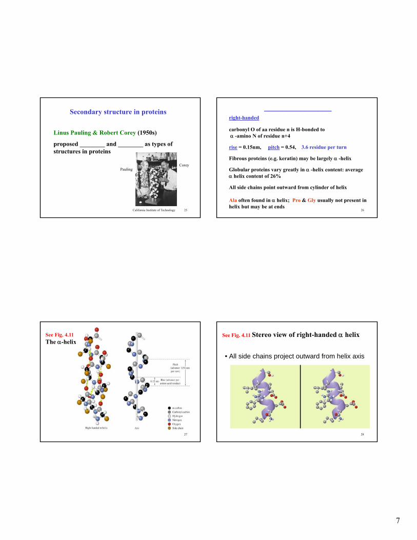
Secondary structure in proteins
Linus Pauling & Robert Corey (1950s)
proposed ________ and ________ as types of structures in proteins
CoreyPauling
California Institute of Technology 26
_______________right-handed
carbonyl O of aa residue n is H-bonded to -amino N of residue n+4
rise = 0.15nm, pitch = 0.54, 3.6 residue per turn
Fibrous proteins (e.g. keratin) may be largely -helix
Globular proteins vary greatly in -helix content: average helix content of 26%
All side chains point outward from cylinder of helix
Ala often found in helix; Pro & Gly usually not present in helix but may be at ends
27
See Fig. 4.11 The -helix
28
See Fig. 4.11 Stereo view of right-handed helix
• All side chains project outward from helix axis
8
29
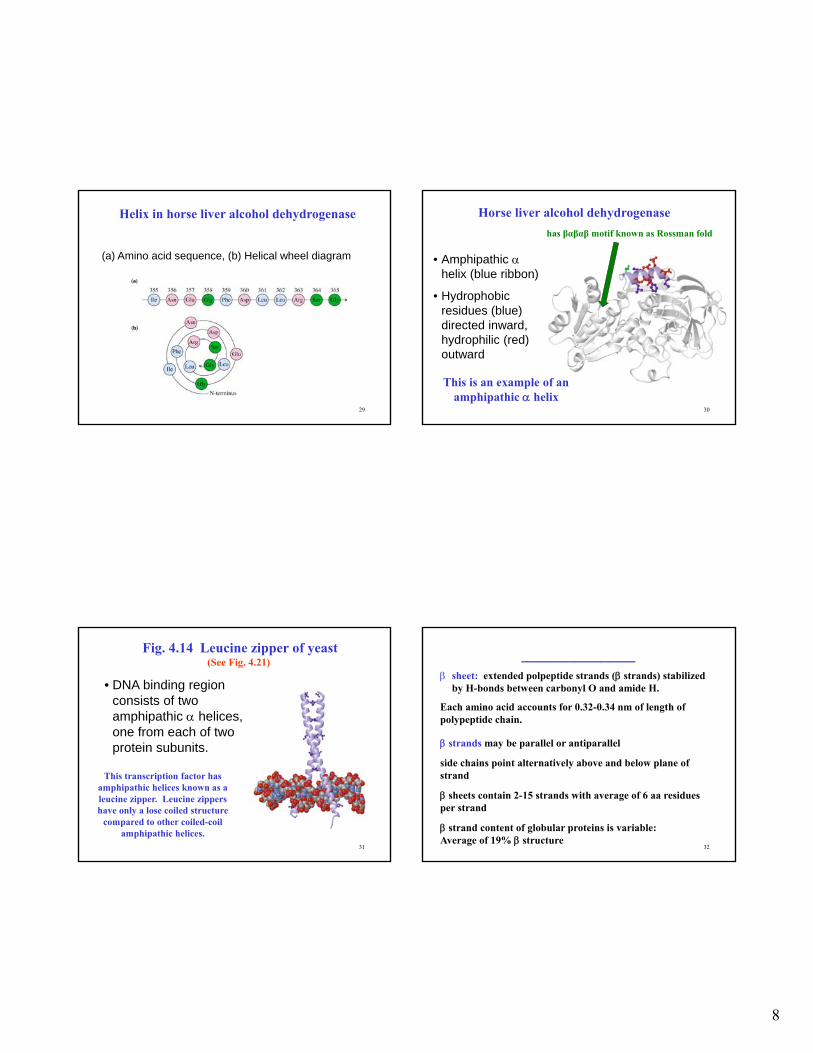
Helix in horse liver alcohol dehydrogenase
(a) Amino acid sequence, (b) Helical wheel diagram
30
Horse liver alcohol dehydrogenase
• Amphipathic helix (blue ribbon)
• Hydrophobic residues (blue) directed inward, hydrophilic (red) outward
This is an example of an amphipathic helix
has βαβαβ motif known as Rossman fold
31
Fig. 4.14 Leucine zipper of yeast(See Fig. 4.21)
• DNA binding region consists of two amphipathic helices, one from each of two protein subunits.
This transcription factor has amphipathic helices known as a leucine zipper. Leucine zippers have only a lose coiled structure
compared to other coiled-coil amphipathic helices.
32
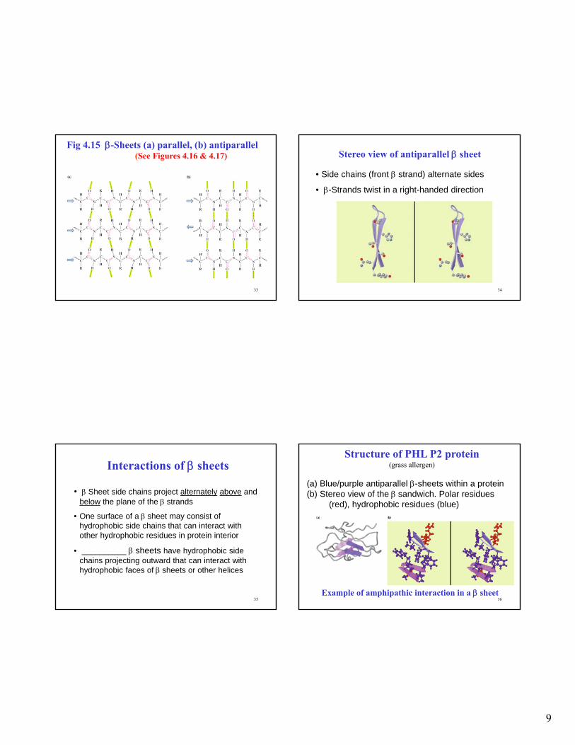
_____________ sheet: extended polpeptide strands ( strands) stabilized
by H-bonds between carbonyl O and amide H.
Each amino acid accounts for 0.32-0.34 nm of length of polypeptide chain.
strands may be parallel or antiparallel
side chains point alternatively above and below plane of strand
sheets contain 2-15 strands with average of 6 aa residues per strand
strand content of globular proteins is variable: Average of 19% structure
9
33
Fig 4.15 -Sheets (a) parallel, (b) antiparallel(See Figures 4.16 & 4.17)
34
Stereo view of antiparallel sheet
• Side chains (front strand) alternate sides
• -Strands twist in a right-handed direction
35
Interactions of sheets
• Sheet side chains project alternately above and below the plane of the strands
• One surface of a sheet may consist of hydrophobic side chains that can interact with other hydrophobic residues in protein interior
• __________ sheets have hydrophobic side chains projecting outward that can interact with hydrophobic faces of sheets or other helices
36
Structure of PHL P2 protein(grass allergen)
(a) Blue/purple antiparallel -sheets within a protein(b) Stereo view of the sandwich. Polar residues
(red), hydrophobic residues (blue)
Example of amphipathic interaction in a sheet

10
37
Loops and Turns (1)
• Loops and turns connect helices and strands and allow a peptide chain to fold backon itself to make a compact structure
• ________ - often contain hydrophilic residues and are found on protein surfaces
• ______ - loops containing 5 residues or less
• - connect different antiparallel strands (also called hairpin loop)
38
Loops and Turns (2)
__________: 4 aa, stabilized by H-bonds between -carbonyl O of residue n and -NH of residue (n+3); Pro is often 2nd residue
____________: (also called glycine turn, bend) like Type I Turn but 3rd residue is Gly
39
Reverse turns
(a) Type I, and (b) Type II
See Fig. 4.20
40
• Supersecondary Structures (Motifs), Domains, Folds, Quaternary Structure
• Anfinsen’s Experimentdenaturation, reduction & refolding
• Protein folding, Chaperones
• Collagen, Myoglobin & Hemoglobin
BCMB 3100 Partial Lecture Notes for Chapter 4 (Part 2)

11
41
Common motifs
Helix-loop-helix
Coiled-coilHelix bundle
Unit
Hairpin
Meander
Greek key Sandwich
Motifs -recurring protein structures
42
_____________
• independently folded, compact, distinct structural unit in proteins
• ~25 to ~300 amino acid residues
• connected to each other by loops, bound by weak interactions between side chains
• may have separate functions
• illustrate evolutionary conservation of protein structure
43
Pyruvate Kinase
• Main polypeptide chain folds into three distinct domains
116-219
1-115220-388
389-550
Last enzyme in glycolysis
DOMAIN: Independently
folded, compact, distinct structural
unit in proteins44
Cytochrome c
• Conservation of cyt c structure
(a) Tuna (+heme)(b) Tuna (c) Rice(d) Yeast (e) Bacteria
See Last slides of Chapter 3 Lecture for Phylogenic Tree
12
45
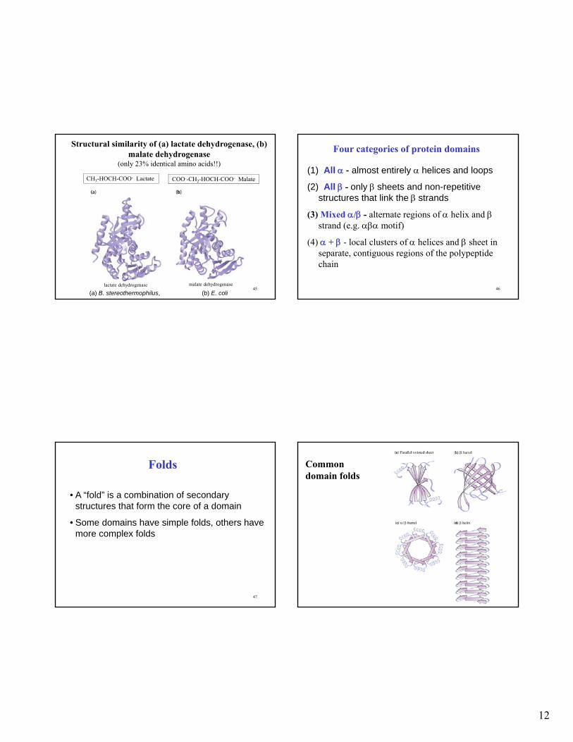
Structural similarity of (a) lactate dehydrogenase, (b) malate dehydrogenase
(only 23% identical amino acids!!)
(a) B. stereothermophilus, (b) E. coli
lactate dehydrogenase malate dehydrogenase
CH3-HOCH-COO- Lactate COO--CH2-HOCH-COO- Malate
46
Four categories of protein domains
(1) All - almost entirely helices and loops
(2) All - only sheets and non-repetitive structures that link the strands
(3) Mixed / - alternate regions of helix and strand (e.g. motif)
(4) + - local clusters of helices and sheet in separate, contiguous regions of the polypeptide chain
47
Folds
• A “fold” is a combination of secondary structures that form the core of a domain
• Some domains have simple folds, others have more complex folds
48
Common domain folds

13
49
Examples of tertiary structure
all
helix bundles
50
Tertiary Protein Structures (cont)
Jellyfish green fluorescent protein
all
barrel & central
helix
all
barrel
51
Domain Structure and Function
• A _______ ________ may have a particular function
• __________ between 2 domains provide crevices, grooves, and pockets on the surface of a protein for binding or catalytic sites
• In multifunctional enzymes, each catalytic activity can be on one of several domains
52
Quaternary Structure
• organization of subunits in a protein with multiplesubunits (an “oligomer”)
• Subunits (may be identical () or different () ) have a defined stoichiometry and arrangement
• Subunits are held together by many weak, noncovalent interactions (hydrophobic, electrostatic)

14
53
Quaternary structure of multidomain proteins
Subunits = 2(each with / folds)
Subunits = 2(each subunit is all structure)
54
(continued)
Subunits = 4 Subunits = 3
55
(cont.)
Rhodopseudomonas photosynthetic reaction center
(f)
Subunits = 22Subunits = 2 56
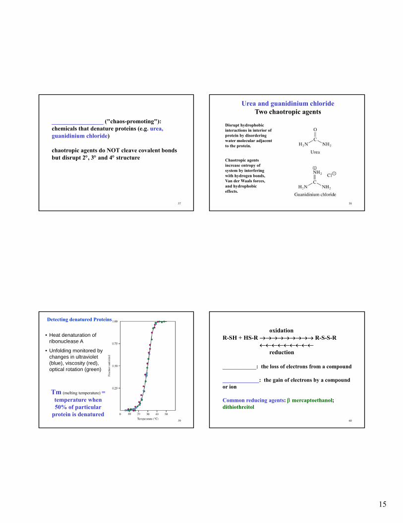
Protein Denaturation and Renaturation
______________:•partial or complete unfolding of native
conformation
•causes loss of biological activity
•caused by heat, extreme pH, detergents, chaotropic agents; due to disruption of non-covalent interactions
• some proteins can be refolded or renatured
15
57
_________________ ("chaos-promoting"): chemicals that denature proteins (e.g. urea, guanidinium chloride)
chaotropic agents do NOT cleave covalent bonds but disrupt 2, 3 and 4 structure
58
Urea and guanidinium chlorideTwo chaotropic agents
Disrupt hydrophobic interactions in interior of protein by disordering water molecular adjacent to the protein.
Chaotropic agents increase entropy of system by interfering with hydrogen bonds, Van der Waals forces, and hydrophobic effects.
59
• Heat denaturation of ribonuclease A
• Unfolding monitored by changes in ultraviolet (blue), viscosity (red), optical rotation (green)
Tm (melting temperature) = temperature when 50% of particular
protein is denatured
Detecting denatured Proteins
60
oxidationR-SH + HS-R R-S-S-R
reduction
____________: the loss of electrons from a compound
_____________: the gain of electrons by a compound or ion
Common reducing agents: mercaptoethanol; dithiothreitol

16
61
Disulfide bridges in bovine ribonuclease A
(a) Location of disulfide bridges
(b) Stereo view of Cys-26 and Cys-84
Ribonuclease:
124 aa,
mainly sheet,
4 disulfide bonds
62
_______________ showed that the information necessary to specify 3D structure of ribonuclease came from the amino acid sequence
The amino acid sequence specifies 3D structure!!
The native form of a protein (e.g. ribonuclease) appears to be the thermodynamically most stable structure
63
Denaturation and renaturation of ribonuclease A
see Fig. 4.33
64
Protein Folding and stabilization (1)
Cooperativity of folding: formation of one part of structure (e.g. initial aa interactions) leads to formation of remaining structure.
__________________ is MAJOR driving force in protein folding
17
65
Protein Folding and stabilization (2)
Protein folding:*nonpolar side chains inside*most polar chains outside*those polar chains inside H-bond with each other 2 structure
H-bonds and van der Waals forces stabilize globular protein folding
Covalent cross links ( R-S-S-R) and ionic interactions may stabilize some globular proteins
66
CURRENT THEORY OF PROTEIN FOLDING
1. not random
2. cooperative and sequential
3. dependent on 1 structure
4. for some proteins 1 structure alone determines folding
5. some proteins require help to fold (e.g. enzymes or chaperones)
6. extremely rapid, native conformation is generally reached < 1 second
7. NOTE: most protein have single native 3D shape; but some exceptions
67
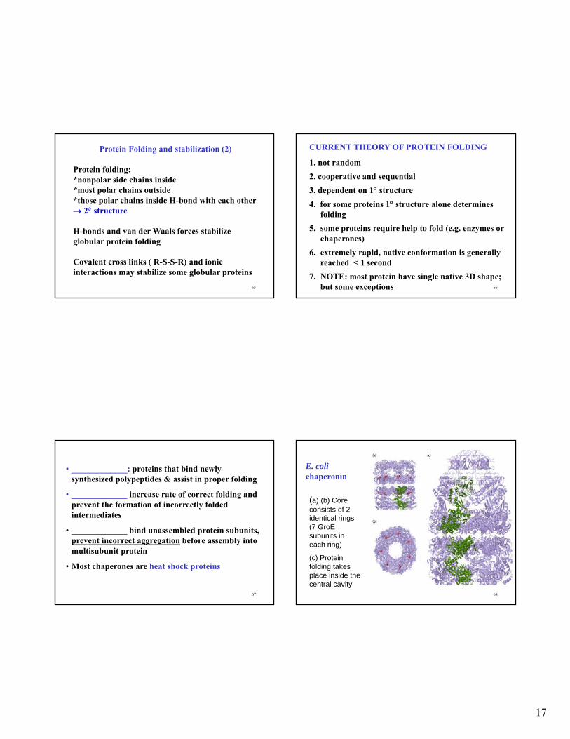
• _____________: proteins that bind newly synthesized polypeptides & assist in proper folding
• _____________ increase rate of correct folding and prevent the formation of incorrectly folded intermediates
• _____________ bind unassembled protein subunits, prevent incorrect aggregation before assembly into multisubunit protein
• Most chaperones are heat shock proteins
68
E. colichaperonin
(a) (b) Core consists of 2 identical rings (7 GroE subunits in each ring)
(c) Protein folding takes place inside the central cavity
18
69
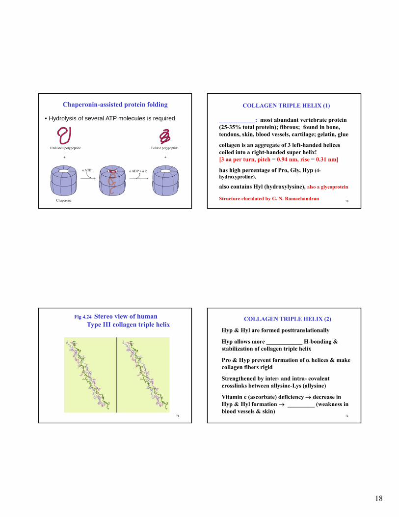
Chaperonin-assisted protein folding
• Hydrolysis of several ATP molecules is required
70
COLLAGEN TRIPLE HELIX (1)
____________: most abundant vertebrate protein (25-35% total protein); fibrous; found in bone, tendons, skin, blood vessels, cartilage; gelatin, glue
collagen is an aggregate of 3 left-handed helices coiled into a right-handed super helix![3 aa per turn, pitch = 0.94 nm, rise = 0.31 nm]
has high percentage of Pro, Gly, Hyp (4-hydroxyproline),
also contains Hyl (hydroxylysine), also a glycoprotein
Structure elucidated by G. N. Ramachandran
71
Fig 4.24 Stereo view of human Type III collagen triple helix
72
COLLAGEN TRIPLE HELIX (2)
Hyp & Hyl are formed posttranslationally
Hyp allows more ____________ H-bonding & stabilization of collagen triple helix
Pro & Hyp prevent formation of helices & make collagen fibers rigid
Strengthened by inter- and intra- covalent crosslinks between allysine-Lys (allysine)
Vitamin c (ascorbate) deficiency decrease in Hyp & Hyl formation _________ (weakness in blood vessels & skin)
19
73
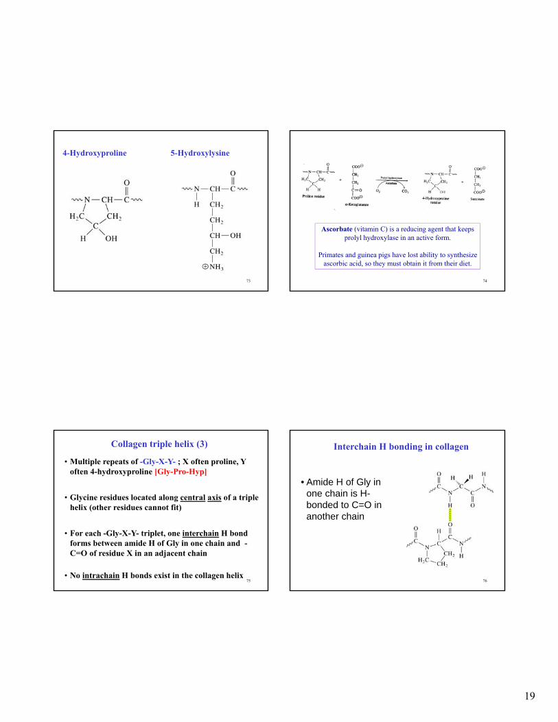
4-Hydroxyproline 5-Hydroxylysine
74
Ascorbate (vitamin C) is a reducing agent that keeps prolyl hydroxylase in an active form.
Primates and guinea pigs have lost ability to synthesize ascorbic acid, so they must obtain it from their diet.
75
Collagen triple helix (3)
• Multiple repeats of -Gly-X-Y- ; X often proline, Y often 4-hydroxyproline [Gly-Pro-Hyp]
• Glycine residues located along central axis of a triple helix (other residues cannot fit)
• For each -Gly-X-Y- triplet, one interchain H bond forms between amide H of Gly in one chain and -C=O of residue X in an adjacent chain
• No intrachain H bonds exist in the collagen helix76
Interchain H bonding in collagen
• Amide H of Gly in one chain is H-bonded to C=O in another chain
20
77
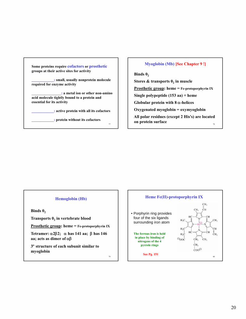
Some proteins require cofactors or prostheticgroups at their active sites for activity
____________: small, usually nonprotein molecule required for enzyme activity
________________: a metal ion or other non-amino acid molecule tightly bound to a protein and essential for its activity
____________: active protein with all its cofactors
____________: protein without its cofactors78
Myoglobin (Mb) [See Chapter 9 !]
Binds 02
Stores & transports 02 in muscle
Prosthetic group: heme = Fe-protoporphyrin IX
Single polypeptide (153 aa) + heme
Globular protein with 8 -helices
Oxygenated myoglobin = oxymyoglobin
All polar residues (except 2 His's) are located on protein surface
79
Hemoglobin (Hb)
Binds 02
Transports 02 in vertebrate blood
Prosthetic group: heme = Fe-protoporphyrin IX
Tetramer: 22; has 141 aa; has 146 aa; acts as dimer of
3 structure of each subunit similar to myoglobin
80
Heme Fe(II)-protoporphyrin IX
• Porphyrin ring provides four of the six ligands surrounding iron atom
The ferrous iron is held in place by binding of
nitrogens of the 4 pyrrole rings
See Pg. 151
21
81
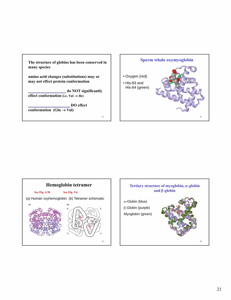
The structure of globins has been conserved in many species
amino acid changes (substitutions) may or may not effect protein conformation
___________________ do NOT significantly effect conformation (i.e. Val Ile)
_____________________ DO effect conformation (Glu Val)
82
Sperm whale oxymyoglobin
• Oxygen (red)
• His-93 and His-64 (green)
83
Hemoglobin tetramer
(a) Human oxyhemoglobin (b) Tetramer schematic
See Fig. 4.30 See Fig. 9.6
84
Tertiary structure of myoglobin, -globin and -globin
-Globin (blue)
-Globin (purple)
Myoglobin (green)
22
85
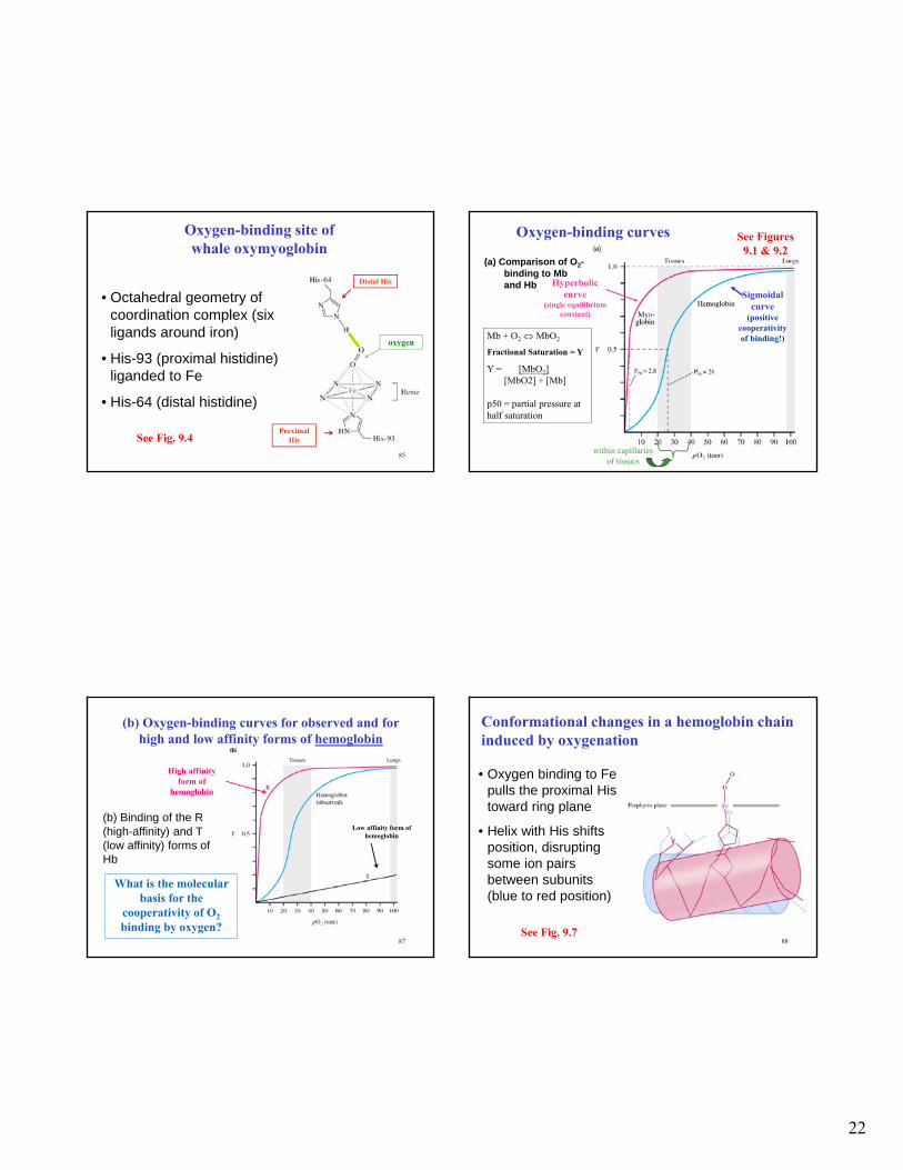
Oxygen-binding site of whale oxymyoglobin
• Octahedral geometry of coordination complex (six ligands around iron)
• His-93 (proximal histidine) liganded to Fe
• His-64 (distal histidine)
See Fig. 9.4
Distal His
ProximalHis
oxygen
86
Oxygen-binding curves
(a) Comparison of O2-binding to Mb and Hb
Sigmoidal curve
(positive cooperativity of binding!)
Hyperbolic curve
(single equilibrium constant)
within capillaries of tissues
Mb + O2 MbO2
Fractional Saturation = Y
Y = [MbO2][MbO2] + [Mb]
p50 = partial pressure at half saturation
See Figures 9.1 & 9.2
87
(b) Binding of the R (high-affinity) and T (low affinity) forms of Hb
(b) Oxygen-binding curves for observed and for high and low affinity forms of hemoglobin
High affinity form of
hemoglobin
Low affinity form of hemoglobin
What is the molecular basis for the
cooperativity of O2
binding by oxygen?88
Conformational changes in a hemoglobin chain induced by oxygenation
• Oxygen binding to Fe pulls the proximal His toward ring plane
• Helix with His shifts position, disrupting some ion pairs between subunits (blue to red position)
See Fig. 9.7
23
89
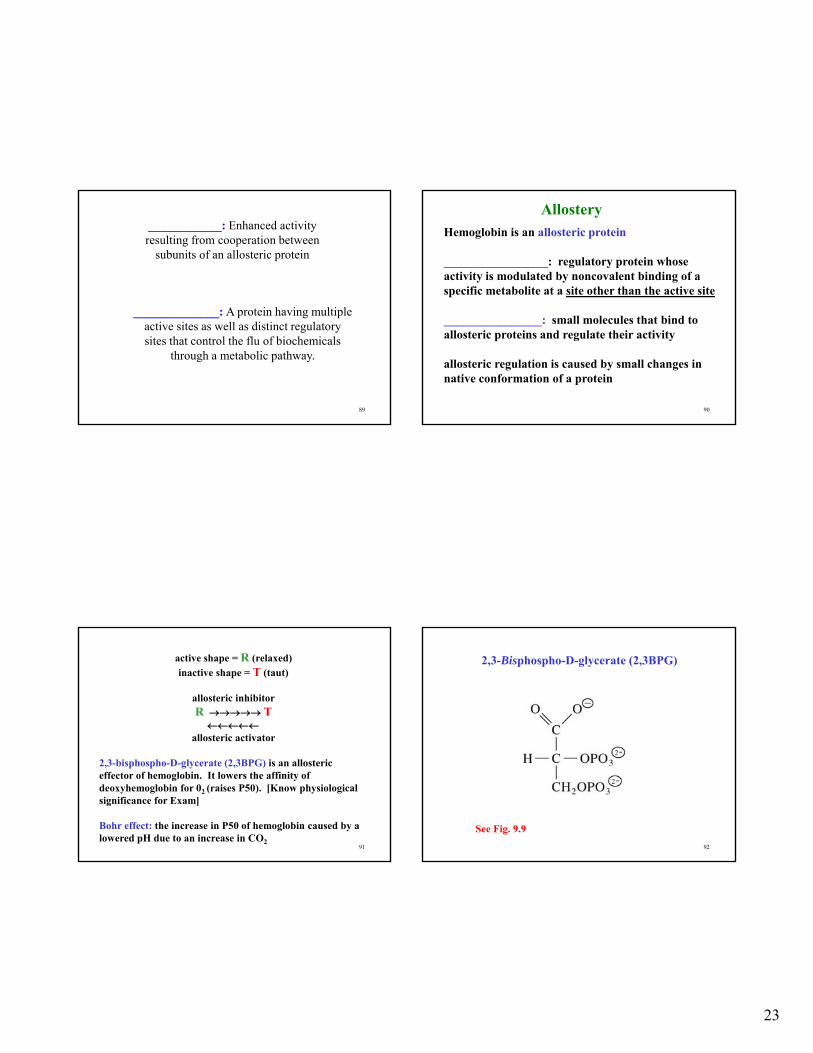
____________: Enhanced activity resulting from cooperation between
subunits of an allosteric protein
______________: A protein having multiple active sites as well as distinct regulatory sites that control the flu of biochemicals
through a metabolic pathway.
90
Hemoglobin is an allosteric protein
_________________: regulatory protein whose activity is modulated by noncovalent binding of a specific metabolite at a site other than the active site
________________: small molecules that bind to allosteric proteins and regulate their activity
allosteric regulation is caused by small changes in native conformation of a protein
Allostery
91
active shape = R (relaxed)
inactive shape = T (taut)
allosteric inhibitorR T
allosteric activator
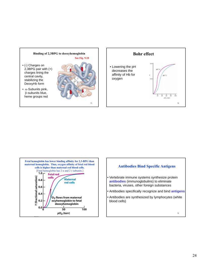
2,3-bisphospho-D-glycerate (2,3BPG) is an allosteric effector of hemoglobin. It lowers the affinity of deoxyhemoglobin for 02 (raises P50). [Know physiological significance for Exam]
Bohr effect: the increase in P50 of hemoglobin caused by a lowered pH due to an increase in CO2
92
2,3-Bisphospho-D-glycerate (2,3BPG)
See Fig. 9.9
24
93
Binding of 2,3BPG to deoxyhemoglobin
• (-) Charges on 2,3BPG pair with (+) charges lining the central cavity, stabilizing the DeoxyHb form
• -Subunits pink,-subunits blue, heme groups red
See Fig. 9.10
94
Bohr effect
• Lowering the pH decreases the affinity of Hb for oxygen
Fetal hemoglobin has lower binding affinity for 2,3-BPG than maternal hemoglobin. Thus, oxygen affinity of fetal red blood
cells is higher than maternal red blood cells.[Fetal hemoglobin has 2 α and 2 γ subunits.]
96
Antibodies Bind Specific Antigens
• Vertebrate immune systems synthesize protein antibodies (immunoglobulins) to eliminate bacteria, viruses, other foreign substances
• Antibodies specifically recognize and bind antigens
• Antibodies are synthesized by lymphocytes (white blood cells)
25
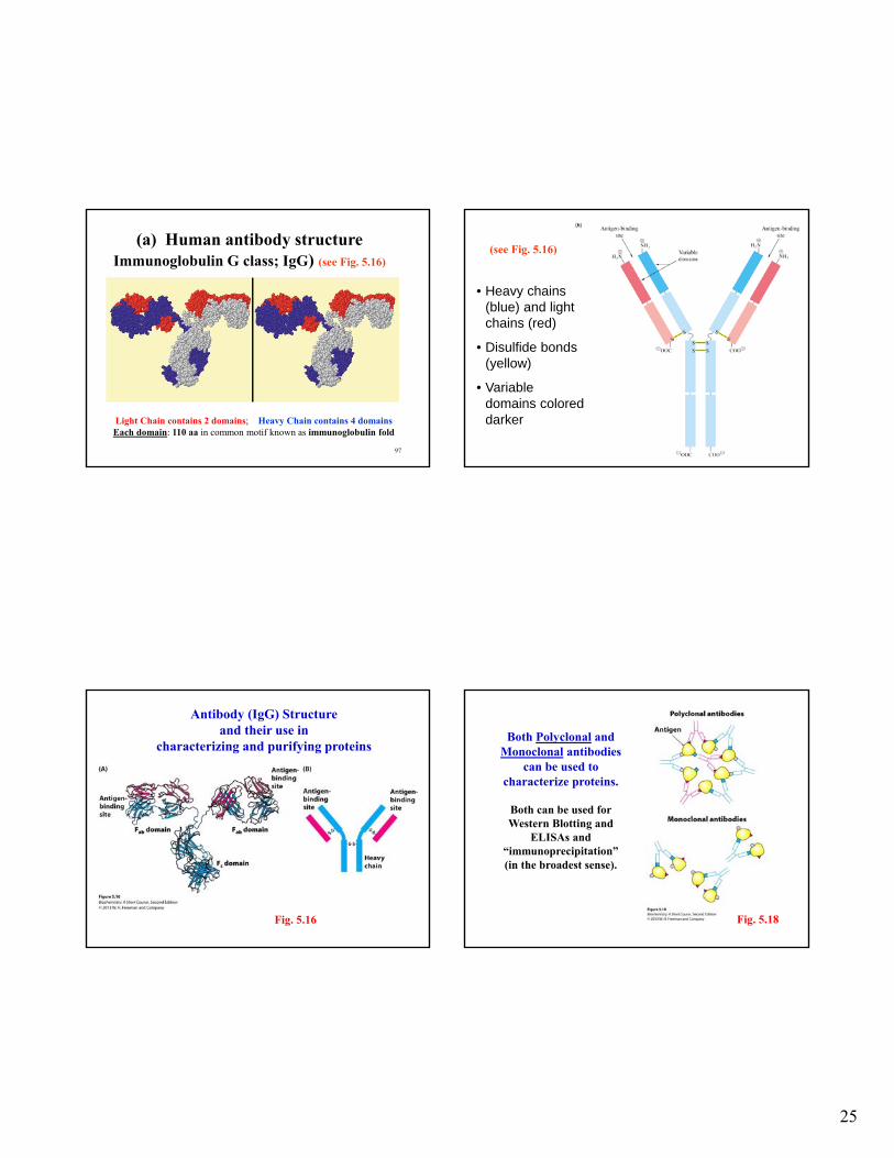
97
Light Chain contains 2 domains; Heavy Chain contains 4 domainsEach domain: 110 aa in common motif known as immunoglobulin fold
(a) Human antibody structureImmunoglobulin G class; IgG) (see Fig. 5.16)
98
• Heavy chains (blue) and light chains (red)
• Disulfide bonds (yellow)
• Variable domains colored darker
(see Fig. 5.16)
Fig. 5.16
Antibody (IgG) Structure and their use in
characterizing and purifying proteins
Fig. 5.18
Both Polyclonal and Monoclonal antibodies
can be used to characterize proteins.
Both can be used for Western Blotting and
ELISAs and “immunoprecipitation”(in the broadest sense).
26
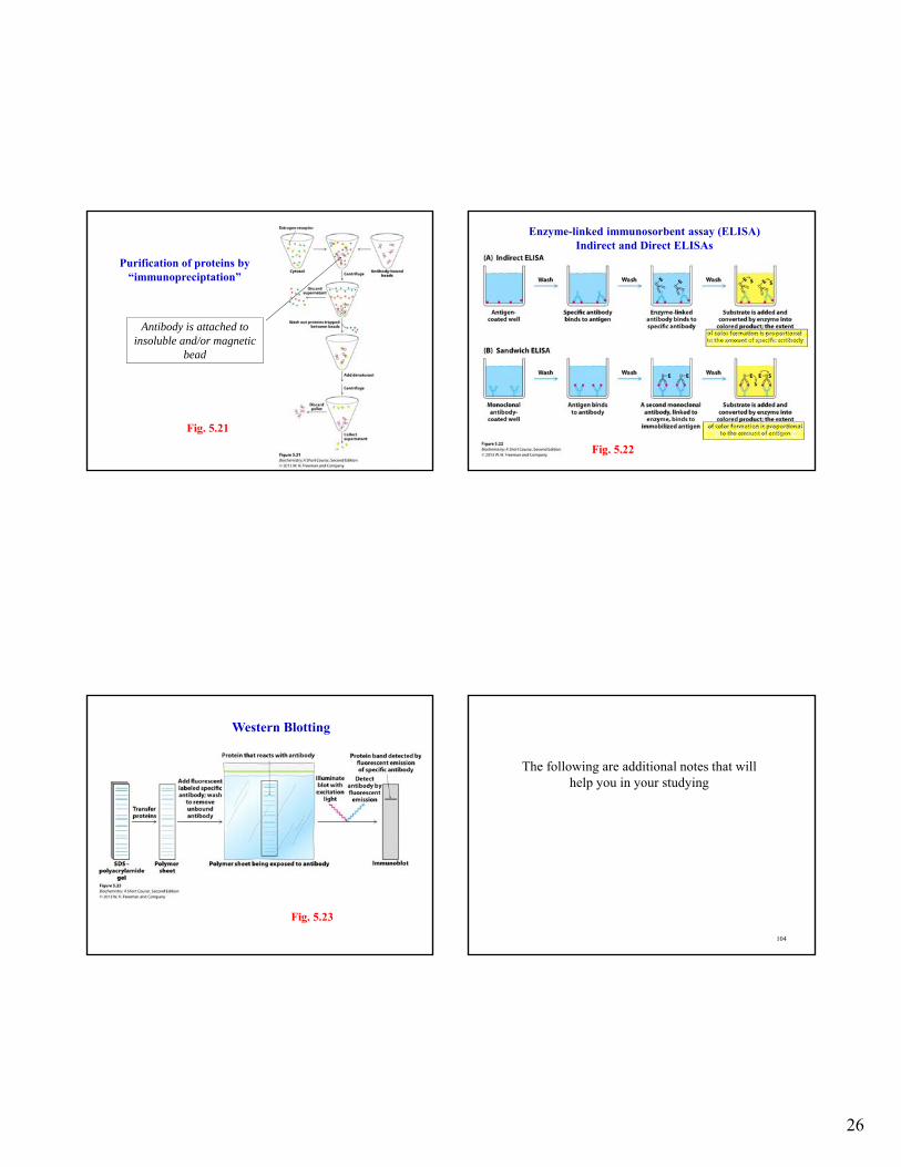
Fig. 5.21
Purification of proteins by “immunopreciptation”
Antibody is attached to insoluble and/or magnetic
bead
Fig. 5.22
Enzyme-linked immunosorbent assay (ELISA)Indirect and Direct ELISAs
Fig. 5.23
Western Blotting
104
The following are additional notes that will help you in your studying
27
105
Review of Globular Protein 3D Structure
Most globular proteins have compact globular shape due to many reversible turns in direction combined with the helix and/or structure. Usually, hydrophobic aa residues are in the interior and hydrophilic aa residues on the exterior of the protein.
The loops & turns contain nonrepetitive regions of 2°structure.
loops: range from ~ 2-16 residues, many hydrophilic residuesfound at surface of protein (can H-bond with water)
turn: loops having only a few residues (<6)
Average structure of Globular Protein helix: 26%; structure: 19%; turns: 15%;
simple loops 21%; complex loops: 10% 106
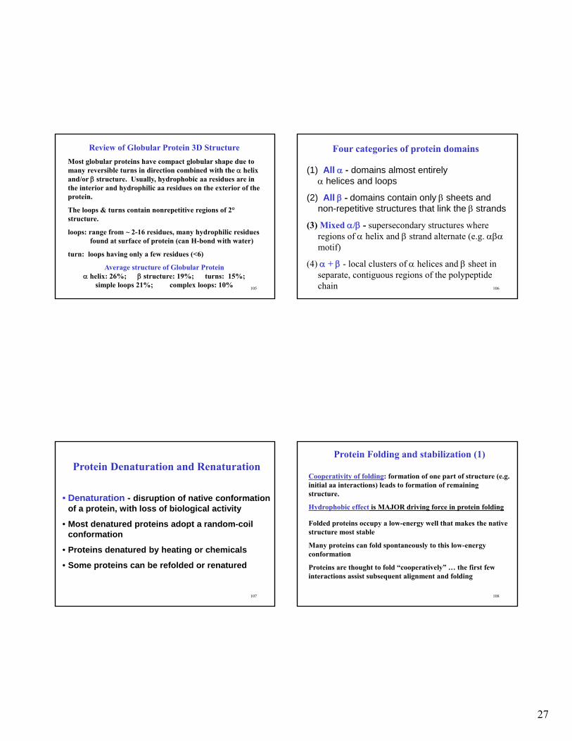
Four categories of protein domains
(1) All - domains almost entirely helices and loops
(2) All - domains contain only sheets and non-repetitive structures that link the strands
(3) Mixed / - supersecondary structures where regions of helix and strand alternate (e.g. motif)
(4) + - local clusters of helices and sheet in separate, contiguous regions of the polypeptide chain
107
Protein Denaturation and Renaturation
• Denaturation - disruption of native conformation of a protein, with loss of biological activity
• Most denatured proteins adopt a random-coil conformation
• Proteins denatured by heating or chemicals
• Some proteins can be refolded or renatured
108
Protein Folding and stabilization (1)
Cooperativity of folding: formation of one part of structure (e.g. initial aa interactions) leads to formation of remaining structure.
Hydrophobic effect is MAJOR driving force in protein folding
Folded proteins occupy a low-energy well that makes the native structure most stable
Many proteins can fold spontaneously to this low-energy conformation
Proteins are thought to fold “cooperatively” … the first few interactions assist subsequent alignment and folding
28
109
Protein folding (more detail)
• extremely rapid, native conformation is generally reached < 1 second
• During folding the polypeptide collapses in upon itself due to the hydrophobic effect
• An intermediate “molten globule” forms with elements of secondary structure
• The backbone is rearranged to achieve a stable native conformation
110
• Chaperones: proteins that bind newly synthesized polypeptides & assist in proper folding
• Chaperones increase rate of correct folding and prevent the formation of incorrectly folded intermediates
• Chaperones bind to unassembled protein subunits to prevent incorrect aggregation before they are assembled into a multisubunit protein
• Most chaperones are heat shock proteins(synthesized as temperature increases)
111
Two conformations of hemoglobin: T and R
• Active (R state) and inactive (T state) forms are in rapid equilibrium in allosteric proteins
• Binding of substrates and allosteric activatorsstabilize the R state and shift the equilibrium in the R direction
• Allosteric inhibitors stabilize the T state and shift the equilibrium in the T direction