Embed Size (px)
Citation preview

Axillary lymph nods..Group b2

Adel Majed Almunlf433025168
1

Structures of the lymphatic system
The lymphatic system is composed of several specialized structures and substances. The substance the entire lymphatic system revolves around is the fluid circulating through the lymphatic system known as lymph. Lymph, a fluid that is typically colorless, is similar to blood and contains plasma. Lymph may contain lipids, macrophages, hormones, bacteria, viruses, cellular wasteproducts and other cells.
The lymph travels through an extensive network of vessels known as the lymphatic vessels. These vessels are generously spread throughout the entire body, present in nearly every tissue of the body. These vessels, similar to the circulatory vessels, are composed of smaller lymphatic capillaries that converge to form collecting vessels. These vessels then merge even further to form what is known as a lymphatic trunks, which give rise to the lymphatic ducts. There are two major lymphatic ducts in the body; the right lymphatic duct, which drains the right arm and right side of the trunk, head and neck, and the thoracic duct, which drains the remaining portions of the body.

Lastly, the lymph nodes are a small organ the size of a pea, located numerously throughout the body. These nodes serve as the site of filtration for the myph fluid and activate specialized cells found within the lymph. The lymph nodes can become swollen or enlarged when infection is present.

Abdulrahman Alomair 434000049
2

Lymphatic system functions
The primary function of the lymphatic system is to transport lymph, a clear, colorless fluid containing white blood cells that helps the body to get rid of toxins, waste and other unwanted materials.

lymphatic system has multiple interrelated
functions lymphatic system has
multiple interrelated functions It is responsible for the removal of interstitial fluid from tissues.
It absorbs and transports fatty acids and fats as chyle from the digestive system .

It transports white blood cells to and from the lymph nodes into the bones .
The lymph transports antigen-presenting cells (APCs), such as dendritic cells, to the lymph nodes where an immune response is stimulated.

Adel Ali AlKenani434000041
3

Anatomy of Axillary Lymph Node

Anterior Group
Lie along the lower border of the pectoralis minor behind the pectoralis major muscle.
Receive lymph from the lateral part of the breast and the superficial vessels from the thoracoabdominal wall above the level of the umbilicus.


Posterior Group
Lie in front of the subscapularis muscle on the posterior wall of the axilla.
They receive superficial lymph vessels from the back, down as far as the level of the iliac crests.


Lateral Group
Lie along the medial side of
the axillary vein.
They receive most of the
lymph vessels of the upper limb.


Central Group
Lie in the central of the axilla and it it is embedded in fat.
They receive lymph from :Anterior group Posterior group Lateral group


Infraclavicular Group
Lie in the interval between the deltoid and pectoralise major muscle along the course of the cephalic vein.
They receive lymph from the
superficial vessels from the lateral side of the hand, the forearm and the arm.
The lymph vessels accompany the cephalic vein.
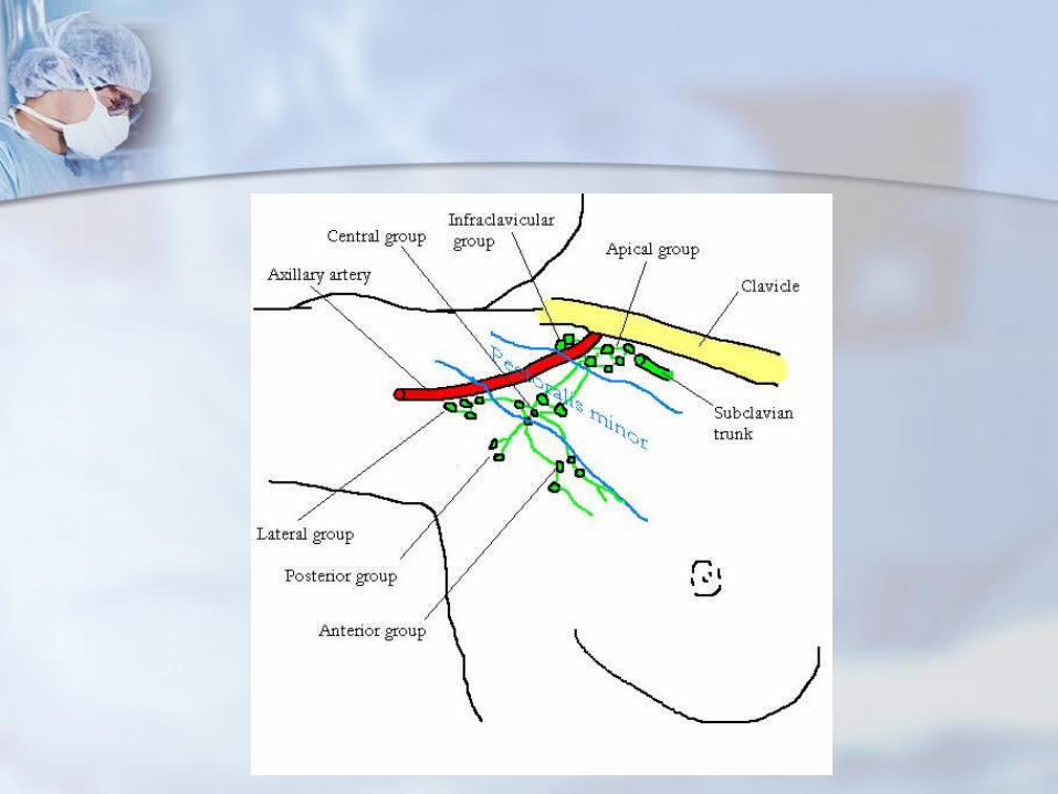
Apical group
Lie at the apex of the axilla at the out border of the first rib.
They receive lymph from all the other axillary nodes.


Apical group
It drain into right lymphatic trunk on the right side.
It drain into the thoracic duct on the left side.

Saud fahad abdulmajeed434021079
4

Histology of lymph node
Histology : is the study of the microscopic anatomy of cells and tissues of plants and animals.

The lymph node is surrounded by a fibrous capsule. The substance of the lymph node is divided into the outer cortex and the inner medulla surrounded by the cortex all around except at the hilum, where the medulla comes in direct contact with the surface.

Lymphocytes
Variable size (6-18um) • smallest are quiescent• Nucleus• Intensely stained• Slightly indented• Spherical in shape• Cytoplasm appears as a pale blue rim around nucleus

There are basically two different types of lymphocytes, T lymphocytes (T cells) that are involved in cell-mediated immunity and B lymphocytes (B cells) that are involved in humoral immunity, both types originate from stem cells in bone marrow• T Lymphocytes (thymus-dependent)• Have a long life span• Involved in cell mediated immunity• B Lymphocytes (B cells)• Variable life span• Involved in the production of circulating antibodies

cortex
The outer cortex consists mainly of the B cells arranged as follicles, which may develop a germinal center when challenged with an antigen, and the deeper cortex mainly consisting of the T cells. There is a zone known as the subcortical zone

medulla
The medulla contains large blood vessels, sinuses and medullary cords that contain plasma cells secreting antibody.

Sayaf H. Alshareef434000024
5

Functions of lymph nods
1. Defense functions Filtration which is divided into two
kinds.. Mechanical filtration: physically stopping particles from progressing.
Biological filtration: biological activity of cells to destroy and remove particles Phagocytosis
If overwhelmed, lymph nods can become infected or damaged. (adenitis)

2. Hematopoiesis:Lymphoid tissue of the lymph nods is the site for final stages of maturation of some lymphocytes and monocytes ( white blood cells which produce antibodies)


Lymph. Interstitial fluid.(surrounds cells before entering lymph
vessels)
Lymphocytes. Lymph return to blood stream with
some lymphocytes to aid in the identification and destruction of pathogens.
B-cell and antibodies.

Axillary lymph nodes
About 75% of lymph from the breasts drains into the axillary lymph nodes, making them important in the diagnosis of breast cancer.
A doctor will usually refer a patient to a surgeon to have an axillary lymph node dissection to see if the cancer cells have been trapped in the nodes.


Suliman Aljabbary434023433
6

Lymphopoiesis
Lymphopoiesis is the process in which lymphocytes are created
The end product of this process is NK cells, T cells, B cells and plasma

Lymphopoiesis for NK cells
NK cells or natural killer cells are developed in the bone marrow
NK also mature in the bone marrow unlike the other lymphocytes
NK lack antigen specific receptors but have granules which gives them their ability to kill cells
They look for they look cancerous cells and infected cells to kill

Lymphopoiesis for T cells
T cells are formed in bone marrowThen they move to the thymus to mature in a clean enviorment.
During maturation almost all the T cells are killed because of defects only about 4% survive.
The dead T cells are then quickly recycledWhen they are activated the undergo more development.
During this development they pass through three phases .

Lymphopoiesis for B cells
B cells are formed and mature in bone marrow and spleen
B cells leave the bone marrow and move to peripheral lymph node
then antigens are introduced into the B cells
They then divide into plasma and B cells
The final B cells produce antibodies that protect the body

Abdulrahman Ibrahim Khardli
433016503
7


What Is Breast Cancer ?
Breast cancer is a kind of cancer that develops from breast cells.
A malignant tumor can spread to other parts of the body.
A breast cancer that started off in the lobules is known as lobular carcinoma, while one that developed from the ducts is called ductal carcinoma.


What are the causes of breast cancer ?
Getting older : Over 80% of all female breast cancers
occur among .women aged 50+ years Genetics : women who have a close relative who
has/had breast or ovarian cancer are more likely to develop breast cancer.
A history of breast cancer :women who have had breast cancer, are
more likely to develop the disease again.


Dense breast tissue : women with more dense breast tissue have a
greater chance of developing breast cancer.
Obesity :post-menopausal obese and overweight women
may have a higher risk of developing breast cancer.
Having had certain types of breast lumps :women who have had some types of benign (non-
cancerous) breast lumps are more likely to develop cancer later on.

Abdulelah Alahmari434000031
8

Symptoms and diagnosis of breast cancer

symptoms
a lump or area of thickened tissue in either breast.
a change in the size or shape of one or both breasts.
discharge from either nipples (which may be streaked with blood).
a lump or swelling in either armpits. dimpling on the skin of breasts. a rash on or around the nipple. a change in the appearance of the nipple, such
as becoming sunken into the breast. pain in either breasts or armpits not related to
period.

Diagnosis
Fine Needle Aspiration. Core Needle Biopsy. Breast Tumor Pathology. Sentinel Lymph Node Biopsy.

Salah Alharbi434029601
9

WHAT TYPES OF CANCER ARE DIAGNOSED AS STAGE 0 AND 1 BREAST CANCER?
The stage of cancer indicates the size of the tumor of abnormal cells and whether or not those cells are contained to the place of origin. The most common type of breast cancer is ductal carcinoma in situ (DCIS), indicating the cancer cell growth starts in the milk ducts. WHAT DOES THE TERM, “IN SITU” MEAN?” Three possible types of “in situ carcinoma” of the breast tissue are:• DCIS - Ductal carcinoma in situ• LCIS - Lobular carcinoma in situ• Paget disease of the nipple

WHAT DOES IT MEAN TO HAVE STAGE 1 BREAST CANCER?
Stage 1 can be divided into Stage 1A and Stage 1B• STAGE 1A BREAST
CANCER • STAGE 1B BREAST
CANCER

STAGE 0 OR STAGE 1 BREAST CANCER TREATMENT?

WHAT DOES IT MEAN TO HAVE STAGE 2 BREAST CANCER?
This stage is divided into groups: • Stage 2A • Stage 2B

STAGE 2A BREAST CANCER MEANS ONE OF THE FOLLOWING DESCRIPTIONS APPLIES.
STAGE 2B BREAST CANCER MEANS ONE OF THE FOLLOWING DESCRIPTIONS APPLIES.

WHAT DOES IT MEAN TO HAVE STAGE 3 BREAST CANCER?
This stage is divided into three groups:
Stage 3A Stage 3B Stage 3C

STAGE 3A BREAST CANCER MEANS ONE OF THE FOLLOWING DESCRIPTIONS APPLIES.
STAGE 3B BREAST CANCER MEANS THE FOLLOWING DESCRIPTIONS APPLIES.

STAGE 3C BREAST CANCER MEANS ONE OF THE FOLLOWING DESCRIPTIONS APPLIES.
STAGE 3 BREAST TREATMENT
Stage 3C breast cancer is divided into operable and inoperable stage 3C breast cancer. However, the term “inoperable” is not the same as “untreatable.”

WHAT DOES IT MEAN TO HAVE STAGE 4 BREAST CANCER?
Stage 4 breast cancer means that the cancer has spread to other areas of the body, such as the brain, bones, lung and liver.

The End
Thank you