Embed Size (px)
DESCRIPTION
Axial muscles: Head and Neck Trunk: Chest wall Abdominal wall Pelvic wall Diaphragm Back. 9/ Sternocleidomastoid muscle. A/ external oblique c/ internal oblique d/ transversus abdominis. E. B. D. C. A. G. F. Appendicular muscles Muscles of upper limb Muscles of lower limb. - PowerPoint PPT Presentation
Citation preview
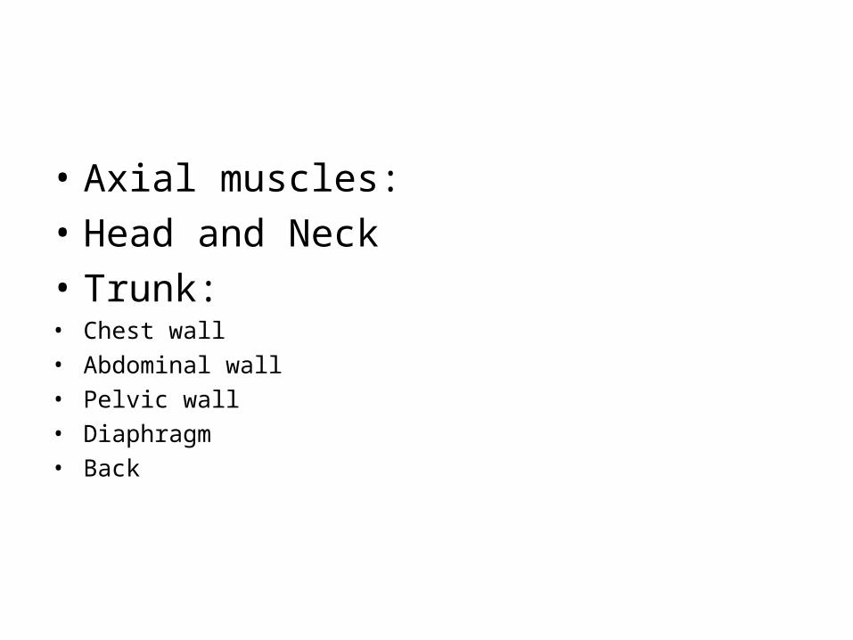
• Axial muscles:• Head and Neck• Trunk:• Chest wall• Abdominal wall• Pelvic wall• Diaphragm• Back
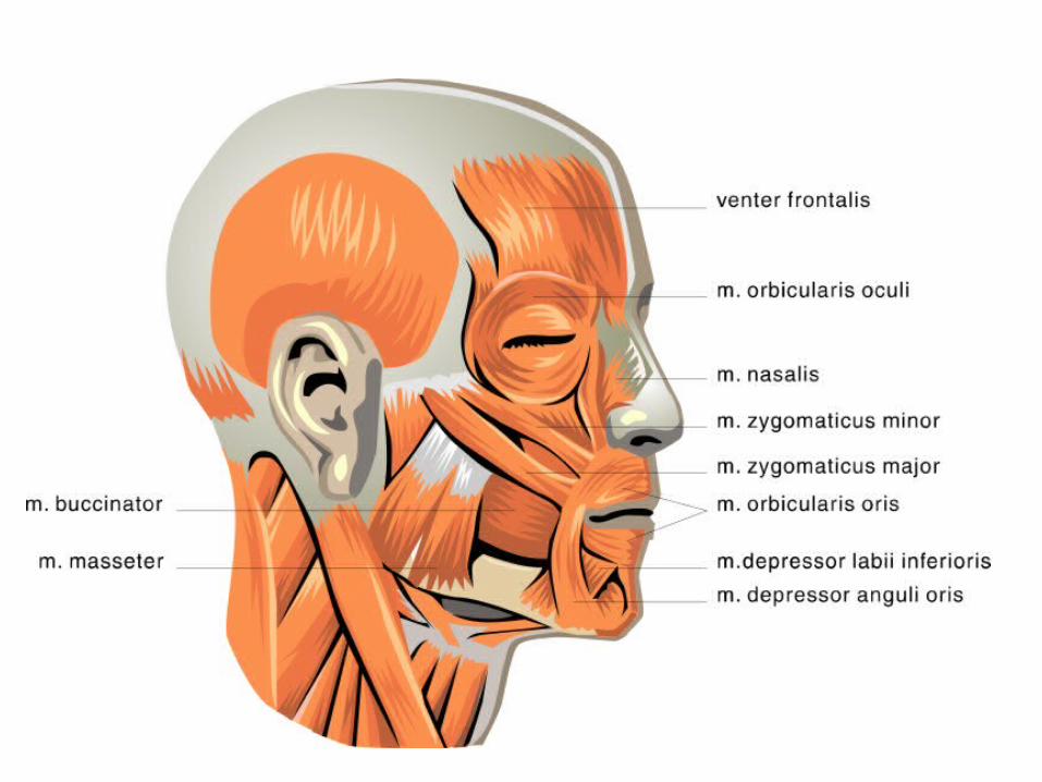
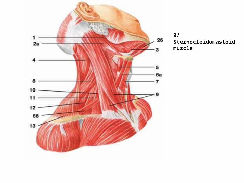
9/ Sternocleidomastoid muscle
G F
E
D
C
A
B
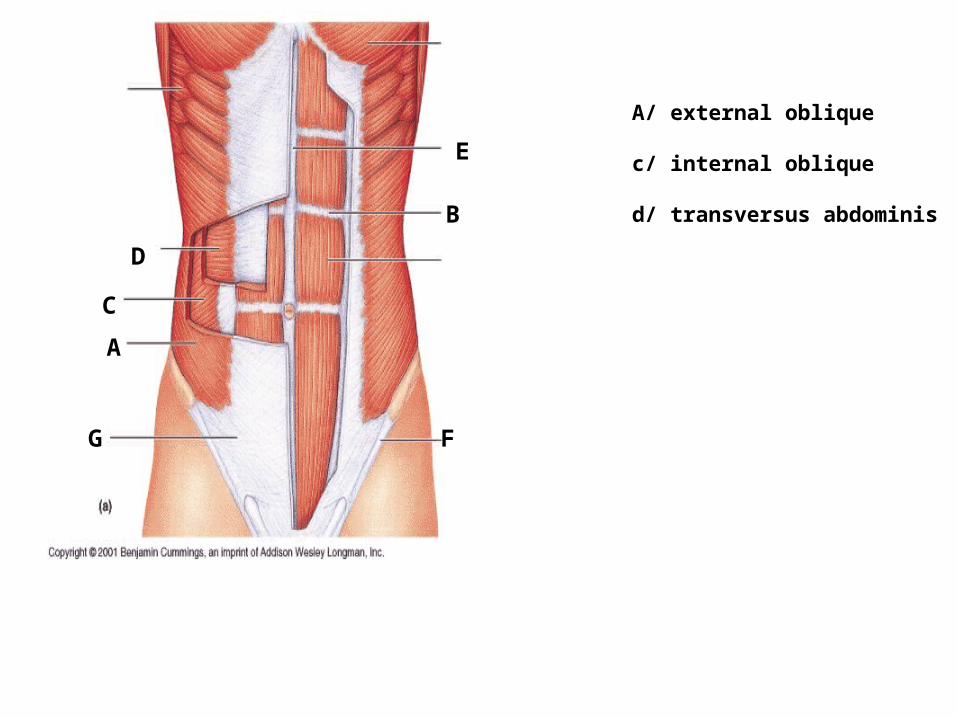
A/ external oblique
c/ internal oblique
d/ transversus abdominis
• Appendicular muscles• Muscles of upper limb• Muscles of lower limb
B
A
C
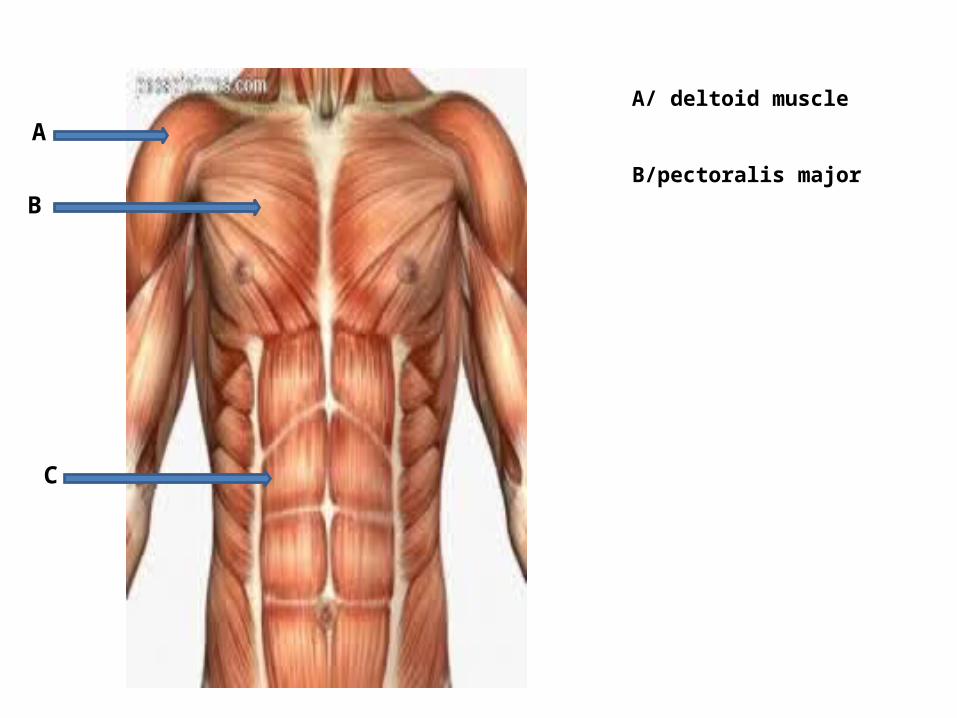
A/ deltoid muscle
B/pectoralis major
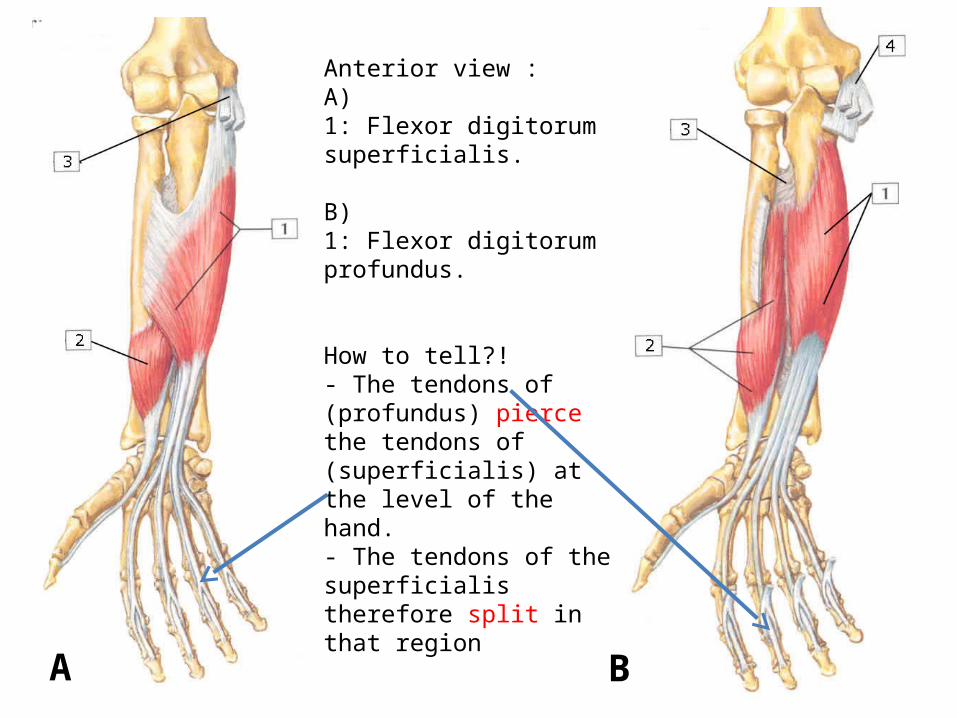
Anterior view : A)1: Flexor digitorum superficialis.
B)1: Flexor digitorum profundus.
How to tell?!- The tendons of (profundus) pierce the tendons of (superficialis) at the level of the hand.- The tendons of the superficialis therefore split in that region
A B
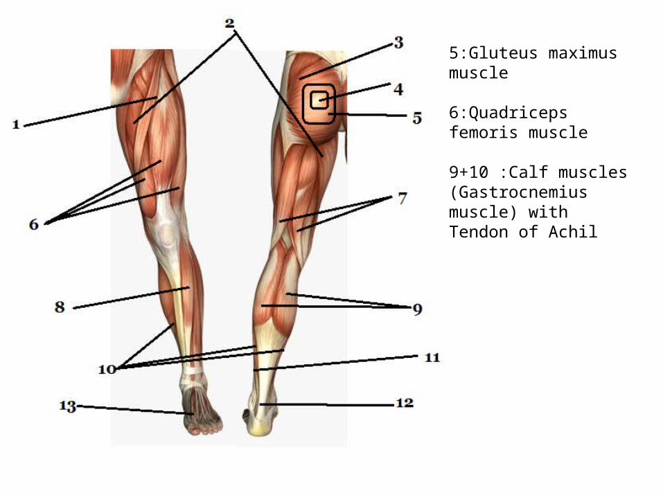
5:Gluteus maximus muscle
6:Quadriceps femoris muscle
9+10 :Calf muscles (Gastrocnemius muscle) with Tendon of Achil
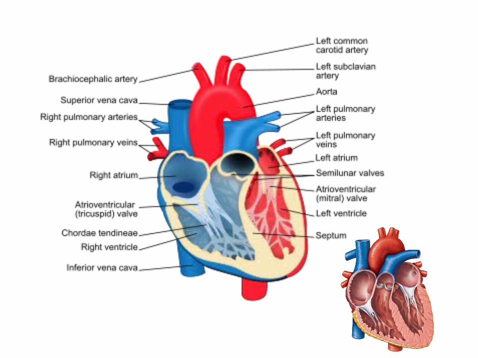
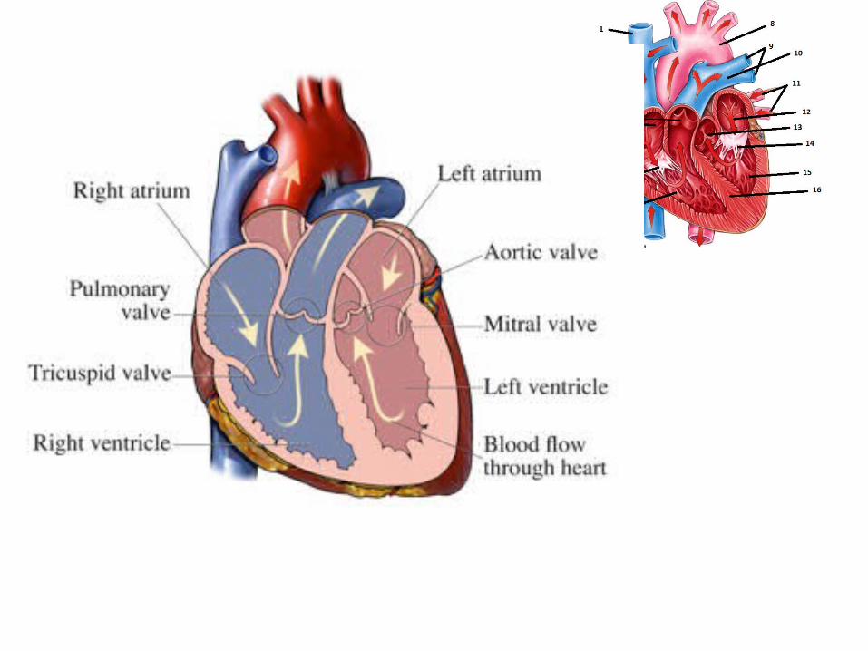
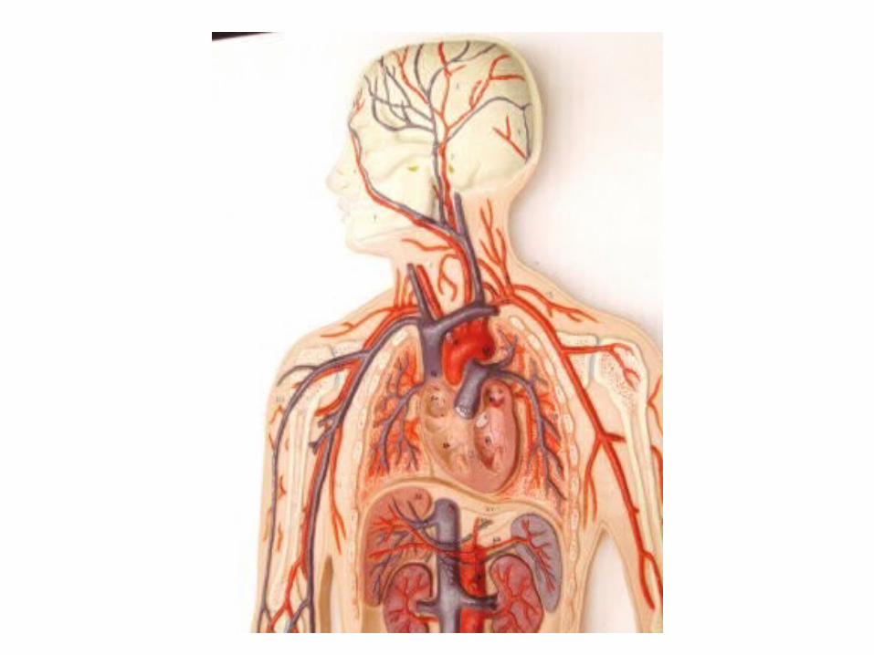
• Cardiovascular system• Heart• Blood vessels:• Arteries• Veins
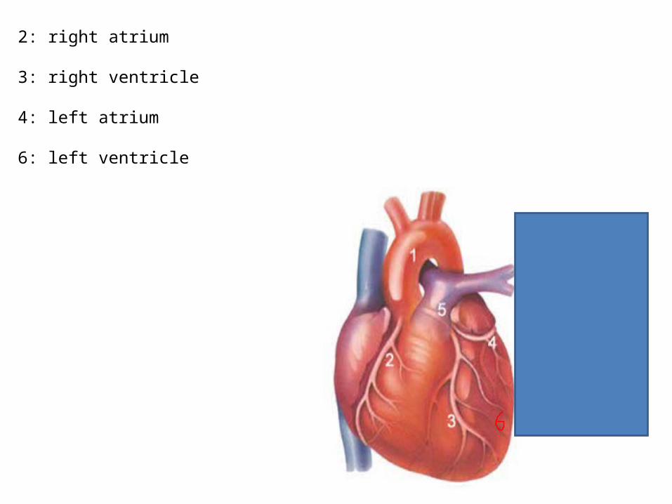
2: right atrium
3: right ventricle
4: left atrium
6: left ventricle
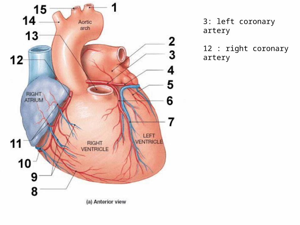
3: left coronary artery
12 : right coronary artery
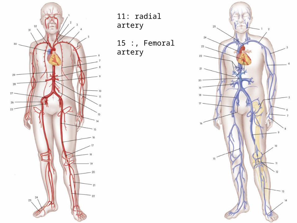
11: radial artery
15 :, Femoral artery
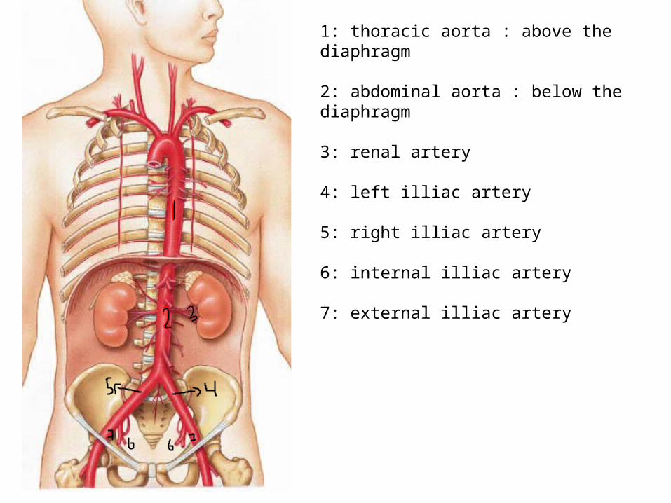
1: thoracic aorta : above the diaphragm
2: abdominal aorta : below the diaphragm
3: renal artery
4: left illiac artery
5: right illiac artery
6: internal illiac artery
7: external illiac artery
• Histology of cardiovascular system• Cardiac muscle• Blood vessels
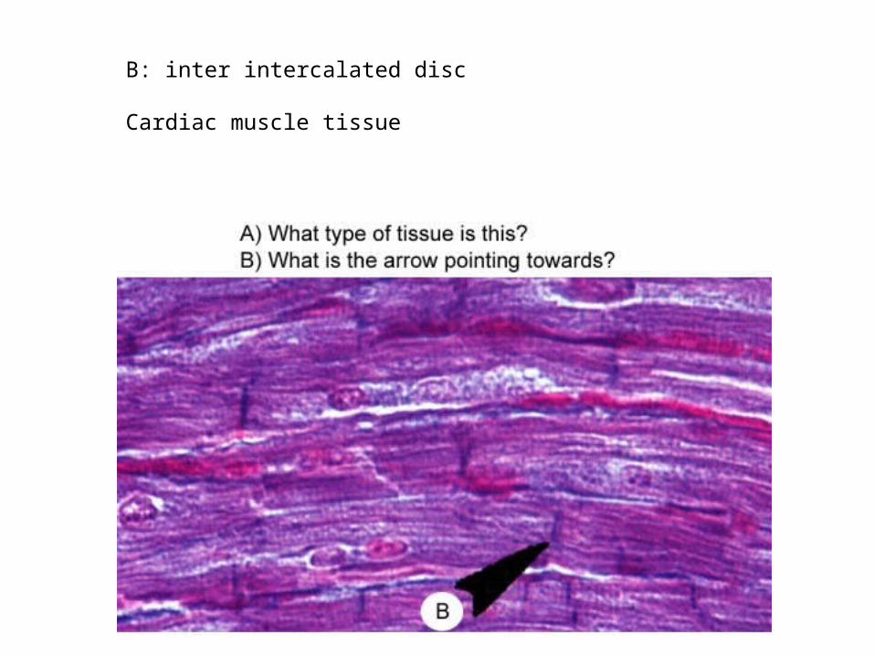
B: inter intercalated disc
Cardiac muscle tissue
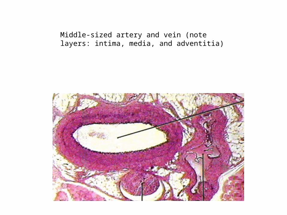
Middle-sized artery and vein (note layers: intima, media, and adventitia)
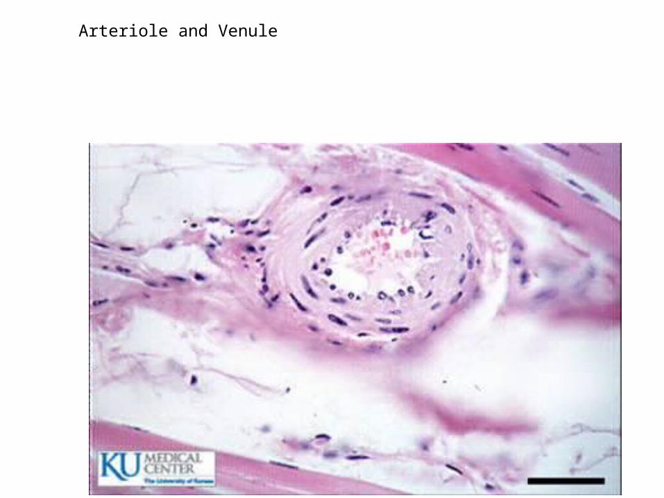
Arteriole and Venule