Embed Size (px)
Citation preview

Award Top Quizzes For Residents
Giovanni Negri, MD, Bolzano (Italy) Eva M. Wojcik MD, Department of Pathology, Loyola University, Chicago (USA) Esther D. Rossi MD, PhD, MIAC, Division of Anatomic Pathology and Cytology, Catholic University of Sacred Heart, Rome (Italy) Pinar Firat MD, MIAC, Department of Pathology, Koc University, School of Medicine, Istanbul (Turkey) Pio Zeppa MD, Department of Pathology, University of Salerno (Italy)
Quiz N°1 (Giovanni Negri)
• 44y, HPV test positive. • Previous pap-test: negative
• ThinPrep LBC
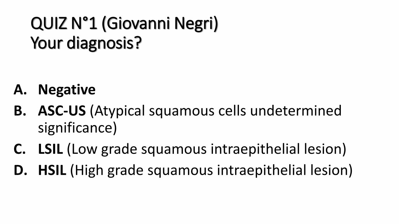
QUIZ N°1 (Giovanni Negri) Your diagnosis?
A. Negative
B. ASC-US (Atypical squamous cells undetermined significance)
C. LSIL (Low grade squamous intraepithelial lesion)
D. HSIL (High grade squamous intraepithelial lesion)
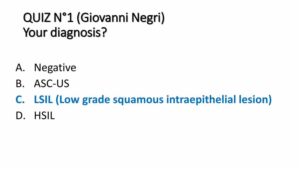
QUIZ N°1 (Giovanni Negri) Your diagnosis?
A. Negative
B. ASC-US
C. LSIL (Low grade squamous intraepithelial lesion)
D. HSIL
Explanation
• Squamous epithelia with some fungal organisms (pseudohyphae) consistent with Candida, associated with binucleations and some more obvious nuclear changes with hyperchromasia, smudged chromatin, and nuclear enlargement.
• In spite of the fungi and although the cytological features are still not completely typical for a LSIL, in HPV-positive women these changes may be classified as low-grade squamous intraepithelial lesion (LSIL)
• Subsequent biopsy confirmed a LSIL (CIN1).
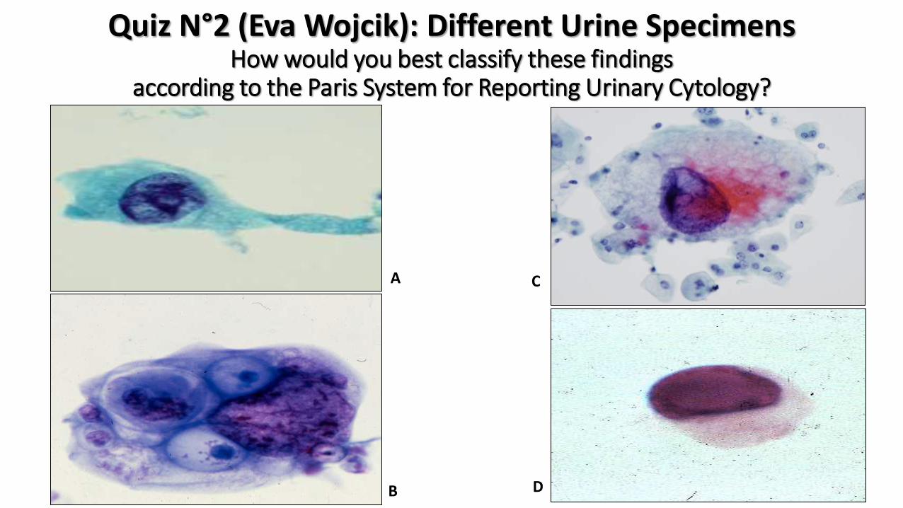
How would you best classify these findings according to the Paris System for Reporting Urinary Cytology?
Quiz N°2 (Eva Wojcik): Different Urine Specimens
A
B D
C
QUIZ N°2 (Eva Wojcik) Your diagnosis?
A. Negative for high grade urothelial carcinoma
B. Atypical urothelial cells present
C. Suspicious for high grade urothelial carcinoma
D. High grade urothelial carcinoma
QUIZ N°2 (Eva Wojcik) Your diagnosis?
A. Negative for high grade urothelial carcinoma
B. Atypical urothelial cells present
C. Suspicious for high grade urothelial carcinoma
D. High grade urothelial carcinoma

A
B D
C
Quiz N°3 (Eva Wojcik): Different Urine Specimens How would you best classify these findings
according to the Paris System for Reporting Urinary Cytology?

QUIZ N°3 (Eva Wojcik) Your diagnosis?
A. Negative for high grade urothelial carcinoma
B. Atypical urothelial cells present
C. Suspicious for high grade urothelial carcinoma
D. High grade urothelial carcinoma

QUIZ N°2 (Eva Wojcik) Your diagnosis?
A. Negative for high grade urothelial carcinoma
B. Atypical urothelial cells present
C. Suspicious for high grade urothelial carcinoma
D. High grade urothelial carcinoma
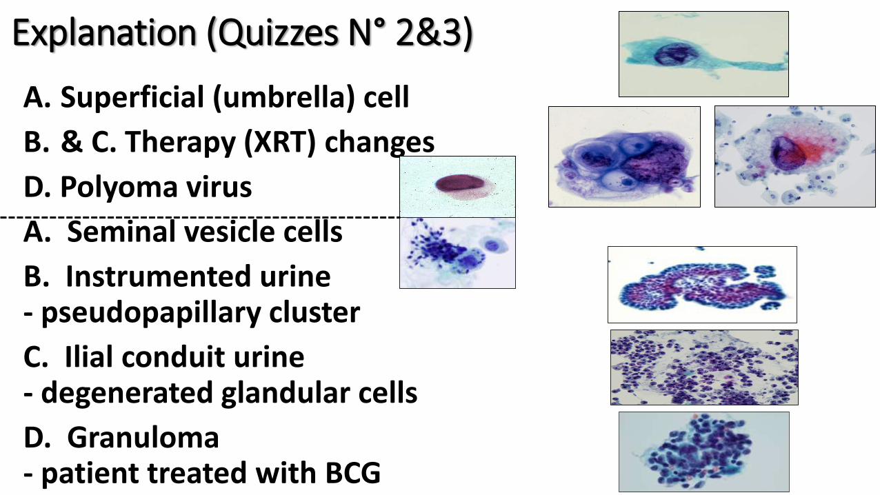
Explanation (Quizzes N° 2&3)
A. Superficial (umbrella) cell
B. & C. Therapy (XRT) changes
D. Polyoma virus
A. Seminal vesicle cells
B. Instrumented urine - pseudopapillary cluster
C. Ilial conduit urine - degenerated glandular cells
D. Granuloma - patient treated with BCG

• F, 48 y, 1.5 cm left parotid nodule • US: hyperpervasculated solid encapsulated nodule with a dis-homogenous pattern • FNAC, 25G, conventional and LB cytology
Quiz N°4 (Esther D. Rossi)
QUIZ N°4 (Esther D. Rossi) Your diagnosis?
A. Non-neoplastic
B. Atypia of undetermined significance (AUS)
C. Neoplasm-Benign
D. Neoplasm-SUMP (Salivary Gland Neoplasm of Undetermined Malignant Potential)
QUIZ N°4 (Esther D. Rossi) Your diagnosis?
A. Non-neoplastic
B. Atypia of undetermined significance (AUS)
C. Neoplasm-Benign
D. Neoplasm-SUMP (Salivary Gland Neoplasm of Undetermined Malignant Potential)

• Male, 55 yrs, smoker • Dyspnea, chest pain • Chest X-ray and CT: Massive left sided pleural effusion, Infiltrative mediastinal mass 7cm in diameter located left to aorta, Calcified nodules/plaques on the right pleura (asbestos exposure?) Preliminary radiologic diagnosis: Lung cancer?
• Bronchoscopy: No endobronchial lesion • Bronchial lavage: Negative for malignancy • The pleural fluid was sent for cytologic examination
Quiz N°5 (Pinar Firat)

QUIZ N°5

Calretinin
CK 5/6
WT-1
EMA
Glut-1 p53
Ber-Ep4
CEA
TTF-1
Desmin

QUIZ N°5 (Pinar Firat) Do you expect any molecular alteration in this case ? If yes, which one?
A. NO; malignancy is not present
B. YES; p16/CDKN2A loss
C. YES; KRAS or EGFR mutation
D. YES; HER2 amplification
A. NO; malignancy is not present
B. YES; p16/CDKN2A loss
C. YES; KRAS or EGFR mutation
D. YES; HER2 amplification
QUIZ N°5 (Pinar Firat) Do you expect any molecular alteration in this case ? If yes, which one?

US: solid mass > 50 mm, involving the right jaw and a lymph node (LN), 18 mm, hypoechoic, roundish. An US-guided FNC from the mass and the LN and rapid on-site evaluation (ROSE) is performed.
Quiz N°6 (Pio Zeppa) Female, 40 yrs, from Philippines, in apparently good health, complains of a rapid swelling of the right cheek. Two weeks before she lost an inferior molar. The dentist asked for an US evaluation.

Nuclei irregular and fragile, clumped chromatin, nucleoli almost absent, mitoses (arrows), small globules in the background (arrows)
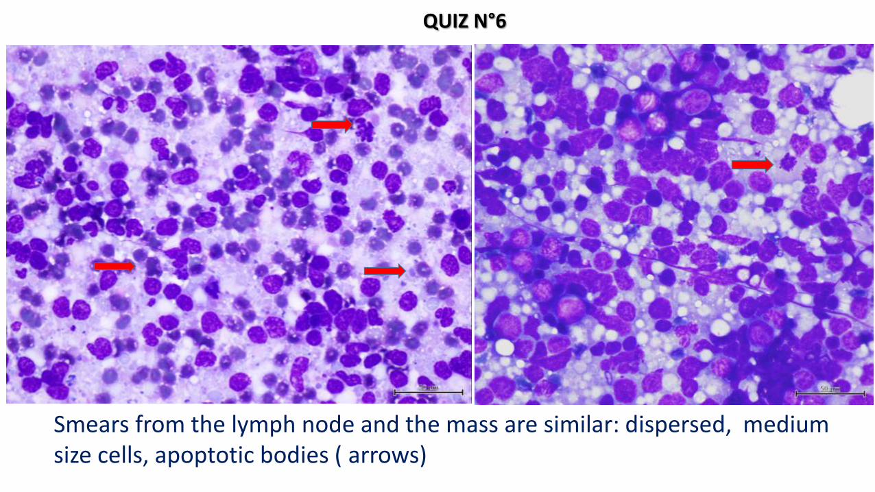
Smears from the lymph node and the mass are similar: dispersed, medium size cells, apoptotic bodies ( arrows)
QUIZ N°6
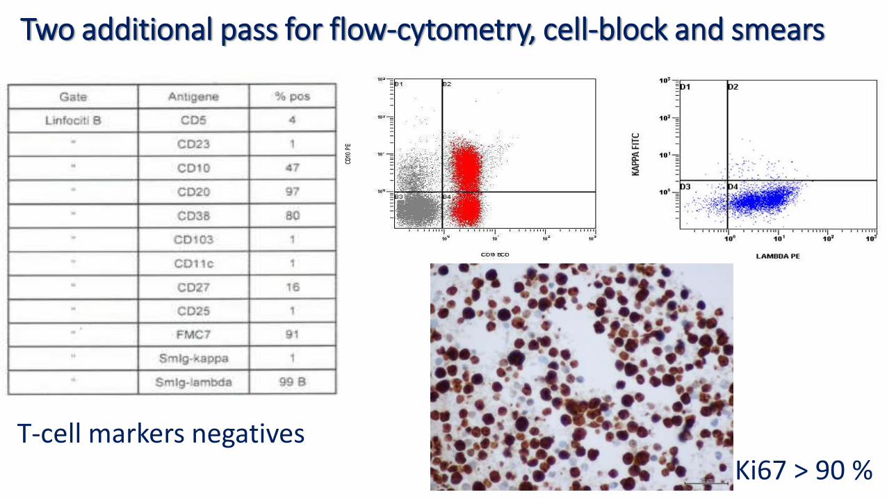
Two additional pass for flow-cytometry, cell-block and smears
T-cell markers negatives Ki67 > 90 %
QUIZ N°6 (Pio Zeppa) Your diagnosis?
A. Undifferentiated neoplasia
B. Lymphoblastic lymphoma
C. High grade follicular lymphoma
D. High grade B-cell lymphoma “Burkitt-like”
QUIZ N°6 (Pio Zeppa) Your diagnosis?
A. Undifferentiated neoplasia
B. Lymphoblastic lymphoma
C. High grade follicular lymphoma
D. High grade B-cell lymphoma “Burkitt-like”