Embed Size (px)
Citation preview
British Journal ofOphthalmology, 1983, 67, 529-534
Autosomal dominant iridogoniodysgenesis withassociated somatic anomalies: four-generation familywith Rieger's syndromeI. A. CHISHOLM AND A. E. CHUDLEY
From the Department ofOphthalmology and Division ofMedical Genetrics, Department of Pediatrics,University ofSaskatchewan
SUMMARY A family extending over 4 generations showed iridogoniodysgenesis accompanied bysomatic malformations inherited in an autosomal dominant fashion. Iridogoniodysgenesis waspresent in 10 members, of whom 5 had established glaucoma; 4 youthful members are likely todevelop glaucoma. Somatic malformations were present in 5 members from the 3rd and 4thgenerations who did not manifest iridogoniodysgenesis. A possible polygenic basis is discusssed,though the variable expression of an autosomal dominant inheritance is still the more likelyexplanation.
The contribution of goniodysgenesis to the patho-genesis of hereditary juvenile glaucoma has been des-cribed. I2 The tern embraces a maldevelopment ofthe trabecular meshwork, hypoplasia of the anteriorstromal layer of the iris, and anomaly of the peripheralcornea-Rieger's ocular malformation. For thosepatients in whom the iris hypoplasia is marked andcomeal involvement absent iridogoniodysgenesis isthe preferred descriptive term.3 Families withhereditary juvenile glaucoma are known in whom adominantly inherited iridogoniodysgenesis is bothprominent and a significant indicator of the accom-panying glaucoma,47 but in none have somatic mal-formations been reported. We report a family over 4generations showing marked iris hypoplasia presentfrom birth accompanied by glaucoma in early adult-hood, associated with somatic malformations of anautosomal dominant inheritance.
Materials aind methods
Access to the family was obtained via III ,0and siblings(Fig. 1). He had undergone bilateral thermal scler-ostomy for the control of glaucoma at the age of 25years and was seeking advice on behalf of his children.He was aware of a connection between iris colour andCorrespondence to Dr 1. A. Chisholm, FRCS Ed, Department ofOphthalmology, College of Medicine, University of Saskatchewan,Saskatoon, Canada S7N OXO.
blindness, as his grandfather, father, and an aunt whohad similar dark brown irides to his were blind bytheir 40s. Two of his children had a similar iris colour.Our assessment of the subjects entailed an ocular
examination, general physical examination, removalof blood samples for chromosome studies, and photo-graphy. The ocular examination consisted of a re-cording of visual acuity and refraction if indicated,slit-lamp examination of the anterior segment,gonioscopy, measurement of anterior chamber depthand comeal diameter, observation of pupil shape andmotility, and recording of the applanation intraocularpressure. Detailed examination of the visual field wasalso performed where indicated. A diagnosis ofglaucoma was reached on finding raised intraocularpressure with cupping of the optic disc supplementedwith the finding of classical nerve bundle defects inthe field of vision.
Results
529
Twenty-eight surviving members of the family wereassessed and data obtained (Fig. 1). Fourteen weremale and 14 female, of whom 10 (35-7%), namely, 4male and 6 female, had marked iris hypoplasia and adistinct facial resemblance (Fig. 2). I, though deceasedpossessed features compatible with the disorder,which were documented by history and examinationof photographs.
on May 22, 2020 by guest. P
rotected by copyright.http://bjo.bm
j.com/
Br J O
phthalmol: first published as 10.1136/bjo.67.8.529 on 1 A
ugust 1983. Dow
nloaded from
1. A. Chisholm and A. E. Chudley
II
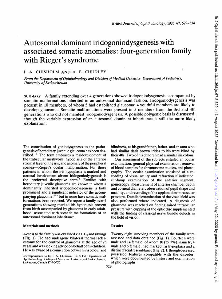
Fig. 1 Pedigree. Note theautosomal dominant Dattern ofinheritance and wide variation inexpression ofthe componentmanifestations.
III
IV
Key:MN (i Examined and normal Ei! e Failure of umbilical involution
Fi 6 Iridogoniodysgenesis [ ( Inguinal hernia
El* ( *Iridogoniodysgenesis -21and glaucoma
0 0) Maxillary hypoplasia, 0l? Chypodontia, microdontia
0il' 0Y Isolated maxillary hypoplasia
Hypospadius
)? Not examined
OCULAR FINDINGS
Glaucoma was found in 5 members, 2 female and 3male, all ofwhom showed iris hypoplasia. Intraocularpressure control was achieved by medical meansalone in 1 (III), repeated surgical procedures in 2(II,) and (II4), and by a combination of surgery withcontinued medical therapy in 1 (II ,o). The glaucomarecently diagnosed in IV20 appears to be unresponsive
Fig. 2 Family group withbetween I., Il,, and 114.
to medical therapy. Glaucoma was not diagnosed inII, and II4 until they were in their 40s, by which timevisual loss was severe. Glaucoma was diagnosed inII9 and III,. when they were in their early 20s and inIV20 at 18 years of age. None of the subjects who hadnormal iris structure show glaucoma. The sister of thepropositus (III 12) is the only subject in her generationwith iris hypoplasia who does not show glaucoma.Subjects IV4, IVs, IV9, and IV,1 who have iris hypo-plasia but not as yet glaucoma must be considered athigh risk of developing the disease.The iris hypoplasia found in 10 members of the
family was bilateral, present from birth, and imparteda characteristic dark brown colour to the iris. Slit-lamp examination revealed absence of the anteriorleaf of the iris stroma (Figs. 3-6). The posterior leafwas gossamer thin, throwing into sharp prominencethe pale band of the sphincter muscle and numerousradial strands. Retroillumination of the iris revealedno dehiscences of the posterior epithelium.Minor developmental changes were identified in
the lens of eyes with iris hypoplasia. These consistedof epicapsular stars on the anterior lens capsule, andsmall white flake opacities in the anterior lens suture.Eyes with normal iris structure did not show thesechanges.The eyes with iris hypoplasia had normal sized
corneas and no evidence of posterior embryrotoxon(Figs. 3-6). The anterior chamber appeared deep,but the depth centrally as measured by the Haag-Streit pachometer in the affected subjects from the3rd and 4th generation was 3 4, SD 0-8 mm (normalfor 15-35-year group is 3 60, SD 0-039 mm8).
530
on May 22, 2020 by guest. P
rotected by copyright.http://bjo.bm
j.com/
Br J O
phthalmol: first published as 10.1136/bjo.67.8.529 on 1 A
ugust 1983. Dow
nloaded from
Autosomal dominant iridogoniodysgenesis with associated somatic anomalies
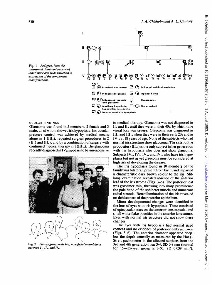
Fig. 3 Typical Rieger's ocular malformation (forcomparison).
Fig. 4 Right eye 11110~showing peripheral iridectomy, ovalpupil, prominent sphincter band, iris hypoplasia.
Fig. 5 Fig. 6
Figs. 5 and 6 Right and left eye V,20 showing iris hypoplasia and prominentsphincter band; rightshowing broad anteriorsynechium below with corneal opacities.
Gonioscopy showed an anterior origin of the iris fromapproximately the scleral spur with a network of finestrands passing forwards towards Schwalbe's lineapparently internal to the trabeculum. The angleswere wide.The pupil of eyes with iris hypoplasia was solitary,
central, and active to light stimulation. In 3 subjectsthe eyes were myopic and the pupils in those eyestended to be horizontally oval and to show pupil-lotonia (IIIlo, IV4 and IV20) (Figs. 3-6).Of the 20 eyes with iris hypoplasia only one differed
from the above. The right eye of IV20 showed down-ward displacement of the pupil because of a broadanterior adhesion of the iris to the posterior peripheryof the cornea, obliterating the angle of the anteriorchamber in that area (Figs. 3-6). The remainder ofthe angle in this eye was as described above. Whitishspots were present in the deep layer of the cornealstroma. This eye was the only one in the series which
approximated to the description of Rieger's ocularmalformation.
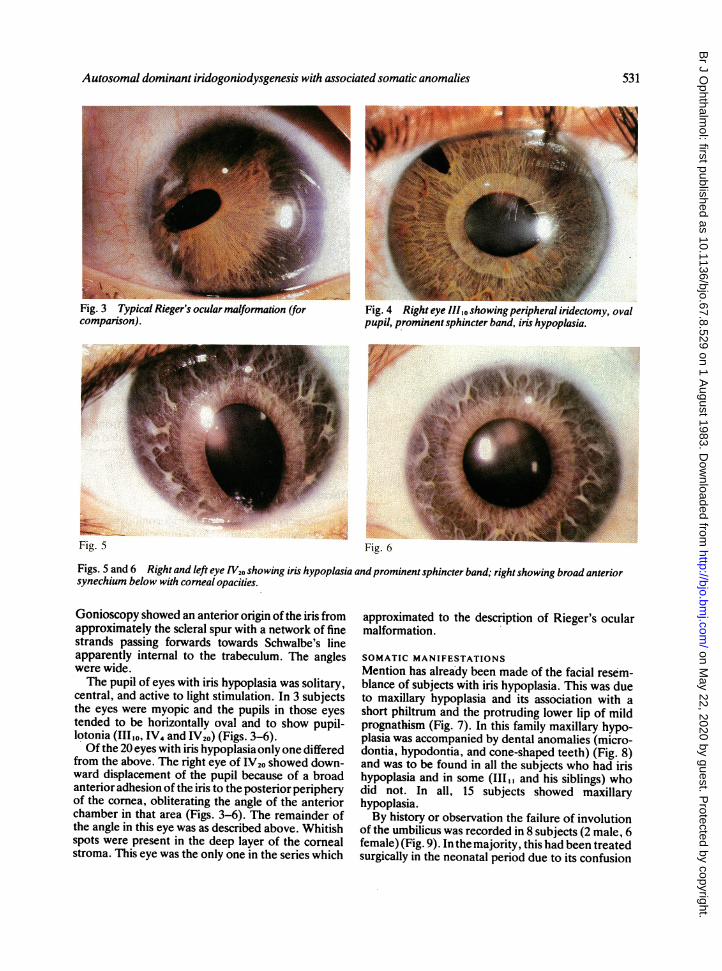
SOMATIC MANIFESTATIONSMention has alrea'dy been made of the facial resem-blance of subjects with iris hypoplasia. This was dueto maxillary hypoplasia and its association with ashort philtrum and the protruding lower lip of mildprognathism (Fig. 7). In this family maxillary hypo-plasia was accompanied by dental anomalies (micro-dontia, hypodontia, and cone-shaped teeth) (Fig. 8)and was to be found in all the subjects who had irishypoplasia and in some (11,, and his siblings) whodid not. In all, 15 subjects showed maxillaryhypoplasia.By history or observation the failure of involution
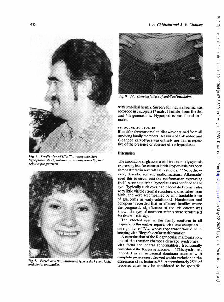
of the umbilicus was recorded in 8 subjects (2 male, 6female) (Fig. 9). In the majority, this had been treatedsurgically in the neonatal period due to its confusion
531
-a
on May 22, 2020 by guest. P
rotected by copyright.http://bjo.bm
j.com/
Br J O
phthalmol: first published as 10.1136/bjo.67.8.529 on 1 A
ugust 1983. Dow
nloaded from
I. A. Chisholm and A. E. Chudley
Fig. 7 Profile view ofIIIo illushypoplasia, short philtrum, proirelative prognathism.
Fig. 8 Facial view IV4; illustratand dental amomalies.
with umbilical hernia. Surgery for inguinal hernia wasrecorded in 8 subjects (7 male, 1 female) from the 3rdand 4th generations. Hypospadias was found in 4males.
CYTOGENETIC STUDIESBlood for chromosomal studies was obtained from allsurviving family members. Analysis of G-banded andC-banded karyotypes was entirely normal, irrespec-tive of the presence or absence of iris hypoplasia.
DiscussionFtrating maxillarytruding lower lip, and The association of glaucoma with iridogoniodysgenesis
expressing itself as connatal iridal hypoplasia has beendemonstrated in several family studies.24I7 None, how-ever, describe somatic malformations; Alkemade9used this to stress that the malformation expressingitself as connatal iridal hypoplasia was confined to theeye. Typically such eyes had chocolate brown irideswith little visible stromal structure, did not alter frombirth, and were accompanied by an intractable formof glaucoma in early adulthood. Hambresen andSchepens' recorded that in affected families wherethe prognostic significance of the iris colour wasknown the eyes of newborn infants were scrutinisedfor this tell-tale sign.The affected eyes in this family conform in all
respects to the earlier reports with one exception-the right eye of IV20, whose appearance would be inkeeping with Rieger's ocular malformation.A combination of the Rieger ocular malformation,
one of the anterior chamber cleavage syndromes,10with facial and dental abnormalities, traditionallyconstituted the Rieger syndrome.1 12This syndrome,inherited in an autosomal dominant manner withcomplete penetrance, showed a wide variation in the
'ing typical dark eyes, facial expression of its features.'3 1' Approximately 25% ofreported cases may be considered to be sporadic.
I532
on May 22, 2020 by guest. P
rotected by copyright.http://bjo.bm
j.com/
Br J O
phthalmol: first published as 10.1136/bjo.67.8.529 on 1 A
ugust 1983. Dow
nloaded from
Autosomal dominant iridogoniodysgenesis with associated somatic anomalies
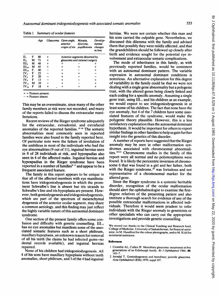
Table 1 Summary ofocularfeatures
Age Glaucoma Open angle, Myopia, Develop-anterior dyscoria, mentalorigin of iris pupillotonia changes
in lens
II, F 80 + Anterior segments distorted byII4 M 71 + glaucoma and cataract surgery1119 M 48 + + - +111,0 M 45 + + + +III 12 F 40 - + - +IV4 F 23 - + + -IVs F 20 - + - -IV9 F 13 - + - +IV, M 10 - + - +1V20 F 18 + * + + +
+ = Feature present.- = Feature absent.
This may be an overestimate, since many of the otherfamily members at risk were not recorded, and manyof the reports failed to discuss the extraocular mani-festations.
Recent reviews of the Rieger syndrome adequatelylist the extraocular, extradental, and extrafacialanomalies of the reported families.5 16 The somaticabnormalities most commonly seen in reportedfamilies were also found in the family reported here.Of particular note were the failure of involution ofthe umbilicus in most of the individuals who had theeye abnormalities (9 out of 11), inguinal hernias seenin 8 of 28 individuals at risk, and hypospadias alsoseen in 4 of the affected males. Inguinal hernias andhypospadius in the Rieger syndrome have beenreported in a number of families9 17 and appear to be afrequent associated feature.The family in this report appears to be unique in
that all of the affected members with eye manifesta-tions have iridogoniodysgenesis in which the prom-inent Schwalbe's line is absent but iris strands toSchwalbe's line and iris hypoplasia are present. How-ever, both goniodysgenesis and iridogoniodysgenesis,which are part of the spectrum of mesenchymaldysgenesis of the anterior ocular segment, may sharea common aetiology, and this finding may just reflectthe highly variable nature of this autosomal dominantsyndrome.One section of the present family offers some con-
fusion and difficulty with genetic counselling. III.,has no eye anomalies but manifests some of the asso-ciated somatic features such as a short philtrum,maxillary hypoplasia, an unknown reason for removalof all his teeth (he claims he had infected gums-nodental records available), and inguinal herniasrepaired.None of his children had iridogoniodysgenesis. All
4 of his sons have maxillary hypoplasia without teethanomalies, short philtrum, and 3 of the 4 had inguinal
hernias. We were not certain whether this man andhis sons carried the culpable gene. Nevertheless, wediscussed this dilemma with the family and advisedthem that possibly they were mildly affected, and thatthe grandchildren should be followed up closely afterbirth and evidence sought for the potential eye in-volvement and extraocular somatic complications.The mode of inheritance in this family, as with
previously reported families, would be consistentwith an autosomal dominant pattern. The variableexpression in autosomal dominant conditions isnotorious. An alternative explanation for this degreeof variability in the family could be that we were notdealing with a single gene abnormality but a polygenictrait, with the altered genes being closely linked andeach coding for a specific anomaly. Assuming a singlegene trait, using IIIl,, and his children as an example,we would expect to see iridogoniodysgenesis in atleast some of his children. The fact that none have theeye anomaly, but 4 of the 7 children have some asso-ciated features of the syndrome, would make thepolygenic theory plausible. However, this is a lesssatisfactory explanation than the autosomal dominanthypothesis. It would be important for others to reportsimilar findings in other families to helpusgain furtherinsight into the genetics of this disorder.A number of reports have suggested that the Rieger
anomaly may be seen in other malformation syn-dromes associated with chromosomal abnormali-ties.'82' Chromosome studies in the family of thisreport were all normal and no polymorphisms werefound. It is likely the pericentric inversion of chromo-some 6 that was found in a girl and her father, bothwith the Rieger syndrome,20 was fortuitous and notrepresentative of a chromosomal marker for thealtered gene.
Since the Rieger syndrome is a systemic heritabledisorder, recognition of the ocular malformationshould alert the ophthalmologist to examine the first-degree relatives of the presenting patient and alsoinstitute a thorough search for evidence of any of thepossible extraocular malformations in affected indi-viduals. Therefore it would seem prudent to referindividuals with the Rieger anomaly to geneticists orother specialists who can carry out the appropriateinvestigations and provide genetic counselling.
We record our thanks to the Clinical Teaching and Research fund,College of Medicine, University of Saskatchewan, for financial assist-ance, to M. Handford for the colour photographs, and to M. Kidd forsecretarial assistance.
References
1 Cromble AL, Cullen JF. Hereditary glaucoma: occurrence in fivegenerations of an Edinburgh family. Br J Ophthalmol 1964; 48:143-7.
2 Jerndal T. Goniodysgenesis and hereditary juvenile glaucoma.Acta Ophthalmol (Kbh) 1970; suppl 107.
533
on May 22, 2020 by guest. P
rotected by copyright.http://bjo.bm
j.com/
Br J O
phthalmol: first published as 10.1136/bjo.67.8.529 on 1 A
ugust 1983. Dow
nloaded from
1. A. Chisholm and A. E. Chudley
3 Waring GO, Rodriques MM, Laibson PR. Anterior chambercleavage syndrome. A stepladder classification. Surv Ophthalmol1975; 20:3-27.
4 Berg F. Erbliches jugendliches Glaukom. Acta Ophthalmol(Kbh) 1932; 10: 568-87.
5 Hambresen L, Schepens C. Glaukome familiale. Bull SocOphtalmol Fr 1932; 59: 219-23.
6 Frangois J, Deweer JP, vanden Berghe J. Chronic simpleglaucoma with dominent heredity. Bull Soc Belge Ophtalmol1950; 96: 665-83.
7 Weatherill JR, Hart CT. Familial hypoplasia of the iris stromaassociated with glaucoma. BrJ Ophthalmol 1969; 53: 433-8.
8 Weekers R, Grieten J, Lavergne G. Study of the dimensions ofthe human anterior chamber. Ophthalmologica 1961; 142:650-62.
9 Alkemade PPH. Dysgenesis mesodermalis of the iris and thecornea. Assen; Van Gorcum, 1969.
10 Reese AB, Ellsworth RM. The anterior chamber cleavagesyndrome. Arch Ophthalmol 1966; 75: 307-18.
11 Rieger H. Beitrage zur Kenntnis seltner Missbildungen der Iris;Membrana iridopupillaris persistens. Albrecht von Graefes ArchKlin Ophthalmol 1934; 131: 523-30.
12 Rieger H. Beitrage zur Kenntnis seltner Missbildungen der Iris;ueber Hypoplasie des Irisvorderblattes mit Verlatgerung und
Entrundung der Pupillie. Albrecht von Graefes Arch KlinOphthalmol 1935; 133: 602-35.
13 Jorgenson RJ, VoderFE, Levin LS. The Reiger syndrome. JMedGenet 1979; 16: 236-7.
14 Cross HE, Penetrance and variability and anterior chambermalformations. Birth Defects 1979; 15: 131-44.
15 Jorgenson RJ, Levin LS, Cross HE, Yoder F, Kelly TE. TheRieger syndrome. Am J Med Genet 1978; 2: 307-18.
16 Fitch N, Kaback M. The Axenfeld syndrome and the Riegersyndrome. J Med Genet 1978; 15: 30-4.
17 Ferngold M, Shiere F, Fogels H, Donaldson D. The Riegersyndrome. Pediatrics 1969; 44: 564-9.
18 Tabbara KF, Khour FP, DerKaloustian VM. Rieger's syndromewith chromosomal anomaly. Can J Ophthalmol 1973; 8: 488-91.
19 Wilcos LM, Bercovitch L, Howard RO. Ophthalmic features ofchromosome deletion 4p-(Wolf-Hirschhom syndrome). Am JOphthalmol 1978; 86: 834-9.
20 Heinemann MH, Breg R, Cotbier E. Rieger's syndrome withpericentric inversion of chromosome 6. BrJ Ophthalmol 1979; 63:40-4.
21 Akazawak Yamane S, Shiota H, NaitoE. A case ofretinoblastomaassociated with Rieger's anomally and 13q deletion. Jpn JOphthalmol 1981; 25: 321-5.
534
on May 22, 2020 by guest. P
rotected by copyright.http://bjo.bm
j.com/
Br J O
phthalmol: first published as 10.1136/bjo.67.8.529 on 1 A
ugust 1983. Dow
nloaded from