Embed Size (px)
Citation preview
Autosomal Dominant Inheritance
More than half of all mendelian disorders are inherited as
autosomal dominant traits. The incidence of some
autosomal dominant disorders is high, at least in specific
geographical areas: for example, 1 in 500 for familial
hypercholesterolemia in populations of European or
Japanese descent.
Dr. Mohamed Saad Daoud 1
Many autosomal dominant disorders are individually much
less common, they are so numerous in the aggregate that
their total incidence is appreciable.
When they are transmitted through families, they become
problems not only for individuals but also for whole
kindreds, often through many generations.
Dr. Mohamed Saad Daoud 2
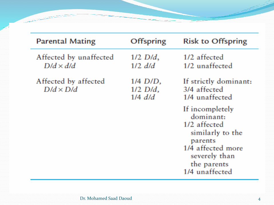
The risk and severity of dominantly inherited disease
depend on whether one or both parents are affected and
whether the trait is strictly dominant or incompletely
dominant. Denoting D as the mutant allele and d as the
normal allele, matings that produce children with an
autosomal dominant disease can be between two
heterozygotes (D/d) for the mutation or, more frequently,
between a heterozygote for the mutation (D/d) and a
homozygote for a normal allele (d/d):
Dr. Mohamed Saad Daoud 3
Dr. Mohamed Saad Daoud 4
Each child of a D/d by d/d mating has a 50% chance of receiving
the affected parent’s abnormal allele D and a 50% chance of
receiving the normal allele d. In the population as a whole, the
offspring of D/d by d/d parents are approximately 50% D/d and
50% d/d. theoretical expected ratio of 1:1, especially if the
sibship is small. Typical autosomal dominant inheritance can be
seen in the pedigree of a family with a dominantly inherited
form of hereditary deafness
Dr. Mohamed Saad Daoud 5
In medical practice, homozygotes for dominant phenotypes
are not often seen because mating's that could produce
homozygous offspring are rare. Again denoting the mutant
allele as D and the normal allele as d, the mating's that can
produce a D/D homozygote might theoretically be D/d by
D/d, D/D by D/d, or D/D by D/D.
Dr. Mohamed Saad Daoud 6
Practically, only the mating of two heterozygotes need be
considered because D/D homozygotes are very rare and
generally too severely affected to reproduce. In the case of
two heterozygotes mating, 3/4 of the offspring of a D/d by
D/d mating will be affected to some extent and 1/4
unaffected. In theory, the 3/4 affected could all have the
same condition if it is a pure dominant, or 1/3 of the
affected would be homozygotes and much more severely
affected than the D/d heterozygotes if it is an incompletely
dominant condition.
Dr. Mohamed Saad Daoud 7

Characteristics of Autosomal Dominant Inheritance
The phenotype usually appears in every generation, each affected
person having an affected parent.
Any child of an affected parent has a 50% risk of inheriting the
trait.
Phenotypically normal family members do not transmit the
phenotype to their children.
Males and females are equally likely to transmit the phenotype, to
children of either sex. In particular, male-to-male transmission can
occur, and males can have unaffected daughters.
Dr. Mohamed Saad Daoud 8
X-LINKED INHERITANCE
The X and Y chromosomes, which are responsible for sex
determination, are distributed unequally to males and
females in families. For this reason, phenotypes determined
by genes on the X have a characteristic sex distribution and
a pattern of inheritance that is usually easy to identify.
Approximately 1100 genes are thought to be located on the
X chromosome, of which approximately 40% are presently
known to be associated with disease phenotypes.
Dr. Mohamed Saad Daoud 9
Because males have one X chromosome but females have two,
there are only two possible genotypes in males and three in
females with respect to a mutant allele at an X-linked locus. A
male with a mutant allele at an X-linked locus is hemizygous for
that allele, whereas females may be homozygous for either the
wild-type or mutant allele or may be heterozygous. For example,
if XH is the wild-type allele for the gene for coagulation factor
VIII and a mutant allele, Xh, causes hemophilia A, the genotypes
expected in males and females would be as follows:
Dr. Mohamed Saad Daoud 10
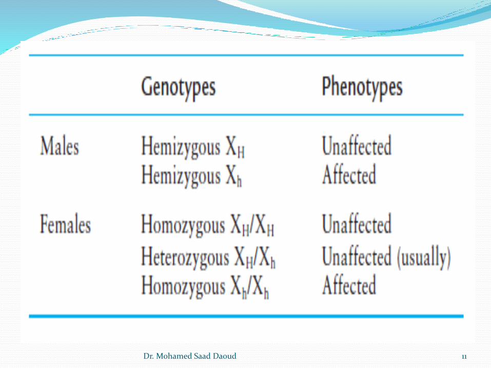
Dr. Mohamed Saad Daoud 11
Recessive and Dominant Inheritance of X-Linked Disorders
X-linked “dominant” and “recessive” patterns of inheritance
are distinguished on the basis of the phenotype in
heterozygous females. Some X-linked phenotypes are
consistently expressed in carriers (dominant), whereas
others usually are not (recessive).
Dr. Mohamed Saad Daoud 12
The difficulty in classifying an X-linked disorder as dominant
or recessive arises because females who are heterozygous
for the same mutant allele in the same family may or may
not demonstrate the disease, depending on the pattern of
random X inactivation and the proportion of the cells in
pertinent tissues that have the mutant allele on the active
versus inactive chromosome.
Dr. Mohamed Saad Daoud 13
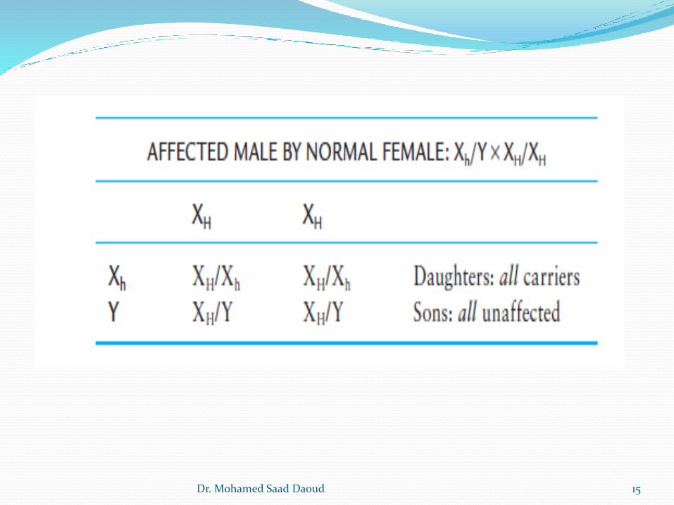
X-Linked Recessive Inheritance:
The inheritance of X-linked recessive phenotypes follows a
well-defined and easily recognized pattern. An X-linked
recessive mutation is typically expressed phenotypically in
all males who receive it but only in those females who are
homozygous for the mutation. Consequently, X-linked
recessive disorders are generally restricted to males and
rarely seen among females.
Dr. Mohamed Saad Daoud 14
Dr. Mohamed Saad Daoud 15
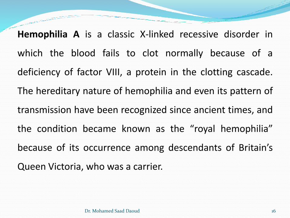
Hemophilia A is a classic X-linked recessive disorder in
which the blood fails to clot normally because of a
deficiency of factor VIII, a protein in the clotting cascade.
The hereditary nature of hemophilia and even its pattern of
transmission have been recognized since ancient times, and
the condition became known as the “royal hemophilia”
because of its occurrence among descendants of Britain’s
Queen Victoria, who was a carrier.
Dr. Mohamed Saad Daoud 16
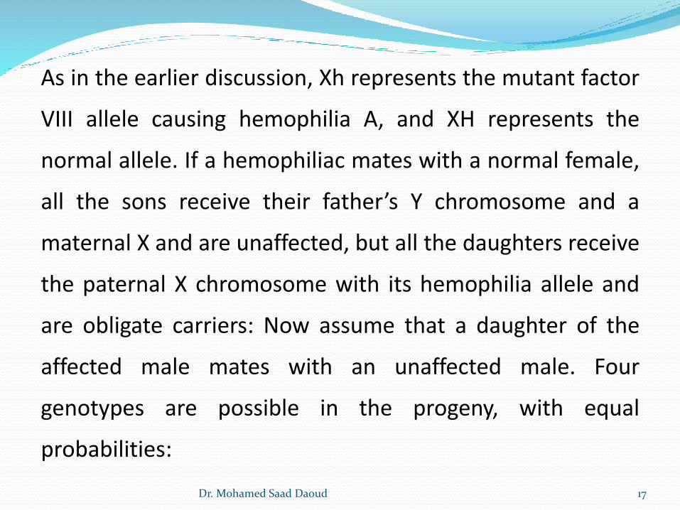
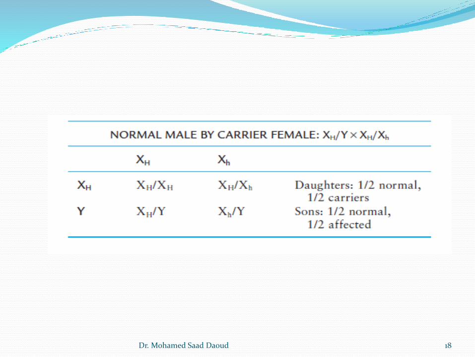
As in the earlier discussion, Xh represents the mutant factor
VIII allele causing hemophilia A, and XH represents the
normal allele. If a hemophiliac mates with a normal female,
all the sons receive their father’s Y chromosome and a
maternal X and are unaffected, but all the daughters receive
the paternal X chromosome with its hemophilia allele and
are obligate carriers: Now assume that a daughter of the
affected male mates with an unaffected male. Four
genotypes are possible in the progeny, with equal
probabilities:
Dr. Mohamed Saad Daoud 17
Dr. Mohamed Saad Daoud 18
Mitochondrial Inheritance
The Mitochondrial Genome
A small and important fraction of proteins is encoded by
genes within the mitochondrial genome.
Mitochondrial genome consists of a circular chromosome,
16.5 kb in size, that is located inside the mitochondrial
organelle, not in the nucleus.
Most cells contain at least 1000 mtDNA molecules,
distributed among hundreds of individual mitochondria.
Dr. Mohamed Saad Daoud 19
A remarkable exception is the mature oocyte, which has more
than 100,000 copies of mtDNA, composing about one third of
the total DNA content of these cells.
Mitochondrial DNA (mtDNA) contains 37 genes. The genes
encode 13 polypeptides that are subunits of enzymes of
oxidative phosphorylation, two types of ribosomal RNA, and 22
transfer RNAs required for translating the transcripts of the
mitochondria-encoded polypeptides. The remaining
polypeptides of the oxidative complex are encoded by the
nuclear genome.
Dr. Mohamed Saad Daoud 20
More than 100 different rearrangements and 100 different point
mutations have been identified in mtDNA that can cause human
disease, often involving the central nervous and musculoskeletal
systems (e.g., myoclonic epilepsy with ragged-red fibers). The
diseases that result from these mutations show a distinctive
pattern of inheritance because of three unusual features of
mitochondria: replicative segregation, homoplasmy and
heteroplasmy, and maternal inheritance.
Dr. Mohamed Saad Daoud 21

Replicative Segregation
The first unique feature of the mitochondrial chromosome
is the absence of the tightly controlled segregation seen
during mitosis and meiosis of the 46 nuclear chromosomes.
At cell division, the multiple copies of mtDNA in each of the
mitochondria in a cell replicate and sort randomly among
newly synthesized mitochondria. The mitochondria, in turn,
are distributed randomly between the two daughter cells.
This process is known as replicative segregation.
Dr. Mohamed Saad Daoud 22
Homoplasmy and Heteroplasmy
The second unique feature of the genetics of mtDNA arises from
the fact that most cells contain many copies of mtDNA
molecules. When a mutation arises in the mtDNA, it is at first
present in only one of the mtDNA molecules in a mitochondrion.
With replicative segregation, however, a mitochondrion
containing a mutant mtDNA will acquire multiple copies of the
mutant molecule. With cell division, a cell containing a mixture
of normal and mutant mtDNAs can distribute very different
proportions of mutant and wild-type
Dr. Mohamed Saad Daoud 23
Mitochondrial DNA to its daughter cells. One daughter cell may,
by chance, receive mitochondria that contain only a pure
population of normal mtDNA or a pure population of mutant
mtDNA (a situation known as homoplasmy). Alternatively, the
daughter cell may receive a mixture of mitochondria, some with
and some without mutation. Because the phenotypic expression
of a mutation in mtDNA depends on the relative proportions of
normal and mutant mtDNA in the cells making up different
tissues, reduced penetrance, variable expression, and pleiotropy
are all typical features of mitochondrial disorders.
Dr. Mohamed Saad Daoud 24
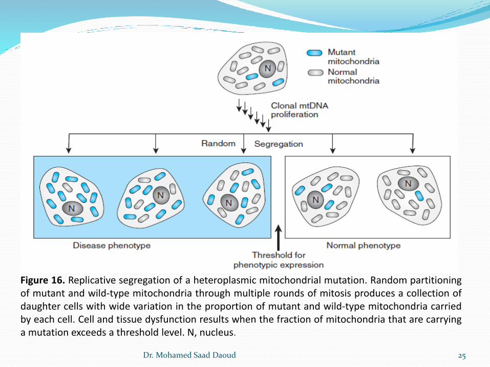
Figure 16. Replicative segregation of a heteroplasmic mitochondrial mutation. Random partitioningof mutant and wild-type mitochondria through multiple rounds of mitosis produces a collection ofdaughter cells with wide variation in the proportion of mutant and wild-type mitochondria carriedby each cell. Cell and tissue dysfunction results when the fraction of mitochondria that are carryinga mutation exceeds a threshold level. N, nucleus.
Dr. Mohamed Saad Daoud 25
Maternal Inheritance of mtDNA
The final defining characteristic of the genetics of mtDNA is its
maternal inheritance. Sperm mitochondria are generally
eliminated from the embryo, so that mtDNA is inherited from
the mother. Thus, all the children of a female who is
homoplasmic for a mtDNA mutation will inherit the mutation,
whereas none of the offspring of a male carrying the same
mutation will inherit the defective DNA. The maternal
inheritance of a homoplasmic mtDNA mutation causing Leber
hereditary optic neuropathy.
Dr. Mohamed Saad Daoud 26
![An interstitial deletion-insertion involving chromosomes 2p25.3 … · 2014-01-30 · may also be inherited as autosomal dominant (Online Mendelian Inheritance in Man [OMIM] 146200),](https://img.pdfslide.us/doc/110x75/5e89419298195b31d277c9f0/an-interstitial-deletion-insertion-involving-chromosomes-2p253-2014-01-30-may.jpg)