Embed Size (px)
Citation preview

Atypical function of a centrosomal module in WNT signalling drives contextual cancer cellmotility
Luo, Yi; Barrios-Rodiles, Miriam; Gupta, Gagan D.; Zhang, Ying Y.; Ogunjimi, Abiodun A.;Bashkurov, Mikhail; Tkach, Johnny M.; Underhill, Ainsley Q.; Zhang, Liang; Bourmoum,Mohamed; Wrana, Jeffrey L.; Pelletier, LaurencePublished in:Nature Communications
Published: 01/01/2019
Document Version:Final Published version, also known as Publisher’s PDF, Publisher’s Final version or Version of Record
License:CC BY
Publication record in CityU Scholars:Go to record
Published version (DOI):10.1038/s41467-019-10241-w
Publication details:Luo, Y., Barrios-Rodiles, M., Gupta, G. D., Zhang, Y. Y., Ogunjimi, A. A., Bashkurov, M., Tkach, J. M., Underhill,A. Q., Zhang, L., Bourmoum, M., Wrana, J. L., & Pelletier, L. (2019). Atypical function of a centrosomal modulein WNT signalling drives contextual cancer cell motility. Nature Communications, 10, [2356].https://doi.org/10.1038/s41467-019-10241-w
Citing this paperPlease note that where the full-text provided on CityU Scholars is the Post-print version (also known as Accepted AuthorManuscript, Peer-reviewed or Author Final version), it may differ from the Final Published version. When citing, ensure thatyou check and use the publisher's definitive version for pagination and other details.
General rightsCopyright for the publications made accessible via the CityU Scholars portal is retained by the author(s) and/or othercopyright owners and it is a condition of accessing these publications that users recognise and abide by the legalrequirements associated with these rights. Users may not further distribute the material or use it for any profit-making activityor commercial gain.Publisher permissionPermission for previously published items are in accordance with publisher's copyright policies sourced from the SHERPARoMEO database. Links to full text versions (either Published or Post-print) are only available if corresponding publishersallow open access.
Take down policyContact [email protected] if you believe that this document breaches copyright and provide us with details. We willremove access to the work immediately and investigate your claim.
Download date: 18/11/2021

ARTICLE
Atypical function of a centrosomal module in WNTsignalling drives contextual cancer cell motilityYi Luo1, Miriam Barrios-Rodiles1, Gagan D. Gupta1,3, Ying Y. Zhang 1,2, Abiodun A. Ogunjimi1,
Mikhail Bashkurov1, Johnny M. Tkach1, Ainsley Q. Underhill1,2, Liang Zhang1,4, Mohamed Bourmoum1,
Jeffrey L. Wrana1,2 & Laurence Pelletier 1,2
Centrosomes control cell motility, polarity and migration that is thought to be mediated by
their microtubule-organizing capacity. Here we demonstrate that WNT signalling drives a
distinct form of non-directional cell motility that requires a key centrosome module, but not
microtubules or centrosomes. Upon exosome mobilization of PCP-proteins, we show that
DVL2 orchestrates recruitment of a CEP192-PLK4/AURKB complex to the cell cortex where
PLK4/AURKB act redundantly to drive protrusive activity and cell motility. This is mediated
by coordination of formin-dependent actin remodelling through displacement of cortically
localized DAAM1 for DAAM2. Furthermore, abnormal expression of PLK4, AURKB and
DAAM1 is associated with poor outcomes in breast and bladder cancers. Thus, a centrosomal
module plays an atypical function in WNT signalling and actin nucleation that is critical for
cancer cell motility and is associated with more aggressive cancers. These studies have broad
implications in how contextual signalling controls distinct modes of cell migration.
https://doi.org/10.1038/s41467-019-10241-w OPEN
1 Lunenfeld-Tanenbaum Research Institute, Mount Sinai Hospital, Toronto, ON M5G 1X5, Canada. 2 Department of Molecular Genetics, University of Toronto,Toronto, ON M5S 1A8, Canada. 3Present address: Department of Chemistry and Biology, Ryerson University, Toronto, ON M5B 2K3, Canada. 4Presentaddress: Department of Biomedical Sciences, City University of Hong Kong, Hong Kong, China. Correspondence and requests for materials should beaddressed to L.P. (email: [email protected])
NATURE COMMUNICATIONS | (2019) 10:2356 | https://doi.org/10.1038/s41467-019-10241-w | www.nature.com/naturecommunications 1
1234
5678
90():,;
Cell motility and invasive behaviour of tumour cells is a keymechanism underlying cancer metastasis. To invade thestroma, cancer cells activate signalling pathways that
control cytoskeletal reorganisation, cell matrix interactions andcell–cell junction turnover1. Dynamic remodelling of the corticalactin cytoskeleton into a filamentous configuration is a criticalstep that facilitates cell elongation and migration2. This generatesactin-rich membrane projections such as lamellipodia, filopodia,blebs and invadopodia that in turn promote cell motility, invasionand extracellular matrix (ECM) degradation3. At the molecularlevel, spatiotemporal control of actin dynamics is mediated bysignalling networks that act on Rho GTPase family members,profilin, ADF/cofilin, Wiskott–Aldrich syndrome protein(WASP), WASP-family verprolin-homologous protein (WAVE)family proteins, actin-related protein 2/3 (ARP2/3) complex andformins4,5. The formation of lamellipodia is predominantlymediated by the ARP2/3 complex, which induces actin mesh-works in protrusions, and RAC, RHOA and CDC42 GTPases thatare key regulators of cytoskeletal organisation6. Filopodia,another important protrusive element can either emerge fromlamellipodia or can form independently via formin-dependentregulation of actin polymerisation7.
Microenvironmental signals emanating from cancer-associatedfibroblasts (CAFs) are important for controlling metastasis8. Wepreviously demonstrated that exosomes secreted from activatedfibroblasts and CAFs stimulate breast cancer cell (BCC) pro-trusive activity, motility, and metastasis through WNT-planar cellpolarity (PCP) signalling9. In the receiving BCC, exosomesmobilise autocrine WNT11 to stimulate its association withFZD6, and asymmetric accumulation of FZD-DVL in protru-sions, while VANGL and PRICKLE localise to the flanking lateralnon-protrusive cortical regions9, where they associate withARHGAP21/23 to restrict RHOA activity, thereby regulatingactomyosin activation and focal adhesion (FA) dynamics10.However, the exact molecular mechanisms that initiate thereorganisation of the actin cytoskeleton in response to exosomesand how this process controls cell migration speed remainunclear.
The centrosome is the primary microtubule-organising centre(MTOC) in animal cells, and it is important for many cellularprocesses, including cell motility, polarity, division and signal-ling11. Centrioles are duplicated once per cell cycle where thekey regulating kinase, PLK4, is recruited to the mother cen-trioles via CEP192 and CEP152 at the G1-S transition11.Impaired centriole duplication results in numerical centrosomalaberrations, which is a common feature of many humantumours12. As such, PLK4 activity is tightly regulated throughtrans-autophosphorylation, which generates a phosphodegrontargeted by the SCF E3 ubiquitin ligase complex13,14. Centro-some aberrations can alter the motile properties of cells, forexample, PLK4-driven centrosome amplification increasesRAC1 activity at the cortex and promotes cell invasion15.During directional cell migration, the centrosome is positionedat the front of the cell between the nucleus and the leading edge,where it nucleates MTs and serves as a hub for directionaltransport of membrane components and signalling molecules.Regulation of cell migration by centrosomes is typically exploredin the context of MT network organisation; however, centro-somes can also act as actin nucleation sites through therecruitment of the actin nucleation-promoting factor WASHand ARP2/3 by the centriolar satellite protein PCM116. Fur-thermore, PLK4 was proposed to participate in cell migrationindependently of its role in controlling the MT landscape bystimulating directional cell migration through ARP2/3-mediatedactin rearrangement, independent of RAC1 and CDC4217,18.However, a mechanistic understanding of how centrosomes
exert contextual control of actin remodelling and cell motility isthus far lacking.
Here, we reveal an atypical function for a centrosomal modulein exosome-WNT signalling stimulated cancer cell motility. Weshow that while exosome-induced cancer cell motility requiresneither centrosomes nor the MT network, it is critically depen-dent on CEP192, the CEP192 interacting kinase PLK4, and theChromosomal Passenger Complex (CPC) protein Aurora KinaseB (AURKB). In response to ACM stimulation, the PCP proteinDVL2 initiates recruitment of a CEP192-PLK4-AURKB moduleto protrusions. This promotes CEP192 localisation to the cellcortex, PLK4 stability and AURKB nuclear exit. In turn, PLK4and AURKB promote switching of the formins DAAM1 forDAAM2 in cell protrusions, which then drives actin re-organisation and cell migration. We further show that keycomponents of this axis are aberrantly expressed in the mostaggressive forms of breast and bladder cancers. Globally, ourstudies unravel an unexpected function for a centrosomal modulein WNT signalling and highlight how the contextual activation ofthis signalling axis by microenvironmental cues regulates themotility and invasive properties of cancer cells.
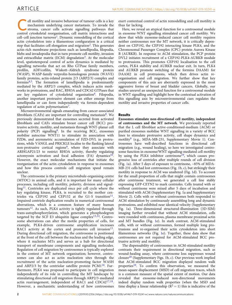
ResultsExosomes stimulate non-directional cell motility, independentof centrosomes and the MT network. We previously reportedthat the L cell fibroblast active-conditioned-medium (ACM) orpurified exosomes mobilise WNT signalling in a variety of BCClines to stimulate protrusive activity, cell shape dynamics andmotility9 (e.g., MDA-MB-231, Supplementary Movie 1). Cen-trosomes have well-described functions in directional cellmigration (e.g., wound healing), so here we investigated centro-some function in exosome/WNT signalling-mediated cell motilityby using the PLK4 inhibitor centrinone19, which causes pro-gressive loss of centrioles after multiple rounds of cell division(Fig. 1a). After 5 days of exposure to centrinone, ~85% of MDA-MB-231 cells had lost centrosomes (Fig. 1b, c), but strikingly theirmotility in response to ACM was unaltered (Fig. 1d). To accountfor the small proportion of cells that might contain centrosomesafter centrinone treatment, we generated a cell line stablyexpressing GFP-CETN2 to mark centrioles. Cells treated with orwithout centrinone were mixed after 5 days of incubation andstimulated with ACM (Supplementary Fig. 1a and SupplementaryMovie 2). Cells with or without centrosomes both responded toACM stimulation by continuously assembling long and dynamicprotrusions, and exhibited near identical velocity (SupplementaryFig. 1a). Three-dimensional structured-illumination (3D-SIM)imaging further revealed that without ACM stimulation, cellswere rounded with continuous, plasma membrane proximal actinand MT bundles (Fig. 1e). In stark contrast, ACM-stimulatedcells, with or without centrosomes, formed multiple long pro-trusions and re-organised their actin cytoskeleton into shortfilamentous networks (Fig. 1e). Together, these data show thatcentrosomes are not required for ACM-stimulated BCC pro-trusive activity and motility.
The dispensability of centrosomes in ACM-stimulated motilitycontrasts their requirement in directional migration, such aswound-healing assay, where centrosome loss suppresses woundclosure20 (Supplementary Figs. 1b, c). Our previous work impliedthat ACM-stimulated BCC migration displayed random walkproperties10. To confirm this observation, we measured themean-square displacement (MSD) of cell migration traces, whichis a common measure of the spatial extent of motion. Our datarevealed that exosome-induced non-directional movementsindeed display random walk properties (when the MSD andtime display a linear relationship (R2= 1) this is indicative of the
ARTICLE NATURE COMMUNICATIONS | https://doi.org/10.1038/s41467-019-10241-w
2 NATURE COMMUNICATIONS | (2019) 10:2356 | https://doi.org/10.1038/s41467-019-10241-w | www.nature.com/naturecommunications

total random movement) (Fig. 1f; Supplementary 1d)9,10. Wetherefore hypothesised that ACM-stimulated BCC migrationreflects an intrinsically different mechanism of cell motilitycompared with directional migration. Accordingly, we observedthat ACM significantly reduced closure rates (~13% at 18 h) inwound-healing assays, which suggests that ACM-induced non-
directional migration might compete with cellular pathwayscontrolling directional migration21 (Fig. 1g; SupplementaryFig. 1e). Surprisingly, disrupting the MT network, which iscrucial in directional migration20, with nocodazole (SupplementaryFig. 1f), did not inhibit ACM-induced motility (Fig. 1h).Furthermore, actin and MT network rearrangement at the cell
b
e
Cen
trin
one
CETN1CEP192Merge
Merge
a
α-Tubulin
α-T
ubul
in
Actin PCNT
DM
EM
(D
MS
O)
AC
M (
DM
SO
)A
CM
(ce
ntrin
one)
Act
in
DMSO DMSO (ROI) NOC NOC (ROI)i
Cancer cell
5 days centrinone treatment
Centriole
Exosome
Active-conditioned medium (ACM)
Cancer cell
ACM/culture medium (1:1)
Fibroblast (L cells)
Highly motilecancer cell
Spe
ed (
μm/h
)
h
Time (h)0 1 2 3 4 5 6 7 8 119 10 12 13 14 15 16 17 18
DMSO/NOC DMEM 6 h, + DMEM (DMSO) 12 hACM 6 h, + ACM (DMSO) 12 hACM 6 h, + ACM (NOC) 12 h
0
10
20
30
40
Spe
ed (
μm/h
)
j
Time (h)
DMSO/cytochalasin B/cytochalasin D
0 1 2 3 4 5 6 7 8 119 10 12 13 14 15 16 17 18
DMEM 3 h, + DMEM (DMSO) 15 hACM 3 h, + ACM (DMSO) 15 hACM 3 h, + ACM (cytochalasin B) 15 h
ACM 3 h, + ACM (cytochalasin D) 15 h
0
10
20
30
40
50
nsS
peed
(μm
/h)
c d
Cel
ls (
%)
DMSO Centrinone
Centriole number **** DMEMACM
DMSO DMSO Centrinone
0
20
40
60
80
100
120
0
20
40
= 01–4> 4 60
f
Mea
n sq
uare
d di
spla
cem
ent
120 2 4 6 8 10 14 16
PBSExosome R2 = 0.9912
R2 = 0.9926
Time (h)
0
4 × 10⁴
3 × 10⁴
2 × 10⁴
1 × 10⁴
18Time (h)
Are
a re
cove
red
(%)
ns
**
gDMEMACM
6 1200
20
40
60
80
100
DM
SO
Mer
ge
NATURE COMMUNICATIONS | https://doi.org/10.1038/s41467-019-10241-w ARTICLE
NATURE COMMUNICATIONS | (2019) 10:2356 | https://doi.org/10.1038/s41467-019-10241-w | www.nature.com/naturecommunications 3

cortex occurred in a similar fashion than control-stimulated cells(Fig. 1i). In stark contrast, disruption of the actin network usingcytochalasin B or cytochalasin D caused strong inhibition of cellmotility (Fig. 1j). Overall, these results show that ACM-stimulatednon-directional migration is actin dependent, but independent ofintact centrosomes and the MT network. Furthermore, theysuggest that ACM-WNT signalling driven motility response toextrinsic cues is distinct from conventional directional cell motilitypathways.
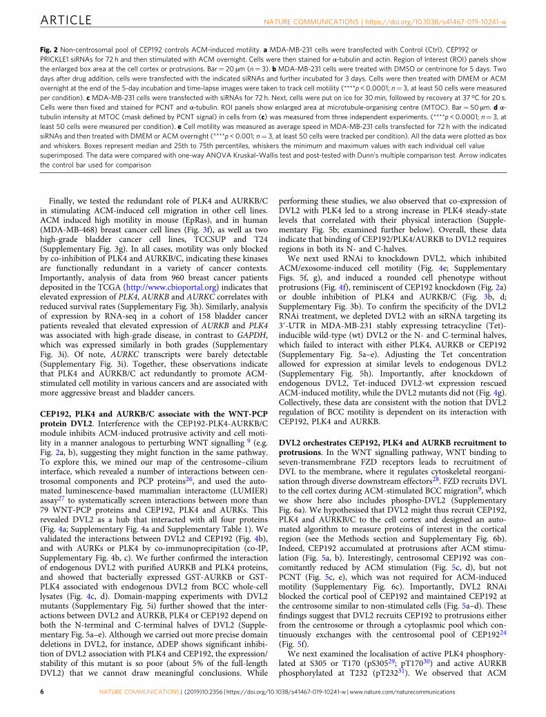
Centrosomal components are required for exosome-stimulatedcancer cell motility. The centriole duplication/maturation factorCEP192 exerts control on directed cell motility through increasedMT nucleation at centrosomes15,22. Surprisingly, in contrast tocentrosome loss, siRNA depletion of CEP192 inhibited ACM-induced protrusive activity (Fig. 2a; Supplementary Fig. 2a, b),and exosome-induced cell motility (Supplementary Fig. 2d).Moreover, in 3D culture, ACM induced protrusion formation in ahighly dynamic manner in siCtrl-treated cells, in contrast tosiCEP192-treated cells that were ACM stimulated (Supplemen-tary Fig. 2c, Supplementary Movie 3). Furthermore, CEP192depletion phenocopied knockdown of the PCP protein,PRICKLE1 (Fig. 2a)9,10, leading us to hypothesise that CEP192may affect WNT signalling in response to ACM in a centrosome-independent manner. To confirm this, we depleted centrosomesusing centrinone, and then knocked-down CEP192 prior to ACMstimulation (Fig. 2b). In contrast to control RNAi-treated cells,CEP192 depletion inhibited cell motility in the absence of cen-trosomes (Fig. 2b). We next evaluated the requirement ofCEP192-mediated MT nucleation in the context of ACM-stimulated motility. CEP192 is necessary for the recruitment ofNEDD1 to centrosomes, which in turn recruits gamma-tubulinring complexes and subsequent MT nucleation23,24. To assess MTregrowth activity, CEP192 or NEDD1 siRNA-treated MDA-MB-231 cells (Supplementary Fig. 2e) were incubated on ice to dis-assemble the MT network before shifting back to 37 oC to allowregrowth. As expected, knockdown of CEP192 orNEDD1 significantly reduced MT nucleation from centrosomes(Fig. 2c, d). However, unlike CEP192 RNAi treatment, knock-down of NEDD1 did not inhibit ACM-induced motility (Fig. 2e).Furthermore, to exclude the possibility that CEP192 down-regulation might alter exosome uptake, modification and
secretion, we determined the levels of endogenous WNT11loaded into exosomes upon their uptake into MDA-MB-231cells (Supplementary Fig. 2f). Our results indicate that controland CEP192 RNAi-treated BCCs process similar amounts ofCD81-positive exosomes (released by L cell fibroblasts) thatare loaded with endogenous WNT11 (Supplementary Fig. 2g).Thus, CEP192 is required for ACM-induced BCC motility in amanner independent of centrosomes and their MT nucleationactivity.
PLK4-AURKB/C redundantly regulate ACM-stimulated cellmotility. Elevated PLK4 expression is proposed to stimulate theinvasive properties of breast cancer cells via CEP192-mediatedMT nucleation at centrosomes15. Although centrinone, a highlyselective PLK4 inhibitor, did not affect motility (see Fig. 1a–e) inthe course of these studies, we observed that another PLK4inhibitor, CFI-40094525 potently inhibited ACM-induced move-ment (Fig. 3a) and reduced protrusive activity (Fig. 3b). SinceCFI-400945 also inhibits AURKB and AURKC25, we reasonedthat PLK4 and AURKB and/or C may act redundantly to regulateBCC motility in response to exosomes. To test this, we usedAZD1152 (AZD), an AURKB/C inhibitor together with cen-trinone (or centrinone B), and found that only co-inhibition ofAURKB/C and PLK4 prevented ACM-induced BCC migration(Fig. 3c) and suppressed protrusive activity (Fig. 3d). We thentargeted either PLK4 or the Aurora kinase family members (A, Band C) using siRNA. Knockdown of PLK4, single AURKs, or thecomplement of AURKs had no effect on protrusive activity andmotility (Supplementary Fig. 3a–d). However, when PLK4 andboth AURKB and AURKC were targeted, ACM-induced pro-trusive activity and cell motility were inhibited (SupplementaryFig. 3a, b).
We next exploited PLK4 and AURKB/C small-moleculeinhibitors to assess the reversibility of inhibition by performingwash-out experiments, where we tracked ACM-stimulated cellstreated with centrinone and AZD for 16 h before and afterwashout (Fig. 3e). While control cells remained highly motilethroughout, addition of centrinone and AZD suppressed motilitythat was rapidly recovered after washout (Fig. 3e). We noticedthat dual inhibition of PLK4 and AURKB required about 14 h tofully suppress ACM-induced cell motility, with a half-life decay at1.7 h. To rule out that this rather slow inhibition of motility
Fig. 1 Exosomes stimulate actin-based non-directional migration in a centrosome and MT-independent manner. a Experimental schematic. MDA-MB-231cells were treated with centrinone for 5 days to induce centrosome loss during division. Centrosome-less cells were treated with ACM. The protrusiveactivity and motility of individual cells were imaged and tracked. b MDA-MB-231 cells were treated with DMSO or centrinone for 5 days. CETN1(centrioles) and CEP192 (centrosomes) staining is shown. Bar= 20 µm. c Percentage of cells from (b) with > 4 centrioles, 1≤ centrioles≤ 4 and 0centrioles (n= 3 independent experiments analysing at least 100 cells per each replicate). dMDA-MB-231 cells were treated with DMSO or centrinone for5 days, followed by overnight incubation with the DMEM or ACM. Cell motility was measured as described in Methods and plotted as box and whiskers.Boxes represent median and 25th to 75th percentiles, whiskers the minimum and maximum values with each individual cell value superimposed. The datawere compared with one-way ANOVA Kruskal–Wallis test and post-tested with Dunn’s multiple comparison test. Arrow indicates the control bar used forcomparison (****p < 0.0001; n= 4, at least 50 cells were tracked per condition). e MDA-MB-231 cells from (d) were stained for α-tubulin, actin(phalloidin) and centrosomes (PCNT). Arrowhead indicates cell protrusion. Bar= 20 µm, inset box 3.88 × 3.88 μm (n= 3). f Mean-squared displacement(MSD) of cells stimulated with PBS (light blue) or purified exosomes (light red). Linear regression analysis of MSD was preformed (dark blue line is PBS,dark red line is purified exosomes). MSD R2= 1 indicates a total random movement (n= 3, at least 40 cells were tracked per condition). g Quantitativedata from wound-healing assay on MDA-MB-231 cells treated overnight with DMEM or ACM are shown in Supplementary Fig. 1e. Graph shows mean arearecovered. The data were analysed with two-way ANOVA post-tested with Bonferroni test (*p < 0.05; n= 3, at least 30 regions were measured percondition). h MDA-MB-231 cells were stimulated with DMEM or ACM overnight. Cell motility was tracked for 6 h. Then, the same cells were furthertracked after being treated with DMSO or Nocodazole for 12 h. Running average speed was plotted (n= 3, at least 40 cells were tracked per condition).i MDA-MB-231 cells were stimulated with ACM overnight. DMSO or NOC were added for the last 2 h of stimulation. Actin (red) and α-tubulin (green)staining are shown. Region of interest (ROI) shows the enlarged area of the selected cell protrusion. Bar= 20 µm (n= 3) j MDA-MB-231 cells werestimulated with DMEM or ACM overnight. Cell motility was tracked for 3 h, then cells were treated with DMSO, cytochalasin B or cytochalasin D andfurther tracked for 15 h. Running average speed was plotted (n= 3, at least 40 cells were tracked per condition). The data from c, f, g, h and j are plotted asmean ± s.e.m.
ARTICLE NATURE COMMUNICATIONS | https://doi.org/10.1038/s41467-019-10241-w
4 NATURE COMMUNICATIONS | (2019) 10:2356 | https://doi.org/10.1038/s41467-019-10241-w | www.nature.com/naturecommunications

occurred indirectly through the inhibition of AURKB/PLK4-controlled transcription, we therefore tested the effect ofinhibiting transcription using actinomycin D on cell motility.Although actinomycin D treatment did inhibit ACM-induced cellmotility, the inhibition kinetics was much slower (about 5.5 h fora half-life decay, Supplementary Fig. 3e, f). Although we cannot
exclude the possibility at this stage that AURKB/PLK4 alsoregulate cell motility via transcription, our preferred hypothesis,considering that AURKB/PLK4 localisation and their activation isinduced upon ACM stimulation (see below), is that these kinaseslikely have direct roles in actin rearrangement that require furtherinvestigation.
a c
Ctr
lC
EP
192
ActinMerge α-Tubulin
PR
ICK
LE1
Ctr
l (R
OI)
CE
P19
2 (R
OI)
PR
ICK
LE1
(RO
I)
(RNAi)
(RNAi)
α-Tubulin α-Tubulin (ROIs)
1
1
1
1
2
12
2
2
1
2
2
Ctrl
CEP192
NEDD1
b
e
d
Ctrl CEP192
α-T
ubul
in in
tens
ity ****
****
NEDD1(RNAi)0
1.2 × 10⁶
8 × 10⁵
4 × 10⁵
****
ns DMEM
ACM
Spe
ed (
μm/h
)
Ctrl Ctrl CEP192 NEDD1(RNAi)0
20
40
50
10
30****
****
Ctrl CtrlCtrl CtrlCEP192 CEP192PRICKLE1 PRICKLE1
Spe
ed (
μm/h
)
(RNAi)
Centrosome present Centrosome depleted
0
20
40
10
30
50
****
****
****
****
****
DMEM (DMSO)ACM (DMSO)
DMEM (centrinone)ACM (centrinone)
Transfect siRNAsAdd DMSO/centrinone
2 days
Stimulate with DMEM or ACM (DMSO/centrinone)
48 h 104 h3 days
Imaging
0 h 120 h
NATURE COMMUNICATIONS | https://doi.org/10.1038/s41467-019-10241-w ARTICLE
NATURE COMMUNICATIONS | (2019) 10:2356 | https://doi.org/10.1038/s41467-019-10241-w | www.nature.com/naturecommunications 5
Finally, we tested the redundant role of PLK4 and AURKB/Cin stimulating ACM-induced cell migration in other cell lines.ACM induced high motility in mouse (EpRas), and in human(MDA-MB-468) breast cancer cell lines (Fig. 3f), as well as twohigh-grade bladder cancer cell lines, TCCSUP and T24(Supplementary Fig. 3g). In all cases, motility was only blockedby co-inhibition of PLK4 and AURKB/C, indicating these kinasesare functionally redundant in a variety of cancer contexts.Importantly, analysis of data from 960 breast cancer patientsdeposited in the TCGA (http://www.cbioportal.org) indicates thatelevated expression of PLK4, AURKB and AURKC correlates withreduced survival rates (Supplementary Fig. 3h). Similarly, analysisof expression by RNA-seq in a cohort of 158 bladder cancerpatients revealed that elevated expression of AURKB and PLK4was associated with high-grade disease, in contrast to GAPDH,which was expressed similarly in both grades (SupplementaryFig. 3i). Of note, AURKC transcripts were barely detectable(Supplementary Fig. 3i). Together, these observations indicatethat PLK4 and AURKB/C act redundantly to promote ACM-stimulated cell motility in various cancers and are associated withmore aggressive breast and bladder cancers.
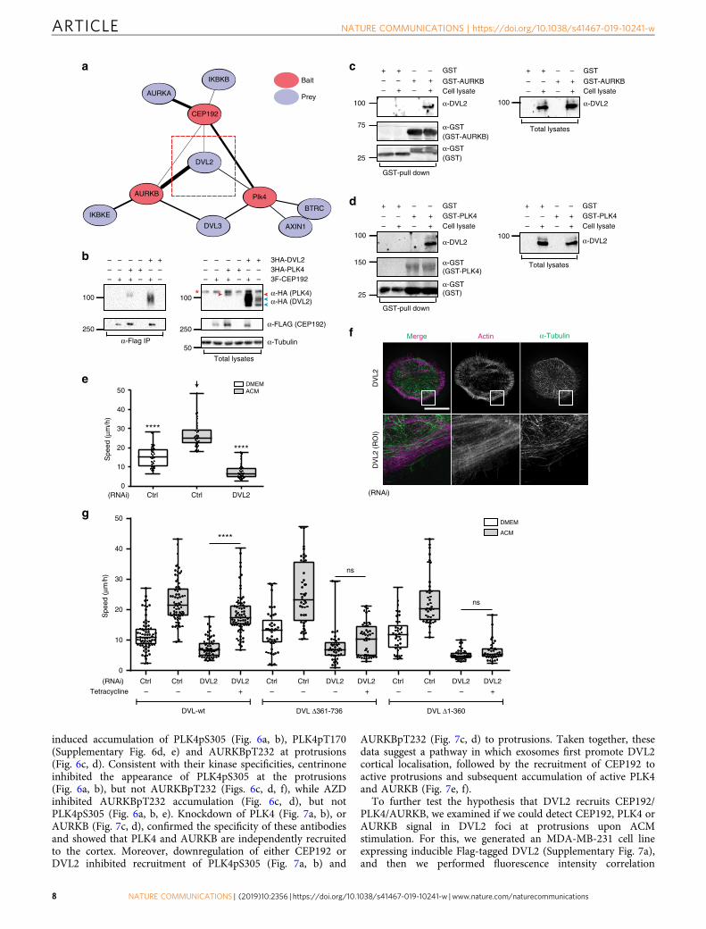
CEP192, PLK4 and AURKB/C associate with the WNT-PCPprotein DVL2. Interference with the CEP192-PLK4-AURKB/Cmodule inhibits ACM-induced protrusive activity and cell moti-lity in a manner analogous to perturbing WNT signalling 9 (e.g.Fig. 2a, b), suggesting they might function in the same pathway.To explore this, we mined our map of the centrosome–ciliuminterface, which revealed a number of interactions between cen-trosomal components and PCP proteins26, and used the auto-mated luminescence-based mammalian interactome (LUMIER)assay27 to systematically screen interactions between more than79 WNT-PCP proteins and CEP192, PLK4 and AURKs. Thisrevealed DVL2 as a hub that interacted with all four proteins(Fig. 4a; Supplementary Fig. 4a and Supplementary Table 1). Wevalidated the interactions between DVL2 and CEP192 (Fig. 4b),and with AURKs or PLK4 by co-immunoprecipitation (co-IP,Supplementary Fig. 4b, c). We further confirmed the interactionof endogenous DVL2 with purified AURKB and PLK4 proteins,and showed that bacterially expressed GST-AURKB or GST-PLK4 associated with endogenous DVL2 from BCC whole-celllysates (Fig. 4c, d). Domain-mapping experiments with DVL2mutants (Supplementary Fig. 5i) further showed that the inter-actions between DVL2 and AURKB, PLK4 or CEP192 depend onboth the N-terminal and C-terminal halves of DVL2 (Supple-mentary Fig. 5a–e). Although we carried out more precise domaindeletions in DVL2, for instance, ΔDEP shows significant inhibi-tion of DVL2 association with PLK4 and CEP192, the expression/stability of this mutant is so poor (about 5% of the full-lengthDVL2) that we cannot draw meaningful conclusions. While
performing these studies, we also observed that co-expression ofDVL2 with PLK4 led to a strong increase in PLK4 steady-statelevels that correlated with their physical interaction (Supple-mentary Fig. 5b; examined further below). Overall, these dataindicate that binding of CEP192/PLK4/AURKB to DVL2 requiresregions in both its N- and C-halves.
We next used RNAi to knockdown DVL2, which inhibitedACM/exosome-induced cell motility (Fig. 4e; SupplementaryFigs. 5f, g), and induced a rounded cell phenotype withoutprotrusions (Fig. 4f), reminiscent of CEP192 knockdown (Fig. 2a)or double inhibition of PLK4 and AURKB/C (Fig. 3b, d;Supplementary Fig. 3b). To confirm the specificity of the DVL2RNAi treatment, we depleted DVL2 with an siRNA targeting its3′-UTR in MDA-MB-231 stably expressing tetracycline (Tet)-inducible wild-type (wt) DVL2 or the N- and C-terminal halves,which failed to interact with either PLK4, AURKB or CEP192(Supplementary Fig. 5a–e). Adjusting the Tet concentrationallowed for expression at similar levels to endogenous DVL2(Supplementary Fig. 5h). Importantly, after knockdown ofendogenous DVL2, Tet-induced DVL2-wt expression rescuedACM-induced motility, while the DVL2 mutants did not (Fig. 4g).Collectively, these data are consistent with the notion that DVL2regulation of BCC motility is dependent on its interaction withCEP192, PLK4 and AURKB.
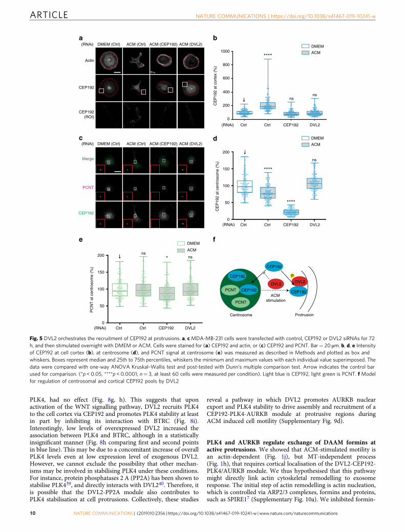
DVL2 orchestrates CEP192, PLK4 and AURKB recruitment toprotrusions. In the WNT signalling pathway, WNT binding toseven-transmembrane FZD receptors leads to recruitment ofDVL to the membrane, where it regulates cytoskeletal reorgani-sation through diverse downstream effectors28. FZD recruits DVLto the cell cortex during ACM-stimulated BCC migration9, whichwe show here also includes phospho-DVL2 (SupplementaryFig. 6a). We hypothesised that DVL2 might thus recruit CEP192,PLK4 and AURKB/C to the cell cortex and designed an auto-mated algorithm to measure proteins of interest in the corticalregion (see the Methods section and Supplementary Fig. 6b).Indeed, CEP192 accumulated at protrusions after ACM stimu-lation (Fig. 5a, b). Interestingly, centrosomal CEP192 was con-comitantly reduced by ACM stimulation (Fig. 5c, d), but notPCNT (Fig. 5c, e), which was not required for ACM-inducedmotility (Supplementary Fig. 6c). Importantly, DVL2 RNAiblocked the cortical pool of CEP192 and maintained CEP192 atthe centrosome similar to non-stimulated cells (Fig. 5a–d). Thesefindings suggest that DVL2 recruits CEP192 to protrusions eitherfrom the centrosome or through a cytoplasmic pool which con-tinuously exchanges with the centrosomal pool of CEP19224
(Fig. 5f).We next examined the localisation of active PLK4 phosphory-
lated at S305 or T170 (pS30529; pT17030) and active AURKBphosphorylated at T232 (pT23231). We observed that ACM
Fig. 2 Non-centrosomal pool of CEP192 controls ACM-induced motility. a MDA-MB-231 cells were transfected with Control (Ctrl), CEP192 orPRICKLE1 siRNAs for 72 h and then stimulated with ACM overnight. Cells were then stained for α-tubulin and actin. Region of Interest (ROI) panels showthe enlarged box area at the cell cortex or protrusions. Bar= 20 µm (n= 3). b MDA-MB-231 cells were treated with DMSO or centrinone for 5 days. Twodays after drug addition, cells were transfected with the indicated siRNAs and further incubated for 3 days. Cells were then treated with DMEM or ACMovernight at the end of the 5-day incubation and time-lapse images were taken to track cell motility (****p < 0.0001; n= 3, at least 50 cells were measuredper condition). c MDA-MB-231 cells were transfected with siRNAs for 72 h. Next, cells were put on ice for 30min, followed by recovery at 37 oC for 20 s.Cells were then fixed and stained for PCNT and α-tubulin. ROI panels show enlarged area at microtubule-organising centre (MTOC). Bar= 50 µm. d α-tubulin intensity at MTOC (mask defined by PCNT signal) in cells from (c) was measured from three independent experiments. (****p < 0.0001; n= 3, atleast 50 cells were measured per condition). e Cell motility was measured as average speed in MDA-MB-231 cells transfected for 72 h with the indicatedsiRNAs and then treated with DMEM or ACM overnight (****p < 0.001; n= 3, at least 50 cells were tracked per condition). All the data were plotted as boxand whiskers. Boxes represent median and 25th to 75th percentiles, whiskers the minimum and maximum values with each individual cell valuesuperimposed. The data were compared with one-way ANOVA Kruskal–Wallis test and post-tested with Dunn’s multiple comparison test. Arrow indicatesthe control bar used for comparison
ARTICLE NATURE COMMUNICATIONS | https://doi.org/10.1038/s41467-019-10241-w
6 NATURE COMMUNICATIONS | (2019) 10:2356 | https://doi.org/10.1038/s41467-019-10241-w | www.nature.com/naturecommunications

a b
AC
M (
CF
I-40
0945
)A
CM
(D
MS
O)
Merge Actin α-Tubulin
Spe
ed (
μm/h
)
DMSO DMSO 10 nM 20 nM 50 nM 100 nM 200 nM 1 μM
****
ns****
****
****
CFI-400945
DMEM
ACM
0
10
20
30
40
50
nsns
c
Spe
ed (
μm/h
)
DMEM
ACM
DMSO DMSO AZD A B AZD+A AZD+B
A = centrinoneB = centrinone B
0
10
20
30
40
e
0 10 20 30 40
Spe
ed (
μm/h
)
Time (h)
ACM (centrinone + AZD) ACM (DMSO)
Add drugsWashout
DMEM (DMSO)
0
10
20
30
40
Merge Actin α-Tubulin
DM
SO
Cen
trin
one
AZ
DC
entr
inon
e +
AZ
D
d50
f
Spe
ed (
μm/h
)
DMEM
ACM
DMSO DMSO Centrinone AZD Centrinone + AZD
EpRas
****
ns ns
****
0
20
40
60
Spe
ed (
μm/h
)
DMEM
ACM
DMSO DMSO Centrinone AZD Centrinone + AZD
MDA-MB-468
**
ns
ns
****
0
10
20
30
****
ns
****
**** ****
ns
Fig. 3 PLK4 and AURKB/C redundantly modulate ACM-induced cancer cell motility. aMDA-MB-231 cells were treated with DMSO or different concentrationsof CFI-400945 (PLK4 and AURKB/C inhibitor), in the presence of DMEM or ACM (****p < 0.0001; n= 3, at least 50 cells were tracked per condition). bMDA-MB-231 cells were treated with DMSO or 100 nM CFI-400945 in the presence of ACM stimulation. Cells were stained for α-tubulin and actin, and enlargedimages showed a cortical region of the cell. Bar= 20 µm (n= 3). c MDA-MB-231 cells were treated with centrinone, centrinone B (PLK4 inhibitors), AZD(AURKB/C inhibitor) or their combination, in the presence of DMEM or ACM overnight (***p < 0.0001; n= 3, at least 50 cells were tracked per condition).d Cells from (c) were stained for α-tubulin and actin, and cortical regions were enlarged on right side panels. Bar= 20 µm. e Cells were incubated with DMEMor ACM overnight. At 6 h of taking time-lapse images, DMSO or inhibitors were added to cells and incubation continued for 14 h more, then DMSO or inhibitorswere washed out and cells were further incubated in DMEM or ACM for another 16 h. Running average speed was plotted as mean ± s.e.m. (n= 4, at least 50cells were tracked per condition) f Breast cancer cell lines EpRas and MDA-MB-468 were treated with DMEM or ACM in the presence of DMSO or inhibitors(**p < 0.01, ****p < 0.0001; n= 3, at least 40 cells were tracked per condition). All the data were plotted as box and whiskers. Boxes represent median and 25thto 75th percentiles, whiskers the minimum and maximum values with each individual cell value superimposed. The data were compared with one-way ANOVAKruskal–Wallis test and post-tested with Dunn’s multiple comparison test. Arrow indicates the control bar used for comparison
NATURE COMMUNICATIONS | https://doi.org/10.1038/s41467-019-10241-w ARTICLE
NATURE COMMUNICATIONS | (2019) 10:2356 | https://doi.org/10.1038/s41467-019-10241-w | www.nature.com/naturecommunications 7
induced accumulation of PLK4pS305 (Fig. 6a, b), PLK4pT170(Supplementary Fig. 6d, e) and AURKBpT232 at protrusions(Fig. 6c, d). Consistent with their kinase specificities, centrinoneinhibited the appearance of PLK4pS305 at the protrusions(Fig. 6a, b), but not AURKBpT232 (Figs. 6c, d, f), while AZDinhibited AURKBpT232 accumulation (Fig. 6c, d), but notPLK4pS305 (Fig. 6a, b, e). Knockdown of PLK4 (Fig. 7a, b), orAURKB (Fig. 7c, d), confirmed the specificity of these antibodiesand showed that PLK4 and AURKB are independently recruitedto the cortex. Moreover, downregulation of either CEP192 orDVL2 inhibited recruitment of PLK4pS305 (Fig. 7a, b) and
AURKBpT232 (Fig. 7c, d) to protrusions. Taken together, thesedata suggest a pathway in which exosomes first promote DVL2cortical localisation, followed by the recruitment of CEP192 toactive protrusions and subsequent accumulation of active PLK4and AURKB (Fig. 7e, f).
To further test the hypothesis that DVL2 recruits CEP192/PLK4/AURKB, we examined if we could detect CEP192, PLK4 orAURKB signal in DVL2 foci at protrusions upon ACMstimulation. For this, we generated an MDA-MB-231 cell lineexpressing inducible Flag-tagged DVL2 (Supplementary Fig. 7a),and then we performed fluorescence intensity correlation
Spe
ed (
μm/h
)
Ctrl Ctrl DVL2 DVL2 Ctrl Ctrl DVL2 DVL2 Ctrl Ctrl DVL2 DVL2– – – + – –– + – – – +Tetracycline
DVL-wt DVL Δ361-736 DVL Δ1-360
(RNAi)
0
10
20
30
40
50
a
AURKB
IKBKEBTRC
Plk4
DVL3 AXIN1
DVL2
IKBKB
AURKA
CEP192
Bait
Prey
g
****
ns
ns
d
c
f
e
(RNAi)
DV
L2D
VL2
(R
OI)
α-TubulinActinMerge
GST
Cell lysateGST-PLK4
α-DVL2
– + – ++ +– –
+ + – –
Total lysates
GST
Cell lysateGST-PLK4
α-DVL2
– + – ++ +– –
+ + – –
GST-pull down
b ++– – – –– – ++ – –– ++ –+–
α-Flag IP
++– – – –– – ++ – –– ++ –+–
3HA-DVL23HA-PLK43F-CEP192
α-HA (PLK4)α-HA (DVL2)
α-FLAG (CEP192)
α-Tubulin
Total lysates
*
Ctrl Ctrl DVL2
Spe
ed (
μm/h
)
****
****
(RNAi)0
10
20
30
40
50
GST
Cell lysateGST-AURKB
α-DVL2
– + – ++ +– –
+ + – –
Total lysates
GST
Cell lysateGST-AURKB
α-DVL2
(GST-AURKB)
– + – ++ +– –
+ + – –
GST-pull down
α-GST
(GST)α-GST
α-GST(GST-PLK4)
α-GST(GST)
100
250
100
250
50
100
75
25
100
100
150
25
100
DMEMACM
DMEM
ACM
ARTICLE NATURE COMMUNICATIONS | https://doi.org/10.1038/s41467-019-10241-w
8 NATURE COMMUNICATIONS | (2019) 10:2356 | https://doi.org/10.1038/s41467-019-10241-w | www.nature.com/naturecommunications
profiling between DVL2 (Flag) and the signal from endogenousCEP192 or the active kinases PLK4 and AURKB at protrusionsusing the algorithm illustrated in Supplementary Fig. 7b (seeMethods section for details). The signal correlation coefficienttakes into consideration not only the overlapping area ofcolocalization but also the signal intensity. Our data indicatethat upon ACM stimulation, the induced DVL2(Flag) signalassociated with CEP192 or active PLK4 and AURKB wassignificantly higher than the random signal detected by the Flagantibody in uninduced cells at protrusions. This indicates that afraction of endogenous CEP192 and the active kinases colocalizewith DVL2 at the cell cortex in ACM-treated cells (Supplemen-tary Fig. 7c–g).
We next investigated the biological significance of theinteractions between components of the DVL2-CEP192-PLK4/AURKB module. As mentioned above, we observed similarbinding patterns between DVL2 mutants and CEP192 (LUMIERassay, Supplementary Fig. 5c), PLK4 and AURKB (co-IP,Supplementary Fig. 5a, b; LUMER assay, Supplementary Fig. 5d,e). Moreover, depletion of DVL2 or CEP192 leads to loss ofaccumulation of active PLK4 and AURKB at the cell cortex inACM-treated cells (Fig. 7a–d). We thus hypothesised thatCEP192 could bridge DVL2 and PLK4/AURKB. In support ofthis hypothesis, we found that CEP192 depletion by RNAisignificantly reduced DVL2 binding to PLK4 and AURKB usingLUMIER assay (Supplementary Fig. 8a). To build on this finding,we took advantage of previous work on CEP192 isoforms, whichindicated that only the longest isoform (Isoform-1, AA 1–2537)interacts with PLK4 at AA 190–24032 (Supplementary Fig. 8k).This led us to posit that CEP192 isoform-1 specifically recruitsPLK4 to cell protrusions. To test this, we first determined thebinding profile of two CEP192 isoforms to DVL2, PLK4 andAURKB. Our LUMIER data show that both, CEP192 isoform-1and a shorter isoform-2 (1–1941) interact with DVL2 andAURKB, but only isoform-1 associates with PLK4 (Supplemen-tary Fig. 8b). Strikingly, specific knockdown of CEP192 isoform-1(Supplementary Fig. 8c) resulted in no recruitment of active PLK4at cell protrusions upon ACM treatment, while the recruitment ofactive AURKB and other CEP192 isoforms was not affected(Supplementary Fig. 8e–j). Furthermore, cells depleted of CEP192isoform-1 treated with ACM displayed high cell motility that isdependent on AURKB, as AZD treatment inhibited cell migration(Supplementary Fig. 8d). Taken together, our data indicate thatupon ACM treatment, DVL2 is required for the recruitment of allCEP192 isoforms to cell protrusions, where CEP192 isoform-1specifically recruits active PLK4, while other CEP192 isoformscan recruit active AURKB.
Precise spatial and temporal control of AURKB is critical for itsfunction during M phase to ensure cell cycle progression andduring interphase is predominantly in the nucleus33,34. Since weobserved active AURKB at protrusions, we hypothesised thatAURKB might undergo nuclear export upon stimulation ofBCCs. To test this, we measured the nuclear to cytoplasmic ratioof AURKB and observed nuclear AURKB decreased ~35% afterACM stimulation in a DVL2-dependent manner (Fig. 8a, c).Importantly, ACM stimulation did not alter cell cycle progres-sion, indicating that the spatial distribution of AURKB inmigrating BCC is a direct consequence of ACM stimulation(Supplementary Fig. 9a). Although the precise mechanism of howACM induces DVL2-dependent AURKB nuclear export remainsunclear, DVL2 is known to shuttle between the nucleus andcytoplasm35. DVL2 contains a conserved nuclear export sequenceand a nuclear localisation sequence, and its nuclear localisation isrequired in the canonical WNT signalling pathway36. Interest-ingly, a recent study demonstrated that mutating DVL2 nuclearexport sequence results in nuclear YAP accumulation37. It will beintriguing to further investigate if DVL2 also directly regulatesAURKB nuclear export though a similar mechanism.
Next, we investigated the significance of AURKB nuclearexport in controlling ACM-induced cell motility. AURKB isexported from the nucleus via CRM1-mediated nucleocytoplas-mic shuttling34, which can be blocked by leptomycin B (LMB).LMB treatment prior to ACM stimulation significantly inhibitedAURKB nuclear export (Fig. 8b, d), but did not inhibit cellmotility on its own (Fig. 8e). This was not surprising, since weestablished that PLK4 and AURKB act redundantly. Accordingly,we observed that the addition of both centrinone and LMBsignificantly reduced cell motility (Fig. 8e). These data indicatethat ACM stimulation induces DVL2-dependent AURKB exportfrom the nucleus and its subsequent accumulation at protrusions.
PLK4 levels are tightly controlled by a negative feedback loopthat prevents centriole overduplication by coupling activation andautophosphorylation to degradation38. As noted in Supplemen-tary Fig. 5b, co-expression of DVL2 and PLK4 significantlyincreased PLK4 steady-state levels, suggesting DVL2 mightstabilise PLK4 at protrusions. Moreover, induced expression ofDVL2 in a tetracycline-responsive cell line also noticeablyincreased endogenous PLK4 level (p= 0.125, SupplementaryFig. 9b, c). We thus hypothesised that DVL2 may disrupt PLK4interaction with BTRC, the substrate-recognition moiety of theSCF complex of its E3 ubiquitin ligase13,14. We observed thatincreased expression of exogenous DVL2-wt led to increasedPLK4 levels with a concomitant loss of PLK4-BTRC interaction,while expression of the DVL2 N-terminus half, that fails to bind
Fig. 4 Dishevelled 2 controls ACM-induced cancer cell motility by binding to PLK4, AURKB and CEP192. a Network graph for selected protein interactionsidentified from a LUMIER screen testing CEP192, PLK4 and AURKs against a collection of 3 Flag-tagged WNT-PCP and centrosomal factors. Edge widthreflects the normalised LUMIER intensity ratio that indicates interaction strength (n= 2). b HEK293T cells were co-transfected with 3Flag-CEP192 and3HA-PLK4 (positive control) or 3HA-DVL2 as indicated. Cell lysates were immunoprecipitated with anti-Flag antibody and 3HA-DVL2 or 3HA-PLK4 weredetected by western blotting. Red arrowhead indicates band corresponding to 3HA-PLK4, blue arrowheads indicate 3HA-DVL2 bands (non-phosphorylatedand phosphorylated forms), red star on the left side indicates non-specific band (n= 3). c, d GST, GST-AURKB (c) and GST-PLK4 (d) were bacteriallyexpressed, purified and incubated with or without cell lysates from MDA-MB-231 cells. GST pull down was performed and endogenous DVL2 was detectedby western blotting. (n= 4 for each). e MDA-MB-231 cells were treated with control or DVL2 siRNAs for 72 h and then stimulated with DMEM or ACMovernight. Cell motility was measured as described in Methods and analysed with one-way ANOVA Kruskal–Wallis test and post-tested with Dunn’smultiple comparison test. Arrow indicates the control bar used for comparison (****p < 0.0001; n= 3, at least 40 cells were tracked per condition). f Cellsfrom (e) were stained with actin and α-tubulin. Enlarged box areas of cortical regions are shown as ROIs in lower panels. Bar= 20 µm (n= 3). gMDA-MB-231 cells stably expressing tetracycline inducible C-terminally HA-tagged DVL2-wt, DVL2-N terminus (Δ361–736) or DVL2-C terminus (Δ1–360) weretransfected with control or DVL2 siRNA. After 72 h, cells were treated with or without tetracycline in the presence of DMEM or ACM as indicated. Cellmotility was measured as described in Methods and analysed by Mann–Whitney U test two-tailed (****p < 0.0001; n= 3, at least 50 cells were tracked percondition). All the data are plotted as box and whiskers. Boxes represent median and 25th to 75th percentiles, whiskers the minimum and maximum valueswith each individual cell value superimposed
NATURE COMMUNICATIONS | https://doi.org/10.1038/s41467-019-10241-w ARTICLE
NATURE COMMUNICATIONS | (2019) 10:2356 | https://doi.org/10.1038/s41467-019-10241-w | www.nature.com/naturecommunications 9
PLK4, had no effect (Fig. 8g, h). This suggests that uponactivation of the WNT signalling pathway, DVL2 recruits PLK4to the cell cortex via CEP192 and promotes PLK4 stability at leastin part by inhibiting its interaction with BTRC (Fig. 8i).Interestingly, low levels of overexpressed DVL2 increased theassociation between PLK4 and BTRC, although in a statisticallyinsignificant manner (Fig. 8h comparing first and second pointsin blue line). This may be due to a concomitant increase of overallPLK4 levels even at low expression level of exogenous DVL2.However, we cannot exclude the possibility that other mechan-isms may be involved in stabilising PLK4 under these conditions.For instance, protein phosphatases 2 A (PP2A) has been shown tostabilise PLK439, and directly interacts with DVL240. Therefore, itis possible that the DVL2-PP2A module also contributes toPLK4 stabilisation at cell protrusions. Collectively, these studies
reveal a pathway in which DVL2 promotes AURKB nuclearexport and PLK4 stability to drive assembly and recruitment of aCEP192-PLK4-AURKB module at protrusive regions duringACM induced cell motility (Supplementary Fig. 9d).
PLK4 and AURKB regulate exchange of DAAM formins atactive protrusions. We showed that ACM-stimulated motility isan actin-dependent (Fig. 1j), but MT-independent process(Fig. 1h), that requires cortical localisation of the DVL2-CEP192-PLK4/AURKB module. We thus hypothesised that this pathwaymight directly link actin cytoskeletal remodelling to exosomeresponse. The initial step of actin remodelling is actin nucleation,which is controlled via ARP2/3 complexes, formins and proteins,such as SPIRE17 (Supplementary Fig. 10a). We inhibited formin-
b
d
fe
a
c
Actin
CEP192
CEP192(ROI)
(RNAi) ACM (Ctrl) ACM (CEP192) ACM (DVL2)
CEP192
PCNT
Merge
CE
P19
2 at
cen
tros
ome
(%)
****
****
ns
PC
NT
at c
entr
osom
e (%
)
nsns*
DMEM
ACM
(RNAi)
Ctrl Ctrl CEP192 DVL2
Ctrl Ctrl CEP192 DVL2
Ctrl Ctrl CEP192 DVL2
CE
P19
2 at
cor
tex
(%)
****
nsns
DMEM
ACM
(RNAi)
0
200
800
400
0
50
100
150
ProtrusionCentrosome
CEP192
CEP192
DVL2
ACM stimulation
PCNT
PCNT
DVL2
CEP192
CEP192
P
600
1000
0
50
100
150
200
200
(RNAi)
DMEM
ACM
DMEM (Ctrl)
(RNAi) ACM (Ctrl) ACM (CEP192) ACM (DVL2)DMEM (Ctrl)
Fig. 5 DVL2 orchestrates the recruitment of CEP192 at protrusions. a, c MDA-MB-231 cells were transfected with control, CEP192 or DVL2 siRNAs for 72h, and then stimulated overnight with DMEM or ACM. Cells were stained for (a) CEP192 and actin, or (c) CEP192 and PCNT. Bar= 20 µm. b, d, e Intensityof CEP192 at cell cortex (b), at centrosome (d), and PCNT signal at centrosome (e) was measured as described in Methods and plotted as box andwhiskers. Boxes represent median and 25th to 75th percentiles, whiskers the minimum and maximum values with each individual value superimposed. Thedata were compared with one-way ANOVA Kruskal–Wallis test and post-tested with Dunn’s multiple comparison test. Arrow indicates the control barused for comparison. (*p < 0.05, ****p < 0.0001; n= 3, at least 60 cells were measured per condition). Light blue is CEP192, light green is PCNT. f Modelfor regulation of centrosomal and cortical CEP192 pools by DVL2
ARTICLE NATURE COMMUNICATIONS | https://doi.org/10.1038/s41467-019-10241-w
10 NATURE COMMUNICATIONS | (2019) 10:2356 | https://doi.org/10.1038/s41467-019-10241-w | www.nature.com/naturecommunications

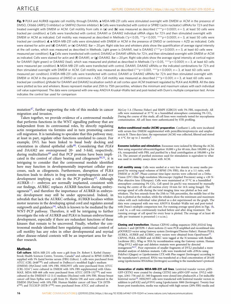
and ARP2/3-dependent actin nucleation using the selectiveinhibitors SIMFH2 or CK666, respectively (Fig. 9a), while theSPIRE1 pathway was inhibited using RNAi (Fig. 9b; Supple-mentary Fig. 10b). Our results showed that SMIFH2, but notCK666 or SPIRE1 RNAi, inhibited ACM-induced motility,
suggesting this is a formin-dependent process. Two formins,DAAM1 and DAAM2, have thus far been linked to DVL andWNT signalling in the context of convergent extension move-ments41. Interestingly. downregulation of DAAM1 by RNAi(Supplementary Fig. 10g) increased cell motility (Fig. 9c) in
b
DMSO AZD
pPLK
4 (S
305)
at c
orte
x (%
)
DMEM
ACM
********
ns
aDMEM (DMSO) ACM (DMSO) ACM (centrinone) ACM (AZD)
Actin
pPLK4S305
pPLK4S305(ROI) 0
200
400
600
800
1000
CentrinoneDMSO
d
e f
c
PLK4
PLK4
P
For
mpr
otru
sion
F
orm
prot
rusi
on
Protrusion
AZD1152
AURKB
P
P
Centrinone AURKB
For
mpr
otru
sion
F
orm
prot
rusi
on
Protrusion
AURKB
PLK4
P
P
P
DMSO AZD
pAU
RK
B (
T23
2) a
t cor
tex
(%)
DMEM
ACM
**** ****
ns
0
100
300
400
200
Actin
pAURKBT232
pAURKBT232(ROI)
DMEM (DMSO) ACM (DMSO) ACM (centrinone) ACM (AZD)
CentrinoneDMSO
Fig. 6 PLK4 and AURKB accumulation at cell protrusions depends on their specific activity. a, cMDA-MB-231 cells were stimulated with DMEM or ACM inthe presence of DMSO, centrinone or AZD. Cells were stained for actin and (a) phospho-PLK4 (S305), or (c) phospho-AURKB (T232). Bar= 20 µm. b, dIntensity at cortical region for (b) phospho-PLK4 (S305), or (d) phospho-AURKB (T232) from cells in (a) and (c) was measured. The data was analysed asdescribed in Methods and plotted as box and-whiskers. Boxes represent median and 25th to 75th percentiles, whiskers the minimum and maximum valueswith each individual value superimposed. The data were compared with one-way ANOVA Kruskal–Wallis test and post-tested with Dunn’s multiplecomparison test. Arrow indicates the control bar used for comparison (****p < 0.0001; n= 3, at least 60 cells were measured per condition). Magenta isphospho-PLK4, dark green is phospho-AURKB. Models for phospho-PLK4 (S305) (e) and phospho-AURKB (T232) (f) localisation at the cell cortex topromote protrusion formation that occurs independently from the other kinase activity
NATURE COMMUNICATIONS | https://doi.org/10.1038/s41467-019-10241-w ARTICLE
NATURE COMMUNICATIONS | (2019) 10:2356 | https://doi.org/10.1038/s41467-019-10241-w | www.nature.com/naturecommunications 11

unstimulated cells and was not further stimulated by ACM. Incontrast, DAAM2 RNAi (Supplementary Fig. 10h) blocked ACM-induced cell motility (Fig. 9c). Similar results were obtained usingthree independent siRNAs targeting either DAAM1 or DAAM2.These data indicate that ACM-induced cell migration is an actin-dependent process that is regulated by the formins DAAM1and DAAM2.
As DAAM2 was required for ACM-induced motility, andDAAM1 restrained motility in unstimulated cells (Fig. 9c), weconsidered that DAAM1 and DAAM2 might act in a switch-likemanner during protrusion formation in motile cells. To test this,we monitored DAAM1 and DAAM2 levels, which revealed thatwhile DAAM1 was at the cell cortex in unstimulated cells, it wasdepleted from ACM-induced protrusions (Fig. 9d, f). In stark
c
aRNAi
RNAi
DMEM (Ctrl) ACM (Ctrl) ACM (PLK4) ACM (AURKB) ACM (CEP192) ACM (DVL2)
DMEM (Ctrl) ACM (Ctrl) ACM (PLK4) ACM (AURKB) ACM (CEP192) ACM (DVL2)
Actin
pPLK4S305
pPLK4S305(ROI)
pAURKB(T232) at cortex (%)
DMEM
ACM
********
nsns
ns
pPLK4(S305) at cortex (%)****
****
ns**
*
DMEM
ACM
f
Actin
pAURKBT232
pAURKBT232(ROI)
e
0 200 600
Plasma membrane
DVL2
PLK4
WNT
FZD
PLK4CEP192
P
P PAURKBAURKB
Plasma membrane
DVL2
WNT
FZD
CEP192
PP P
400
0 200 300 400100
(RNAi)
DVL2
CEP192
AURKB
PLK4
Ctrl
Ctrl
DVL2
CEP192
AURKB
PLK4
Ctrl
Ctrl
b
(RNAi)
d
Fig. 7 DVL2 and CEP192 orchestrate the recruitment of PLK4 and AURKB at cell protrusions. a, c MDA-MB-231 cells transfected with control, PLK4,AURKB, CEP192 or DVL2 siRNAs were stimulated overnight with DMEM or ACM. Cells were stained for actin and (a) phospho-PLK4 (S305), or (c)phospho-AURKB (T232). Bar= 20 µm. b, d Intensity of (b) phospho-PLK4 (S305), or (d) phospho-AURKB (T232) at cortical regions was measured fromcells in (a) and (c), and plotted as box and whiskers. Boxes represent median and 25th to 75th percentiles, whiskers the minimum and maximum valueswith each individual value superimposed. The data are compared with one-way ANOVA Kruskal–Wallis test and post-tested with Dunn’s multiplecomparison test. Arrow indicates the control bar used for comparison (*p < 0.05, **p < 0.01, ****p < 0. 0001; n= 3, at least 60 cells were measured percondition). Magenta is phospho-PLK4, dark green is phospho-AURKB. Models for (e) phospho-PLK4 (S305) and (f) phospho-AURKB (T232) recruitmentand localisation at the cell cortex to promote protrusion formation via DVL2 and CEP192
ARTICLE NATURE COMMUNICATIONS | https://doi.org/10.1038/s41467-019-10241-w
12 NATURE COMMUNICATIONS | (2019) 10:2356 | https://doi.org/10.1038/s41467-019-10241-w | www.nature.com/naturecommunications

contrast, cortical DAAM2 levels were low in unstimulated cells,but markedly increased at protrusions after ACM stimulation(Fig. 9e, g). The specificity and sensitivity of the DAAM1 andDAAM2 antibodies were confirmed (Supplementary Fig. 10c–f).Furthermore, downregulation of DAAM2 prevented ACM-
induced reduction of DAAM1 at the cortex (Fig. 9f), whiledownregulation of DAAM1 led to DAAM2 accumulation atprotrusions, even in the absence of ACM stimulation (Fig. 9g)concomitant with increased basal levels of cell motility (Fig. 9c, h).We next monitored DAAM levels upon centrinone and AZD
b
h
e
a
g
3HA-PLK4
T7-DVL2
++ +– + +
– –
3F-BTRC+ +
++ ++ + + + +
WT Δ361–736
α-FLAG IP
α-FLAG (BTRC)
α-HA (PLK4)
Total lysates
α-FLAG(BTRC)
α-HA (PLK4)
α-T7(DVL2-wt)
α-Tubulin
α-T7(DVL2Δ361–736)
f
ProtrusionNucleus
DVL2
ACM stimulation
DVL2
CEP192
AURKBAURKB
AURKBAURKB
DVL2 (μg)
PLK
4 an
d B
TR
C a
ssoc
iatio
n (%
) DVL2-Wt
DVL2Δ361–736
AC
M (
DM
SO
)A
CM
(LM
B)
DM
EM
(D
MS
O)
Merge DAPI α-Tubulin AURKBMerge DAPI Actin AURKB
DM
EM
(si
Ctr
l)A
CM
(si
Ctr
l)A
CM
(si
DV
L2)
BTRC
PLK4 destabilization
DVL2PLK4
AU
RK
B N
/C r
atio
ns
***
DMEM
ACM
(RNAi)
ns
***
DMEM
ACM
AU
RK
B N
/C r
atio
ns
**
***
Spe
ed (
μm/h
)
DMEM
ACM
c d
i
0
5
10
15
0
5
10
15
0
10
20
30
0
50
100
150
200
RNAi
PP
P
P
40
****
ns
ns
ns
ns
100
75
100
75
100
50
50
Ctrl Ctrl DVL2 DMSO DMSO LMB DMSO DMSO LMB LMB+centrinone
NATURE COMMUNICATIONS | https://doi.org/10.1038/s41467-019-10241-w ARTICLE
NATURE COMMUNICATIONS | (2019) 10:2356 | https://doi.org/10.1038/s41467-019-10241-w | www.nature.com/naturecommunications 13

treatment, which showed that dual inhibition of PLK4 andAURKB sustained DAAM1 at the cell cortex in ACM-treated cellsand prevented DAAM2 recruitment (Fig. 9d, e). Importantly,ACM-induced cell motility was not blocked by PLK4 andAURKB inhibition in DAAM1-depleted cells, sharply contrastingcontrol cells (Fig. 9i and see above). These data suggest that PLK4or AURKB activates cell migration in response to ACMstimulation, by removing DAAM1 and recruiting DAAM2 atcell protrusions. Consequently, artificial depletion of DAAM1 byRNAi can bypass the requirement of PLK4/AURKB in ACM-induced BCC migration.
Consistent with the notion that DAAM1 inhibits and DAAM2promotes ACM-induced BCC migration (Fig. 9j), decreasedexpression of DAAM1 in breast cancer patients resulted in asignificant reduction in survival (Supplementary Fig. 10i).Furthermore, reduced expression of DAAM1 and elevatedexpression of DAAM2 were associated with high-grade bladdercancer (Supplementary Fig. 10j). Taken together, these datasuggest a pathway in which exosomes mobilise WNT signalling atthe cell cortex, which initiates a DVL2-dependent local assemblyof a CEP192-PLK4/AURKB module that in turn mediates akinase-dependent switch of DAAM1 for DAAM2 to promoteprotrusive activity and cell motility (Fig. 9j).
DiscussionWe have previously shown that exosome-induced BCC migrationand invasive behaviour are regulated by the WNT signallingpathway which requires interplay with the PCP pathway com-ponents9. We also found that at the non-protrusive lateralmembrane of protrusions, the PCP protein PK1 cooperates withthe RhoGAPs Arhgap 21/23 to promote cell motility and pro-trusion formation. The work presented here defines an unex-pected role for a discrete module of centrosomal proteinsrecruited by the WNT-PCP protein DVL2 to protrusions inresponse to WNT signalling, promoting exosome/ACM-drivennon-directional cell migration that occurs independently of MTsand centrosomes. We find that the two major facilitators ofcentriole duplication (CEP192 and PLK4) and PCM assembly(CEP192) act downstream of the WNT-PCP protein DVL2 thatorchestrates their recruitment to the cell cortex in response toexosome-stimulated WNT signalling. Our findings thus revealunexpected crosstalk between these two biological pathways anddefine the role for this module in contextual control of cellmotility.
We further show that this pathway relies on the coordinatedactions of active PLK4 or AURKB kinases at the cell cortex, whichare specifically recruited via different CEP192 isoforms (Supple-mentary Fig. 8). In addition, we demonstrate that exosome/ACM-induced cell motility is non-directional and likely its underlyingmechanisms compete with directional migration (Fig. 1f, g;
Supplementary Fig. 1d, e). This is rather important, as PLK4 hasbeen shown to regulate directional migration/invasion in either2D and 3D cell cultures15,17,18, by ARP2/3- and MT-dependentprocesses, respectively. Remarkably, we found these mediators tobe dispensable in exosome/ACM-induced cell motility. The dis-tinct roles of PLK4 in both types of migration raise the tantalisinghypothesis that this kinase plays a key role in the contextualcontrol of cell migration in response to specific stimulatory cues.Independent from PLK4 activity, AURKB is also recruited to cellprotrusions by DVL2-CEP192 to activate migration. Inhibition ofAURKB has been shown to suppress osteosarcoma and HepG2cell migration and invasion42,43, while overexpressed AURKBlocalises to the nucleus and cortical actin filaments of interphasenormal rat kidney cells44. Although the emerging role of AURKBin cell migration remains to be fully defined, these studies high-light the importance of AUKRB in cell migration.
Our results suggest that at the cell cortex, AURKB and PLK4act redundantly to regulate protrusion formation and cellmigration. Consistent with this, expression of either PLK4 orAURKB in an inducible system is sufficient to promote high cellmotility in the absence of ACM stimulation (data not shown).Interestingly, clinical data show that PLK4 and AURKB expres-sion are increased in high-grade bladder cancer (SupplementaryFig. 3i) and correlate with reduced survival in breast cancerpatients (Supplementary Fig. 3h). Therefore, it will be importantto determine whether PLK4 and AURKB also act redundantlyin vivo, and if co-inhibition of PLK4 and AURKB can block orreduce metastasis, since PLK4/AURKB are predominantly viewedas cell cycle regulators and on this basis have been the subject ofconsiderable drug development efforts. Our studies suggest thattargeting contextual non-directional cell motility through dualinhibition of PLK4 and AURKB might be an important ther-apeutic goal in the cancer clinic.
We find that exosome-stimulated cell motility is mainly anactin-dependent process. Interestingly, an emerging, yet poorlyunderstood property of centrosomes is their ability to act as actin-organising centre16. Moreover, PLK4 phosphorylates ARP2/3 topromote directional migration17,18, while AURKB interacts withMT- and/or actin-related proteins and in turn regulates theirfunction/localisation45. We therefore propose that AURKB/PLK4may function in actin reorganisation at the cortex. Indeed, ACM-stimulated BCC migration depends on a cortical switch orexchange between DAAM1 and DAAM2, which is regulated byPLK4 or AURKB activities. Most importantly, depletion ofDAAM1 by RNAi bypassed the requirement of these two kinasesin cell migration, suggesting they function in the same pathway.However, how AURKB/PLK4 regulates DAAM1 removal fromcell protrusions upon ACM treatment remains unknown and willbe an important topic for further investigation.
Our study thus reveals an atypical function for a centrosomalmodule in WNT signalling and actin nucleation that is critical for
Fig. 8 DVL2 facilitates AURKB export from the nucleus and PLK4 stabilisation. a MDA-MB-231 cells were treated with control or DVL2 siRNAs for 72 h,and then stimulated overnight with DMEM or ACM. Cells were stained for actin, AURKB and nucleus (DAPI). Bar= 20 µm. b MDA-MB-231 cells werestimulated overnight with DMEM or ACM in the presence of DMSO or Leptomycin (LMB), a nuclear export inhibitor. Cells were stained with α-tubulin,AURKB and DAPI. Bar= 20 µm. c, d Quantification of the nuclear to cytoplasmic (N/C) ratio of AURKB from cells in (a) and (b), respectively. The datawere plotted as described in Methods (***p < 0. 001; n= 3, at least 60 cells were measured per condition). e Cells were stimulated overnight with DMEMor ACM in the presence of DMSO, LMB or LMB+ centrinone. Cell motility was measured as described in Methods (**p < 0.01, ***p < 0.001; n= 3, at least40 cells were tracked per condition). f Model for DVL2 regulation of the nuclear and cytoplasmic/cortical pools of AURKB. g HEK293T cells weretransfected with 3HA-PLK4, 3 Flag-BTRC and increasing amounts of T7-DVL2-wt or T7-DVL2 (Δ361–736) mutant. Co-immunoprecipitation wasperformed using anti-Flag antibody. h Quantification of the binding between PLK4 and BTRC in the presence of increasing amounts of DVL2 is shown asaverage signal ratio of HA-PLK4/3F-BTRC in immunoprecipitates. Data were analysed with two-way ANOVA post-tested with Bonferroni test (n= 4, errorbars are ± s.e.m.). i Model for DVL2-mediated PLK4 stabilisation. The data from c, d and e were plotted as box and whiskers. Boxes represent median and25th to 75th percentiles, whiskers the minimum and maximum values with each individual cell value superimposed. Data were compared with one-wayANOVA Kruskal–Wallis test and post-tested with Dunn’s multiple comparison test. Arrow indicates the control bar used for comparison
ARTICLE NATURE COMMUNICATIONS | https://doi.org/10.1038/s41467-019-10241-w
14 NATURE COMMUNICATIONS | (2019) 10:2356 | https://doi.org/10.1038/s41467-019-10241-w | www.nature.com/naturecommunications

cancer cell motility. More importantly, deregulation of genes inthis module including PLK4, AURKB, DAAM1 and DAAM2 arecorrelated with aggressive forms of human cancer (Supplemen-tary Fig. 3h, i; Supplementary Fig. 10i, j). It is also notable thatDAAM1 is one of 47 genes with decreased expression in a
subgroup of MDA-MB-231 cells displaying a higher propensity oflung metastasis in orthotopic models46. Moreover, topographicsingle cell sequencing comparison between in situ versus invasiveductal carcinoma breast cancers revealed that CEP192 is one ofeleven misregulated genes of an invasive signature from a specific
DAAM2 at cortex (%)
********
****
DAAM1 at cortex (%)
ns
*
****
0 100 200 300
****
****
DAAM2 at cortex (%)
0 50 100 150 200
0 50 100 150 200
DAAM1 at cortex (%)
****
ns
e
f
DM
EM
(D
MS
O)
AC
M (
DM
SO
)A
CM
(ce
ntrin
one+
AZ
D)
DM
EM
(C
trl)
AC
M (
Ctr
l)D
ME
M (
DA
AM
2)A
CM
(D
AA
M2)
Actin DAAM1 DAAM1 (ROI) Actin DAAM2 DAAM2 (ROI)
Actin DAAM1 DAAM1 (ROI) Actin DAAM2 DAAM2 (ROI)
AC
M (
DA
AM
1)D
ME
M (
DA
AM
1)D
ME
M (
Ctr
l)A
CM
(C
trl)
baS
peed
(μm
/h)
Spe
ed (
μm/h
)
Spe
ed (
μm/h
)
ns
********
DMEM
ACM
****
ns
****
DMEM
ACM
1DMSO DMSO CK666 SMIFH2 (RNAi) Ctrl Ctrl SPIRE1 SPIRE1
(RNAi) Ctrl Ctrl 2 3 1 2 3 1 2 3 1 2 3
DAAM1 DAAM2
**** *******
**** ******** ****
*
*
ns
ns
ns ns
d
g
DMEM
ACM
(RNAi) (RNAi)
DM
EM
(D
MS
O)
AC
M (
DM
SO
)A
CM
(ce
ntrin
one+
AZ
D)
c
0
10
20
0
10
20
0
20
40
60
40
3040
30
50
h j
(RNAi) Ctrl Ctrl Ctrl DAAM1 DAAM2
Spe
ed (
μm/h
)
DMSO(DMEM)
****
nsns
DMSO(ACM)Centrinone+AZD(ACM)
Spe
ed (
μm/h
)
****** ****
ns ns
ns
ns
(RNAi) Ctrl Ctrl DAAM1 DAAM1 DAAM2 DAAM2 DAAM1+
DAAM2
DAAM1+
DAAM2
DMEM
ACM
i
orAURKBPLK4
DAAM1
DAAM2
DAAM2
Protrusion
DAAM1
DVL2
WNTFZD
CEP192
Regulate
0
10
20
50
P
P P
****
40
30
0
10
20
50
40
30
0 100 200 300 400
NATURE COMMUNICATIONS | https://doi.org/10.1038/s41467-019-10241-w ARTICLE
NATURE COMMUNICATIONS | (2019) 10:2356 | https://doi.org/10.1038/s41467-019-10241-w | www.nature.com/naturecommunications 15
mutation47, further supporting the role of this module in cancermigration and invasion.
Taken together, we provide evidence of a centrosomal modulethat performs functions in the WNT signalling pathway that areindependent from its centrosomal roles, by directly regulatingactin reorganisation via formins and in turn promoting cancercell migration. It is tantalising to speculate that this pathway may,at least in part, regulate other functions unrelated to cancer. Forexample, DVL has been linked to basal body docking andorientation in ciliated epithelial cells48. Considering that PLK4and DAAM2 are overexpressed 20- and 3-fold, respectively,during multiciliation49 and that PCP proteins have been impli-cated in the control of ciliary beating and ciliogenesis50,51, it isintriguing to consider that the centrosomal module identifiedhere may function in developmentally important cellular pro-cesses, such as ciliogenesis. Furthermore, disruption of PLK4function leads to defects in frog somite morphogenesis and eyedevelopment implying a role in the PCP pathway52. AlthoughAURKB−/− embryos are normally implanted, consistent withour findings, AURKC replaces AUKRB function during embry-ogenesis53, and therefore the importance of AURKB in embryo-nic development may still be under appreciated. Indeed, inzebrafish that lack the AURKC ortholog, AURKB localises withinmotor neurons in the developing spinal cord and regulates axonaloutgrowth and guidance54, which is known to be mediated by theWNT-PCP pathway. Therefore, it will be intriguing to furtherinvestigate the role of AURKB and PLK4 in human embryo/tissuedevelopment, especially if there are redundant functions of thesekinases that remain to be uncovered. Finally, whether the cen-trosomal module identified here regulating contextual control ofcell motility has any roles in other developmental and myriadother disease-related contexts will be an important area for fur-ther research.
MethodsCell culture. MDA-MB-231 cells were a gift from Dr. Robert S. Kerbel (Sunny-brook Health Sciences Centre, Toronto, Canada9 and cultured in RPMI (GIBCO)supplied with 5% foetal bovine serum (FBS) (Gibco). L cells were purchased fromATCC (CRL-2648TM) and cultured in Dulbecco’s modified Eagle’s medium(DMEM) (HyClone) with 10% FBS. HEK293T cells were purchased from ATCC(CRL-3216™) were cultured in DMEM with 10% FBS supplemented with Gluta-MAX. MDA-MB-468 cells were purchased from ATCC (HTB-132TM) and werecultured in the DMEM/F-12 (1:1) medium (Hyclone) with 10% FBS. Mouse breastcancer cell lines EpRas were a gift from Dr. Martin Oft and were cultured in theDMEM (HyClone) with 10% FBS. Human bladder cancer cell lines T24 (HTB-4TM) and TCCSUP (HTB-5TM) were purchased from ATCC and cultured in
McCoy 5 A (Thermo Fisher) and RMPI (GIBCO) with 5% FBS, respectively. Allcells were maintained at 37 oC in a humidified atmosphere containing 5% CO2.During the course of this study, all cell lines were routinely tested for mycoplasmacontamination. All cell lines were authenticated by STR profiling.
Active-conditioned media (ACM) preparation. Confluent L cells were incubatedwith serum-free DMEM supplemented with penicillin/streptomycin and ampho-tericin B. Three days later, the supernatant (ACM) was collected, filtered and storedat 4 oC for up to 2 months9.
Exosome isolation and stimulation. Exosomes were isolated by filtering the ACM,then using sequential ultracentrifugation 10,000 × g for 40 min, then 100,000 × g for4 h, resuspended with PBS, and purified by a final centrifugation at 100,000 × g for2 h. The amount of purified exosomes used for stimulation is equivalent to whatwas used in motility assays done with ACM.
Cell motility assay. Cells were seeded at a very low density in assay media pre-pared by mixing equal volumes of RPMI (5% FBS), this is at a 1:1 ratio, withDMEM or ACM9. Phase-contrast time-lapse movies were collected on a Delta-Vision (DV) Elite high-resolution Microscope (Applied Precision) using a ×20 N-Plan objective lens (Olympus). Cells were maintained at 37 oC in a humidifiedatmosphere containing 5% CO2. Cell speed (in µm/h) was measured by manuallytracing the centre of the cell nucleus every 10 min for 16 h using ImageJ. Theaverage speed of cells during the total imaging time was plotted as box andwhiskers. The box extends from the 25th to 75th percentiles, the middle line of thebox is plotted at the median, while the whiskers show the minimum and maximumvalues with each individual value plotted as a dot superimosed on the graph. Thedata were compared with one-way ANOVA Kruskal–Wallis test and post-testedwith Dunn’s multiple comparison test. For running average speed plots in Figs. 1h,j and 3e, a cell was continuously tracked before and after drug treatment. Therunning average of cell speed for every hour is plotted. The average of at least 40cells per treatment is presented ( ± s.e.m.).
Cloning and transfection. Human CEP192 coding sequences (NM_032142 longisoform-1 and Q8TEP8–1 short isoform-2) were PCR amplified and recombined intopDONR223 vector using Gateway system (Invitrogen/Thermo Fisher). Human PLK4,AURKA, AURKB and AURKC entry vectors were obtained from OpenFreezer55.CEP192, PLK4, AURKB and AURKC were tagged at the N-terminus with RenillaLuciferase (RL), 3Flag or 3HA by recombination using the Gateway system. Human3Flag-DVL2 wild-type and deletion mutants were generated by directedmutagenesis56,57. Poor expression of smaller fragments of DVL2 precluded a moredetailed analysis of deletion mutants. Cells were transfected with cDNAs usingPolyFect (Qiagen) or Lipofectamine LTX with Plus reagent (Invitrogen) according tothe manufacturer’s protocol. RNAi was transfected at a final concentration of 20 nMusing Lipofectamine RNAiMax (Invitrogen) according to the manufacturer’s protocol.
Generation of stable MDA-MB-231 cell lines. Lentiviral transfer vectors pSIN-GFP-CETN2 were created by cloning CETN2 into pSIN-GFP vector. DVL2 wild-type, Δ361–736 and Δ1–360 fragments were cloned into pInducer20, a Tet-induciblelentiviral vector58. HEK293T cells were transfected with lentiviral transfer vectors inaddition to psPAX2 and pVSVG using Lipofectamin 3000 (Invitrogen). Twenty fourhours post transfection, media was replaced with high-serum (20% FBS) media and
Fig. 9 PLK4 and AURKB regulate cell motility through DAAMs. a MDA-MB-231 cells were stimulated overnight with DMEM or ACM in the presence ofDMSO, CK666 (ARP2/3 inhibitor) or SMIFH2 (formin inhibitor). b Cells were transfected with control or SPIRE1 (actin nucleator) siRNAs for 72 h and thentreated overnight with DMEM or ACM. Cell motility from cells in (a) and (b) was measured as described (****p < 0.0001; n= 3, at least 50 cells weretracked per condition). c Cells were transfected with control, DAAM1 or DAAM2 individual siRNA oligos for 72 h and then stimulated overnight withDMEM or ACM as indicated. Cell motility was measured as described in Methods (*p < 0.05, ***p < 0.001, ****p < 0.0001; n= 3, at least 50 cells weretracked per condition). d, e MDA-MB-231 cells were stimulated with DMEM or ACM in the presence of DMSO or centrinone+AZD as indicated. Cellswere stained for actin and (d) DAAM1, or (e) DAAM2. Bar= 20 µm. Right side box and whiskers plots show the quantification of average signal intensityat the cell cortex, which was measured as described in Methods. Light green is DAAM1, teal is DAAM2 (****p < 0.0001; n= 3, at least 60 cells weremeasured per condition). f, g Cells were transfected with control, DAAM2 or DAAM1 siRNAs for 72 h, and then stimulated overnight with DMEM or ACMas indicated. Cells were stained for actin and (f) DAAM1, or (g) DAAM2. Bar= 20 µm. Right side plots show the average signal intensity at cortical regionfor DAAM1 (light green) or DAAM2 (teal), which was measured and plotted as described in Methods (*p < 0.05, ****p < 0.0001; n= 3, at least 60 cellswere measured per condition). h MDA-MB-231 cells were transfected with control, DAAM1, DAAM2 siRNAs or the indicated combinations for 72 h andthen stimulated overnight with DMEM or ACM. Cell motility was measured as described (***p < 0.001, ****p < 0.0001; n= 3, at least 60 cells weremeasured per condition). i MDA-MB-231 cells were transfected with control, DAAM1 or DAAM2 siRNAs for 72 h and then stimulated overnight withDMEM or ACM in the presence of DMSO or centrinone+AZD. Cell motility was measured as described (***p < 0.001; n= 3, at least 60 cells weretracked per condition). jModel for DAAM1 and DAAM2 localisation switch at cell cortex upon ACM treatment regulated by PLK4 and AURKB. All the datawere plotted as box and whiskers. Boxes represent median and 25th to 75th percentiles, whiskers the minimum and maximum values with each individualcell value superimposed. The data were compared with one-way ANOVA Kruskal–Wallis test and post-tested with Dunn’s multiple comparison test. Arrowindicates the control bar used for comparison
ARTICLE NATURE COMMUNICATIONS | https://doi.org/10.1038/s41467-019-10241-w
16 NATURE COMMUNICATIONS | (2019) 10:2356 | https://doi.org/10.1038/s41467-019-10241-w | www.nature.com/naturecommunications
virus was collected after 48 h. MDA-MB-231 cells were then infected with viralsupernatant. GFP-CETN2-positive cells were sorted by FACS, and DVL2 cell lineswere selected by G418.
LUMIER protein interaction screening. We performed LUMIER screens27 todiscover interactors using as baits Renilla luciferase (RL) fusions for CEP192,PLK4, AURKB and AURKC. They were tested against 79 Flag-tagged WNT-PCPpathway components, and 5 known interactors of the baits (positive controls).HEK293T cells were co-transfected in 96-well plates with a unique bait and Flag-tagged prey combination per well. After 48 h, cell lysates were immunoprecipitatedin plates coated with M2 anti-Flag antibody (Sigma cat# F-1804). Renilla luciferasesubstrate was added to detect the presence of the bait in the immunoprecipitates.The screens were performed in duplicates (biological replicates), and positiveinteractions with NLIR values ≥ 659,60 in both screens were selected for con-firmation by co-immunoprecipitation and western blot, using 3HA-tagged baitsexpressed in HEK293T cells.
Immunoprecipitation and western blotting. HEK293T cells were transfectedusing PolyFect (QIAGEN), and 48 h later they were lysed using TNTE 0.5% buffer(50 mM Tris-HCl pH 7.4, 150 mM NaCl, 1 mM tetrasodium EDTA, 0.5% TritonX-100) in the presence of protease inhibitors. Cleared supernatants were incubatedwith M2 anti-flag antibody (Sigma) for 1–2 h rocking at 4 oC. Protein G-Sepharose(GE Healthcare) was added, and then samples were rocked for 1 h with the beads.Protein immunecomplexes were washed using TNTE 0.1% buffer (50 mM Tris-HCl pH 7.4, 150 mM NaCl, 1 mM EDTA, 0.1% Triton X-100). For western blot-ting, protein lysates in Laemmli buffer were loaded on 7.5%, 8% or 12% SDSpolyacrylamide gels and then transferred to 0.45- µm nitrocellulose membranes(Biorad, cat#1620115) or PVDF (Biorad, cat#1620177). After transfer, the mem-branes were blocked with 5% non-fat dry milk in Tris buffered saline containing0.1% Tween (TBST) and incubated with primary antibodies overnight at 4 °C.Membranes were incubated with goat anti-rabbit IgG coupled with horseradishperoxidase (1:10,000, GE Healthcare cat# RPN4301) or sheep anti-mouse IgGcoupled with horseradish peroxidase (1:10,000, GE Healthcare cat# RPN4301) anddeveloped with Femto (Thermo Scientific) or Clarity™ Western ECL Substrate(Biorad). Images were acquired and quantified where indicated with Biorad Che-midoc™ MP Imaging System. For publication purposes, original files were exportedas 300 dpi TIFF images, sporadically brightness and contrast settings were adjustedfor the whole image without disturbing the image integrity (no Gamma settingswere changed). When HEK293T or MDA-MB-231 cells were transfected withRNAiMax (Life Technologies/Thermo Scientific), protein concentration wasmeasured using the Pierce BCA Protein Assay Kit (Thermo Scientific) to load equalamounts per lane for SDS-PAGE. Uncropped and unprocessed scans of the blotsfrom main figures can be found in Supplementary Fig. 11.
Microscopy. Super-resolution microscopy was performed on a three-dimensionalstructured-illumination microscope (3D-SIM) (OMX Blaze v4, GE Biosciences PA)as described61. Multi-channel 3D-SIM image Z stacks were reconstructed, three-dimensionally aligned using calibrations based on a GE reference slide and 100 -nmdiameter TetraSpeck Microspheres and maximum intensity projected using thesoftWoRx 6.0 software package (GE).
Immunofluorescence. Cells were treated with siRNA or kinase inhibitors asindicated, and then seeded on coverslips and supplemented with the DMEM orACM for overnight treatment. Cells were fixed with 4% PFA in the presence orabsence of 0.1% glutaraldehyde, then permeabilized in 0.2% Triton/PBS with orwithout a prior quenching step for 7 min with 0.1% NaBH4 (Sigma), respectively,and blocked in 0.2% gelatin-2%BSA/PBS. Samples were incubated with primaryantibody for 2 h and with secondary antibody for 45 min at room temperature.Samples were washed with 0.1% PBS-Tween 20 between each incubation. For allquantitatively compared images, identical exposure times and imaging conditionswere maintained, and maximum/average intensity projections of Z stacks wereused as indicated in Quantification of immunofluorescence images.
Quantification of immunofluorescence images. To quantify the intensity ofprotein of interest (POI), 30 z-planes of 6 µm from more than 50 cells per samplewere acquired at 60 ×/1.4NA on a Deltavision Elite DV with a 2048 × 2048 sCMOScamera (GE Life Sciences, PA). Image analysis employing adaptive thresholdingwas carried out on deconvolved, z-projected stacks with MATLAB to identifyprotrusions, centrosome, nucleus or cytoplasm using specific markers as describedbelow. The data were presented as box and whiskers as described in Cell MotilityAssay. The data were compared using Kruskal–Wallis test and post-tested withDunn’s multiple comparison test.
For quantification of the cell aspect ratio, actin and nucleus (DAPI) signal(average projecrion) was used to segment cells using MATLAB (MathWorks). Theaspect ratio of each cell was then measured.
For quantifications of the cell cortex and protrusions, and as the diagram inSupplementary Fig. 6b shows, the cell outline was segmented according to the actinsignal (average projection) using MATLAB (MathWorks). Then a ~2-μm-thickring was generated by walking from cell outline towards the cell centre to isolate
the cortical region. This cortical region mask generated from the actin signal wasthen applied to protein of interest (POI). For the cells with protrusions, theprotrusions were manually sliced off in ImageJ using actin signal as a reference. Forthe cells that failed to make protrusions, we first dilated the nucleus using DAPI toremove the cortical regions that were close to the nucleus so that selected regionswould be similarly distanced from the nucleus as the regions selected for cells withprotrusions. After removing these areas, the algorithm randomly picks two boxesper cell, which have the same size as the ones used in selecting protrusions, fromthe remaining cortical region. The mean signal intensity of POI (maximumprojection) under the selected region was then measured.
For the analysis of colocalization between DVL2–3Flag and endogenousCEP192, PLK4 or AURKB, signal quantification was performed in MDA-MB-231cells expressing Tet-inducible DVL2–3Flag. Cells were treated with tetracycline (0.5μg/ml) and ACM overnight and processed for immunofluorescence using a mouseanti-flag antibody and the specific rabbit antibodies recognising CEP192, activePLK4 and active AURKB to detect all the POIs at cell protrusions. Secondary goatanti-mouse AlexaFluor® 594 (Channel 1) and goat anti-rabbit AlexaFluor® 647 orgoat anti-mouse AlexaFluor® 647 (Channel 2) was then applied. We designed analgorithm illustrated in Supplementary Fig. 7b in which, after manually croppingthe protrusions, masks were generated to capture the signals for Flag (DVL2)(Channel 1) and the other POI (Channel 2). The masks were then combined andthe correlation coefficient of the two channels was calculated. As the normalisingreference, we used the correlation coefficient of the induced DVL2(Flag) signalcolocalizing with itself at cell protrusions in both channels using two non-overlapping fluorescently labelled secondary antibodies (anti-mouse AlexaFluor®
594, and anti-mouse AlexaFluor® 647) recognising the Flag (DVL2) signal.For centrosomal quantification, the centrosome mask was generated using the
pericentrin (PCNT) signal (maximum projection) in MATLAB. The mask of thecentrosome was then applied to the POI (maximum projection) and the totalintensity was measured.
For quantification of the nucleus and cytoplasm, the cell nucleus was selectedusing the signal from DAPI staining (average projection) and the whole cell wassegmented using either actin or tubulin signal (average projection) in MATLAB.The mean intensity of POI (maximum projection) at the nucleus or cytoplasm(defined as the whole cell excluding nucleus region) was then measured.
To avoid fluorescence signal varations from different experiments, all theimages from a given experiment were collected the same day using identicalimaging parameters, and the POI intensity was normalised to control cells from thesame experiment as percentage (fold) changes.
Wound-healing assay. MDA-MB-231 cells were treated with inhibitors or siRNAas indicated. The day before imaging, 840,000 cells were seeded into six-well plateswith 0.5% serum. DMEM or ACM was added to each well in a 1:1 ratio, if needed.A single wound was made the next day with a P20 pipette tip, and then cells weretransferred to the DV microscope as described above. Live imaging was capturedusing a ×10 objective lens (Olympus) at every 30 min up to 24 h, and 10 randomlypicked locations of a single wound were imaged. Image analysis was carried out byImageJ measuring the wound area at t= 0, 6, 12, 18 and 24 h. The residual woundarea was expressed as a percent of original wound at t= 0.
Matrigel 3D cultures. MDA-MB-231 cells were harvested 48 h after RNAitransfection using trypsin and transferred as a single cell suspension into culturemedia which was then mixed with growth factor reduced Matrigel (BD BioS-ciences) Matrigel to achieve a final 75%v/v Matrigel. Cells were then incubated withassay media for further 24 h before imaging. Live imaging was then captured asdescribed in Cell motility assay section, at every 15 min during a 16 h incubation.
In vitro protein interaction assay. Human PLK4 was obtained from AddgenepFastBac GST-PLK4 (plasmid # 8026462), and human AURKB was amplified froman entry vector obtained from Openfreezer55. Both PLK4 and AURKB full-lengthwere subcloned into BamHI-NotI restriction enzyme sites in bacterial expressionvector pET30M and sequenced-verified. PLK4 and AURKB were expressed as aGST-fusion, the fusion protein expression was induced with 0.5 mM IPTG over-night at 17 oC, bacterial culture harvested and lysed in a buffer containing 50 mMTris-HCL pH 7.5, 150 mM NaCl, 1 mM DTT and cocktail of protease inhibitors.GST-PLK4 or GST-AURKB fusion protein was pulled down with glutathioneSepharose beads (GE Healthcare). GST-PLK4 or GST-AURKB pulling downexperiments for endogenous DVL2 from MDA-MB-231 lysates were performed inTNTE buffer containing 50 mM Tris-HCl (pH 7.5), 150 mM NaCl, 1 mM EDTA,1 mM PMSF and 0.1% Triton X-100. The GST-fusion proteins were incubated withthe MDA-MB-231 lysates for 4 h at 4 oC. Samples were washed with TNTE andprocessed for western blotting56.
Real-time quantitative PCR. To confirm the knockdown of proteins of interestwhen no suitable antibodies could be found, we performed real-time-quantitativePCR (RT-qPCR)10. The total RNA was extracted from MDA-MB-231 cells usingthe RNeasy Mini Kit (QIAGEN) and reveres transcribed using oligo (dT) primersand SuperScript III Reverse Transcriptase (Life Technologies). Real-time PCR wasconducted using SYBR Green master mix (Roche) in a Light Cycler 480 (Roche).
NATURE COMMUNICATIONS | https://doi.org/10.1038/s41467-019-10241-w ARTICLE
NATURE COMMUNICATIONS | (2019) 10:2356 | https://doi.org/10.1038/s41467-019-10241-w | www.nature.com/naturecommunications 17
Gene expression levels were normalised to human HPRT and quantified usingthe 2−ΔΔCT method. The primers used were as follows: hAURKC63; forward:5′- TATAACTATTTCCATGATGCACGCC-3′, reverse: 5′-ACTTTCTTGTCATGGCAGTAGGTC-3′. hHPRT64; forward: 5′-TCCAAAGATGGTCAAGGTCGCAAG-3′ reverse: 5′- TGGCGATGTCAATAGGACTCCAGA-3′.
Flow cytometry. MDA-MB-231 cells were cultured with DMEM or ACM over-night. Approximately 1–2 × 10⁶ cells were then harvested as a single-cell suspen-sion, washed twice in ice-cold PBS and re-suspended in 1 -ml ice-cold PBS in a 15-ml v-bottom tube (Falcon 352096). In total, 3 ml of cold ethanol (−20 oC) was thenadded dropwise while vortexing to minimise clumping and the cells fixed at −20 oCovernight. Fixed cells were then washed twice in PBS and re-suspended in a 1 -mlsolution of PI (40 µg/ml) and RNAse A (50 µl of 10 mg/ml stock) and incubated fora minimum of 3 h at 4 oC. Cells were then acquired on a flow cytometer (BeckmanCoulter Gallios 10 colour/4laser) to measure DNA content (PI intensity). DNAcontent was modelled using ModFit LT V4.1 (Verity Software House) with gates toremove acquisition irregularities (time vs. PI lin) and doublets (PI Integral lin. vs.PI Peak lin.).
Bladder cancer RNA-seq. The study was carried out with institutional ethicsboard approval from two institutions (University Health Network: 11–0134-T andMount Sinai Hospital: 11–0015-E). Formalin-fixed paraffin-embedded (FFPE)archival urothelial bladder cancer (UBC) specimens from the University HealthNetwork (UHN) departmental database were subjected to serial sectioning. TheFFPE sections were reviewed by a pathologist to confirm the assigned pathologicalgrades. The total RNA was extracted from the FFPE tumour samples; and cDNAlibraries were prepared and sequenced by HiSeq2000. Transcript reads weremapped onto the human genome (hg19), and expression levels of each gene wereestimated by counting the number of reads mapped to the gene normalised tototal mapped reads to obtain transcript union Reads Per Million total reads(truRPMs)65.
Antibodies, siRNAs and other reagents. Phospho-PLK4 (S305) antibody (1:500)was a gift from Dr. Tak W. Mak (Princess Margaret Cancer Centre). Phospho-AURKB (T232) antibody (1:200, cat# 636101) was purchased from BioLegend.CEP192 antibody (1:500 Immunofluorescence) was generated as described24.Phospho-DVL2 (S143) (1:500, cat# ab124933), PCNT (1:1000, cat# ab4448), andAURKA (1:1000, cat# ab12875) antibodies were purchased from Abcam. Anti-CEP192 antibody (1:500 Western blotting, cat# A302–324A) was purchased fromBethyl Laboratories. Anti-AURKB (1:500, cat# 611082) antibody was purchasedfrom BD Biosciences. Anti-PLK4 (1:500, cat# MABC544) was purchased fromMillipore. Anti-alpha tubulin (1:500 Immunofluorescence, 1:10000 western blot-ting, cat# T9026), anti-gamma tubulin (1:500, cat# T6557), anti-beta actin (1:1000,cat# A5316), anti-flag (1:10000, cat# F1804), anti-GST (1:5000, cat# G7781) andanti-HA-HRP (1:10000, cat# 11867423001) were purchased from Sigma. DAAM1(1:500, cat# sc-100942) was purchased from Santa Cruz. Antibody for DVL2 (1:500Immunofluorescence, 1:1000 Western blotting, cat# 3324) was purchased from CellSignalling Technology. DAAM2 (1:500, cat# NBP2–47496) and SPIRE1 (1:500,cat# H00056907-M01) antibodies were purchased from Novus Biologicals. Alex-aFluorTM 488 Phalloidin (A12379) was purchased from Thermo Fisher. Addi-tionally, polyclonal phospho-PLK4 (S305) (1:5000) and phospho-PLK4 (T170)(1:1000) antibodies were generated by Covance. Peptides (SISGpSLFDKRRLLCand HEKHYpTLCGTP, for PLK4 S305 and T170, respectively) were conjugated toKLH and used to immunise rabbits.
The siRNA control was the GL2 Duplex targeting Luciferase(CGUACGCGGAAUACTTCG) from Dharmacon. The siRNAs targeting humanPLK4 (L-005036–00), CEP192 (all isoforms) (L-032250–01), PRICKLE (L-016677–02), NEDD1 (L-008306–01), AURKA (L-003545–01), SPIRE1 (L-023397–02) and were SMART pools from Dharmacon (Supplementary Table 2).The siRNAs targeting human AURKB (CCAAACUGCUCAGGCAUAA) andAURKC (GGGAGAACAUAUGAUGAAA) were ON-TARGETplus fromDharmacon. DVL2 was depleted using STEALTH siRNA targeting the3′untranslated region CACAGUGGCCACAAUCUCCUGUAUG from LifeTechnologies. The siRNA targeting CEP192 isoform-1 N‐terminal region (5′‐GCUUAAACUGCAAGUUUCAAUCAGA‐3′) was from Life Technoloies22,66.The siRNAs targeting DAAM1 and DAAM2 were ON-TARGETplus fromDharmacon, with the following sequences: GGACCGAAAUUGAUGAUAC(DAAM1–1), GUGGAGAGUUUGGACUUUA (DAAM1–2), AGAUAGGCAUUUAGACUUU (DAAM1–3), GCAAUAAACCGGCUAAAUU (DAAM2–1),GGCUUGACCUGUCUGCUAA (DAAM2–2) andGGACACAGCCUAUGAGGUU (DAAM2–3).
PLK4 kinase inhibitors centrinone and centrinone B19 were gifts from Dr.Andrew Shiau (Ludwig Cancer Research). The PLK4 inhibitor CFI-400945 andAURKB/C kinase inhibitor AZD115267 were gifts from Dr. Tak W. Mak (PrincessMargaret Cancer Centre). Tetracycline was purchased from Bioshop. Leptomycin B(L2913), Cytochalasin B (C6762), Cytochalasin D (C8273), CK-666 (SML0006) andSMIFH2 (S4826) were purchased from Sigma-Aldrich. Propidium Iodide (P4170)was purchased from Sigma. RNase A (LS005649) was purchased fromWorthingtonBiochemicals.
Statistics and reproducibility. All quantitative data are presented as box-and-whisker plots, with the boxes extending from the 25th to 75th percentiles, and theline in the middle of the box indicating the median. The whiskers go from theminimum up to the maximum values and all individual data points analysed percondition are superimposed on each plotted bar. Statistical tests were performedusing GraphPad Prism 7.04. Mann–Whitney U test two-tailed were used forcomparisons of the means of data between two groups. For multipleindependent groups, one-way ANOVA Kruskal–Wallis test subsequently post-tested with Dunn’s multiple comparison test was used. Values of P < 0.05 wereconsidered significant. Sample size was generally chosen on the basis ofpreliminary data indicating the variance within each group and the differencesbetween groups.
Reporting summary. Further information on research design is available inthe Nature Research Reporting Summary linked to this article.
Data availabilityThe RNA-seq data generated and analysed from bladder cancer FFPE samples areavailable at the NCBI GEO database with the accession number GSE129871. The datafrom breast cancer patients were obtained from the TCGA website at http://www.cbioportal.org68,69. All other data generated during this study are available from thecorresponding author upon reasonable request.
Code availabilityThe algorithms generated for the quantification of proteins of interest at the cell cortexwere generated using MATLAB version r2015b with the image analysis toolbox. Thecode is available from the corresponding author upon reasonable request.
Received: 16 August 2018 Accepted: 29 April 2019
References1. Friedl, P. & Alexander, S. Cancer invasion and the microenvironment:
plasticity and reciprocity. Cell 147, 992–1009 (2011).2. Yilmaz, M. & Christofori, G. Mechanisms of motility in metastasizing cells.
Mol. Cancer Res 8, 629–642 (2010).3. Ridley, A. Life at the leading edge. Cell 145, 1012–1022 (2011).4. Goode, B. & Eck, M. Mechanism and function of formins in the control of
actin assembly. Annu Rev. Biochem 76, 593–627 (2007).5. Yamaguchi, H. & Condeelis, J. Regulation of the actin cytoskeleton in cancer
cell migration and invasion. Biochim Biophys. Acta 1773, 642–652 (2007).6. Machacek, M. et al. Coordination of Rho GTPase activities during cell
protrusion. Nature 461, 99–103 (2009).7. Campellone, K. & Welch, M. A nucleator arms race: cellular control of actin
assembly. Nat. Rev. Mol. Cell Biol. 11, 237–251 (2010).8. McAllister, S. & Weinberg, R. Tumor-host interactions: a far-reaching
relationship. J. Clin. Oncol. 28, 4022–4028 (2010).9. Luga, V. et al. Exosomes mediate stromal mobilization of autocrine Wnt-PCP
signaling in breast cancer cell migration. Cell 151, 1542–1556 (2012).10. Zhang, L. et al. A lateral signalling pathway coordinates shape volatility during
cell migration. Nat. Commun. 7, 11714 (2016).11. Conduit, P., Wainman, A. & Raff, J. Centrosome function and assembly in
animal cells. Nat. Rev. Mol. Cell Biol. 16, 611–624 (2015).12. Chan, J. A clinical overview of centrosome amplification in human cancers.
Int J. Biol. Sci. 7, 1122–1144 (2011).13. Guderian, G., Westendorf, J., Uldschmid, A. & Nigg, E. Plk4 trans-
autophosphorylation regulates centriole number by controlling betaTrCP-mediated degradation. J. Cell Sci. 123, 2163–2169 (2010).
14. Cunha-Ferreria, I. et al. The SCF/Slimb ubiquitin ligase limits centrosomeamplification through degradation of SAK/PLK4. Curr. Biol. 19, 43–49 (2009).
15. Godinho, S. et al. Oncogene-like induction of cellular invasion fromcentrosome amplification. Nature 510, 167–171 (2014).
16. Farina, F. et al. The centrosome is an actin-organizing centre. Nat. Cell Biol.18, 65–76 (2016).
17. Kazazian, K. et al. Plk4 promotes cancer invasion and metastasis throughArp2/3 complex regulation of the actin cytoskeleton. Cancer Res. 77, 434–447(2017).
18. Rosario, C. et al. A novel role for Plk4 in regulating cell spreading andmotility. Oncogene 34, 3441–3451 (2015).
19. Wong, Y. et al. Cell biology. Reversible centriole depletion with an inhibitor ofPolo-like kinase 4. Science 348, 1155–1160 (2015).
20. Etienne-Manneville, S. Microtubules in cell migration. Annu Rev. Cell Dev.Biol. 29, 471–499 (2013).
ARTICLE NATURE COMMUNICATIONS | https://doi.org/10.1038/s41467-019-10241-w
18 NATURE COMMUNICATIONS | (2019) 10:2356 | https://doi.org/10.1038/s41467-019-10241-w | www.nature.com/naturecommunications
21. Petrie, R., Doyle, A. & Yamada, K. Random versus directionally persistent cellmigration. Nat. Rev. Mol. Cell Biol. 10, 538–549 (2009).
22. O’Rourke, B. et al. Cep192 controls the balance of centrosome and non-centrosomal microtubules during interphase. PLoS One 9, e101001 (2014).
23. Lüders, J., Patel, U. & Stearns, T. GCP-WD is a gamma-tubulin targetingfactor required for centrosomal and chromatin-mediated microtubulenucleation. Nat. Cell Biol. 8, 137–147 (2006).
24. Zhu, F. et al. The mammalian SPD-2 ortholog Cep192 regulates centrosomebiogenesis. Curr. Biol. 18, 136–141 (2008).
25. Mason, J. et al. Functional characterization of CFI-400945, a Polo-like kinase 4inhibitor, as a potential anticancer agent. Cancer Cell 26, 163–176 (2014).
26. Gupta, G. et al. A dynamic protein interaction landscape of the humancentrosome-cilium interface. Cell 163, 1484–1499 (2015).
27. Barrios-Rodiles, M. et al. High-throughput mapping of a dynamic signalingnetwork in mammalian cells. Science 307, 1621–1625 (2005).
28. Yang, Y. & Mlodzik, M. Wnt-Frizzled/planar cell polarity signaling: cellularorientation by facing the wind (Wnt). Annu Rev. Cell Dev. Biol. 31, 623–646(2015).
29. Sillibourne, J. et al. Autophosphorylation of polo-like kinase 4 and its role incentriole duplication. Mol. Biol. Cell 21, 547–561 (2010).
30. Nakamura, T., Saito, H. & Takekawa, M. SAPK pathways and p53cooperatively regulate PLK4 activity and centrosome integrity under stress.Nat. Commun. 4, 1775 (2013).
31. Yasui, Y. et al. Autophosphorylation of a newly identified site of Aurora-B isindispensable for cytokinesis. J. Biol. Chem. 279, 12997–13003 (2004).
32. Sonnen, K., Gabryjonczyk, A., Anselm, E., Stierhof, Y. & Nigg, E. HumanCep192 and Cep152 cooperate in Plk4 recruitment and centriole duplication.J. Cell Sci. 126, 3223–3233 (2013).
33. Murata-Hori, M., Tatsuka, M. & Wang, Y. Probing the dynamics andfunctions of Aurora B Kinase in living cells during mitosis and cytokinesis.Mol. Biol. Cell 13, 1099–1108 (2002).
34. Rannou, Y. et al. Localization of aurora A and aurora B kinases duringinterphase: role of the N-terminal domain. Cell Cycle 7, 3012–3020 (2008).
35. Sharma, M., Castro-Piedras, I., Simmons, G. J. & Pruitt, K. Dishevelled: amasterful conductor of complex Wnt signals. Cell Signal 47, 52–64 (2018).
36. Itoh, K., Brott, B., Bae, G., Ratcliffe, M. & Sokol, S. Nuclear localization isrequired for Dishevelled function in Wnt/beta-catenin signaling. J. Biol. 4, 3(2005).
37. Lee, Y. et al. Dishevelled has a YAP nuclear export function in a tumorsuppressor context-dependent manner. Nat. Commun. 9, 2301 (2018).
38. Holland, A. et al. The autoregulated instability of Polo-like kinase 4 limitscentrosome duplication to once per cell cycle. Genes Dev. 26, 2684–2689 (2012).
39. Brownlee, C., Klebba, J., Buster, D. & Rogers, G. The protein phosphatase 2Aregulatory subunit twins stabilizes Plk4 to induce centriole amplification. J.Cell Biol. 195, 231–243 (2011).
40. Yokoyama, N. & Malbon, C. Phosphoprotein phosphatase-2A docks toDishevelled and counterregulates Wnt3a/beta-catenin signaling. J. Mol. Signal2, 12 (2007).
41. Habas, R., Kato, Y. & He, X. Wnt/Frizzled activation of Rho regulatesvertebrate gastrulation and requires a novel Formin homology protein Daam1.Cell 107, 843–854 (2001).
42. Shan, R., Zhou, Y., Peng, A. & Jie, Z. Inhibition of Aurora-B suppressesHepG2 cell invasion and migration via the PI3K/Akt/NF-κB signalingpathway in vitro. Exp. Ther. Med 8, 1005–1009 (2014).
43. Zhu, L. et al. Knockdown of Aurora-B inhibits osteosarcoma cell invasion andmigration via modulating PI3K/Akt/NF-κB signaling pathway. Int J. Clin. Exp.Pathol. 7, 3984–3991 (2014).
44. Abdullah, A., Foong, C. & Murata-Hori, M. Specific distribution ofoverexpressed aurora B kinase during interphase of normal epithelial cells.Cancer Cell Int. 5, 31 (2005).
45. Ozlü, N. et al. Binding partner switching on microtubules and aurora-B in themitosis to cytokinesis transition. Mol. Cell Proteom. 9, 336–350 (2010).
46. Minn, A. et al. Genes that mediate breast cancer metastasis to lung. Nature436, 518–524 (2005).
47. Casasent, A. et al. Multiclonal invasion in breast tumors identified bytopographic single cell sequencing. Cell 172, 205–217 (2018).
48. Vladar, E. & Axelrod, J. Dishevelled links basal body docking and orientationin ciliated epithelial cells. Trends Cell Biol. 18, 517–520 (2008).
49. Hoh, R., Stowe, T., Turk, E. & Stearns, T. Transcriptional program of ciliatedepithelial cells reveals new cilium and centrosome components and links tohuman disease. PloS One 7, e52166 (2012).
50. Kim, S. et al. Planar cell polarity acts through septins to control collective cellmovement and ciliogenesis. Science 329, 1337–1340 (2010).
51. Wallingford, J. Planar cell polarity signaling, cilia and polarized ciliary beating.Curr. Opin. Cell Biol. 22, 597–604 (2010).
52. Rapchak, C., Patel, N., Hudson, J., Crawford, M. Developmental role of plk4 inXenopus laevis and Danio rerio: implications for Seckel Syndrome. BiochemCell Biol. 93, 396–404 (2015).
53. Femandez-Miranda, G. et al. Genetic disruption of aurora B uncovers anessential role for aurora C during early mammalian development.Development 138, 2661–2672 (2011).
54. Gwee, S. et al. Aurora kinase B regulates axonal outgrowth and regeneration inthe spinal motor neurons of developing zebrafish. Cell Mol. Life Sci. 75,4269–4285 (2018).
55. Olhovsky, M. et al. OpenFreezer: a reagent information management softwaresystem. Nat. Methods 8, 612–613 (2011).
56. Shnitsar, I. et al. PTEN regulates cilia through Dishevelled. Nat. Commun. 6,8388 (2015).
57. Narimatsu, M. et al. Regulation of planar cell polarity by Smurf ubiquitinligases. Cell 137, 295–307 (2009).
58. Meerbrey, K. et al. The pINDUCER lentiviral toolkit for inducible RNAinterference in vitro and in vivo. Proc. Natl. Acad. Sci. USA 108, 3665–3670(2011).
59. Barrios-Rodiles, M., Ellis, J., Blencowe, B. & Wrana, J. LUMIER: A discoverytool for mammalian protein interaction networks. Methods Mol. Biol. 1550,137–148 (2017).
60. Braun, P. et al. An experimentally derived confidence score for binary protein-protein interactions. Nat. Methods 6, 91–97 (2009).
61. Mojarad, B. et al. CEP19 cooperates with FOP and CEP350 to drive early stepsin the ciliogenesis programme. Open Biol. 6, 170114 (2017).
62. Moyer, T., Clutario, K., Lambrus, B., Daggubati, V. & Holland, A. Binding ofSTIL to Plk4 activates kinase activity to promote centriole assembly. J. CellBiol. 209, 863–878 (2015).
63. Ulisse, S. et al. Expression of Aurora kinases in human thyroid carcinoma celllines and tissues. Int J. Cancer 119, 275–282 (2006).
64. Beyer, T. et al. Switch enhancers interpret TGF-β and Hippo signaling tocontrol cell fate in human embryonic stem cells. Cell Rep. 5, 1611–1624(2013).
65. Liu, Y. et al. Next-generation RNA sequencing of archival formalin-fixedparaffin-embedded urothelial bladder cancer. Eur. Urol. 66, 982–986 (2014).
66. Fung, E. et al. FBXL13 directs the proteolysis of CEP192 to regulatecentrosome homeostasis and cell migration. EMBO Rep. 19, e44799 (2018).
67. Wilkinson, R. et al. AZD1152, a selective inhibitor of Aurora B kinase, inhibitshuman tumor xenograft growth by inducing apoptosis. Clin. Cancer Res. 13,3682–3688 (2007).
68. Gao, J. et al. Integrative analysis of complex cancer genomics and clinicalprofiles using the cBioPortal. Sci. Signal 6, pl1 (2013).
69. Cerami, E. et al. The cBio cancer genomics portal: an open platform forexploring multidimensional cancer genomics data. Cancer Disco. 2, 401–404(2012).
AcknowledgementsWe thank Dr. T.W. Mak, and Jacqueline Mason for providing the phospho-PLK4 (S305)antibody, PLK4 inhibitor CFI-400945, AURKB inhibitor AZD1152 and helpful discus-sions. We thank Dr. D. Schramek and Geraldine Mbamalu for providing MCF10A cellsand M. Parsons at the LTRI Flow Cytometry Facility for training, technical support andhelp in generating FACS data. Dr. Alessandro Datti and Jenny Wang at the SMARTfacility (LTRI) for assistance with LUMIER screening. Jess Shen and Kin Chan at theNetwork Biology Collaborative Centre (https://nbcc.lunenfeld.ca) for the bladder cancerRNAseq data. Dr. K. Oegema for initially providing the PLK4 centrinone inhibitors. Wethank Dr. C.J. Swallow for discussing unpublished work and providing the ARP2/3inhibitor CK-666. 3D-SIM and time-lapse imaging were performed at the NetworkBiology Collaborative Centre (NBCC), a facility supported by Canada Foundation forInnovation, the Ontarian Government, and Genome Canada and Ontario GenomicsInstitute (OGI-139). We thank Dr. Liliana Attisano and Sally Cheung for comments onthe manuscript and Pelletier and Wrana laboratory members for helpful suggestions.This work was supported by the CCSRI (Innovation grant 704378) to L.P., the KrembilFoundation and the CFREF-MbD to L.P. and J.L.W., the CCSRI (Impact grant 702320)to L.P. and a CIHR Foundation grant (FDN 143252) to J.L.W.
Author contributionsY.L. designed research, performed experimental work, and wrote the paper. M.B.R. per-formed LUMIER screen and co-IP experimental work, and co-wrote the manuscript. G.D.G. performed automated and semi-automated protein intensity quantification at centro-some, nucleus/cytoplasm and protrusions. Y.Y.Z. generated the Tet-inducible stable celllines, prepared ACM and provided bladder cancer cell lines. A.A.O. generated and purifiedbacterially expressed PLK4 and AURKB and conducted GST pull-down assays. M.Ba.wrote the scripts for automated random selection of ROIs in cells that failed to formprotrusions, and cell aspect ratio quantification. J.M.T. contributed to generate and validatephospho-PLK4 (S305) and phospho-PLK4 (T170) antibodies, and reviewed the paper. A.Q.U. prepared ACM and purified exosomes from L cells. L.Z. provided the breast cancer celllines and prepared ACM. M.Bo. helped with WB validation. L.P. and J.L.W. providedresources, feedback on experimental design, data analysis and interpretation, and assistedwith data analysis and contributed to the writing of the paper.
NATURE COMMUNICATIONS | https://doi.org/10.1038/s41467-019-10241-w ARTICLE
NATURE COMMUNICATIONS | (2019) 10:2356 | https://doi.org/10.1038/s41467-019-10241-w | www.nature.com/naturecommunications 19

Additional informationSupplementary Information accompanies this paper at https://doi.org/10.1038/s41467-019-10241-w.
Competing interests: The authors declare no competing interests.
Reprints and permission information is available online at http://npg.nature.com/reprintsandpermissions/
Journal peer review information: Nature Communications would like to thank DavidBryant, Fanni Gergely, Daiju Kitagawa, and other anonymous reviewers for theircontributions to the peer review of this work. Peer review reports are available.
Publisher’s note: Springer Nature remains neutral with regard to jurisdictional claims inpublished maps and institutional affiliations.
Open Access This article is licensed under a Creative CommonsAttribution 4.0 International License, which permits use, sharing,
adaptation, distribution and reproduction in any medium or format, as long as you giveappropriate credit to the original author(s) and the source, provide a link to the CreativeCommons license, and indicate if changes were made. The images or other third partymaterial in this article are included in the article’s Creative Commons license, unlessindicated otherwise in a credit line to the material. If material is not included in thearticle’s Creative Commons license and your intended use is not permitted by statutoryregulation or exceeds the permitted use, you will need to obtain permission directly fromthe copyright holder. To view a copy of this license, visit http://creativecommons.org/licenses/by/4.0/.
© The Author(s) 2019
ARTICLE NATURE COMMUNICATIONS | https://doi.org/10.1038/s41467-019-10241-w
20 NATURE COMMUNICATIONS | (2019) 10:2356 | https://doi.org/10.1038/s41467-019-10241-w | www.nature.com/naturecommunications