Embed Size (px)
Citation preview

Atomistic Simulations of Biomolecules at the Water-AmorphousSilica Interface: Application to Peptides and DNA Oligomers
Bobo Shi,1 Yun Kyung Shin,2 Ali Hassanali3 and Sherwin J. Singer1
1Department of Chemistry and Biophysics Program, Ohio State University100 W. 18th Ave., Columbus, OH 43210, [email protected], [email protected]
2Pennsylvania State University, University Park, PA, USA, [email protected] Hochschule Zurich, Lugano, Switzerland, [email protected]
ABSTRACT
Realistic modeling of the behavior of biomolecules nearthe materials commonly used for biomedical device fabri-cation is required for their design and evaluation. Here wereport the development of a practical scheme to integrateour previously developed model for the water-amorphous sil-ica interface [Hassanali, Singer,J.Phys.Chem.B, 2007,111,11181; Hassanaliet al., J. Comput. Theoretical. Chem.,2010,11, 3456] with common biological force fields to treatbiomolecules at this important interface. We then apply themethodology to study binding of the lys-trp-lys and glu-trp-glu tripeptides, and a DNA oligomer, at the water-silica sur-face. Mechanisms for binding of biomolecules at the water-amorphous silica interface are identified.
Keywords: nanofluidics, amorphous silica, biomolecule ad-soprtion, molecular dynamics
1 INTRODUCTION
Understanding amorphous silica [1, 2], and its interac-tion with adsorbates from aqueous solution, is central to abroad range of technologies, including nanotechnology. Sil-ica is the basic material for microchip-based DNA purifica-tion techniques [3, 4]. Recently, interest in the properties ofadsorbates at the amorphous silica/water interface has beenstimulated by applications in nanotechnology and, in particu-lar, nanomedicine, where silica nanoparticles have found usein diagnostics and drug delivery [5–8]. Considerable efforthas been invested in using silica nanochannels to stretch andsequence DNA [9, 10]. The importance of biomolecules atthe water-silica interface in a variety of situations has prompteda number of fundamental investigations of the interactionsof silica with nucleic acids [11–17] and with proteins [18–27]. It should be noted that silicon acquires an oxide coat-ing in contact with aqueous solution [28, 29], so the wa-ter/amorphous silica is also quite relevant for silicon-baseddevices. Finally interaction of biomolecules with crystallineand amorphous silicates is key to understanding the widelyvarying toxicity of the different forms of SiO2 [30,31].
Nucleic acids and many proteins can have a substantialoverall electric charge. The silica surface in contact withwater is negatively charged at all but the lowestpH values.
Overall electric charge of biomolecules and the surface ac-counts for gross trends, but binding of biomolecules to sil-ica is not simply explained as “like molecules repel, unlikemolecules attract.” While a silica surface binds positivelycharged bio-molecules more strongly than negatively chargedones, even molecules with an overall negative charge like al-bumin [32–34] or nucleic acids [9–17, 35, 36] bind to un-treated silica atpH values where the silica surface is nega-tive. For example, binding of bovine serum albumin (BSA)at pH 7 was sufficient to affect the zeta potential of colloidalsilica particles [33]. Also, in quartz crystal microbalance ex-periments, Wolnyet al. confirmed that incubation of silicawith BSA solutions atpH 7.4 and 10 mg/mL (a concentrationtypically used when BSA is employed to block non-specificbinding to silica chromatographic media) produced a stableprotein layer [37]. Furthermore, as mentioned above, bind-ing of negatively charged DNA to silica has been measured innumerous experiments [11–17,35,36]. The studies of tripep-tides presented here illustrate how both positively (lys-trp-lys) and negatively (glu-trp-glu) charged biomolecules canbe stabilized at the amorphous silica/water interface.
2 INTERACTION MODEL
Our strategy to develop of a full model for biomoleculesat the silica/water interface is to interpolate between repre-sentativeab initio fragment calculations. We interpolate intwo senses. First, like many force fields, we assume thatparameters for atoms in similar bonding situations are trans-ferable. Secondly, we use standard Lorentz-Berthelot com-bining rules [38] to infer interactions where we have notgenerated quantum chemical data. The representative casesare a group of small probe molecules which contain func-tional groups often found in biomolecules – methane (CH4),methanol (CH3OH), ammonium (NH+4 ), acetate (CH3COO−)and benzene (C6H6) – whose energetics near several differentfragments from the silica surface (Fig. 1) were investigated.This set of molecules included non-polar, polar, charged, andaromatic species. Nine combinations of probe molecules andsilica fragments of various size were used to determine in-teraction parameters. Allab initio computations were per-formed with Gaussian program packages [39]. For all moleculesbrought up to the silica fragments except benzene, we em-
NSTI-Nanotech 2012, www.nsti.org, ISBN 978-1-4665-6275-2 Vol. 2, 2012 341

Figure 1: A silica fragment with a benzene probe molecule,one of the systemsused inab initio calculations. The col-ors used to represent different atoms in the fragment are red:oxygen, green: silanol oxygen, yellow: silicon, white: hy-drogen.
ployed MP2 level [40–44] electronic structure theory with a6-311G** basis set. We optimized each small molecule incontact with the silica cluster, and then varied the distancebetween the molecule and the silica fragment to generate apotential energy surface. Single point comparisons indicatedthat the 6-311G** basis set was adequate for these cases.However, for benzene-silica interactions we found that dif-fuse functions were needed, and the 6-311++G** basis wasused in this case. Owing to the size of the benzene-silica
a) c)
b) d)
Figure 2: Snapshots of tripeptide binding to the silica surfacesites. (a) KWK, two points of attachment (b) KWK, fourpoints of attachment (c) EWE, two points of attachment (d)EWE, three points of attachment.
Figure 3: Snapshot of KWK tripeptide binding involving hy-drophobic regions ofthe silica surface. The peptide is notshown in the top panel to reveal the nature of the hydropho-bic binding region, which are indicated by dashed curves.The absence of silanol hydrogens (white atoms) and dissoci-ated silanol oxygens (large red spheres) within the indicatedregions confirms their hydrophobic nature. The same reigonwith the bound peptide is shown in bottom panel.
system, we only obtained energies for an optimized bindingconfiguration and separated fragments, i.e. a binding energy.
We optimized Lennard-Jones parameters for three atomtypes of the silica surface OX ,OH,OM (siloxane oxygen, silanoloxygen, and dissociated silanol oxygen) to achieve a best fitto theab initio data when using Lorentz-Berthelot combiningrules [38]. Detailed results and model parameters are givenin a forthcoming publication [45]. As in most common watermodels, there was no Lennard-Jones inteaction center on thehydrogen atoms of silanol groups. Also, the Lennard-Jonesparameter for silicon atoms was not optimized because probemolecules tended not to approach close to silicon atoms.
3 BINDING OF TRIPEPTIDES AT THESILICA/WATER INTERFACE
Our studies of two tripeptides, lysine-tryptophan-lysine(KWK) and glutamic acid-tryptophan-glutamic acid (EWE)
NSTI-Nanotech 2012, www.nsti.org, ISBN 978-1-4665-6275-2 Vol. 2, 2012342
(Figs. 2,3) were motivated by recent experiments by Bren-nan and co-workers probing the binding and fluorescenceanisotropy of these tripeptides bound to Ludox silica nanopar-ticles [18, 46]. These workers observed strong binding ofKWK, and weak binding of EWE to the silica nanoparticles[18]. They attributed binding exclusively to electrostatic in-teractions, based on their observations that KWK and acety-lated KWK (Ac-KWK), the peptides with the most cationicamino groups, are the most strongly bound. They also citedisruption of KWK binding with increased salt as evidenceof the primacy of electrostatic interactions [46].
A total of seven binding cases, four for KWK and threefor EWE, were simulated in the ground state. Representativesnapshots of peptides binding to the surface when the bind-ing occurs through lysine or glutamic acid side or terminalgroups are shown in Fig. 2. Two to four points of attachmentbetween the tripeptides and the silica surface are typical forboth KWK and EWE, and these attachments may arise be-tween a variety of groups. For example, salt bridges betweenthe amino acid groups of lysine side chains and dissociatedsilanol groups on the surface (Fig. 2a) is but one of manybinding configurations observed for KWK. It is significantthat in each trajectory we generated, the negatively chargedEWE peptide stayed attached at the surface for the length ofthe runs of typical length of more than 20ns. This explainshow negatively charged species like albumin or DNA bind tothe negatively charged silica surface.
There is an additional binding mechanism, shown in Fig. 3,in which the indole side group of the tryptophan finds a patchof the silica surface which, from random variations, is defi-cient in silanol groups. We have shown in previous publi-cations [47–49] that regions of the surface with few silanolsare hydrophobic, as evidenced by reduced density of waternear these regions. In this regard, it is interesting to notethat hydrophobic interactions have been proposed as an ex-planation for the stronger binding of single-stranded DNAto silica compared with double-stranded DNA [14, 16]. Wealso found that anionic carboxylic acid groups participate inbinding to the silica surface. Very recently, Zhaoet al. [27]performedab initio self-consistent charge density functionaltight-binding simulations of zwitterionic glycine near a pe-riodic edingtonite surface and found that the carboxylic acidsite strongly bound to the geminal silanol groups on this sur-face.
4 FURTHER STUDIES
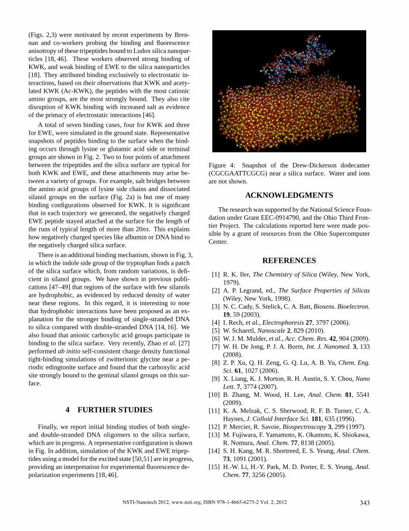
Finally, we report initial binding studies of both single-and double-stranded DNA oligomers to the silica surface,which are in progress. A representative configuration is shownin Fig. In addition, simulation of the KWK and EWE tripep-tides using a model for the excited state [50,51] are in progress,providing an interpretation for experimental fluorescence de-polarization experiments [18,46].
Figure 4: Snapshot of the Drew-Dickerson dodecamer(CGCGAATTCGCG) near asilica surface. Water and ionsare not shown.
ACKNOWLEDGMENTS
The research was supported by the National Science Foun-dation under Grant EEC-0914790, and the Ohio Third Fron-tier Project. The calculations reported here were made pos-sible by a grant of resources from the Ohio SupercomputerCenter.
REFERENCES
[1] R. K. Iler, The Chemistry of Silica (Wiley, New York,1979).
[2] A. P. Legrand, ed.,The Surface Properties of Silicas(Wiley, New York, 1998).
[3] N. C. Cady, S. Stelick, C. A. Batt,Biosens. Bioelectron.19, 59 (2003).
[4] I. Rech,et al., Electrophoresis 27, 3797 (2006).[5] W. Schaertl,Nanoscale 2, 829 (2010).[6] W. J. M. Mulder,et al., Acc. Chem. Res. 42, 904 (2009).[7] W. H. De Jong, P. J. A. Borm,Int. J. Nanomed. 3, 133
(2008).[8] Z. P. Xu, Q. H. Zeng, G. Q. Lu, A. B. Yu,Chem. Eng.
Sci. 61, 1027 (2006).[9] X. Liang, K. J. Morton, R. H. Austin, S. Y. Chou,Nano
Lett. 7, 3774 (2007).[10] B. Zhang, M. Wood, H. Lee,Anal. Chem. 81, 5541
(2009).[11] K. A. Melzak, C. S. Sherwood, R. F. B. Turner, C. A.
Haynes,J. Colloid Interface Sci. 181, 635 (1996).[12] P. Mercier, R. Savoie,Biospectroscopy 3, 299 (1997).[13] M. Fujiwara, F. Yamamoto, K. Okamoto, K. Shiokawa,
R. Nomura,Anal. Chem. 77, 8138 (2005).[14] S. H. Kang, M. R. Shortreed, E. S. Yeung,Anal. Chem.
73, 1091 (2001).[15] H.-W. Li, H.-Y. Park, M. D. Porter, E. S. Yeung,Anal.
Chem. 77, 3256 (2005).
NSTI-Nanotech 2012, www.nsti.org, ISBN 978-1-4665-6275-2 Vol. 2, 2012 343

[16] S. Isailovic, H.-W. Li, E. S. Yeung,J. Chromatogr.A1150, 259 (2007).
[17] S. R. Walter, F. M. Geiger,J. Phys. Chem. Lett. 1, 9(2010).
[18] J. Sui, D. Tleugabulova, J. D. Brennan,Langmuir 21,4996 (2005).
[19] M. Lundqvist,et al., Langmuir 21, 11903 (2005).[20] J. J. Valle-Delgado,et al., Langmuir 21, 9544 (2005).[21] M. van der Veen, W. Norde, M. C. Stuart,Colloids Surf.
B35, 33 (2004).[22] M. Nonella, S. Seeger,ChemPhysChem 9, 414 (2008).[23] H. Stutz,Electrophoresis 30, 2032 (2009).[24] A. Rimola, M. Sodupe, S. Tosoni, B. Civalleri,
P. Ugliengo,Langmuir 22, 6593 (2006).[25] A. Rimola, B. Civalleri, P. Ugliengo,Langmuir 24,
14027 (2008).[26] A. Rimola, M. Sodupe, P. Ugliengo,J. Phys. Chem.
C113, 5741 (2009).[27] Y. L. Zhao, S. Koppen, T. Frauenheim,J. Phys. Chem.
C115, 9615 (2011).[28] J. Dabrowski, H.-J. Mussig,Silicon Surfaces and For-
mation of Interfaces: Basic Science in the IndustrialWorld (World Scientific, River Edge, NJ, 2000).
[29] S. Yao, A. M. Myers, J. D. Posner, K. A. Rose, J. G.Santiago,J. Microelectromech. Syst. 15, 717 (2006).
[30] B. Fubini, The Surface Properties of Silicas, A. P.Legrand, ed. (Wiley, New York, 1998), pp. 415–464.
[31] U. Saffiotti, Acta Bio-Med. Ateneo Parmense 76, 30(2005).
[32] W. R. Bowen, N. Hilal, R. W. Lovitt, C. J. Wright,J.Colloid Interface Sci. 197, 348 (1998).
[33] K. Rezwan, L. P. Meier, L. J. Gauckler,Biomaterials26, 4351 (2005).
[34] S. J. McClellan, E. I. Franses,Langmuir 21, 10148(2005).
[35] P. Towner,Essential Molecular Biology, T. A. Brown,ed. (Oxford, London, 2000), vol. 1, chap. 3, pp. 55–67.
[36] O. Z. Nanassy, P. V. Haydock, M. W. Reed,Anal.Biochem. 365, 240 (2007).
[37] P. M. Wolny, J. P. Spatz, R. P. Richter,Langmuir 26,1029 (2010).
[38] M. P. Allen, D. J. Tildesley,Computer Simulation ofLiquids (Clarendon Press, New York, 1987).
[39] M. J. Frisch,et al., Gaussian 03, Revision C.02. Gaus-sian, Inc., Wallingford, CT, 2004.
[40] C. Møller, M. Plesset,Phys. Rev. 46, 618 (1934).[41] R. J. Bartlett, D. M. Silver,Int. J. Quantum Chem.,
Symp. 8, 271 (1974).[42] R. J. Bartlett, D. M. Silver,Int. J. Quantum Chem.,
Symp. 9, 183 (1975).[43] J. S. Binkley, J. A. Pople,Int. J. Quantum Chem. 9, 229
(1975).[44] R. J. Bartlett, D. M. Silver,Int. J. Quantum Chem. 10,
185 (1976).[45] Y. K. Shin, A. A. Hassanali, S. J. Singer, Biomolecules
at the amorphous silica/water interface: model devel-opment and application to binding and fluorescenceanisotropy of peptides (2012). (submitted).
[46] D. Tleugabulova, J. D. Brennan,Langmuir 22, 1852(2006).
[47] A. A. Hassanali, S. J. Singer,J. Phys. Chem. B111,11181 (2007).
[48] A. A. Hassanali, H. Zhang, C. Knight, Y. K. Shin, S. J.Singer,J. Chem. Theory Comput. 6, 3456 (2010).
[49] H. Zhang, A. A. Hassanali, Y. K. Shin, C. Knight, S. J.Singer,J. Chem. Phys. 134, 024705 (2011).
[50] A. A. Hassanali, T. Li, D. Zhong, S. J. Singer,J. Phys.Chem. B110, 10497 (2006).
[51] T. Li, A. A. Hassanali, S. J. Singer,J. Phys. Chem. 112,16121 (2008).
NSTI-Nanotech 2012, www.nsti.org, ISBN 978-1-4665-6275-2 Vol. 2, 2012344