Embed Size (px)
Citation preview

This article was downloaded by: [Thammasat University Libraries]On: 08 October 2014, At: 13:55Publisher: Taylor & FrancisInforma Ltd Registered in England and Wales Registered Number: 1072954Registered office: Mortimer House, 37-41 Mortimer Street, London W1T 3JH,UK
Molecular Crystals andLiquid Crystals Scienceand Technology. Section A.Molecular Crystals and LiquidCrystalsPublication details, including instructions forauthors and subscription information:http://www.tandfonline.com/loi/gmcl19
Atomic Force MicroscopyInvestigation of Polystyreneand Polystyrene/PMMAComposites SurfacesMarek Nowicki a , Halina Kaczmarek b , RyszardCzajka a & Bronisław Susła a
a Institute of Physics, Poznań University ofTechnology , 60-965, Poznań, Polandb Faculty of Chemistry, Nicolaus CopernicusUniversity , 87-100, Toruń, PolandPublished online: 24 Sep 2006.
To cite this article: Marek Nowicki , Halina Kaczmarek , Ryszard Czajka & BronisławSusła (2000) Atomic Force Microscopy Investigation of Polystyrene and Polystyrene/PMMA Composites Surfaces, Molecular Crystals and Liquid Crystals Science andTechnology. Section A. Molecular Crystals and Liquid Crystals, 354:1, 167-172, DOI:10.1080/10587250008023611
To link to this article: http://dx.doi.org/10.1080/10587250008023611
PLEASE SCROLL DOWN FOR ARTICLE
Taylor & Francis makes every effort to ensure the accuracy of all theinformation (the “Content”) contained in the publications on our platform.

However, Taylor & Francis, our agents, and our licensors make norepresentations or warranties whatsoever as to the accuracy, completeness,or suitability for any purpose of the Content. Any opinions and viewsexpressed in this publication are the opinions and views of the authors, andare not the views of or endorsed by Taylor & Francis. The accuracy of theContent should not be relied upon and should be independently verified withprimary sources of information. Taylor and Francis shall not be liable for anylosses, actions, claims, proceedings, demands, costs, expenses, damages,and other liabilities whatsoever or howsoever caused arising directly orindirectly in connection with, in relation to or arising out of the use of theContent.
This article may be used for research, teaching, and private study purposes.Any substantial or systematic reproduction, redistribution, reselling, loan,sub-licensing, systematic supply, or distribution in any form to anyone isexpressly forbidden. Terms & Conditions of access and use can be found athttp://www.tandfonline.com/page/terms-and-conditions
Dow
nloa
ded
by [
Tha
mm
asat
Uni
vers
ity L
ibra
ries
] at
13:
55 0
8 O
ctob
er 2
014

Mol. Cryr andLiy. Crvsr., 2000, Vol. 354. pp. 167-172 Reprints available directly from the publisher Photocopying permitted by license only
0 2000 OPA (Overseas Publishers Association) N.V. Published by license under the
Cordon and Breach Science Publishers imprint. Printed in Malaysia
Atomic Force Microscopy Investigation of Polystyrene and Polystyrene/PMMA Composites
Surfaces
MAREK NOWICKI~, HALINA KACZMAREK~ RYSZARD CZAJKA~ and BRONISLAW SUSLAa
ahstitUte of Physics, Poznai University of Technology, 60-965 Poznak Poland and bFaculty of Chemistry, Nicolaus Copernicus University, 87-100 Torun: Poland
The surface morphology of polystyrene (PS) before and after u.v.-irradiation was studied by means of Atomic Force Microscopy (AFM). The u.v.-irradiation of PS sample doped with poly(methy1 methacrylate) caused distinct changes in surface morphology. The nanome- ter-scale structures of the rod-like shape bumps of diameter of 300 nm and the height of 75 nm were created. We also showed that the very small forces of 0.2 nN are enough high to modify the surface of PS samples.
Keyw0rd.v; AFM; Polystyrene/PMMA; UV irradiation
INTRODUCTION
Physical and mechanical properties of plastics depend not only on the
chemical structure of polymers but also on their morphology [1,2]. AFM
technique is one of more suitable for characterisation of organic material surface
[3,4]. The polymer surfaces undergo very often changes due to influence of
various environmental factors. Ultraviolet (u.v.) irradiation is one of the most
destructive factors and it involves formation of radicals in macrochains followed
by a breakdown of chemical bonds [5,6]. The main reactions occurring in
polymer upon its exposure to U.V. radiation are main chain scission, oxidation,
side groups abstraction or destruction, etc. [5-81. Because photodegradation of
[755]/167
Dow
nloa
ded
by [
Tha
mm
asat
Uni
vers
ity L
ibra
ries
] at
13:
55 0
8 O
ctob
er 2
014
I68/[756] MAREK NOWICKI er al.
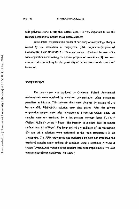
solid polymers starts in very thin surface layer, it is very important to use the
technique enabling to monitor these surface changes
In this letter, we present the results of our study of morphology changes
caused by U.V. irradiation of polystyrene (PS), polystyrendpoly(methyl
methacrylate) blend (PSPMMA). These materials are of interest because of its
wide applications and looking for optimal preparation conditions [9]. We were
also interested in looking for the possibility of the nanometer-scale structures’
formation.
EXPERIMENT
The polystyrene was produced by Oswiecim, Poland. Poly(methy1
methacrylate) were obtained by emulsion polymerisation using ammonium
persulfate as initiator. Thin polymer films were obtained by casting of 2%
benzene (PS, PSPMMA) solution onto glass plates. AAer the solvent
evaporation samples were dried in vacuum to a constant weight. Then, the
samples were u.v.-irradiated by a low-pressure mercury lamp TUV30W
(Philips, Holland) during 8 hours. The intensity of incident light (at sample
surface) was 4.4 mW/cm2. The lamp emitted u.v.-radiation of the wavelength
254 nm. All irradiations were performed at the room temperature in air
atmosphere. The AFM experiment was performed on both non-irradiated and
irradiated samples under ambient air condition using a combined AFM/STM
system (OMICRON) working in the constant force topographic mode. We used
contact mode silicon cantilevers (NT-MDT). Dow
nloa
ded
by [
Tha
mm
asat
Uni
vers
ity L
ibra
ries
] at
13:
55 0
8 O
ctob
er 2
014

ATOMIC FORCE MICROSCOPY [757]/169
RESULTS AND DISCUSSION
Figure 1 shows images of PS surface before and after irradiation.
Measurement conditions in this case were much more difficult due to a high
adhesion force between the AFM tip and the sample surface Non modified PS
surface revealed an array of the randomly distributed holes of the typical
diameter of (100KL5) nm, and the depth of (1M.2) nm. They were probably
formed during fast solvent evaporation during film preparation. The individual
bigger bumps of the diameter from few tens to few hundred nm appeared after
irradiation, as shown in Fig. lb.
FIGURE I . AFM top view images of 2.45 pm x 2.45 pm area of PS surface before (a) and aAer (b) u.v.-irradiation for 8 h.
Their appearance can be explained by a diffusion of low-molecular
gaseous blisters towards the polymer surface When the tip was scanned a few
times at the same area (lpm2) the parts of the top layer were removed and a
small depression (1 nm deep) appeared as shown in Fig. 2. The removed
material is localised mainly along the sides of the scanning area, perpendicularly
to the direction of scanning, however there are some hillocks inside an abrasion
mark. The mechanism of the material removal is most probably the result of
abrasion via direct mechanical tip-sample contact. The study of nanometer scale
modifications proved that using a force of 0.2 nN in AFM is enough high to
modifL the PS surface!
Dow
nloa
ded
by [
Tha
mm
asat
Uni
vers
ity L
ibra
ries
] at
13:
55 0
8 O
ctob
er 2
014

170/[7581 MAREK NOWICKI el al.
FIGURE 2. AFM quasi-3D image of 2 pm x 2 pm area of PS after few scans of the same area of 1 pm2 using a loading force of 0.2 nN
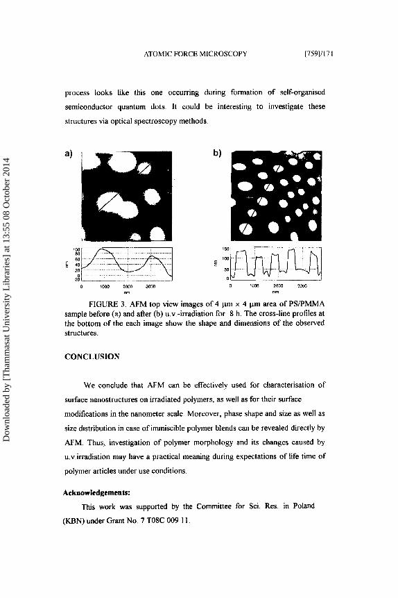
The most spectacular changes after u.v.-irradiation were observed in the
case of PSEMMA sample (Fig. 3). The domains in the form of big rounded
bumps of 1.2 pm diameter and up to 80 nm in height were observed before
irradiation. There were 8 bumps on average on 16 pmZ covering 64% of the
sample area AAer irradiation the bumps become distinctly isolated and their
shape was changed. The edges of these bumps become steep (nearly orthogonal
towards the sample surface) and their height become nearly the same for all of
them and was about (74k1.5) nm. The average diameter of theses rod-like
bumps was (300+30) nm. There were 24 bumps on average on I6 pm2 covering
14% of the sample area Our interpretation of the observed process is as
follows: it is expected, that PMMA component is preferentially adsorbed on
SiO, type substrates (a polar surface) forming homogenous layers with some
protruding bumps [9]. These PMMA bumps are surrounded and covered by PS
phase. The volatile products which appear due to u.v.-irradiation of the PS
component make that the PMMA component is unveiled. The formation
Dow
nloa
ded
by [
Tha
mm
asat
Uni
vers
ity L
ibra
ries
] at
13:
55 0
8 O
ctob
er 2
014
ATOMIC FORCE MICROSCOPY [759]/171
process looks like this one occurring during formation of self-organised
semiconductor quantum dots. It could be interesting to investigate these
structures via optical spectroscopy methods
0 1wo 2 m SUIM “in
FIGURE 3 . AFM top view images of 4 pin x 4 pm area of PWPMMA sample before (a) and afier (b) u.v -irradiation for 8 h. The cross-line profiles at the bottom of the each image show the shape and dimensions of the observed structures.
CONCLUSION
We conclude that AFM can be effectively used for characterisation of
surface nanosttuctures on irradiated polymers, as well as for their surface
modifications in the nanometer scale. Moreover, phase shape and size as well as
size distribution in case of immiscible polymer blends can be revealed directly by
AFM. Thus, investigation of polymer morphology and its changes caused by
u.v.irradiation may have a practical meaning during expectations of life time of
polymer articles under use conditions.
Acknowledgements:
This work was supported by the Committee for Sci. Res. in Poland
(KBN) under Grant No. 7 TOSC 009 I 1.
Dow
nloa
ded
by [
Tha
mm
asat
Uni
vers
ity L
ibra
ries
] at
13:
55 0
8 O
ctob
er 2
014

172/[760] MAREK NOWICKT et al.
References [ I ] Structure und Properties of Polymers, edited by R. W Cahn, P. Haasen, E. J. Kramer,
Material Science and Technology lZ,(VCH, Weinhem, 1993). 121 I . M. Ward, D. W. Hadley, An Introduction to the Mechanical Properties rfSolid Poly-
mers (John Wiley & Sons, Chichester, 1993). [3] S. N. Magonov D. H. Reneker, Annu. Rev. Matec Sci., 27, 175 (1997). [4] M. C. Goh, Advances in Chemicul Physics, Volume XCI, edited by I. Prigogine and
S.A. Rice (John Wiley & Sons, 1995). [ S ] J . F. Rahek, Polymer Photodegradation - Mechanisms and Experimental Methods
(Chapman & Hall, London, 1993). [6] J. F. Rabek, Phorodegradation of Polymers. Physicul Charucteristics and Applications
(Springer Verlag, 1996). 171 H. Zweifel, Chimia, 10,390 (1993). [XI C. Decker, in Handbook of Polymer Science and Technology edited by N. P. Cherem-
isinoff(Marce1 Dekker Inc., N. York, 19x9). vol. 3, p. 541. [91 S. Walheim, M. Boltau, J. Mlynek, G. Krausch, and U. Steiner, Mucrornolecules, 30,
4995 (1997).
Dow
nloa
ded
by [
Tha
mm
asat
Uni
vers
ity L
ibra
ries
] at
13:
55 0
8 O
ctob
er 2
014


![Index [] · Arrhenius equation 395, 399 Arrhenius plot 139 Arrhenius relationship 135 atactic PHB (aPHB) 431 atactic PMMA (aPMMA) 629 atomic force microscopy (AFM) 3, 309, 523,](https://img.pdfslide.us/doc/110x75/5bc3131509d3f29f4d8baf50/index-arrhenius-equation-395-399-arrhenius-plot-139-arrhenius-relationship.jpg)