Embed Size (px)
Citation preview
Asf1 facilitates dephosphorylationof Rad53 after DNA double-strandbreak repairMichael Tsabar, David P. Waterman, Fiona Aguilar,1 Lizabeth Katsnelson,2 Vinay V. Eapen,Gonen Memisoglu, and James E. Haber
Department of Biology, Rosenstiel Basic Medical Sciences Research Center, Brandeis University, Waltham,Massachusetts 02454, USA
To allow for sufficient time to repair DNAdouble-stranded breaks (DSBs), eukaryotic cells activate theDNAdamagecheckpoint. In budding yeast, Rad53 (mammalian Chk2) phosphorylation parallels the persistence of the unrepairedDSB and is extinguished when repair is complete in a process termed recovery or when the cells adapt to the DNAdamage checkpoint. A strain containing a slowly repaired DSB does not require the histone chaperone Asf1 toresume cell cycle progression after DSB repair. When a second, rapidly repairable DSB is added to this strain, Asf1becomes required for recovery. Recovery from two repairable DSBs also depends on the histone acetyltransferaseRtt109 and the cullin subunit Rtt101, both of which modify histone H3 that is associated with Asf1. We show thatdissociation of histone H3 from Asf1 is required for efficient recovery and that Asf1 is required for completedephosphorylation of Rad53 when the upstream DNA damage checkpoint signaling is turned off. Our data suggestthat the requirements for recovery from the DNA damage checkpoint become more stringent with increasedlevels of damage and that Asf1 plays a histone chaperone-independent role in facilitating complete Rad53dephosphorylation following repair.
[Keywords: Asf1; Rad53; Rtt101; Rtt109; DNA damage checkpoint; DNA double-stranded breaks]
Supplemental material is available for this article.
Received March 9, 2016; revised version accepted April 29, 2016.
Cells use several pathways to repair double-strandedbreaks (DSBs). G1-arrested cells repair DSBs mainly bynonhomologous end-joining (NHEJ) (Lewis and Resnick2000; Ferreira and Cooper 2004), but, after CDK1 activa-tion and the initiation of 5′-to-3′ resection of DSB ends,the repair pathway choice shifts to homologous recombi-nation (HR) (Aylon et al. 2004; Ira et al. 2004). HR requiresthat the cell harbor a homologous sequence that can beused as a template to repair the DSB. When the donor se-quence is homologous to both sides of the DSB, cells willprimarily repair using gene conversion (GC), a largely er-ror-free process (Krogh and Symington 2004; Haber2013). When the DSB is created in a region flanked by ho-mologous sequences, the DSB can be repaired by single-strand annealing (SSA) (Krogh and Symington 2004). SSAdepends on extensive 5′-to-3′ resection followed by the an-nealing of complementary single strands and results in adeletion of the sequences between the homologies.
To allow for sufficient time to repair, cells activate theDNA damage checkpoint (Harrison and Haber 2006; Cic-cia and Elledge 2010). In budding yeast, the two PI3K-likekinases Mec1 and Tel1 (homologs of mammalian ATRand ATM, respectively) phosphorylate a cascade of down-stream effectors to enforce G2/M arrest (Harrison and Ha-ber 2006; Gobbini et al. 2013). Tel1 is recruited to the DSBends through its interaction with the MRX complex, acomplex required for the initiation of resection (Nakadaet al. 2003). Mec1 forms a complex with the essential pro-tein Ddc2, the yeast homolog of the mammalian ATRIP(Paciotti et al. 2000). After resection takes place, RPA rap-idly coats the ssDNA, and the RPA-coated ssDNA re-cruits Ddc2 to the sites of damage; this recruitment isessential for checkpoint signaling by Mec1 (Rouse andJackson 2000; Cortez et al. 2001; Melo et al. 2001; Waka-yama et al. 2001; Zou and Elledge 2003).Chk1 and Rad53, the budding yeast homologs of mam-
malian Chk1 and Chk2, are phosphorylated in response to
Present addresses: 1Massachusetts Institute of Technology,Department ofBiology, Cambridge, MA 02139, USA; 2Weill Cornell Medical College,New York City, NY 10065, USACorresponding author: [email protected] is online at http://www.genesdev.org/cgi/doi/10.1101/gad.280685.116.
© 2016 Tsabar et al. This article is distributed exclusively by Cold SpringHarbor Laboratory Press for the first six months after the full-issue publi-cation date (see http://genesdev.cshlp.org/site/misc/terms.xhtml). Aftersix months, it is available under a Creative Commons License (At-tribution-NonCommercial 4.0 International), as described at http://creativecommons.org/licenses/by-nc/4.0/.
GENES & DEVELOPMENT 30:1211–1223 Published by Cold Spring Harbor Laboratory Press; ISSN 0890-9369/16; www.genesdev.org 1211
Cold Spring Harbor Laboratory Press on April 30, 2020 - Published by genesdev.cshlp.orgDownloaded from
damage by Mec1 and Tel1 (Sanchez et al. 1996, 1999; Sunet al. 1996; Gardner et al. 1999), although, in buddingyeast, Mec1 is the dominant kinase for this modification.Another target of Mec1 and Tel1 phosphorylation, Rad9(yeast ortholog of 53BP1), serves as a scaffold for Rad53binding after their phosphorylation (Sun et al. 1998; Gil-bert et al. 2001; Schwartz et al. 2002; Sweeney et al.2005). Upon association, Rad53 undergoes autophosphor-ylation and is released from Rad9 to promote arrest(Gilbert et al. 2001). Rad53 hyperphosphorylation is corre-lated to the duration of repair. Repair events that areresolved rapidly do not trigger Rad53 hyperphosphory-lation and do not cause a noticeable cell cycle delay,whereas DSBs that are slow to repair trigger Rad53 hyper-phosphorylation (Pellicioli et al. 2001; Vaze et al. 2002;Kim and Haber 2009).
Following repair of a DSB, cells turn off the DNA dam-age checkpoint and resume cell cycle progression. Thisprocess, termed recovery, requires dephosphorylation ofRad53 by the PP2C family phosphatases Ptc2 and Ptc3(Leroy et al. 2003). The H3–H4 histone chaperones Asf1and CAF-1 also play a redundant role in recovery, al-though their role remains unclear (Chen et al. 2008; Kimand Haber 2009; Tsabar and Haber 2013). In mammaliancells, Asf1 and CAF-1 function synergistically to re-estab-lish nucleosomes following nucleotide excision repair(Mello et al. 2002). Cells that fail to repair the DSB canalso turn off the DNA damage checkpoint in a processtermed adaptation (Sandell and Zakian 1993; Toczyskiet al. 1997; Lee et al. 1998). In this process, cells arrestfor 12–15 h and then resume cell cycle progression. Cellsthat suffermore than one unrepairedDSB fail to adapt (Leeet al. 1998), perhaps because Rad53 is hyperphosphory-lated to a greater extent. Indeed, the level of hyperphos-phorylation is well correlated to the number of DSBsthat the cells suffer (Mantiero et al. 2007). These findingsdemonstrate that Rad53 acts as an indicator for both thepersistence and the extent of DNA damage.
Although multiple irreparable DSBs elicit a strongercheckpoint response, it is unclear whether multiple re-pairable DSBs trigger a stronger checkpoint response aswell. In this study, we asked whether cells can monitorthe number of repairable DSBs, even if one of theseDSBs is repaired so rapidly that it does not itself elicitRad53 hyperphosphorylation. We show that an additionof a second, repairable DSB does not cause a recoverydefect in wild-type cells. However, unlike our finding fora single DSB where both Asf1 and CAF-1 participate in re-covery, when the cell is challenged by two repairableDSBs, deletion ofASF1 alone is sufficient to cause a recov-ery defect, suggesting that the requirements for recoveryfrom a single DSB and multiple DSBs are different. Thistwo-DSB system provides us with a tool to study the re-quirements for recovery from more than one DSB.
We also explored how proteins that genetically or phys-ically interact with Asf1 affect recovery. After binding toAsf1, histone H3 undergoes acetylation on Lys56 by thehistone acetyltransferase Rtt109 (Collins et al. 2007; Dris-coll et al. 2007; Han et al. 2007; Tsubota et al. 2007; Fil-lingham et al. 2008). Rtt101, a Cul4 subunit of the Roc1-
dependent E3 ubiquitin ligase, ubiquitylates histone H3on Lys121, Lys122, and Lys125, with a preference for his-tone H3 that has been acetylated on Lys56 (Han et al.2013). Rtt101-mediated ubiquitylation of H3 promotesthe handoff of the histone H3–H4 heterodimer fromAsf1 to CAF-1 (Han et al. 2013). We found that RTT101and RTT109 are epistatic to ASF1, suggesting that thehandoff of the histone H3–H4 heterodimer from Asf1 toCAF-1 is required for recovery from the DNA damagecheckpoint.
The modifications of histone H3 also affect the associa-tion of Asf1 with Rad53. Histone H3 and Rad53 share thesame Asf1-binding site and therefore compete for Asf1binding (Emili et al. 2001; Jiao et al. 2012). In buddingyeast, Asf1 binds most of the free and unmodified Rad53in the cell (Emili et al. 2001; Hu et al. 2001). Phosphoryla-tion of Rad53 following DNA damage prevents this inter-action and frees Rad53 to undergo autophosphorylationand allows Asf1 to bind histone H3K56ac (Emili et al.2001; Hu et al. 2001). We show that impairing the interac-tion between histoneH3 andAsf1 rescues the recovery de-fects of rtt101Δ and rtt109Δ but not asf1Δ, demonstratingthat the alleviation of the interaction between Asf1 andhistone H3 is required for recovery. We propose that thedissociation of histone H3 from Asf1 frees Asf1 to pro-mote recovery by binding and sequestering dephosphory-lated Rad53.
Results
Addition of a rapidly repairable DSB to a slowly repairedDSB does not exacerbate the DNA damage checkpointactivation in wild-type cells
Our first goal was to see whether adding a rapidly repairedDSB to a cell in which aDSBwas slowly repaired by ectop-ic GCwould lead tomore robust checkpoint activation. Asecond DSB, even rapidly repaired, might lead to reducedviability compared with a strain with only the slowly re-paired break, analogous to the fact that two DSBs preventadaptation.
We began with strain YJK17, in which an HO endonu-clease-induced DSB within the MATα locus on chromo-some 3 (Chr 3) is repaired by GC, using a donorconsisting of a cloned segment of MATa-inc carrying amutation that prevents HO cleavage, inserted at theARG5,6 locus on the right arm of Chr 5 (Kim and Haber2009). In this strain, the two normal homologous donorsto repair a DSB at MAT (HML and HMR) have been delet-ed. Inducing a DSB in this strain activates the DNA dam-age checkpoint and results in GC repair over a 9-h timespan, with ∼70% viability (Kim and Haber 2009).
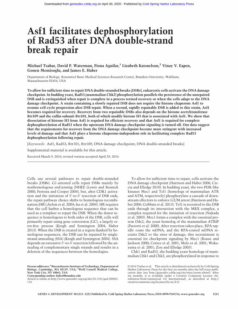
To see whether adding a rapidly repairable DSB wouldcreate a greater checkpoint “load,”we integrated plasmidpNS102 at the ura3-52 locus to produce an SSA substratein which the flanking 1-kb URA3 and ura3-52 homolo-gous sequences are each separated by 2 kb from anHO en-donuclease cleavage site (Fig. 1A; Sugawara and Haber1992). SSA repair was complete in 3–5 h (Sugawara andHaber 1992). Addition of a rapidly repaired DSB (strain
Tsabar et al.
1212 GENES & DEVELOPMENT
Cold Spring Harbor Laboratory Press on April 30, 2020 - Published by genesdev.cshlp.orgDownloaded from
YFA01) did not lead to decreased viability in thewild-typebackground (Fig. 1B), indicating that both recovery and re-pair are proficient when two repairable DSBs are present.
Asf1 is not required for repair of two DSBs
Previously, we published that deletion of the H3–H4 his-tone chaperone ASF1 does not impede recovery in theYJK17 ectopic GC system, but, in conjunction with dele-tion ofCAC1 (the largest subunit of CAF-1), recovery is re-duced (Kim and Haber 2009). However, another studysuggested that deletion ofASF1 alonewas sufficient to im-pede recovery in a single-DSB system (Chen et al. 2008).To address this discrepancy, we tested the effect of asf1Δor cac1Δ on viability of the one and two repairable DSBstrains.We first repeated the viability experiments for the previ-
ously studied systems: YJK17 (Kim and Haber 2009) andYMV80 (Vaze et al. 2002; Chen et al. 2008). As we previ-ously reported, in strain YJK17, deletion of either CAC1orASF1 did not cause a reduction in viability, whereas vi-ability in a cac1Δ asf1Δ double-mutant strain was reducedto 35% (Fig. 1B). In YMV80, repair occurs by SSA, but oneof the flanking repeats is 25-kb away from the HO cleav-age site and requires 25 kb of resection to expose both re-gions of homology; consequently, this repair event doesnot begin until∼6 h and leads to strong checkpoint activa-tion (Vaze et al. 2002). We used a derivative of YMV080 inwhich the NAT cassette had been replaced with HPH(YMT170) (Fig. 1C). Deleting ASF1 did not prevent recov-ery in this system (Fig. 1D), further supporting our previ-ous findings that deletion of ASF1 in a single-DSB
system does not impair recoverywhen the cell needs to re-pair a single DSB.We next tested the effect of deletingASF1 and CAC1 in
the two-DSB system YFA01. As with the single DSB, theviability of cac1Δ was comparable with wild type, butdeletion of ASF1 alone was sufficient to reduce viabilityin the two-DSB system from 70% to 40% (Fig. 1B). The vi-ability of the cac1Δ asf1Δ double mutant was not furtherreduced as compared with a single DSB (35%).We next tested the effect ofASF1 deletion on repair. We
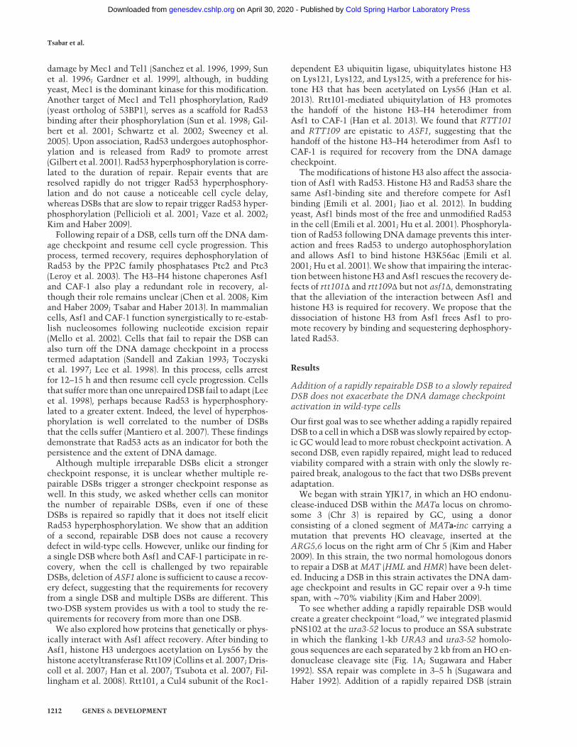
monitored GC and SSA separately by Southern blot. Inwild-type cells, GC was 90% completed by 9 h (Fig. 2A,E). Repair of this DSB in the two-DSB system was similarin outcome and kinetics to those previously reportedwhen only the ectopic GC was present (Kim and Haber2009). Repair of the SSA DSB was 100% completed by3–5 h (Fig. 2A,E), comparable with the kinetics and out-come previously reported in the system that containedonly this SSA event (Sugawara and Haber 1992). Repairin asf1Δ is comparable with wild type for both the ectopicGC and SSA (Fig. 2B,E). The observation that deletion ofASF1 led to a reduction in viability without impeding re-pair suggests that deletion of ASF1 causes a recoverydefect when the cells experience two repairable DSBs.
Asf1 is required for recovery after repair of two DSBs
Checkpoint activation can be monitored by a Westernblot in which Rad53 phosphorylation is seen as moreslowly migrating bands. In strain YJK17, Rad53 was phos-phorylated by 3 h afterHO induction andwas dephosphor-ylated by 9 h (Kim and Haber 2009). Repair of the SSA
Figure 1. The two repairable DSB system.(A) Schematic of the two-DSB system. Thetop panel describes the GC assay (slowerto repair), while the bottom panel describesthe SSA repair construct (faster to repair).(B) Viability of the wild type and thecac1Δ, asf1Δ, and asf1Δ cac1Δ mutants inthe ectopic GC (YJK17) and ectopic GC+SSA (YFA01) backgrounds. n≥ 3. (∗∗) P <0.005, calculated relative to wild type.(C ) Schematic of YMT170. After HOinduction, repair requires a 25-kb resectionand is executed through SSA, as in strainYMV080 (Vaze et al. 2002). StrainYMT170 is similar to YMV080, but theNAT cassette has been switched withHPH. (D) Viability of wild type (YMT170)and asf1Δ (YMT169).
Asf1 facilitates Rad53 dephosphorylation
GENES & DEVELOPMENT 1213
Cold Spring Harbor Laboratory Press on April 30, 2020 - Published by genesdev.cshlp.orgDownloaded from
event alone (Fig. 1A) did not elicit an observable Rad53hyperphosphorylation (Fig. 3A). When a DSB was inducedin strain JKM179 (Lee et al. 1998) lacking donors and witha singleHOcleavage site, Rad53 hyperphosphorylation re-sulted in a mobility shift that was detectable 3 h after in-duction and was lost 12 h after induction when the cellsadapted to the DNA damage (Fig. 3B). In strain YJK17 (1-DSB wild type), Rad53 hyperphosphorylation was detect-able 3 h after HO induction and was lost ∼9 h followinginduction of HO (Fig. 3B). A similar pattern of Rad53 acti-vation was observed in YFA01 (2-DSB wild type) (Fig. 3B),but dephosphorylation was delayed in this strain com-pared with YJK17. Rad53 hyperphosphorylation in theYFA01 was only completely lost 12 h after induction.After Rad53 activation, three distinct bands appeared,the lowest ran as unphosphorylated Rad53, and the twohigher bands indicated apparently increasingly hyper-phosphorylated Rad53. In both YJK17 and YFA01, thetwo higher-migrating bands were most observable 7 h af-ter HO induction. By 9 h, the two upper phosphorylatedbands were still observable, but the lowest-migratingband was much more prominent, indicating the regenera-tion of unphosphorylated Rad53. By 12 h, the slower-mi-grating bands were completely lost. These kinetics ofphosphorylation and dephosphorylation correlate withGC repair kinetics.
Similar to wild type, in the two-DSB asf1Δ cells, HO in-duction led to Rad53 hyperphosphorylation by 3 h (Fig.3B). In asf1Δ, however, phosphorylation of Rad53 wasmore extensive than in wild type, resulting in more prom-inent phosphorylated bands. Strikingly, despite similar re-pair kinetics and outcomes in wild type and asf1Δ, Rad53in asf1Δ remains hyperphosphorylated up to 24 h, long af-ter repair has been completed. In contrast, when only theectopic GC is present, Rad53 phosphorylation in asf1Δcells is similar to wild type (Kim andHaber 2009). This re-sult supports the conclusion reached from the viabilityanalysis above that ASF1 is required for recovery whencells suffer two DSBs.
If failure to turn off the DNA damage checkpoint fol-lowing repair is indeed responsible for the lower viabilityin asf1Δ in the two-DSB system, then alleviating thecheckpoint should rescue this defect. Overexpression ofPTC2 is sufficient to dephosphorylate Rad53 (Leroyet al. 2003). Although overexpression of PTC2 results inlethality, recovery of the cells can be monitored micro-scopically on a galactose plate by observing the ability ofsingle cells to grow beyond the dumbbell (G2/M-arrested)state. Overexpression of PTC2 rescues the arrest of asf1Δcac1Δ cells, allowing them to escape G2/M arrest 24 h af-ter HO induction when a single DSB had to be repaired byectopic GC (Kim and Haber 2009). We found a similar
Figure 2. Repair kinetics in the two-DSBsystem. Southern blot monitoring repair ofthe GC DSB (top panel) and the SSA DSB(bottom panel) in wild-type (YFA01) (A),asf1Δ (YFA02) (B), rtt109Δ (YFA06) (C ),and rtt101Δ (YLK03) (D) cells. (E) Quantifi-cation of repair of both GC and SSA DSBs.GC product was normalized to the uncutrestriction fragment at 0 h. n = 2. SSA wasnormalized to product at 24 h. n = 1. Errorbars in GC are ranges.
Tsabar et al.
1214 GENES & DEVELOPMENT
Cold Spring Harbor Laboratory Press on April 30, 2020 - Published by genesdev.cshlp.orgDownloaded from
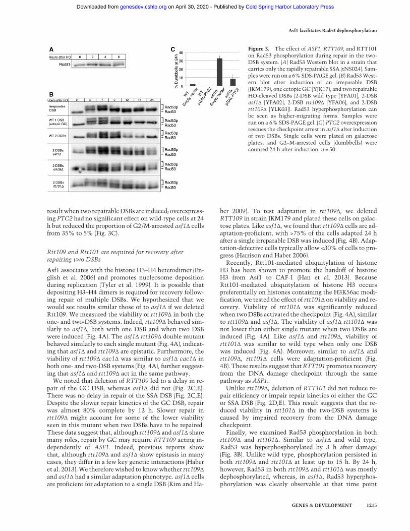
resultwhen two repairableDSBs are induced; overexpress-ing PTC2 had no significant effect on wild-type cells at 24h but reduced the proportion of G2/M-arrested asf1Δ cellsfrom 35% to 5% (Fig. 3C).
Rtt109 and Rtt101 are required for recovery afterrepairing two DSBs
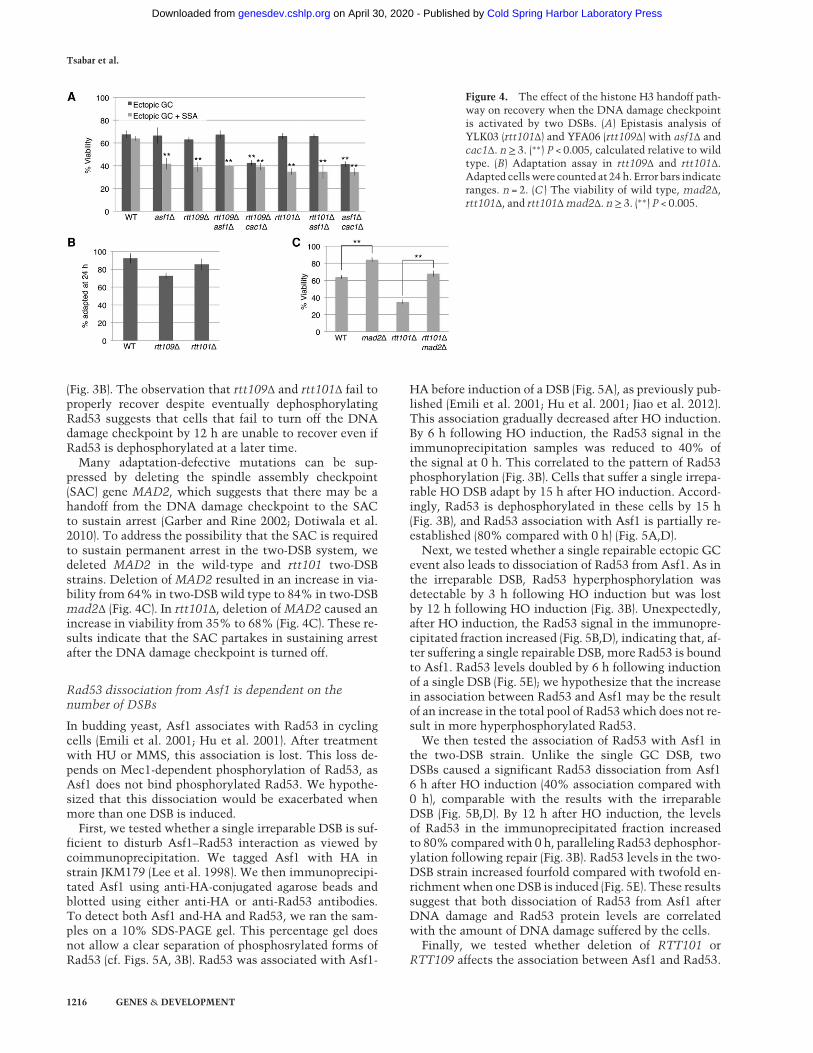
Asf1 associates with the histone H3–H4 heterodimer (En-glish et al. 2006) and promotes nucleosome depositionduring replication (Tyler et al. 1999). It is possible thatdepositing H3–H4 dimers is required for recovery follow-ing repair of multiple DSBs. We hypothesized that wewould see results similar those of to asf1Δ if we deletedRtt109. We measured the viability of rtt109Δ in both theone- and two-DSB systems. Indeed, rtt109Δ behaved sim-ilarly to asf1Δ, both with one DSB and when two DSBwere induced (Fig. 4A). The asf1Δ rtt109Δ double mutantbehaved similarly to each single mutant (Fig. 4A), indicat-ing that asf1Δ and rtt109Δ are epistatic. Furthermore, theviability of rtt109Δ cac1Δ was similar to asf1Δ cac1Δ inboth one- and two-DSB systems (Fig. 4A), further suggest-ing that asf1Δ and rtt109Δ act in the same pathway.We noted that deletion of RTT109 led to a delay in re-
pair of the GC DSB, whereas asf1Δ did not (Fig. 2C,E).There was no delay in repair of the SSA DSB (Fig. 2C,E).Despite the slower repair kinetics of the GC DSB, repairwas almost 80% complete by 12 h. Slower repair inrtt109Δ might account for some of the lower viabilityseen in this mutant when two DSBs have to be repaired.These data suggest that, although rtt109Δ and asf1Δ sharemany roles, repair by GC may require RTT109 acting in-dependently of ASF1. Indeed, previous reports showthat, although rtt109Δ and asf1Δ show epistasis in manycases, they differ in a few key genetic interactions (Haberet al. 2013).We thereforewished to knowwhether rtt109Δand asf1Δ had a similar adaptation phenotype. asf1Δ cellsare proficient for adaptation to a single DSB (Kim and Ha-
ber 2009). To test adaptation in rtt109Δ, we deletedRTT109 in strain JKM179 and plated these cells on galac-tose plates. Like asf1Δ, we found that rtt109Δ cells are ad-aptation-proficient, with >75% of the cells adapted 24 hafter a single irreparable DSB was induced (Fig. 4B). Adap-tation-defective cells typically allow <30% of cells to pro-gress (Harrison and Haber 2006).Recently, Rtt101-mediated ubiquitylation of histone
H3 has been shown to promote the handoff of histoneH3 from Asf1 to CAF-1 (Han et al. 2013). BecauseRtt101-mediated ubiquitylation of histone H3 occurspreferentially on histones containing the H3K56ac modi-fication, we tested the effect of rtt101Δ on viability and re-covery. Viability of rtt101Δ was significantly reducedwhen twoDSBs activated the checkpoint (Fig. 4A), similarto rtt109Δ and asf1Δ. The viability of asf1Δ rtt101Δ wasnot lower than either single mutant when two DSBs areinduced (Fig. 4A). Like asf1Δ and rtt109Δ, viability ofrtt101Δ was similar to wild type when only one DSBwas induced (Fig. 4A). Moreover, similar to asf1Δ andrtt109Δ, rtt101Δ cells were adaptation-proficient (Fig.4B). These results suggest thatRTT101 promotes recoveryfrom the DNA damage checkpoint through the samepathway as ASF1.Unlike rtt109Δ, deletion of RTT101 did not reduce re-
pair efficiency or impair repair kinetics of either the GCor SSA DSB (Fig. 2D,E). This result suggests that the re-duced viability in rtt101Δ in the two-DSB systems iscaused by impaired recovery from the DNA damagecheckpoint.Finally, we examined Rad53 phosphorylation in both
rtt109Δ and rtt101Δ. Similar to asf1Δ and wild type,Rad53 was hyperphosphorylated by 3 h after damage(Fig. 3B). Unlike wild type, phosphorylation persisted inboth rtt109Δ and rtt101Δ at least up to 15 h. By 24 h,however, Rad53 in both rtt109Δ and rtt101Δ was mostlydephosphorylated, whereas, in asf1Δ, Rad53 hyperphos-phorylation was clearly observable at that time point
Figure 3. The effect of ASF1, RTT109, and RTT101on Rad53 phosphorylation during repair in the two-DSB system. (A) Rad53 Western blot in a strain thatcarries only the rapidly repairable SSA (tNS024). Sam-pleswere run on a 6%SDS-PAGE gel. (B) Rad53West-ern blot after induction of an irreparable DSB(JKM179), one ectopic GC (YJK17), and two repairableHO-cleaved DSBs (2-DSB wild type [YFA01], 2-DSBasf1Δ [YFA02], 2-DSB rtt109Δ [YFA06], and 2-DSBrtt109Δ [YLK03]). Rad53 hyperphosphorylation canbe seen as higher-migrating forms. Samples wererun on a 6% SDS-PAGE gel. (C ) PTC2 overexpressionrescues the checkpoint arrest in asf1Δ after inductionof two DSBs. Single cells were plated on galactoseplates, and G2–M-arrested cells (dumbbells) werecounted 24 h after induction. n = 50.
Asf1 facilitates Rad53 dephosphorylation
GENES & DEVELOPMENT 1215
Cold Spring Harbor Laboratory Press on April 30, 2020 - Published by genesdev.cshlp.orgDownloaded from
(Fig. 3B). The observation that rtt109Δ and rtt101Δ fail toproperly recover despite eventually dephosphorylatingRad53 suggests that cells that fail to turn off the DNAdamage checkpoint by 12 h are unable to recover even ifRad53 is dephosphorylated at a later time.
Many adaptation-defective mutations can be sup-pressed by deleting the spindle assembly checkpoint(SAC) gene MAD2, which suggests that there may be ahandoff from the DNA damage checkpoint to the SACto sustain arrest (Garber and Rine 2002; Dotiwala et al.2010). To address the possibility that the SAC is requiredto sustain permanent arrest in the two-DSB system, wedeleted MAD2 in the wild-type and rtt101 two-DSBstrains. Deletion of MAD2 resulted in an increase in via-bility from 64% in two-DSB wild type to 84% in two-DSBmad2Δ (Fig. 4C). In rtt101Δ, deletion of MAD2 caused anincrease in viability from 35% to 68% (Fig. 4C). These re-sults indicate that the SAC partakes in sustaining arrestafter the DNA damage checkpoint is turned off.
Rad53 dissociation from Asf1 is dependent on thenumber of DSBs
In budding yeast, Asf1 associates with Rad53 in cyclingcells (Emili et al. 2001; Hu et al. 2001). After treatmentwith HU or MMS, this association is lost. This loss de-pends on Mec1-dependent phosphorylation of Rad53, asAsf1 does not bind phosphorylated Rad53. We hypothe-sized that this dissociation would be exacerbated whenmore than one DSB is induced.
First, we tested whether a single irreparable DSB is suf-ficient to disturb Asf1–Rad53 interaction as viewed bycoimmunoprecipitation. We tagged Asf1 with HA instrain JKM179 (Lee et al. 1998). We then immunoprecipi-tated Asf1 using anti-HA-conjugated agarose beads andblotted using either anti-HA or anti-Rad53 antibodies.To detect both Asf1 and-HA and Rad53, we ran the sam-ples on a 10% SDS-PAGE gel. This percentage gel doesnot allow a clear separation of phosphosrylated forms ofRad53 (cf. Figs. 5A, 3B). Rad53 was associated with Asf1-
HA before induction of a DSB (Fig. 5A), as previously pub-lished (Emili et al. 2001; Hu et al. 2001; Jiao et al. 2012).This association gradually decreased after HO induction.By 6 h following HO induction, the Rad53 signal in theimmunoprecipitation samples was reduced to 40% ofthe signal at 0 h. This correlated to the pattern of Rad53phosphorylation (Fig. 3B). Cells that suffer a single irrepa-rable HO DSB adapt by 15 h after HO induction. Accord-ingly, Rad53 is dephosphorylated in these cells by 15 h(Fig. 3B), and Rad53 association with Asf1 is partially re-established (80% compared with 0 h) (Fig. 5A,D).
Next, we tested whether a single repairable ectopic GCevent also leads to dissociation of Rad53 from Asf1. As inthe irreparable DSB, Rad53 hyperphosphorylation wasdetectable by 3 h following HO induction but was lostby 12 h following HO induction (Fig. 3B). Unexpectedly,after HO induction, the Rad53 signal in the immunopre-cipitated fraction increased (Fig. 5B,D), indicating that, af-ter suffering a single repairable DSB, more Rad53 is boundto Asf1. Rad53 levels doubled by 6 h following inductionof a single DSB (Fig. 5E); we hypothesize that the increasein association between Rad53 and Asf1 may be the resultof an increase in the total pool of Rad53which does not re-sult in more hyperphosphorylated Rad53.
We then tested the association of Rad53 with Asf1 inthe two-DSB strain. Unlike the single GC DSB, twoDSBs caused a significant Rad53 dissociation from Asf16 h after HO induction (40% association compared with0 h), comparable with the results with the irreparableDSB (Fig. 5B,D). By 12 h after HO induction, the levelsof Rad53 in the immunoprecipitated fraction increasedto 80% compared with 0 h, paralleling Rad53 dephosphor-ylation following repair (Fig. 3B). Rad53 levels in the two-DSB strain increased fourfold compared with twofold en-richmentwhen oneDSB is induced (Fig. 5E). These resultssuggest that both dissociation of Rad53 from Asf1 afterDNA damage and Rad53 protein levels are correlatedwith the amount of DNA damage suffered by the cells.
Finally, we tested whether deletion of RTT101 orRTT109 affects the association between Asf1 and Rad53.
Figure 4. The effect of the histone H3 handoff path-way on recovery when the DNA damage checkpointis activated by two DSBs. (A) Epistasis analysis ofYLK03 (rtt101Δ) and YFA06 (rtt109Δ) with asf1Δ andcac1Δ. n≥ 3. (∗∗) P < 0.005, calculated relative to wildtype. (B) Adaptation assay in rtt109Δ and rtt101Δ.Adapted cellswere counted at 24 h. Error bars indicateranges. n = 2. (C ) The viability of wild type, mad2Δ,rtt101Δ, and rtt101Δmad2Δ. n≥ 3. (∗∗) P < 0.005.
Tsabar et al.
1216 GENES & DEVELOPMENT
Cold Spring Harbor Laboratory Press on April 30, 2020 - Published by genesdev.cshlp.orgDownloaded from
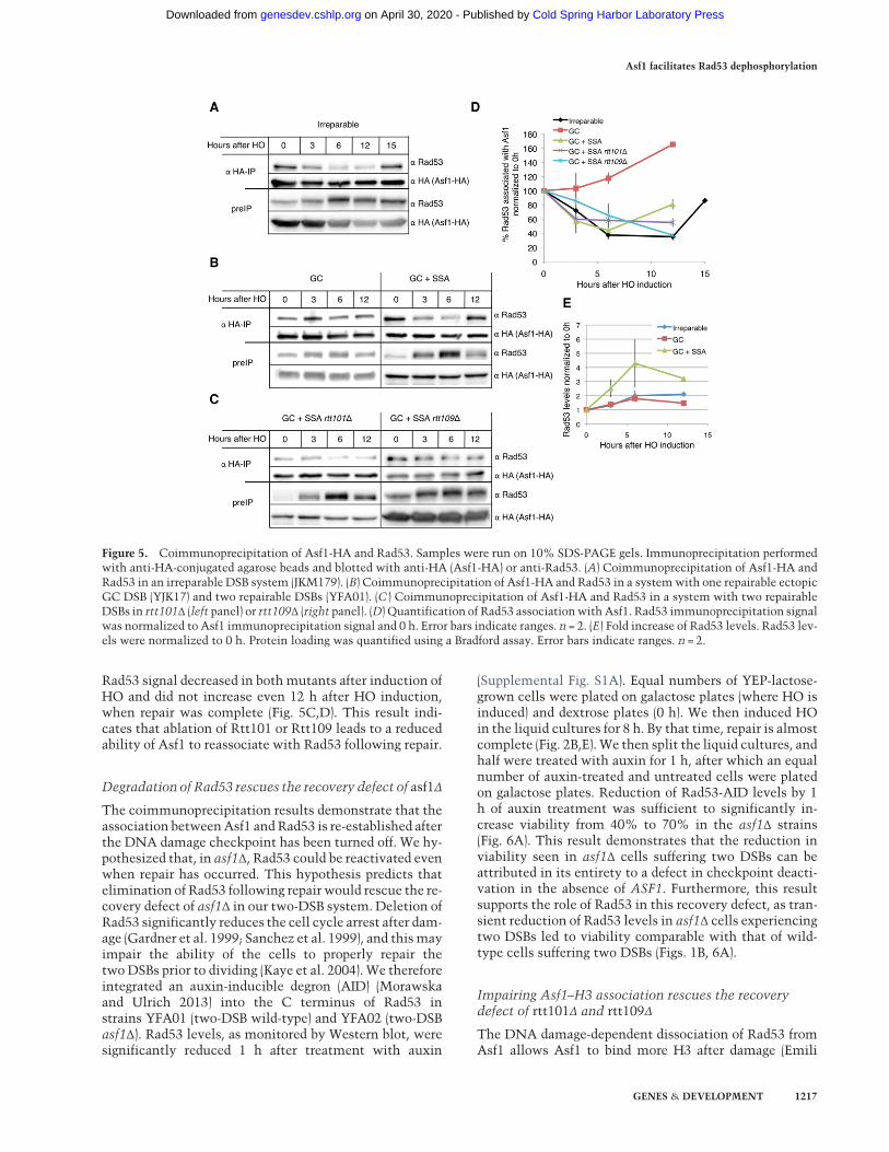
Rad53 signal decreased in bothmutants after induction ofHO and did not increase even 12 h after HO induction,when repair was complete (Fig. 5C,D). This result indi-cates that ablation of Rtt101 or Rtt109 leads to a reducedability of Asf1 to reassociate with Rad53 following repair.
Degradation of Rad53 rescues the recovery defect of asf1Δ
The coimmunoprecipitation results demonstrate that theassociation betweenAsf1 andRad53 is re-established afterthe DNA damage checkpoint has been turned off. We hy-pothesized that, in asf1Δ, Rad53 could be reactivated evenwhen repair has occurred. This hypothesis predicts thatelimination of Rad53 following repair would rescue the re-covery defect of asf1Δ in our two-DSB system. Deletion ofRad53 significantly reduces the cell cycle arrest after dam-age (Gardner et al. 1999; Sanchez et al. 1999), and thismayimpair the ability of the cells to properly repair thetwo DSBs prior to dividing (Kaye et al. 2004). We thereforeintegrated an auxin-inducible degron (AID) (Morawskaand Ulrich 2013) into the C terminus of Rad53 instrains YFA01 (two-DSB wild-type) and YFA02 (two-DSBasf1Δ). Rad53 levels, as monitored by Western blot, weresignificantly reduced 1 h after treatment with auxin
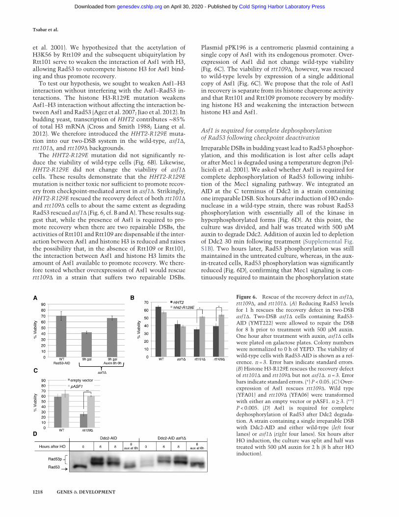
(Supplemental Fig. S1A). Equal numbers of YEP-lactose-grown cells were plated on galactose plates (where HO isinduced) and dextrose plates (0 h). We then induced HOin the liquid cultures for 8 h. By that time, repair is almostcomplete (Fig. 2B,E). We then split the liquid cultures, andhalf were treated with auxin for 1 h, after which an equalnumber of auxin-treated and untreated cells were platedon galactose plates. Reduction of Rad53-AID levels by 1h of auxin treatment was sufficient to significantly in-crease viability from 40% to 70% in the asf1Δ strains(Fig. 6A). This result demonstrates that the reduction inviability seen in asf1Δ cells suffering two DSBs can beattributed in its entirety to a defect in checkpoint deacti-vation in the absence of ASF1. Furthermore, this resultsupports the role of Rad53 in this recovery defect, as tran-sient reduction of Rad53 levels in asf1Δ cells experiencingtwo DSBs led to viability comparable with that of wild-type cells suffering two DSBs (Figs. 1B, 6A).
Impairing Asf1–H3 association rescues the recoverydefect of rtt101Δ and rtt109Δ
The DNA damage-dependent dissociation of Rad53 fromAsf1 allows Asf1 to bind more H3 after damage (Emili
Figure 5. Coimmunoprecipitation of Asf1-HA and Rad53. Samples were run on 10% SDS-PAGE gels. Immunoprecipitation performedwith anti-HA-conjugated agarose beads and blotted with anti-HA (Asf1-HA) or anti-Rad53. (A) Coimmunoprecipitation of Asf1-HA andRad53 in an irreparable DSB system (JKM179). (B) Coimmunoprecipitation of Asf1-HA and Rad53 in a systemwith one repairable ectopicGC DSB (YJK17) and two repairable DSBs (YFA01). (C ) Coimmunoprecipitation of Asf1-HA and Rad53 in a system with two repairableDSBs in rtt101Δ (left panel) or rtt109Δ (right panel). (D) Quantification of Rad53 associationwith Asf1. Rad53 immunoprecipitation signalwas normalized to Asf1 immunoprecipitation signal and 0 h. Error bars indicate ranges. n = 2. (E) Fold increase of Rad53 levels. Rad53 lev-els were normalized to 0 h. Protein loading was quantified using a Bradford assay. Error bars indicate ranges. n = 2.
Asf1 facilitates Rad53 dephosphorylation
GENES & DEVELOPMENT 1217
Cold Spring Harbor Laboratory Press on April 30, 2020 - Published by genesdev.cshlp.orgDownloaded from
et al. 2001). We hypothesized that the acetylation ofH3K56 by Rtt109 and the subsequent ubiquitylation byRtt101 serve to weaken the interaction of Asf1 with H3,allowing Rad53 to outcompete histone H3 for Asf1 bind-ing and thus promote recovery.
To test our hypothesis, we sought to weaken Asf1–H3interaction without interfering with the Asf1–Rad53 in-teractions. The histone H3-R129E mutation weakensAsf1–H3 interaction without affecting the interaction be-tweenAsf1 andRad53 (Agez et al. 2007; Jiao et al. 2012). Inbudding yeast, transcription of HHT2 contributes ∼85%of total H3 mRNA (Cross and Smith 1988; Liang et al.2012). We therefore introduced the HHT2-R129E muta-tion into our two-DSB system in the wild-type, asf1Δ,rtt101Δ, and rtt109Δ backgrounds.
The HHT2-R129E mutation did not significantly re-duce the viability of wild-type cells (Fig. 6B). Likewise,HHT2-R129E did not change the viability of asf1Δcells. These results demonstrate that the HHT2-R129Emutation is neither toxic nor sufficient to promote recov-ery from checkpoint-mediated arrest in asf1Δ. Strikingly,HHT2-R129E rescued the recovery defect of both rtt101Δand rtt109Δ cells to about the same extent as degradingRad53 rescued asf1Δ (Fig. 6, cf. B andA). These results sug-gest that, while the presence of Asf1 is required to pro-mote recovery when there are two repairable DSBs, theactivities of Rtt101 andRtt109 are dispensable if the inter-action between Asf1 and histone H3 is reduced and raisesthe possibility that, in the absence of Rtt109 or Rtt101,the interaction between Asf1 and histone H3 limits theamount of Asf1 available to promote recovery. We there-fore tested whether overexpression of Asf1 would rescuertt109Δ in a strain that suffers two repairable DSBs.
Plasmid pPK196 is a centromeric plasmid containing asingle copy of Asf1 with its endogenous promoter. Over-expression of Asf1 did not change wild-type viability(Fig. 6C). The viability of rtt109Δ, however, was rescuedto wild-type levels by expression of a single additionalcopy of Asf1 (Fig. 6C). We propose that the role of Asf1in recovery is separate from its histone chaperone activityand that Rtt101 and Rtt109 promote recovery by modify-ing histone H3 and weakening the interaction betweenhistone H3 and Asf1.
Asf1 is required for complete dephosphorylationof Rad53 following checkpoint deactivation
IrreparableDSBs in budding yeast lead to Rad53 phosphor-ylation, and this modification is lost after cells adaptor after Mec1 is degraded using a temperature degron (Pel-licioli et al. 2001). We asked whether Asf1 is required forcomplete dephosphorylation of Rad53 following inhibi-tion of the Mec1 signaling pathway. We integrated anAID at the C terminus of Ddc2 in a strain containingone irreparableDSB. Six hours after induction ofHOendo-nuclease in a wild-type strain, there was robust Rad53phosphorylation with essentially all of the kinase inhyperphosphorylated forms (Fig. 6D). At this point, theculture was divided, and half was treated with 500 µMauxin to degrade Ddc2. Addition of auxin led to depletionof Ddc2 30 min following treatment (Supplemental Fig.S1B). Two hours later, Rad53 phosphorylation was stillmaintained in the untreated culture, whereas, in the aux-in-treated cells, Rad53 phosphorylation was significantlyreduced (Fig. 6D), confirming that Mec1 signaling is con-tinuously required to maintain the phosphorylation state
Figure 6. Rescue of the recovery defect in asf1Δ,rtt109Δ, and rtt101Δ. (A) Reducing Rad53 levelsfor 1 h rescues the recovery defect in two-DSBasf1Δ. Two-DSB asf1Δ cells containing Rad53-AID (YMT222) were allowed to repair the DSBfor 8 h prior to treatment with 500 µM auxin.One hour after treatment with auxin, asf1Δ cellswere plated on galactose plates. Colony numberswere normalized to 0 h of YEPD. The viability ofwild-type cells with Rad53-AID is shown as a ref-erence. n = 3. Error bars indicate standard errors.(B) Histone H3-R129E rescues the recovery defectof rtt101Δ and rtt109Δ but not asf1Δ. n = 3. Errorbars indicate standard errors. (∗) P < 0.05. (C ) Over-expression of Asf1 rescues rtt109Δ. Wild type(YFA01) and rtt109Δ (YFA06) were transformedwith either an empty vector or pASF1. n≥ 3. (∗∗)P < 0.005. (D) Asf1 is required for completedephosphorylation of Rad53 after Ddc2 degrada-tion. A strain containing a single irreparable DSBwith Ddc2-AID and either wild-type (left fourlanes) or asf1Δ (right four lanes). Six hours afterHO induction, the culture was split and half wastreated with 500 µM auxin for 2 h (8 h after HOinduction).
Tsabar et al.
1218 GENES & DEVELOPMENT
Cold Spring Harbor Laboratory Press on April 30, 2020 - Published by genesdev.cshlp.orgDownloaded from
of Rad53 (Pellicioli et al. 2001). Checkpoint activationwas then monitored in asf1Δ in the Ddc2-AID strain. Asin wild-type, strong Rad53 phosphorylation was evident6 h after DSB induction. Interestingly, 2 h following auxintreatment, Rad53 phosphorylation was still detected (Fig.6D). While the highest migrating forms of Rad53 werelost, indicating some dephosphorylation, Rad53 persistedin amiddle migrating band and failed to return to the low-est migrating form observed prior to DSB induction (Fig.6D). These results suggest that although Asf1 does not it-self dephosphorylate Rad53, it is required to promotecomplete dephosphorylation of Rad53 following the deac-tivation of the upstream Ddc2–Mec1 signaling. Giventhat Rad53 amplifies its Mec1-dependent phosphoryla-tion by autophosphorylation, these results might implythat Asf1 plays a role in either preventing autophosphory-lation or promoting turnover of phosphorylated forms ofthe protein.
Discussion
Budding yeast cells can sense the number of DSBs thatthey have suffered (Lee et al. 1998; Mantiero et al. 2007).This sensitivity is apparently achieved by regulating thelevel of Rad53 phosphorylation in response to moreDSBs (Mantiero et al. 2007). Maintenance of γ-H2AX inthe 50-kb region around each of two DSBs is regulatedindependently so that it is removed around a repairedDSB even as it persists around an unrepaired DSB (Tsabaret al. 2015). Here we provide the first evidence that cellscan sense the number of repairable DSBs even if one ofthese DSBs repairs rapidly enough so that it does not by it-self elicit detectable Rad53 phosphorylation. In addition,we show that the genetic requirements for recovery aredifferent when there are twoDSBs versus one.We proposethat Asf1 participates in a dynamic DNA damage-sensingmechanism. When the cells sense DSBs, Mec1 phosphor-ylates Rad53, leading to its dissociation from Asf1 andhyperactivation by autophosphorylation. When Rad53 isdephosphorylated by Ptc2 and Ptc3, it can reassociate
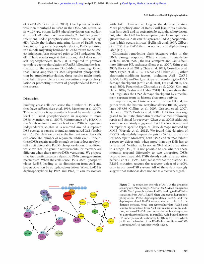
with Asf1. However, so long as the damage persists,Mec1 phosphorylation of Rad53 will lead to its dissocia-tion from Asf1 and its activation by autophosphorylation,but, when the DSB has been repaired, Asf1 can rapidly se-quester Rad53. Asf1 can thus prevent Rad53 phosphoryla-tion (which occurs in trans) (Pellicioli et al. 1999; Gilbertet al. 2001) by Rad53 that has not yet been dephosphory-lated (Fig. 7).Chromatin remodeling plays extensive roles in the
DNA damage response. While chromatin remodelerssuch as Fun30, Ino80, the RSC complex, and Rad54 facil-itate different HR pathways (Kent et al. 2007; Shim et al.2007; Hicks et al. 2011; Chen et al. 2012; Costelloe et al.2012; Eapen et al. 2012; Tsabar and Haber 2013), otherchromatin-modifying factors, including Asf1, CAF-1Rdh54, Ino80, andSwr1, participate in regulating theDNAdamage checkpoint (Emili et al. 2001; Hu et al. 2001; Leeet al. 2001; Papamichos-Chronakis et al. 2006; Kim andHaber 2009; Tsabar and Haber 2013). Here we show thatAsf1 regulates the DNA damage checkpoint by a mecha-nism separate from its histone chaperone activity.In replication, Asf1 interacts with histone H3 and, to-
gether with the histone acetyltransferase Rtt109, acety-lates H3K56 (Collins et al. 2007; Driscoll et al. 2007;Han et al. 2007; Tsubota et al. 2007). H3K56ac was sug-gested to facilitate chromatin re-establishment followingrepair and signal for recovery (Chen et al. 2008), althougha more recent study suggested that H3K56ac is requiredfor repair of specific types of DNA damage induced byMMS (Wurtele et al. 2012). We found that deletion ofRTT09 only slightly impaired repair by GC and did not af-fect SSA repair. Moreover, both asf1Δ and rtt109Δ exhibita recovery defect only when more than one DSB has tobe repaired. Neither asf1Δ nor rtt109Δ affect adaptationto a single DSB; it is not possible to see whether thesemutants respond differently to two unrepaired DSBsbecause two irreparableDSBs already confer an adaptationdefect (Lee et al. 1998). Last, we show that the histoneH3-R129E mutation rescues the recovery defect of rtt109Δcells in our two-DSB system. All of these data stronglysuggest that H3K56ac does not act as a recovery signal.
Figure 7. A model for the role of Asf1 in the dynamicsensing of DNA damage. After a Ddc2–Mec1 recognizesaDSB,Mec1 phosphorylates Rad53, leading toRad53 dis-sociation from Asf1. Rad53 then undergoes hyperphos-phorylation. PP2C dephosphorylates Rad53, and thedephosphorylated Rad53 reassociates with Asf1. If thedamage persists, Mec1 can rephosphorylate Rad53 andlead to dissociation from Asf1 and reactivation. In addi-tion, activated Rad53 can counter the dephosphorylationby autophosphorylation. In parallel, Asf1-bound histoneH3undergoesmodificationsbyRtt109andRtt101,whichfacilitate the handoff of theH3–H4histone dimer toCAF-1, freeing Asf1 to reinteract with Rad53.
Asf1 facilitates Rad53 dephosphorylation
GENES & DEVELOPMENT 1219
Cold Spring Harbor Laboratory Press on April 30, 2020 - Published by genesdev.cshlp.orgDownloaded from
Rtt101 ubiquitylates histone H3 in vitro, with a prefer-ence for histone H3 that has been acetylated on Lys56(Han et al. 2013). This ubiquitylation is suggested to fa-cilitate the handoff of histone H3–H4 heterodimersfrom Asf1 to CAF-1 and Rtt106. In vivo, Rtt101 func-tions in the same genetic pathway as H3K56ac to conferresistance to MMS damage (Wurtele et al. 2012). Likedeletion of RTT109 and ASF1, deletion of RTT101 doesnot confer an adaptation defect or a repair defect. Simi-larly to rtt109Δ, the recovery defect of rtt101Δ in thetwo-DSB system is rescued by weakening the interactionof Asf1 with H3. Together with our genetic data, thisfinding suggests that Rtt101 and Rtt109 act on thesame pathway to promote recovery and that their rolein recovery is to facilitate the removal of histone H3from Asf1.
Rad53 phosphorylation in asf1Δ cells suffering two re-pairable DSBs persisted 24 h after induction of theDSBs, long after repair is completed. This suggests thatthe DNA damage checkpoint responds to the number ofDSBs that it experienced even if one of these DSBs itselfdoes not trigger a checkpoint activation. Rad53 phosphor-ylation in rtt109Δ and rtt101Δ persisted 15 h after HO in-duction but did not persist up to 24 h. It is noteworthythat adaptation occurs ∼12–15 h following induction ofa single irreparable DSB. It is possible that the 12- to 15-h time frame following DNA damage checkpoint activa-tion represents a time window by which the DNA dam-age checkpoint must be resolved by a productivepathway leading to recovery. One possibility raised byour findings is that failure to turn off the DNA damagecheckpoint by 12 h results in a handoff of the arrest tothe SAC, which prevents productive recovery. According-ly, deletion of MAD2 had a more significant effect onrtt101Δ cells exhibiting a longer checkpoint than onwild-type cells.
The association of Asf1 with Rad53 is disrupted byexposure to the genotoxic agents MMS and HU, and thisdisruption is dependent on DNA damage checkpoint sig-naling (Emili et al. 2001; Hu et al. 2001). Interestingly,Rad53 phosphorylation in response to HU is delayed inasf1Δ (Hu et al. 2001). As Asf1 also binds histone H3 andCAF-1, thismay indicate that the role of Asf1 is to concen-trate Rad53 close to the replication forks, which are verysusceptible to DNA damage (Hu et al. 2001). Here weshow that a single irreparable DSB is sufficient to disruptAsf1’s associationwith Rad53 significantly.Moreover, weshow that adaptation and recovery temporally correlatewith the re-establishment of the association betweenAsf1 and Rad53. This, together with the observationthat ablation of Rad53 rescues the viability of asf1Δ cellsin a system with two repairable DSBs and the prolongedRad53 phosphorylation in asf1Δ, may suggest that re-es-tablishment of the Asf1–Rad53 interaction plays a signifi-cant role in regulating the DNA damage checkpointduring recovery and adaptation. Supporting this model,our observation that Rad53 phosphorylation persists inasf1Δ cells even when Ddc2 is depleted demonstratesthat Asf1 is required for the complete turning off of theDNA damage checkpoint.
The role of Asf1 in recovery manifests itself only whenthe cells are exposed to multiple DSBs. The reason forthe requirement for Asf1 when the cell is exposed tomultiple DSBs remains unclear. One clue may comefrom studying the association of Rad53 and Asf1. A sin-gle repairable DSB did not cause dissociation betweenAsf1 and Rad53 that was seen with two DSBs. This dif-ference may reflect how one or two DSBs alter totalRad53 protein levels and its hyperphosphorylation. Theobservation that total Rad53 protein levels increase ina DNA damage-dependent manner (Smolka et al. 2005)and that the magnitude of the increase and phosphoryla-tion of Rad53 are correlated to the number of DSBs sug-gests that, in response to two DSBs, there is a larger poolof free Rad53, and this may lead to more active Rad53 inresponse to two DSBs. In this case, when only one DSBtriggers the DNA damage checkpoint, Ptc2 and Ptc3dephosphorylation of Rad53 is sufficient to deactivatethe checkpoint; however, when more than one DSB is in-duced, Rad53 levels increase further, and there may bemore autophosphorylation. In this event, Asf1 might berequired to sequester Rad53 and prevent it from beingreactivated.
In mammalian cells, the DNA damage checkpoint ishighly sensitive to the amount of damage inflicted onthe cells (Buscemi et al. 2004; Deckbar et al. 2007; Loeweret al. 2013). These studies suggest that studying recoveryfrom more than one DSB in budding yeast may be morephysiologically similar to the condition in mammaliancells. Nevertheless, despite the high level of conservationof Asf1 and Rad53 (mammalian Chk2) between yeast andmammals, the interaction between Asf1 and Chk2 wasnot detected in mammalian cells (Groth et al. 2005).This suggests that, in mammalian cells, a large fractionof Chk2 is free from interaction with Asf1. Another possi-bility is that the interaction between Asf1 and Rad53 iscell cycle-regulated. If this is the case, then we are ableto capture the interaction between Asf1 and Rad53because an asynchronous population of budding yeastcontains cells in all stages of the cell cycle, and this inter-action might be missed in mammalian cells that are pre-dominantly in G0/G1. Another possibility is that theabsence of this interactionmay be a result of fundamentaldifferences between budding yeast and mammalian cellsin themechanism ofDNA damage checkpoint regulation.However, Asf1 has been shown to participate in nucleo-some deposition following repair (Mello et al. 2002) andinteract with tousled-like kinases (TLKs), a family of ki-nases specific to metazoans that plays an extensive rolein the regulation of replication, transcription, and repair(Groth et al. 2003, 2005; Mousson et al. 2007; De Bene-detti 2012). These observations implicate Asf1 with regu-lation of the DNA damage checkpoint in mammaliancells as well. The findings presented in our study demon-strate that the DNA damage checkpoint is regulated byboth the duration of damage persistence and the amountof damage, link chromatin assembly to recovery fromthe DNA damage checkpoint, and contribute to our un-derstanding of mechanisms by which cells sense the com-pletion of repair.
Tsabar et al.
1220 GENES & DEVELOPMENT
Cold Spring Harbor Laboratory Press on April 30, 2020 - Published by genesdev.cshlp.orgDownloaded from
Materials and methods
Strains
Strains were based on strain YJK17 (Kim and Haber 2009), whichcontains a glactose-inducible HO gene and is deleted for the nor-mal HML and HMR donors to repair a DSB at MAT. This MATαstrain carries a 4-kbMATa-inc sequence, which cannot be cleavedby HO, inserted at the arg5,6 locus on Chr 5. In some strains, anadditional HO cut site was inserted at the ura3-52 locus on theopposite arm of Chr 5 such that it was flanked by ∼1 kb of flank-ing homology (Sugawara and Haber 1992).To construct the histone H3-R129E mutation, the region con-
taining HHF2 and HHT2 was amplified using quantitative PCR(qPCR) and introduced to plasmid pRS405 at its HpaI site to cre-ate plasmid pMT05. The R129Emutationwas introduced by site-directed mutagenesis using the QuikChange II kit (Agilent) tocreate plasmid pMT06. HHT2-R129E and the adjacent LEU2were amplified from this plasmid and integrated at the endoge-nous HHT2 in strains YFA01, YFA02, YLK03, and YFA06 sothat LEU2 was downstream from HHT2-R129E. Integration ofLEU2 and the coinserted R129E mutation was verified by PCRfollowed by DNA sequencing.A full list of strains is in Supplemental Table S1. All strains and
sequences of oligos for PCR are available on request.
Plating assays
For viability experiments, cells were grown overnight in 5 mL ofYEPD, washed twice with 5 mL of YEP-lactose, and kept for atleast 6 h in lactose. Next, cells were counted, and ∼100 cellswere plated on YEP galactose plates (where HO is induced) andon YEPD plates as a control. Colonies were counted 3–5 d afterplating.Adaptation assays were conducted by inoculating cells over-
night in 5mL of YEP-lactose and then plating themonYEP-galac-tose plates. Single cells were manipulated and monitored for 24and 48 h following plating. A PTC2 overexpression assay was per-formed similarly to the adaptation assay, using strains containingplasmid pJH2447 with PTC2 under the control a galactosepromoter.
Southern blots
Southern blots were performed as described in Kim and Haber(2009). To monitor the ectopic GC, genomic DNA extracted byphenol extraction from a time course was digested overnightwith EcoRI. A radiolabeled probe for the Z1Z2 region at MATwas used (Kim and Haber 2009). The product was normalized tothe donor signal and 0 h. To monitor SSA repair at ura3, DNAwas digested with BglII. A radiolabeled probe for the 3′ end oftheURA3 gene was used. The signal was normalized to the totalsignal and the signal from a cell that had completed SSA.
Western blots
Western blots were done as described (Pellicioli et al. 2001). Pro-teins were extracted using TCA preparation. An antibody againstRad53 (Abcam, ab166859) was used to detect Rad53. Ddc2-AID-Myc degradation was monitored by an antibody against Myc.
Degradation of proteins using AID
Proteins were degraded using the AID system from H. Ullrich(Morawska and Ulrich 2013). Rad53-AID cells were grown over-
night in YEPD and washed twice in YEP-lactose for 6 h, andthen a larger culturewas inoculated and grown until cells reachedlog-phase growth (5 × 10⁶ cells per milliliter). Cells were thencounted, and ∼100 colony-forming units (CFUs) were plated onYEPD and YEP-galactose plates to monitor viability. The liquidculture was treated with 2% galactose to induce the HO endonu-clease. Eight hours after HO induction, the culture was split intwo, and one of the two cultures was treated with 500 µM auxin.One hour after auxin treatment, cells were counted, and 100CFUs were plated on galactose plates. Western blots were per-formed to confirm loss of the protein (Fig. 5E).Ddc2-AID-Myc cells were grown as described above. A sample
was collected prior to galactose induction, and then 2% galactosewas added to the medium. Six hours after galactose induction, asample was collected, the culture was split, and one half wastreated with 500 µM auxin for 2 h. A final sample was collected8 h after galactose induction (2 h after auxin induction). Westernblots were used to confirm loss of Ddc2.
Coimmunoprecipitation
Coimmunoprecipitation experiments were conducted as previ-ously published (Hu et al. 2001) with a modified lysis buffer(50 mM Tris at pH 7.5, 50 mM NaCl, 5 mM EDTA, 0.5% TritonX-100, proteinase inhibitors).
Acknowledgments
We thankHelle Ulrich for plasmids to generate the AIDs.We alsothank Paul Kaufman for the plasmid pPK196. This work was sup-ported by National Institutes of Health grants GM20056 andGM61766 to J.E.H. D.P.W. is supported by National Institutesof Health Genetics training grant T32GM007122.
References
AgezM,Chen J, Guerois R, vanHeijenoort C, Thuret JY,MannC,Ochsenbein F. 2007. Structure of the histone chaperone ASF1bound to the histone H3 C-terminal helix and functional in-sights. Structure 15: 191–199.
AylonY, Liefshitz B, KupiecM. 2004. TheCDK regulates repair ofdouble-strand breaks by homologous recombination duringthe cell cycle. EMBO J 23: 4868–4875.
BuscemiG, Perego P, CareniniN,NakanishiM,Chessa L, Chen J,Khanna K, Delia D. 2004. Activation of ATM andChk2 kinas-es in relation to the amount of DNA strand breaks.Oncogene23: 7691–7700.
Chen CC, Carson JJ, Feser J, Tamburini B, Zabaronick S, Linger J,Tyler JK. 2008. Acetylated lysine 56 on histone H3 driveschromatin assembly after repair and signals for the comple-tion of repair. Cell 134: 231–243.
Chen X, Cui D, Papusha A, Zhang X, Chu CD, Tang J, Chen K,Pan X, Ira G. 2012. The Fun30 nucleosome remodeller pro-motes resection of DNA double-strand break ends. Nature489: 576–580.
Ciccia A, Elledge SJ. 2010. The DNA damage response: making itsafe to play with knives. Mol Cell 40: 179–204.
Collins SR, Miller KM, Maas NL, Roguev A, Fillingham J, ChuCS, Schuldiner M, Gebbia M, Recht J, Shales M, et al. 2007.Functional dissection of protein complexes involved in yeastchromosome biology using a genetic interaction map.Nature446: 806–810.
Asf1 facilitates Rad53 dephosphorylation
GENES & DEVELOPMENT 1221
Cold Spring Harbor Laboratory Press on April 30, 2020 - Published by genesdev.cshlp.orgDownloaded from
Cortez D, Guntuku S, Qin J, Elledge SJ. 2001. ATR and ATRIP:partners in checkpoint signaling. Science 294: 1713–1716.
Costelloe T, Louge R, Tomimatsu N, Mukherjee B, Martini E,Khadaroo B, Dubois K, Wiegant WW, Thierry A, Burma S,et al. 2012. The yeast Fun30 and human SMARCAD1 chroma-tin remodellers promote DNA end resection. Nature 489:581–584.
Cross SL, SmithMM. 1988. Comparison of the structure and cellcycle expression of mRNAs encoded by two histone H3-H4loci in Saccharomyces cerevisiae. Mol Cell Biol 8: 945–954.
De Benedetti A. 2012. The tousled-like kinases as guardians of ge-nome integrity. ISRN Mol Biol 2012: 627596.
Deckbar D, Birraux J, Krempler A, Tchouandong L, Beucher A,Walker S, Stiff T, Jeggo P, Lobrich M. 2007. Chromosomebreakage after G2 checkpoint release. J Cell Biol 176: 749–755.
Dotiwala F, Harrison JC, Jain S, Sugawara N, Haber JE. 2010.Mad2 prolongs DNA damage checkpoint arrest caused by adouble-strand break via a centromere-dependent mechanism.Curr Biol 20: 328–332.
Driscoll R, Hudson A, Jackson SP. 2007. Yeast Rtt109 promotesgenome stability by acetylating histone H3 on lysine 56. Sci-ence 315: 649–652.
Eapen VV, Sugawara N, Tsabar M, Wu WH, Haber JE. 2012. TheSaccharomyces cerevisiae chromatin remodeler Fun30 regu-lates DNA end resection and checkpoint deactivation. MolCell Biol 32: 4727–4740.
Emili A, Schieltz DM, Yates JR III, Hartwell LH. 2001. Dynamicinteraction of DNA damage checkpoint protein Rad53 withchromatin assembly factor Asf1. Mol Cell 7: 13–20.
English CM, Adkins MW, Carson JJ, Churchill ME, Tyler JK.2006. Structural basis for the histone chaperone activity ofAsf1. Cell 127: 495–508.
Ferreira MG, Cooper JP. 2004. Two modes of DNA double-strandbreak repair are reciprocally regulated through the fissionyeast cell cycle. Genes Dev 18: 2249–2254.
Fillingham J, Recht J, SilvaAC, Suter B, Emili A, Stagljar I, KroganNJ, Allis CD, KeoghMC, Greenblatt JF. 2008. Chaperone con-trol of the activity and specificity of the histone H3 acetyl-transferase Rtt109. Mol Cell Biol 28: 4342–4353.
Garber PM, Rine J. 2002. Overlapping roles of the spindle assem-bly and DNA damage checkpoints in the cell-cycle responseto altered chromosomes in Saccharomyces cerevisiae.Genet-ics 161: 521–534.
Gardner R, Putnam CW, Weinert T. 1999. RAD53, DUN1 andPDS1 define two parallel G2/M checkpoint pathways in bud-ding yeast. EMBO J 18: 3173–3185.
Gilbert CS, Green CM, Lowndes NF. 2001. Budding yeast Rad9 isan ATP-dependent Rad53 activating machine. Mol Cell 8:129–136.
Gobbini E, Cesena D, Galbiati A, Lockhart A, Longhese MP.2013. Interplays between ATM/Tel1 and ATR/Mec1 in sens-ing and signaling DNA double-strand breaks. DNA Repair(Amst) 12: 791–799.
GrothA,LukasJ,NiggEA,SilljeHH,WernstedtC,BartekJ,HansenK. 2003. HumanTousled like kinases are targeted by an ATM-and Chk1-dependent DNA damage checkpoint. EMBO J 22:1676–1687.
Groth A, Ray-Gallet D, Quivy JP, Lukas J, Bartek J, Almouzni G.2005. Human Asf1 regulates the flow of S phase histones dur-ing replicational stress. Mol Cell 17: 301–311.
Haber JE. 2013. Genome stability: DNA repair and recombi-nation. Garland Science, New York.
Haber JE, Braberg H,WuQ, Alexander R, Haase J, Ryan C, Lipkin-Moore Z, Franks-Skiba KE, Johnson T, Shales M, et al. 2013.Systematic triple-mutant analysis uncovers functional con-
nectivity between pathways involved in chromosome regula-tion. Cell Rep 3: 2168–2178.
Han J, Zhou H, Horazdovsky B, Zhang K, Xu RM, Zhang Z. 2007.Rtt109 acetylates histone H3 lysine 56 and functions in DNAreplication. Science 315: 653–655.
Han J, ZhangH,WangZ, ZhouH, ZhangZ. 2013. ACul4 E3 ubiq-uitin ligase regulates histone hand-off during nucleosome as-sembly. Cell 155: 817–829.
Harrison JC, Haber JE. 2006. Surviving the breakup: the DNAdamage checkpoint. Annu Rev Genet 40: 209–235.
Hicks WM, Yamaguchi M, Haber JE. 2011. Real-time analysis ofdouble-strand DNA break repair by homologous recombina-tion. Proc Natl Acad Sci 108: 3108–3115.
Hu F, Alcasabas AA, Elledge SJ. 2001. Asf1 links Rad53 to controlof chromatin assembly. Genes Dev 15: 1061–1066.
Ira G, Pellicioli A, Balijja A, Wang X, Fiorani S, CarotenutoW, Li-beri G, Bressan D, Wan L, Hollingsworth NM, et al. 2004.DNA end resection, homologous recombination and DNAdamage checkpoint activation require CDK1. Nature 431:1011–1017.
Jiao Y, Seeger K, Lautrette A, Gaubert A, Mousson F, Guerois R,Mann C, Ochsenbein F. 2012. Surprising complexity of theAsf1 histone chaperone-Rad53 kinase interaction. Proc NatlAcad Sci 109: 2866–2871.
Kaye JA, Melo JA, Cheung SK, Vaze MB, Haber JE, Toczyski DP.2004. DNA breaks promote genomic instability by impedingproper chromosome segregation. Curr Biol 14: 2096–2106.
Kent NA, Chambers AL, Downs JA. 2007. Dual chromatin re-modeling roles for RSC during DNA double strand break in-duction and repair at the yeast MAT locus. J Biol Chem 282:27693–27701.
Kim JA, Haber JE. 2009. Chromatin assembly factors Asf1 andCAF-1 have overlapping roles in deactivating the DNA dam-age checkpoint when DNA repair is complete. Proc NatlAcad Sci 106: 1151–1156.
Krogh BO, Symington LS. 2004. Recombination proteins in yeast.Annu Rev Genet 38: 233–271.
Lee SE, Moore JK, Holmes A, Umezu K, Kolodner RD, Haber JE.1998. Saccharomyces Ku70, mre11/rad50 and RPA proteinsregulate adaptation to G2/M arrest after DNA damage. Cell94: 399–409.
Lee SE, Pellicioli A, Malkova A, Foiani M, Haber JE. 2001. TheSaccharomyces recombination protein Tid1p is required foradaptation from G2/M arrest induced by a double-strandbreak. Curr Biol 11: 1053–1057.
Leroy C, Lee SE, Vaze MB, Ochsenbein F, Guerois R, Haber JE,Marsolier-Kergoat MC. 2003. PP2C phosphatases Ptc2 andPtc3 are required forDNAcheckpoint inactivation after a dou-ble-strand break. Mol Cell 11: 827–835.
Lewis LK, Resnick MA. 2000. Tying up loose ends: nonhomolo-gous end-joining in Saccharomyces cerevisiae. Mutat Res451: 71–89.
Liang D, Burkhart SL, Singh RK, Kabbaj MH, Gunjan A. 2012.Histone dosage regulates DNA damage sensitivity in a check-point-independentmanner by the homologous recombinationpathway. Nucleic Acids Res 40: 9604–9620.
Loewer A, KaranamK,Mock C, Lahav G. 2013. The p53 responsein single cells is linearly correlated to the number of DNAbreaks without a distinct threshold. BMC Biol 11: 114.
MantieroD,ClericiM, LucchiniG, LongheseMP. 2007.Dual rolefor Saccharomyces cerevisiaeTel1 in the checkpoint responseto double-strand breaks. EMBO Rep 8: 380–387.
Mello JA, Sillje HH, Roche DM, Kirschner DB, Nigg EA,Almouzni G. 2002. Human Asf1 and CAF-1 interact and
Tsabar et al.
1222 GENES & DEVELOPMENT
Cold Spring Harbor Laboratory Press on April 30, 2020 - Published by genesdev.cshlp.orgDownloaded from
synergize in a repair-coupled nucleosome assembly pathway.EMBO Rep 3: 329–334.
Melo JA, Cohen J, Toczyski DP. 2001. Two checkpoint complex-es are independently recruited to sites ofDNAdamage in vivo.Genes Dev 15: 2809–2821.
Morawska M, Ulrich HD. 2013. An expanded tool kit for theauxin-inducible degron system in budding yeast. Yeast 30:341–351.
Mousson F, Ochsenbein F, MannC. 2007. The histone chaperoneAsf1 at the crossroads of chromatin and DNA checkpointpathways. Chromosoma 116: 79–93.
Nakada D, Matsumoto K, Sugimoto K. 2003. ATM-related Tel1associates with double-strand breaks through an Xrs2-depen-dent mechanism. Genes Dev 17: 1957–1962.
Paciotti V, Clerici M, Lucchini G, Longhese MP. 2000. Thecheckpoint protein Ddc2, functionally related to S. pombeRad26, interacts with Mec1 and is regulated by Mec1-depen-dent phosphorylation in budding yeast. Genes Dev 14:2046–2059.
Papamichos-ChronakisM, Krebs JE, Peterson CL. 2006. Interplaybetween Ino80 and Swr1 chromatin remodeling enzymes reg-ulates cell cycle checkpoint adaptation in response to DNAdamage. Genes Dev 20: 2437–2449.
Pellicioli A, Lucca C, Liberi G, Marini F, Lopes M, Plevani P, Ro-mano A, Di Fiore PP, Foiani M. 1999. Activation of Rad53 ki-nase in response to DNA damage and its effect in modulatingphosphorylation of the lagging strand DNA polymerase.EMBO J 18: 6561–6572.
Pellicioli A, Lee SE, Lucca C, Foiani M, Haber JE. 2001. Regula-tion of Saccharomyces Rad53 checkpoint kinase during adap-tation from DNA damage-induced G2/M arrest. Mol Cell 7:293–300.
Rouse J, Jackson SP. 2000. LCD1: an essential gene involved incheckpoint control and regulation of the MEC1 signallingpathway in Saccharomyces cerevisiae. EMBO J 19: 5801–5812.
Sanchez Y, Desany BA, JonesWJ, Liu Q,Wang B, Elledge SJ. 1996.Regulation of RAD53 by the ATM-like kinases MEC1 andTEL1 in yeast cell cycle checkpoint pathways. Science 271:357–360.
Sanchez Y, Bachant J,WangH,Hu F, LiuD, TetzlaffM, Elledge SJ.1999. Control of the DNA damage checkpoint by chk1 andrad53 protein kinases through distinct mechanisms. Science286: 1166–1171.
Sandell LL, Zakian VA. 1993. Loss of a yeast telomere: arrest, re-covery, and chromosome loss. Cell 75: 729–739.
SchwartzMF, Duong JK, Sun Z,Morrow JS, Pradhan D, Stern DF.2002. Rad9 phosphorylation sites couple Rad53 to the Saccha-romyces cerevisiae DNA damage checkpoint. Mol Cell 9:1055–1065.
Shim EY, Hong SJ, Oum JH, Yanez Y, Zhang Y, Lee SE. 2007. RSCmobilizes nucleosomes to improve accessibility of repair
machinery to the damaged chromatin. Mol Cell Biol 27:1602–1613.
Smolka MB, Albuquerque CP, Chen SH, Schmidt KH, Wei XX,Kolodner RD, Zhou H. 2005. Dynamic changes in protein-protein interaction and protein phosphorylation probed withamine-reactive isotope tag. Mol Cell Proteomics 4: 1358–1369.
Sugawara N, Haber JE. 1992. Characterization of double-strandbreak-induced recombination: homology requirements andsingle-stranded DNA formation. Mol Cell Biol 12: 563–575.
Sun Z, Fay DS,Marini F, FoianiM, SternDF. 1996. Spk1/Rad53 isregulated by Mec1-dependent protein phosphorylation inDNA replication and damage checkpoint pathways. GenesDev 10: 395–406.
Sun Z, Hsiao J, Fay DS, Stern DF. 1998. Rad53 FHA domain asso-ciated with phosphorylated Rad9 in the DNA damage check-point. Science 281: 272–274.
Sweeney FD, Yang F, Chi A, Shabanowitz J, Hunt DF, DurocherD. 2005. Saccharomyces cerevisiae Rad9 acts as a Mec1 adap-tor to allow Rad53 activation. Curr Biol 15: 1364–1375.
Toczyski DP, Galgoczy DJ, Hartwell LH. 1997. CDC5 and CKIIcontrol adaptation to the yeast DNA damage checkpoint.Cell 90: 1097–1106.
TsabarM, Haber JE. 2013. Chromatin modifications and chroma-tin remodeling during DNA repair in budding yeast. CurrOpin Genet Dev 23: 166–173.
Tsabar M, Eapen VV, Mason JM, Memisoglu G, Waterman DP,Long MJ, Bishop DK, Haber JE. 2015. Caffeine impairs resec-tion during DNA break repair by reducing the levels of nucle-ases Sae2 and Dna2. Nucleic Acids Res 43: 6889–6901.
Tsubota T, Berndsen CE, Erkmann JA, Smith CL, Yang L, FreitasMA, Denu JM, Kaufman PD. 2007. Histone H3-K56 acetyla-tion is catalyzed by histone chaperone-dependent complexes.Mol Cell 25: 703–712.
Tyler JK, Adams CR, Chen SR, Kobayashi R, Kamakaka RT,Kadonaga JT. 1999. The RCAF complex mediates chromatinassembly during DNA replication and repair. Nature 402:555–560.
Vaze MB, Pellicioli A, Lee SE, Ira G, Liberi G, Arbel-Eden A,Foiani M, Haber JE. 2002. Recovery from checkpoint-mediat-ed arrest after repair of a double-strand break requires Srs2helicase. Mol Cell 10: 373–385.
WakayamaT, KondoT, Ando S,MatsumotoK, SugimotoK. 2001.Pie1, a protein interacting with Mec1, controls cell growthand checkpoint responses in Saccharomyces cerevisiae. MolCell Biol 21: 755–764.
WurteleH, KaiserGS, Bacal J, St-Hilaire E, Lee EH, Tsao S, Dorn J,Maddox P, LisbyM, Pasero P, et al. 2012. HistoneH3 lysine 56acetylation and the response to DNA replication fork damage.Mol Cell Biol 32: 154–172.
Zou L, Elledge SJ. 2003. Sensing DNA damage through ATRIPrecognition of RPA–ssDNA complexes. Science 300: 1542–1548.
Asf1 facilitates Rad53 dephosphorylation
GENES & DEVELOPMENT 1223
Cold Spring Harbor Laboratory Press on April 30, 2020 - Published by genesdev.cshlp.orgDownloaded from
10.1101/gad.280685.116Access the most recent version at doi: 30:2016, Genes Dev.
Michael Tsabar, David P. Waterman, Fiona Aguilar, et al. break repairAsf1 facilitates dephosphorylation of Rad53 after DNA double-strand
Material
Supplemental
http://genesdev.cshlp.org/content/suppl/2016/05/24/30.10.1211.DC1
References
http://genesdev.cshlp.org/content/30/10/1211.full.html#ref-list-1
This article cites 73 articles, 35 of which can be accessed free at:
License
Commons Creative
.http://creativecommons.org/licenses/by-nc/4.0/at Creative Commons License (Attribution-NonCommercial 4.0 International), as described
). After six months, it is available under ahttp://genesdev.cshlp.org/site/misc/terms.xhtmlsix months after the full-issue publication date (see This article is distributed exclusively by Cold Spring Harbor Laboratory Press for the first
ServiceEmail Alerting
click here.right corner of the article or
Receive free email alerts when new articles cite this article - sign up in the box at the top
© 2016 Tsabar et al.; Published by Cold Spring Harbor Laboratory Press
Cold Spring Harbor Laboratory Press on April 30, 2020 - Published by genesdev.cshlp.orgDownloaded from