Embed Size (px)
Citation preview
1
8-1
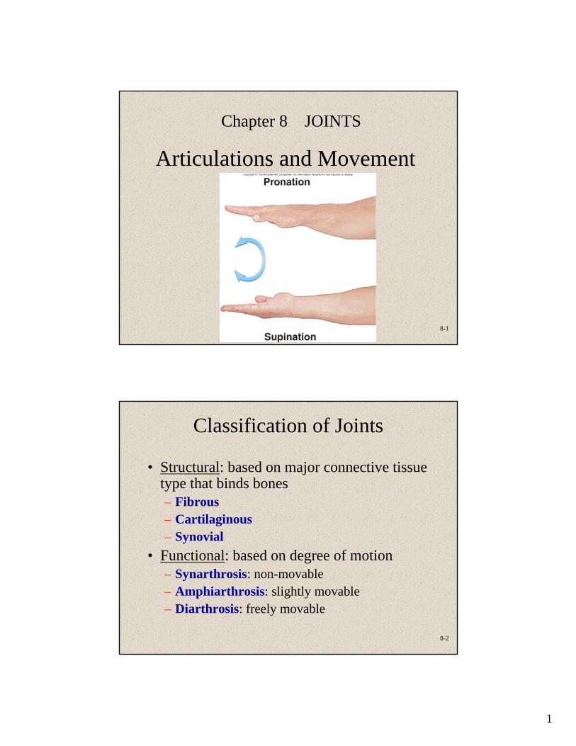
Chapter 8 JOINTS
Articulations and Movement
8-2
Classification of Joints
• Structural: based on major connective tissue type that binds bones– Fibrous– Cartilaginous– Synovial
• Functional: based on degree of motion – Synarthrosis: non-movable– Amphiarthrosis: slightly movable– Diarthrosis: freely movable
2
8-3
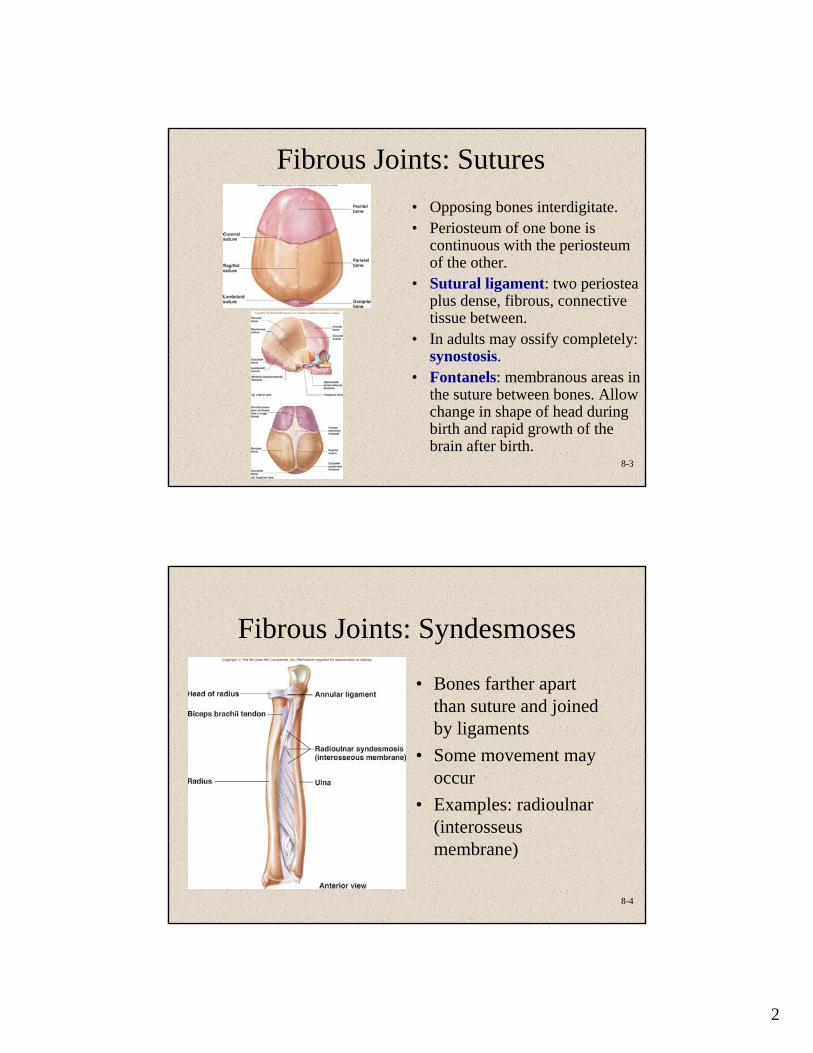
Fibrous Joints: Sutures • Opposing bones interdigitate. • Periosteum of one bone is
continuous with the periosteum of the other.
• Sutural ligament: two periostea plus dense, fibrous, connective tissue between.
• In adults may ossify completely: synostosis.
• Fontanels: membranous areas in the suture between bones. Allow change in shape of head during birth and rapid growth of the brain after birth.
8-4
Fibrous Joints: Syndesmoses
• Bones farther apart than suture and joined by ligaments
• Some movement may occur
• Examples: radioulnar (interosseus membrane)
3
8-5
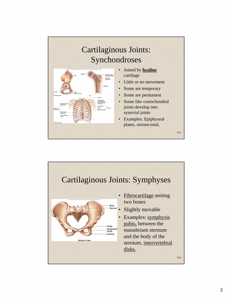
Cartilaginous Joints: Synchondroses
• Joined by hyalinecartilage
• Little or no movement• Some are temporary• Some are permanent• Some like costochondral
joints develop into synovial joints
• Examples: Epiphyseal plates, sternocostal,
8-6
Cartilaginous Joints: Symphyses
• Fibrocartilage uniting two bones
• Slightly movable• Examples: symphysis
pubis, between the manubrium sternum and the body of the sternum, intervertebral disks.
4
8-7
Synovial Joints
• Contain synovial fluid• Allow considerable movement• Most joints that unite bones of
appendicular skeleton reflecting greater mobility of appendicular skeleton compared to axial
• Complex
8-8
Structure of Synovial
Joints
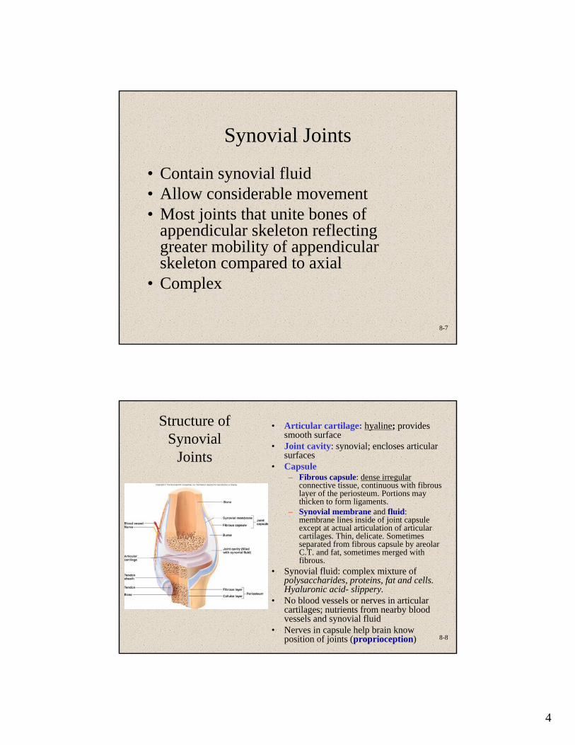
• Articular cartilage: hyaline; provides smooth surface
• Joint cavity: synovial; encloses articular surfaces
• Capsule– Fibrous capsule: dense irregular
connective tissue, continuous with fibrous layer of the periosteum. Portions may thicken to form ligaments.
– Synovial membrane and fluid: membrane lines inside of joint capsule except at actual articulation of articular cartilages. Thin, delicate. Sometimes separated from fibrous capsule by areolar C.T. and fat, sometimes merged with fibrous.
• Synovial fluid: complex mixture of polysaccharides, proteins, fat and cells. Hyaluronic acid- slippery.
• No blood vessels or nerves in articular cartilages; nutrients from nearby blood vessels and synovial fluid
• Nerves in capsule help brain know position of joints (proprioception)
5
8-9
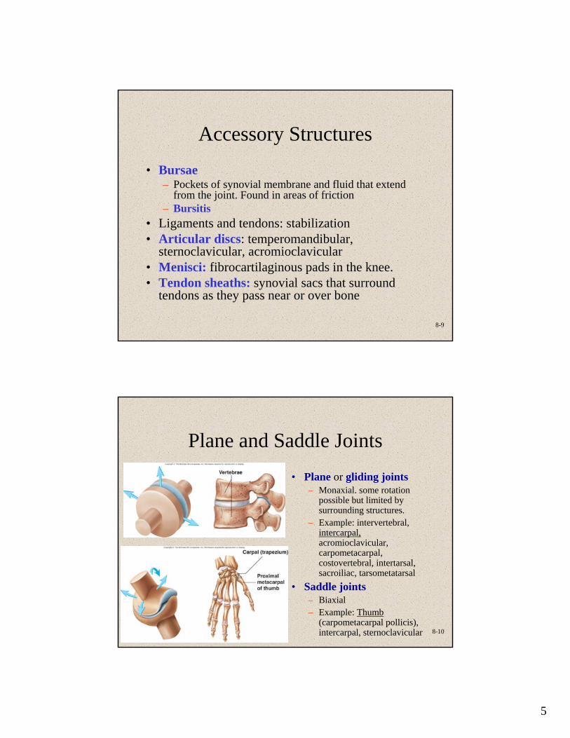
Accessory Structures• Bursae
– Pockets of synovial membrane and fluid that extend from the joint. Found in areas of friction
– Bursitis• Ligaments and tendons: stabilization• Articular discs: temperomandibular,
sternoclavicular, acromioclavicular• Menisci: fibrocartilaginous pads in the knee.• Tendon sheaths: synovial sacs that surround
tendons as they pass near or over bone
8-10
Plane and Saddle Joints• Plane or gliding joints
– Monaxial. some rotation possible but limited by surrounding structures.
– Example: intervertebral, intercarpal,acromioclavicular, carpometacarpal, costovertebral, intertarsal, sacroiliac, tarsometatarsal
• Saddle joints– Biaxial– Example: Thumb
(carpometacarpal pollicis), intercarpal, sternoclavicular
6
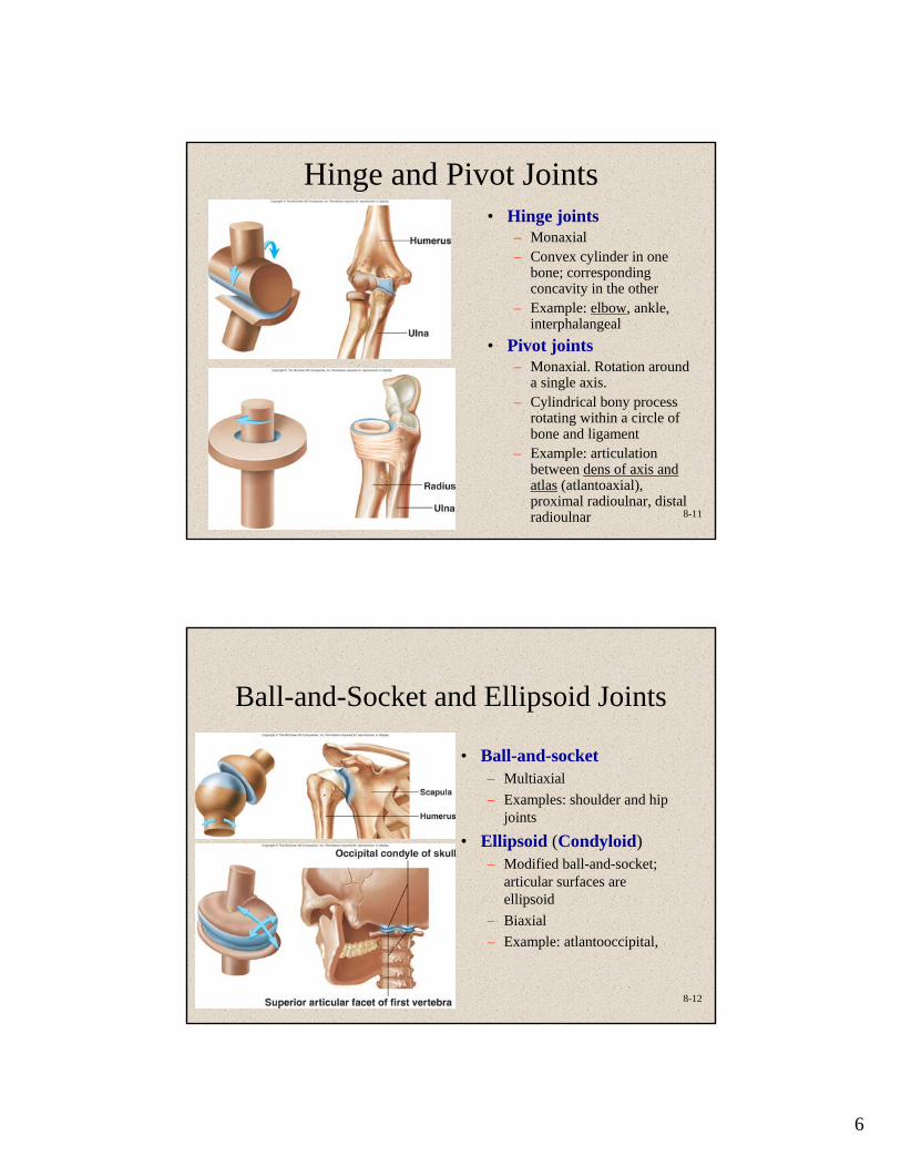
8-11
Hinge and Pivot Joints• Hinge joints
– Monaxial– Convex cylinder in one
bone; corresponding concavity in the other
– Example: elbow, ankle, interphalangeal
• Pivot joints– Monaxial. Rotation around
a single axis.– Cylindrical bony process
rotating within a circle of bone and ligament
– Example: articulation between dens of axis and atlas (atlantoaxial), proximal radioulnar, distal radioulnar
8-12
Ball-and-Socket and Ellipsoid Joints
• Ball-and-socket– Multiaxial– Examples: shoulder and hip
joints
• Ellipsoid (Condyloid)– Modified ball-and-socket;
articular surfaces are ellipsoid
– Biaxial– Example: atlantooccipital,
7
8-13
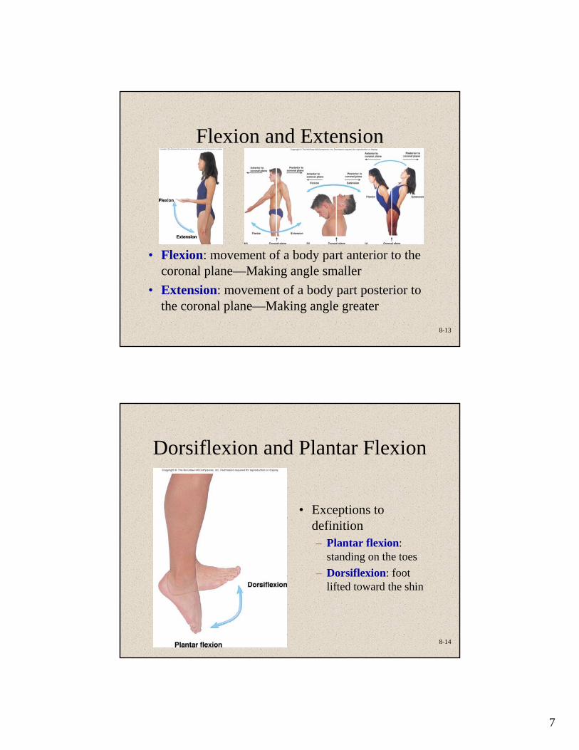
Flexion and Extension
• Flexion: movement of a body part anterior to the coronal plane—Making angle smaller
• Extension: movement of a body part posterior to the coronal plane—Making angle greater
8-14
Dorsiflexion and Plantar Flexion
• Exceptions to definition– Plantar flexion:
standing on the toes– Dorsiflexion: foot
lifted toward the shin
8
8-15
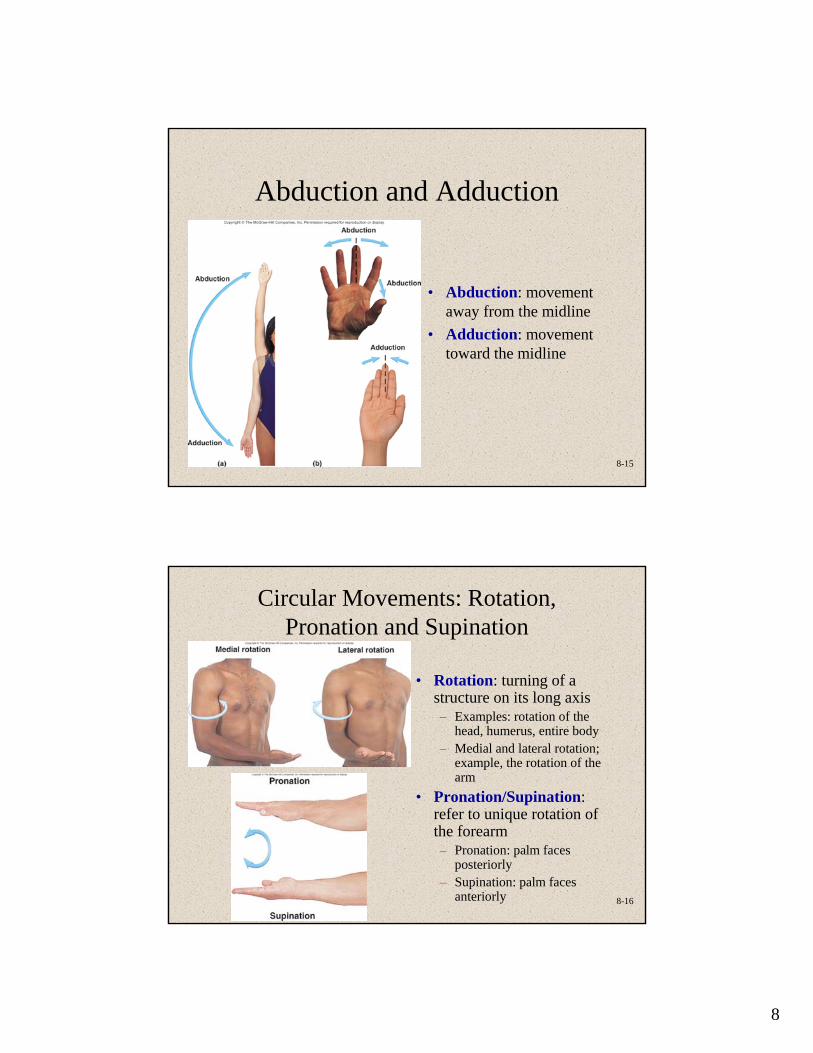
Abduction and Adduction
• Abduction: movement away from the midline
• Adduction: movement toward the midline
8-16
Circular Movements: Rotation, Pronation and Supination
• Rotation: turning of a structure on its long axis– Examples: rotation of the
head, humerus, entire body– Medial and lateral rotation;
example, the rotation of the arm
• Pronation/Supination: refer to unique rotation of the forearm– Pronation: palm faces
posteriorly– Supination: palm faces
anteriorly
9
8-17
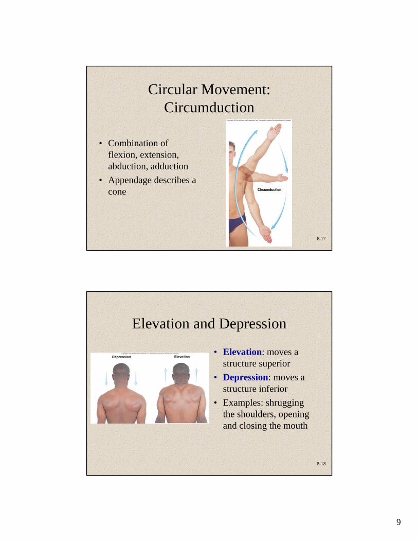
Circular Movement: Circumduction
• Combination of flexion, extension, abduction, adduction
• Appendage describes a cone
8-18
Elevation and Depression
• Elevation: moves a structure superior
• Depression: moves a structure inferior
• Examples: shrugging the shoulders, opening and closing the mouth
10
8-19
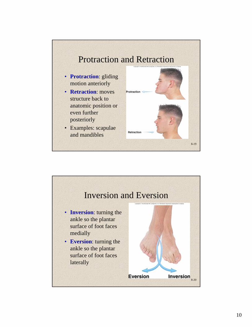
Protraction and Retraction
• Protraction: gliding motion anteriorly
• Retraction: moves structure back to anatomic position or even further posteriorly
• Examples: scapulae and mandibles
8-20
Inversion and Eversion
• Inversion: turning the ankle so the plantar surface of foot faces medially
• Eversion: turning the ankle so the plantar surface of foot faces laterally
11
8-21
Shoulder (Glenohumeral)
Joint
• Ball-and-socket: stability is reduced, mobility is increased compared to hip
• Flexion/extension, abduction/adduction, rotation, circumduction
• Glenoid labrum: rim of fibrocartilage built up around glenoid cavity; joint capsule attachment
• Bursae: subacromial and subscapular
• Rotator cuff: four muscles that along with ligaments give stability to the joint
• Tendon of biceps brachii passes through the joint capsule
8-22
Elbow Joint• Compound hinge joint
– Humeroulnar joint– Humeroradial joint– Proximal radioulnar joint
• Shape of trochlear notch and trochlea limit movement to extension and flexion
• Rounded head of radius allows pronation and supination
• Ligaments– Ulnar collateral ligament– Radial collateral ligament– Radial annular ligament
• Subacromial bursa
12
8-23
Knee Joint
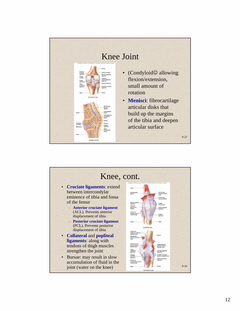
• (Condyloid☺ allowing flexion/extension, small amount of rotation
• Menisci: fibrocartilage articular disks that build up the margins of the tibia and deepen articular surface
8-24
Knee, cont.• Cruciate ligaments: extend
between intercondylar eminence of tibia and fossa of the femur– Anterior cruciate ligament
(ACL). Prevents anterior displacement of tibia
– Posterior cruciate ligament(PCL). Prevents posterior displacement of tibia
• Collateral and popliteal ligaments: along with tendons of thigh muscles strengthen the joint
• Bursae: may result in slow accumulation of fluid in the joint (water on the knee)
13
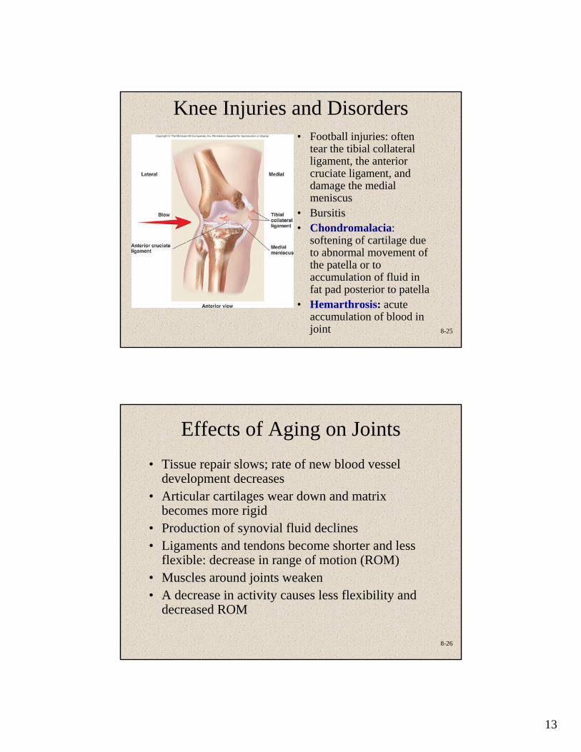
8-25
Knee Injuries and Disorders• Football injuries: often
tear the tibial collateral ligament, the anterior cruciate ligament, and damage the medial meniscus
• Bursitis• Chondromalacia:
softening of cartilage due to abnormal movement of the patella or to accumulation of fluid in fat pad posterior to patella
• Hemarthrosis: acute accumulation of blood in joint
8-26
Effects of Aging on Joints• Tissue repair slows; rate of new blood vessel
development decreases• Articular cartilages wear down and matrix
becomes more rigid• Production of synovial fluid declines• Ligaments and tendons become shorter and less
flexible: decrease in range of motion (ROM)• Muscles around joints weaken• A decrease in activity causes less flexibility and
decreased ROM
14
8-27
Joint Disorders• Arthritis
– Osteoarthritis: wear and tear– Rheumatoid: caused by transient infection or
autoimmune disease• Joint infections. Lyme disease (with ticks as
vector), suppurative arthritis, tuberculous arthritis• Gout. Metabolic disorders of unknown cause
(idiopathic). Increase in uric acid in blood results in deposition of monosodium urate crystals in joints and kidneys
• Hallux valgus and bunion. Caused by ill-fitting shoes
• Joint replacement. Prosthetic joint used to eliminate excruciating pain, usually due to arthritis