Embed Size (px)
Citation preview

Article
Novel Modular Rhodopsins from Green Algae Hold
Great Potential for Cellular Optogenetic Modulation
across the Biological Model Systems
Mayanka Awasthi 1,#, Kumari Sushmita 2,#, Manish Singh Kaushik 2 and Peeyush Ranjan 1,*and
Suneel Kateriya 2,*
1 Department of Cell Biology and Molecular Genetics, University of Maryland, College Park, USA. 2 Laboratory of Optobiology, School of Biotechnology, Jawaharlal Nehru University, New Delhi, India.
# These authors have contributed equally to this work.
* Correspondence: [email protected] (P.R.); [email protected] (S.K.)
Published: xxx
Abstract: Light-gated ion channel and ion pump rhodopsins are widely used as optogenetic tools
and these can control the electrically excitable cells as: (1) they are a single-component system i.e.,
their sensor and effector functions are encoded by the 7-transmembrane domains and (2) they
show fast kinetics with small dark-thermal recovery time. In cellular signaling, a signal receptor,
modulator and effector components are involved for attaining synchronous multicomponent
regulation. Optical modulation of this network requires either receptor to effector encoded in a
single ORF or direct modulation of the effector domain through bypassing all upstream players.
Recently discovered modular rhodopsins like rhodopsin guanine cyclase (RhoGC) and rhodopsin
phosphodiesterase (RhoPDE) paves the way to establish proof of concept. Light sensor coupled
modular system could be expressed in a precise cell type and which holds great potential in the
advancement of optogenetics 2.0. It would enable manipulating entire relevant cell signaling
system. Here, we had identified 50 novels modular rhodopsins with variant rhodopsins domain
and its diverse cognate signaling cascades encoded in a single ORF, which are associated with
specialized functions in the cells. These novel modular algal rhodopsins have been characterized
functionality based on their sequence and structural homology with previously characterized
rhodopsins. Presented novel modular rhodopsins with various effector domains hold potential to
expand optogenetics tool kit to regulate various cellular signaling pathways across the diverse
biological model systems.
Keywords: Enzymerhodopsin; Channelrhodopsins; Optogenetics; Two-component system;
Cyclase; Phosphodiesterase
Abbreviations: Cop-Chlamyopsin (rhodopsin from Chlamydomonas reinhardtii), Vop-Volvoxopsin
(rhodopsin from Volvox carteri), GpRh 1-5 (rhodopsin from Gonium pectorale), AsRh1-4 (Asterochloris
sp.), KnRh1-3 (Klabsormidium nitens), OtRh1-2 (Ostreococcus tauri), MpuRh1&2 (Micromonas pusilla),
MspRh1&2 (Micromonas species), OlRh1-4 (Ostreococcus lucimarinus), CsRh1 (Chlorella sorokiniana),
ApRh1 (Auxenochlorella protothecoides) , BgRh1&2 (Bigelowiella natans), GtRh1-10 (Guillardia theta),
DsRh1 (Dunaliella salina), TsRh1 (Tetraselmis subcordiformis)
1. Introduction
Many photobehavioural responses are mediated by rhodopsin-based photoreceptor(s) that are
distributed across almost all clades of life. Rhodopsins are seven transmembrane helical proteins
which use retinal as a chromophore. Based on the isoforms of the retinal bound in the ground state,
Preprints (www.preprints.org) | NOT PEER-REVIEWED | Posted: 1 September 2020 doi:10.20944/preprints202009.0015.v1
© 2020 by the author(s). Distributed under a Creative Commons CC BY license.
rhodopsins are classified into two broad categories i.e., Type I or microbial type (MTR) and Type II
or animal-type rhodopsins (ATR). MTRs are widely distributed across all kingdoms of life and
perform diverse physiological functions, such as the light-activated ion pumps- Bacteriorhodopsin
(BR) [1] and Halorhodopsin (HR) [2], light-gated channels- Channelrhodopsins (ChR1 & ChR2)
[3,4], and sensory photoreceptors (SRI & II) [5]. Light-gated ion pumps and channels cause
alterations in the membrane potential in a light dependent manner whereas sensory rhodopsins
mediate downstream signaling. SR I and II in halobacteria communicate with the flagellar motor
via transducer proteins HtrI and HtrII respectively [5].
ATR or type II rhodopsins are broadly classified as vertebrate and invertebrate rhodopsins on
the basis of variation in their amino acid sequences [6]. The ATRs (both vertebrate and invertebrate)
mediate the downstream signaling cascade through the G-protein coupled receptor (GPCR)
proteins that involves multiple steps and protein complexes. Both the ATRs and SRs of MTRs are
multi-component systems which require a series of protein complexes to mediate the light-activated
signalling. This poses the limitation to use them as an optogenetic tool for regulating intracellular
signaling process. The success of MTRs as an optogenetic tool is mainly attributed to its property
that both the light sensing and the ion channel activity of the Channelrhodopsins (ChR) are
encoded in a single protein. Recent advancements in the genome database has led to the discovery
of many new MTRs which are directly coupled to effector domains e.g. two-component system and
cyclase in enzyme-rhodopsins [7,8]. This structural diversity imparts great precision, fast kinetics
and low off-target effects that provides an edge to the MTR to target and regulate specific cellular
processes simply by illumination. cAMP and cGMP, the key modulators of cell signaling, are the
secondary messengers that regulate many cellular, metabolic and developmental processes.
However, it is difficult to target/modulate cGMP and cAMP levels precisely in specific cell types
with spatial-temporal resolution using the animal-type rhodopsin signaling cascade because of the
involvement of many player(s) in the cascade. In addition, pharmacological targeting has the
limitation of specificity and temporal issues at the cellular level.
Enzyme-rhodopsins (Rhodopsin phosphodiesterase; RhoPDE and Rhodopsin cyclase; RhoGC)
have emerged as promising optogenetic tools for the precise and non-invasive spatiotemporal
control of cyclic nucleotide signaling pathways. The heterologous expression of RhoPDE [9,10] from
Salpingoeca rosetta in Xenopus oocyte and HEK293 cell lines demonstrated the light-activated cGMP
and cAMP-phosphodiesterase activity [11]. Similarly, RhoGC [12,13] isolated from fungi
Blastocladiella emersonii and Catenaria anguillulae when expressed in various mammalian cell lines,
could generate substantial cGMP, and were used as an optogenetic tool [14,15]. Since then
significant interest has developed towards the identification, characterization and testing of novel
modular rhodopsins [7,16,17] as optogenetic tool candidates for tweaking the cell signaling process.
The identified modular rhodopsins coupled with other domains in a single ORF have shown the
potential to overcome the limitation of SRs to be used as an optogenetic tool. Characterizing the
physiological role of the existing and newly identified multidomain rhodopsins is tempting but
limited because of their large transcript size, poor heterologous expression of transmembrane
domain and lack of the established functional assays for these modular rhodopsins. Recently, we
have identified 24 new modular rhodopsins from different algae [7]. In the present study, we have
identified many new modular rhodopsins and ChRs fused with new domains that were previously
unknown and analysed their evolutionary pattern and sequence homology as well as the structural
and functional potential of these domains coupled to rhodopsin (based on available experimental
evidences). We have also investigated the diversity of multidomain rhodopsins and the recruitment
of signaling component in a single ORF in relation to its prokaryotic counterpart. This extensive
analysis of MTRs defines a future roadmap towards the involvement of modular rhodopsin-based
photoreceptors in the photophysiological response of the relevant organism. Evolutionary pattern
analysis of the MTRs suggests the evolution of multi-domain rhodopsins in the microalgal system
after evolution of the ChRs with extended C-terminus of unknown function by lateral gene transfer.
Moreover, these novel modular rhodopsins with different effector domains hold potential to
Preprints (www.preprints.org) | NOT PEER-REVIEWED | Posted: 1 September 2020 doi:10.20944/preprints202009.0015.v1
expand optogenetics tool kit 2.0 to regulate various cellular signaling pathways across the manifold
biological model systems.
2. Materials and Methods
2.1. Identification of rhodopsin domain, homology and structural analysis.
Extensive genome database search for MTRs and modular rhodopsins were performed on JGI
genome database, metagenome database and NCBI portal using BR and Chlamydomonas
rhodopsin as template. The rhodopsin identity, sequence accession number, homology, conserved
domains are summarized in Table S1. Multiple sequence alignment was performed using Clustal_X
program [18] and BioEdit (http://www.mbio.ncsu.edu/bioedit/bioedit.html). All colour editing was
done by using the BioEdit program. The rhodopsin domains of new MTRs were identified by
sequence alignment with canonical rhodopsins and analysis with conserved domain architecture
retrieval tool (CDART) [19] and conserved domain database [20] program. The rhodopsin with
conserved seven transmembrane helices and retinal binding motif in the seventh helix was
considered for further analysis. The number indicating the position of amino acid is referred with
respect to BR unless mentioned in the text.
2.2. Evolutionary analysis of rhodopsin domains of modular proteins
Molecular evolutionary analysis of typical MTR and rhodopsin domains of modular proteins
were performed computationally with protein sequences. Multiple sequence alignment of
rhodopsin domain was done on Clustal X 2.0 [18]. Phylogenetic analysis was performed by
Neighbour – joining (NJ) method using MEGA X [21] with a thousand bootstrap replicates. The
same was also verified by maximum likelihood ML method on MEGA X and topology was viewed
by MEGA X as well as tree view and NJ plot [22].
2.3. Protein-protein interaction analysis of novel domains from modular algal rhodopsins
The interactomes for domains associated with ChRs, i.e. FimV, MED15 and UL36, were
constructed. The interacting partners for each of the effector domains were predicted using the
String version 11 [23] and the output was further used to generate the network by employing
Cytoscape 3.7.2 [24].
3. Results and Discussion
3.1. Microbial rhodopsins with modular domain organization
Mining the genome database of the organisms from diverse taxa and strata has revealed the
presence of MTRs from archaea to algae inhabiting in diverse habitats from freshwater to terrestrial
environments. The phototactic green alga C. reinhardtii has been extensively studied for learning
various aspects of cell biology from photobehavioural responses (especially ChR-mediated) to
photosynthesis, cilia biology, intraflagellar transport to vesicle, and membrane-bound trafficking
and dynamics [25,26]. The early modular rhodopsins were identified in this green alga and since
then very few have been reported in other organisms. Owing to its cellular optogenetic potential, a
thorough and extensive genome database search was performed to identify novel rhodopsin(s) with
modular nature, better kinetics and fast recovery time.
Here, we have identified new microbial modular ChRs (Figure 1A and table 1A&B) and SRs
(Figure 1B-D and table 2A&B) across different taxa and analysed their critical features that
segregate MTRs from other seven transmembrane protein families. Based on the modular domain
coupled to the rhodopsin, we evaluated the possible function of these proteins in the respective
organism and their potential optogenetic application in cell and developmental biology of the
different model systems.
Preprints (www.preprints.org) | NOT PEER-REVIEWED | Posted: 1 September 2020 doi:10.20944/preprints202009.0015.v1
3.2. Modular Channelrhodopsins and their optogenetic potential
Our targeted search for the modular ChR yielded three modular ChRs as shown in Figure 1A.
These are (i) KnRh3 from Klebsormidium nitens (terrestrial alga) which is coupled with the
peptidoglycan binding protein, FimV, (ii) the blue-shifted ChR, TsRh1 from Tetraselmis
subcordiformis, for which the rhodopsin domain has been characterized [TsRh1 is coupled with the
mediator subunit, MED15 (Mediator of RNA polymerase II subunit 15)] [27], however its modular
nature has not been discussed and (iii) GpRh1 from Gonium pectorale, which is coupled with UL36
(large tegument protein). The optogenetic potential of these modular domains (FimV, MED15, and
UL36) is summarized in table 1A. The Rhodopsin domains of KnRh3, TsRh1 and GpRh1 were
aligned with well characterized ChRs taken as the reference for sequence analysis (Figure 2). The
conserved residues essential for photocycle are marked in Figure 2, and the same have been
analysed for four main functionalities namely: (1) retinal-binding lysine, (2) counter ion/proton
acceptor of RSB, (3) proton-release complex and (4) DC-gate present in helix 3 and 4. Based on these
amino acid residues, we evaluated the rhodopsin domain and summarized the details in table 1B
and 2B for modular ChRs and SRs respectively.
Preprints (www.preprints.org) | NOT PEER-REVIEWED | Posted: 1 September 2020 doi:10.20944/preprints202009.0015.v1
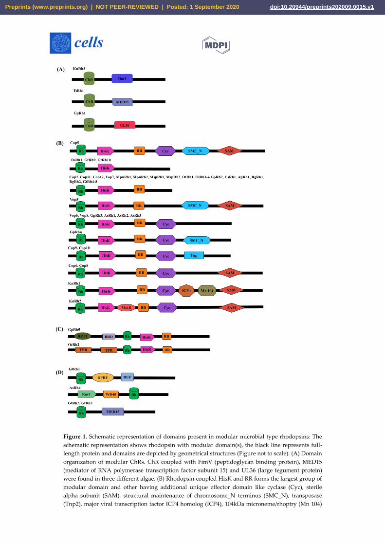
Figure 1. Schematic representation of domains present in modular microbial type rhodopsins: The
schematic representation shows rhodopsin with modular domain(s), the black line represents full-
length protein and domains are depicted by geometrical structures (Figure not to scale). (A) Domain
organization of modular ChRs. ChR coupled with FimV (peptidoglycan binding protein), MED15
(mediator of RNA polymerase transcription factor subunit 15) and UL36 (large tegument protein)
were found in three different algae. (B) Rhodopsin coupled HisK and RR forms the largest group of
modular domain and other having additional unique effector domain like cyclase (Cyc), sterile
alpha subunit (SAM), structural maintenance of chromosome_N terminus (SMC_N), transposase
(Tnp2), major viral transcription factor ICP4 homolog (ICP4), 104kDa microneme/rhoptry (Mn 104)
Preprints (www.preprints.org) | NOT PEER-REVIEWED | Posted: 1 September 2020 doi:10.20944/preprints202009.0015.v1
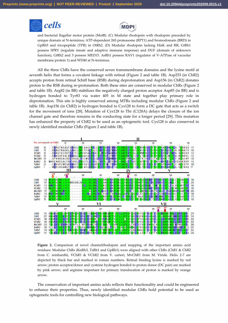
and bacterial flagellar motor protein (MotB). (C) Modular rhodopsin with rhodopsin preceded by
unique domain at N-terminus; ATP-dependent 26S proteasome (RPT1) and bromodomain (BRD) in
GpRh5 and tricopeptide (TPR) in OtRh2. (D) Modular rhodopsin lacking HisK and RR; GtRh1
possess SPRY (regulate innate and adaptive immune response) and DUF (domain of unknown
function), GtRh2 and 3 possess MED15. AsRh1 possess RAV1 (regulator of V-ATPase of vacuolar
membrane protein 1) and WD40 at N-terminus.
All the three ChRs have the conserved seven transmembrane domains and the lysine motif at
seventh helix that forms a covalent linkage with retinal (Figure 2 and table 1B). Asp253 (in ChR2)
accepts proton from retinal Schiff base (RSB) during deprotonation and Asp156 (in ChR2) donates
proton to the RSB during re-protonation. Both these sites are conserved in modular ChRs (Figure 2
and table 1B). Arg82 (in BR) stabilizes the negatively charged proton acceptor Asp85 (in BR) and is
hydrogen bonded to Tyr83 via water 405 in M state and together play primary role in
deprotonation. This site is highly conserved among MTRs including modular ChRs (Figure 2 and
table 1B). Asp156 (in ChR2) is hydrogen bonded to Cys128 to form a DC gate that acts as a switch
for the movement of ions [28]. Mutation of Cys128 to Thr (C128A) delays the closure of the ion
channel gate and therefore remains in the conducting state for a longer period [29]. This mutation
has enhanced the property of ChR2 to be used as an optogenetic tool. Cys128 is also conserved in
newly identified modular ChRs (Figure 2 and table 1B).
Figure 2. Comparison of novel channelrhodopsin and mapping of the important amino acid
residues: Modular ChRs (KnRh3, TsRh1 and GpRh1) were aligned with other ChRs (ChR1 & ChR2
from C. reinhardtii, VChR1 & VChR2 from V. carteri, MvChR1 from M. Viride. Helix 2-7 are
depicted by black bar and marked in roman numbers. Retinal binding lysine is marked by red
arrow; proton acceptor/donor and cysteine hydrogen bonded to proton donor (DC pair) are marked
by pink arrow; and arginine important for primary translocation of proton is marked by orange
arrow.
The conservation of important amino acids reflects their functionality and could be engineered
to enhance their properties. Thus, newly identified modular ChRs hold potential to be used as
optogenetic tools for controlling new biological pathways.
Preprints (www.preprints.org) | NOT PEER-REVIEWED | Posted: 1 September 2020 doi:10.20944/preprints202009.0015.v1
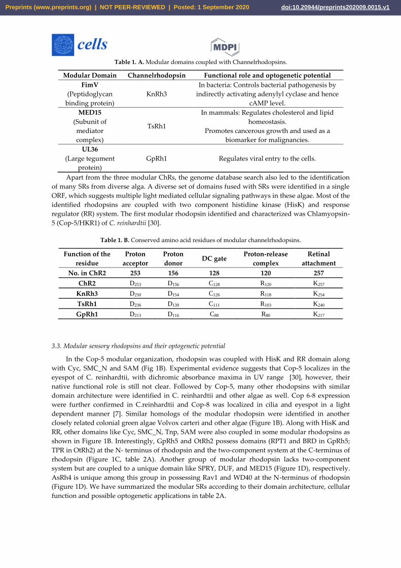
Table 1. A. Modular domains coupled with Channelrhodopsins.
Modular Domain Channelrhodopsin Functional role and optogenetic potential
FimV
(Peptidoglycan
binding protein)
KnRh3
In bacteria: Controls bacterial pathogenesis by
indirectly activating adenylyl cyclase and hence
cAMP level.
MED15
(Subunit of
mediator
complex)
TsRh1
In mammals: Regulates cholesterol and lipid
homeostasis.
Promotes cancerous growth and used as a
biomarker for malignancies.
UL36
(Large tegument
protein)
GpRh1 Regulates viral entry to the cells.
Apart from the three modular ChRs, the genome database search also led to the identification
of many SRs from diverse alga. A diverse set of domains fused with SRs were identified in a single
ORF, which suggests multiple light mediated cellular signaling pathways in these algae. Most of the
identified rhodopsins are coupled with two component histidine kinase (HisK) and response
regulator (RR) system. The first modular rhodopsin identified and characterized was Chlamyopsin-
5 (Cop-5/HKR1) of C. reinhardtii [30].
Table 1. B. Conserved amino acid residues of modular channelrhodopsins.
Function of the
residue
Proton
acceptor
Proton
donor DC gate
Proton-release
complex
Retinal
attachment
No. in ChR2 253 156 128 120 257
ChR2 D253 D156 C128 R120 K257
KnRh3 D250 D154 C126 R118 K254
TsRh1 D236 D139 C111 R103 K240
GpRh1 D213 D116 C88 R80 K217
3.3. Modular sensory rhodopsins and their optogenetic potential
In the Cop-5 modular organization, rhodopsin was coupled with HisK and RR domain along
with Cyc, SMC_N and SAM (Fig 1B). Experimental evidence suggests that Cop-5 localizes in the
eyespot of C. reinhardtii, with dichromic absorbance maxima in UV range [30], however, their
native functional role is still not clear. Followed by Cop-5, many other rhodopsins with similar
domain architecture were identified in C. reinhardtii and other algae as well. Cop 6-8 expression
were further confirmed in C.reinhardtii and Cop-8 was localized in cilia and eyespot in a light
dependent manner [7]. Similar homologs of the modular rhodopsin were identified in another
closely related colonial green algae Volvox carteri and other algae (Figure 1B). Along with HisK and
RR, other domains like Cyc, SMC_N, Tnp, SAM were also coupled in some modular rhodopsins as
shown in Figure 1B. Interestingly, GpRh5 and OtRh2 possess domains (RPT1 and BRD in GpRh5;
TPR in OtRh2) at the N- terminus of rhodopsin and the two-component system at the C-terminus of
rhodopsin (Figure 1C, table 2A). Another group of modular rhodopsin lacks two-component
system but are coupled to a unique domain like SPRY, DUF, and MED15 (Figure 1D), respectively.
AsRh4 is unique among this group in possessing Rav1 and WD40 at the N-terminus of rhodopsin
(Figure 1D). We have summarized the modular SRs according to their domain architecture, cellular
function and possible optogenetic applications in table 2A.
Preprints (www.preprints.org) | NOT PEER-REVIEWED | Posted: 1 September 2020 doi:10.20944/preprints202009.0015.v1
Table 2. A: Modular domains coupled with sensory rhodopsins.
Modular Domain Modular Rhodopsins Cellular role and optogenetic
potential
HisK
DsRh1, GtRh4-10, Cop5-12,
Vop5-8, AsRh1-3, GpRh2-5,
KnRh1 & 2, OtRh1&2,
OlRh1-4, MpuRh1&2,
Msp1&2, CsRh1, ApRh1,
BgRh1&2
Part of two-component
signaling; regulates gene
expression
HisK-RR (Histidine kinase-
response regulator)
Two-component signaling
system
GtRh4-8, Cop5-12, Vop5-8,
AsRh1-3, GpRh2-5, KnRh1 &
2, OtRh1&2, OlRh1-4,
MpuRh1&2, Msp1&2,
CsRh1, ApRh1, BgRh1&2
Regulates gene expression and
various other cell processes via
output domain like helix-turn-
helix (HTH), RNA, enzyme or
ligand-binding domain.
Cyc (Cyclase)
Cop5, 6, 8, 9 &10, Vop6&8,
AsRh1-3, GpRh3&4, KnRh1
& 2
Regulates the level of secondary
messengers: cAMP and cGMP.
SMC_N (Structural
Maintenance of Chromosome
_N terminal)
Cop5, Vop5, GpRh4
Stabilizes the chromosome,
helps in its proper segregation
during cell division and DNA
repair.
Tnp (Transposase) Cop9 & 10
Recognizes the transposable
elements in DNA and catalyses
their movement to another
DNA.
SAM (Sterile alpha motif) Cop5-8, Vop5, KnRh1 & 2
Mediate protein-protein
interactions, RNA and lipid
binding; regulates transcription
factor
ICP4 (Infected-cell polypeptide
4) KnRh1
Major transcription factor of
herpes simplex virus type1
(HSV-1)
Mn104 (Microneme/rhoptry) KnRh1
Helps in invading host cell by
apicomplexan parasites; N-
terminal region proposed to
serve as signal peptide for ER
MotB (Flagellar motor protein) KnRh2 MotB acts as a stator in proton
pump.
RPT1 (Regulatory Particle
Triple ATPase) GpRh5
Forms a part of 26S proteasomal
complex
BRD (Bromodomain) GpRh5
Modulate gene expression by
associating with acetylated
lysine on histone
TPR (Tetracopeptide repeat) OtRh2
Regulates virulence in bacteria;
translocation of receptors to
their respective organelles in
different systems
SPRY [Spore lysis A (Spl A) in
Dictyostelium discoideum and
mammalian Ryanodine receptor
(RYR)]
GtRh1
Substrate binding for
ubiquitination in ubiquitin
ligase family proteins; involved
in various immune response
Preprints (www.preprints.org) | NOT PEER-REVIEWED | Posted: 1 September 2020 doi:10.20944/preprints202009.0015.v1
DUF GtRh1 Mediate protein-protein
interaction
Rav1
(Regulator of V-ATPase of
vacuole membrane protein 1)
AsRh4
Regulates the assembly of V-
ATPase (ATP powered H+
pump in vacuole forming
organelles)
WD40 AsRh4 Mediate protein-protein
interaction
3.4. Light-gated ion pump and photo-sensory function prediction based on conserved residues of rhodopsins
Amino acids in the proximity of retinal are the key determinants in the activation and function
of rhodopsin. The crystal structure of BR suggests that Asp85 is the proton acceptor from RSB
during deprotonation. Thr89 is hydrogen bonded to Asp85 (Figure 3 and Table 2B). Asp212 also
remains protonated and thus plays a role during the primary proton transfer event. Asp96 donates
proton to the RSB during reprotonation. Glu194 and 204 are the terminal amino acids responsible
for the outward release of into extracellular side. These positions were analysed in the modular
rhodopsins to assign their functionality. Out of 47 modular rhodopsins at position 85, 14 had
conserved Asp/Glu while 17 had Gln (Figure 3 and Table 2B). Position 89 is well conserved with 43
out of 47 modular rhodopsins possessing Ser/Thr at this position (Figure 3 and Table 2B). Asp96 is
only conserved in AsRh4 (Table 2B). Asp212 is well conserved among modular rhodopsin except 6
of them which possess Asn at this position (Figure 3 and Table 2B). Only 4 modular rhodopsins
possess Asp at 194th position while 25 modular rhodopsins have Glu at 204th position (Figure 3
and Table 2B). Since the retinal attachment lysine is conserved among all modular rhodopsin, these
rhodopsins seem to be functional (Figure 3 and Table 2B). AsRh4 is the only modular rhodopsin
with an amino acid conserved for proton pump. Other modular rhodopsins seem to form a new
group with different mechanism for activation and relay of signals. Despite lacking the proton
acceptor Asp85, Cop5 was found to be active in UV A and blue light (Figure 3 and Table 2B).
Cop6/Vop6 was suggested to be a light inhibited guanylate cyclase upon supplementation of ATP
when expressed in Xenopus oocyte [31] Although it lacks Asp85, Asp96 and Asp212 (Figure 3 and
Table 2B). Signal relay in Cop6/Vop6 proceeds through HisK and RR. OtRh1/Ot-HKR is a green
absorbing modular rhodopsin controlling the circadian clock of O. tauri. The photophysical
properties of OtRh1/Ot-HKR are affected by salt concentration indicating this rhodopsin might
provide input for adaptation in salt environment [32]. These examples suggest that the important
amino acids are substituted but these rhodopsins are functional. Unique domains coupled with
rhodopsin might regulate specific function in cell/organism and hold potential to be used as
optogenetic tool and therefore should be explored in detail.
Preprints (www.preprints.org) | NOT PEER-REVIEWED | Posted: 1 September 2020 doi:10.20944/preprints202009.0015.v1
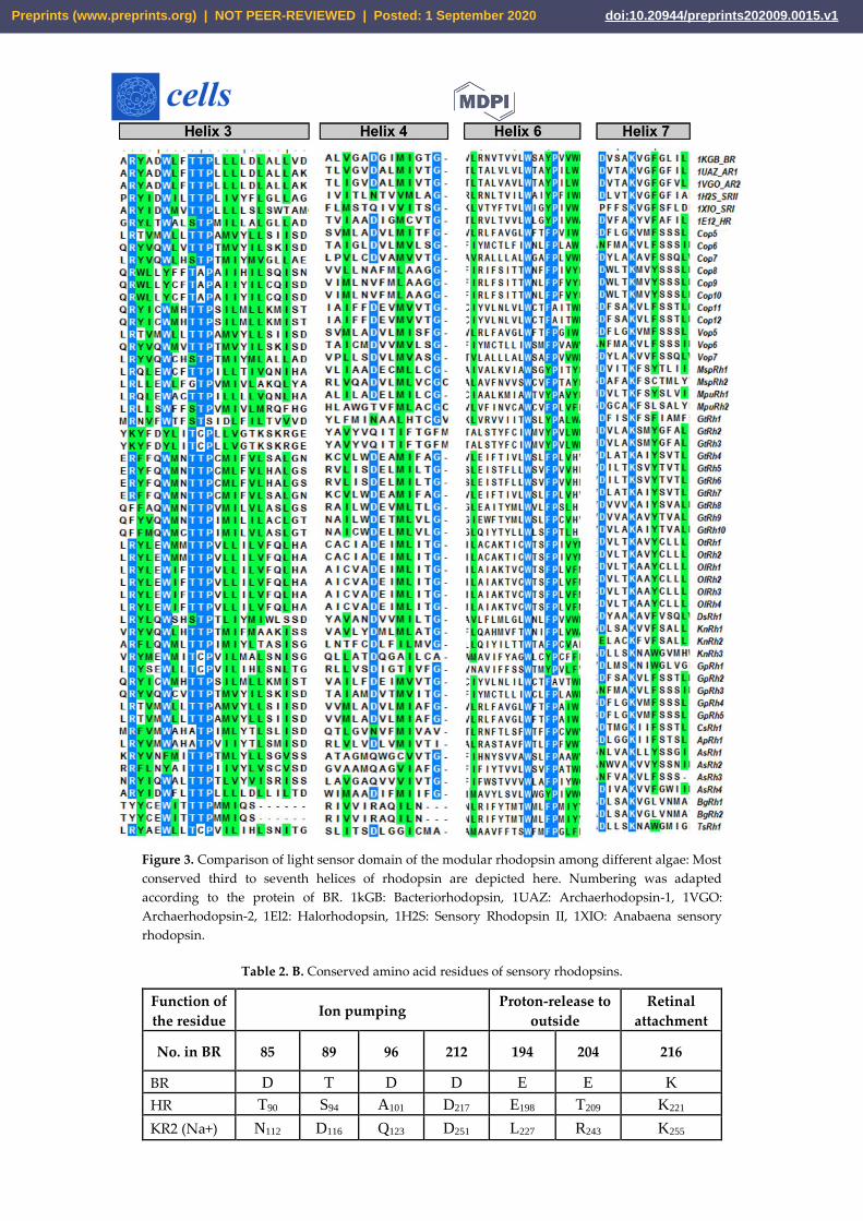
Figure 3. Comparison of light sensor domain of the modular rhodopsin among different algae: Most
conserved third to seventh helices of rhodopsin are depicted here. Numbering was adapted
according to the protein of BR. 1kGB: Bacteriorhodopsin, 1UAZ: Archaerhodopsin-1, 1VGO:
Archaerhodopsin-2, 1El2: Halorhodopsin, 1H2S: Sensory Rhodopsin II, 1XIO: Anabaena sensory
rhodopsin.
Table 2. B. Conserved amino acid residues of sensory rhodopsins.
Function of
the residue Ion pumping
Proton-release to
outside
Retinal
attachment
No. in BR 85 89 96 212 194 204 216
BR D T D D E E K
HR T90 S94 A101 D217 E198 T209 K221
KR2 (Na+) N112 D116 Q123 D251 L227 R243 K255
Preprints (www.preprints.org) | NOT PEER-REVIEWED | Posted: 1 September 2020 doi:10.20944/preprints202009.0015.v1
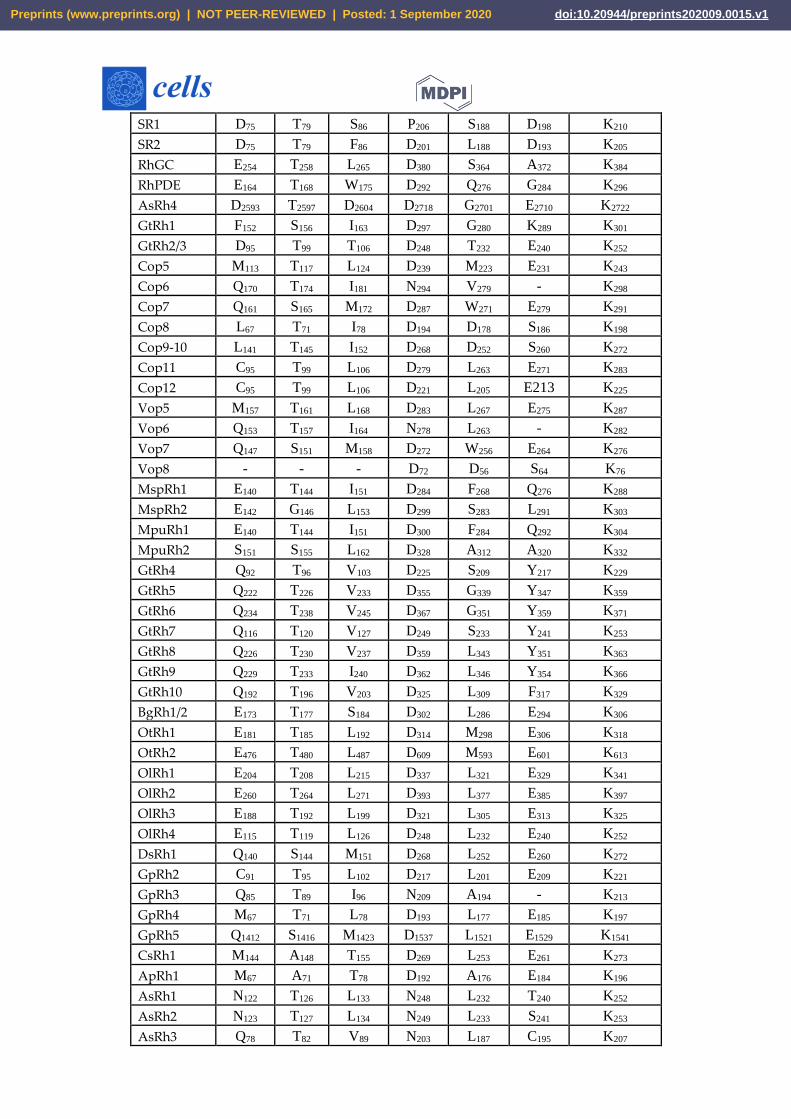
SR1 D75 T79 S86 P206 S188 D198 K210
SR2 D75 T79 F86 D201 L188 D193 K205
RhGC E254 T258 L265 D380 S364 A372 K384
RhPDE E164 T168 W175 D292 Q276 G284 K296
AsRh4 D2593 T2597 D2604 D2718 G2701 E2710 K2722
GtRh1 F152 S156 I163 D297 G280 K289 K301
GtRh2/3 D95 T99 T106 D248 T232 E240 K252
Cop5 M113 T117 L124 D239 M223 E231 K243
Cop6 Q170 T174 I181 N294 V279 - K298
Cop7 Q161 S165 M172 D287 W271 E279 K291
Cop8 L67 T71 I78 D194 D178 S186 K198
Cop9-10 L141 T145 I152 D268 D252 S260 K272
Cop11 C95 T99 L106 D279 L263 E271 K283
Cop12 C95 T99 L106 D221 L205 E213 K225
Vop5 M157 T161 L168 D283 L267 E275 K287
Vop6 Q153 T157 I164 N278 L263 - K282
Vop7 Q147 S151 M158 D272 W256 E264 K276
Vop8 - - - D72 D56 S64 K76
MspRh1 E140 T144 I151 D284 F268 Q276 K288
MspRh2 E142 G146 L153 D299 S283 L291 K303
MpuRh1 E140 T144 I151 D300 F284 Q292 K304
MpuRh2 S151 S155 L162 D328 A312 A320 K332
GtRh4 Q92 T96 V103 D225 S209 Y217 K229
GtRh5 Q222 T226 V233 D355 G339 Y347 K359
GtRh6 Q234 T238 V245 D367 G351 Y359 K371
GtRh7 Q116 T120 V127 D249 S233 Y241 K253
GtRh8 Q226 T230 V237 D359 L343 Y351 K363
GtRh9 Q229 T233 I240 D362 L346 Y354 K366
GtRh10 Q192 T196 V203 D325 L309 F317 K329
BgRh1/2 E173 T177 S184 D302 L286 E294 K306
OtRh1 E181 T185 L192 D314 M298 E306 K318
OtRh2 E476 T480 L487 D609 M593 E601 K613
OlRh1 E204 T208 L215 D337 L321 E329 K341
OlRh2 E260 T264 L271 D393 L377 E385 K397
OlRh3 E188 T192 L199 D321 L305 E313 K325
OlRh4 E115 T119 L126 D248 L232 E240 K252
DsRh1 Q140 S144 M151 D268 L252 E260 K272
GpRh2 C91 T95 L102 D217 L201 E209 K221
GpRh3 Q85 T89 I96 N209 A194 - K213
GpRh4 M67 T71 L78 D193 L177 E185 K197
GpRh5 Q1412 S1416 M1423 D1537 L1521 E1529 K1541
CsRh1 M144 A148 T155 D269 L253 E261 K273
ApRh1 M67 A71 T78 D192 A176 E184 K196
AsRh1 N122 T126 L133 N248 L232 T240 K252
AsRh2 N123 T127 L134 N249 L233 S241 K253
AsRh3 Q78 T82 V89 N203 L187 C195 K207
Preprints (www.preprints.org) | NOT PEER-REVIEWED | Posted: 1 September 2020 doi:10.20944/preprints202009.0015.v1
KnRh1 Q166 T170 M177 D292 L276 E284 K296
KnRh2 Q95 T99 L106 E221 T205 E213 K225
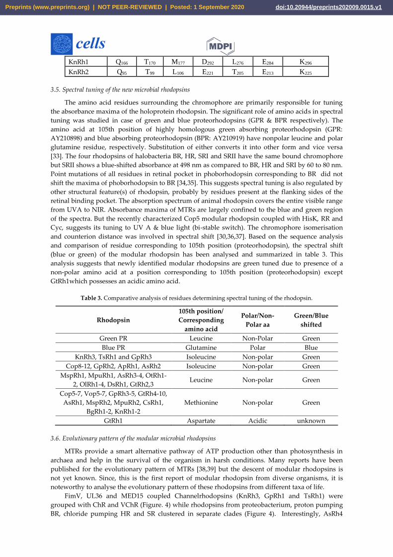
3.5. Spectral tuning of the new microbial rhodopsins
The amino acid residues surrounding the chromophore are primarily responsible for tuning
the absorbance maxima of the holoprotein rhodopsin. The significant role of amino acids in spectral
tuning was studied in case of green and blue proteorhodopsins (GPR & BPR respectively). The
amino acid at 105th position of highly homologous green absorbing proteorhodopsin (GPR:
AY210898) and blue absorbing proteorhodopsin (BPR: AY210919) have nonpolar leucine and polar
glutamine residue, respectively. Substitution of either converts it into other form and vice versa
[33]. The four rhodopsins of halobacteria BR, HR, SRI and SRII have the same bound chromophore
but SRII shows a blue-shifted absorbance at 498 nm as compared to BR, HR and SRI by 60 to 80 nm.
Point mutations of all residues in retinal pocket in phoborhodopsin corresponding to BR did not
shift the maxima of phoborhodopsin to BR [34,35]. This suggests spectral tuning is also regulated by
other structural feature(s) of rhodopsin, probably by residues present at the flanking sides of the
retinal binding pocket. The absorption spectrum of animal rhodopsin covers the entire visible range
from UVA to NIR. Absorbance maxima of MTRs are largely confined to the blue and green region
of the spectra. But the recently characterized Cop5 modular rhodopsin coupled with HisK, RR and
Cyc, suggests its tuning to UV A & blue light (bi-stable switch). The chromophore isomerisation
and counterion distance was involved in spectral shift [30,36,37]. Based on the sequence analysis
and comparison of residue corresponding to 105th position (proteorhodopsin), the spectral shift
(blue or green) of the modular rhodopsin has been analysed and summarized in table 3. This
analysis suggests that newly identified modular rhodopsins are green tuned due to presence of a
non-polar amino acid at a position corresponding to 105th position (proteorhodopsin) except
GtRh1which possesses an acidic amino acid.
Table 3. Comparative analysis of residues determining spectral tuning of the rhodopsin.
Rhodopsin
105th position/
Corresponding
amino acid
Polar/Non-
Polar aa
Green/Blue
shifted
Green PR Leucine Non-Polar Green
Blue PR Glutamine Polar Blue
KnRh3, TsRh1 and GpRh3 Isoleucine Non-polar Green
Cop8-12, GpRh2, ApRh1, AsRh2 Isoleucine Non-polar Green
MspRh1, MpuRh1, AsRh3-4, OtRh1-
2, OlRh1-4, DsRh1, GtRh2,3 Leucine Non-polar Green
Cop5-7, Vop5-7, GpRh3-5, GtRh4-10,
AsRh1, MspRh2, MpuRh2, CsRh1,
BgRh1-2, KnRh1-2
Methionine Non-polar Green
GtRh1 Aspartate Acidic unknown
3.6. Evolutionary pattern of the modular microbial rhodopsins
MTRs provide a smart alternative pathway of ATP production other than photosynthesis in
archaea and help in the survival of the organism in harsh conditions. Many reports have been
published for the evolutionary pattern of MTRs [38,39] but the descent of modular rhodopsins is
not yet known. Since, this is the first report of modular rhodopsin from diverse organisms, it is
noteworthy to analyse the evolutionary pattern of these rhodopsins from different taxa of life.
FimV, UL36 and MED15 coupled Channelrhodopsins (KnRh3, GpRh1 and TsRh1) were
grouped with ChR and VChR (Figure. 4) while rhodopsins from proteobacterium, proton pumping
BR, chloride pumping HR and SR clustered in separate clades (Figure 4). Interestingly, AsRh4
Preprints (www.preprints.org) | NOT PEER-REVIEWED | Posted: 1 September 2020 doi:10.20944/preprints202009.0015.v1
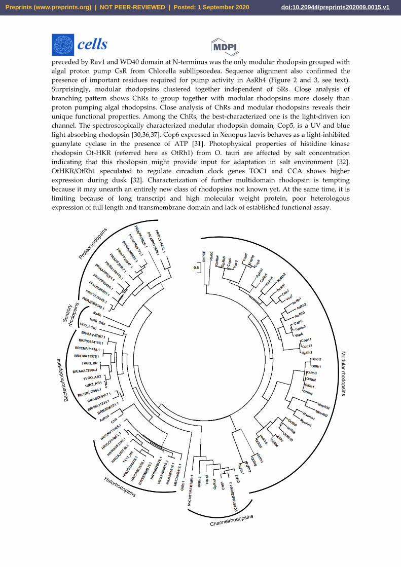
preceded by Rav1 and WD40 domain at N-terminus was the only modular rhodopsin grouped with
algal proton pump CsR from Chlorella subllipsoedea. Sequence alignment also confirmed the
presence of important residues required for pump activity in AsRh4 (Figure 2 and 3, see text).
Surprisingly, modular rhodopsins clustered together independent of SRs. Close analysis of
branching pattern shows ChRs to group together with modular rhodopsins more closely than
proton pumping algal rhodopsins. Close analysis of ChRs and modular rhodopsins reveals their
unique functional properties. Among the ChRs, the best-characterized one is the light-driven ion
channel. The spectroscopically characterized modular rhodopsin domain, Cop5, is a UV and blue
light absorbing rhodopsin [30,36,37]. Cop6 expressed in Xenopus laevis behaves as a light-inhibited
guanylate cyclase in the presence of ATP [31]. Photophysical properties of histidine kinase
rhodopsin Ot-HKR (referred here as OtRh1) from O. tauri are affected by salt concentration
indicating that this rhodopsin might provide input for adaptation in salt environment [32].
OtHKR/OtRh1 speculated to regulate circadian clock genes TOC1 and CCA shows higher
expression during dusk [32]. Characterization of further multidomain rhodopsin is tempting
because it may unearth an entirely new class of rhodopsins not known yet. At the same time, it is
limiting because of long transcript and high molecular weight protein, poor heterologous
expression of full length and transmembrane domain and lack of established functional assay.
Preprints (www.preprints.org) | NOT PEER-REVIEWED | Posted: 1 September 2020 doi:10.20944/preprints202009.0015.v1
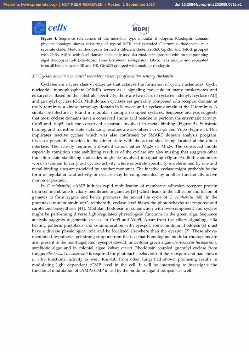
Figure 4. Sequence relatedness of the microbial type modular rhodopsin: Rhodopsin domain
phyletic topology shows clustering of typical MTR and extended C-terminus rhodopsins in a
separate clade. Modular rhodopsins formed a different clade. KnRh3, GpRh1 and TsRh1 grouped
with ChRs. AsRh4 with Rav1 domain is the only modular rhodopsin grouped with proton pumping
algal rhodopsin CsR (Rhodopsin from Coccomyxa subllipsodea). GtRh1 was unique and separated
from all lying between BR and HR. Gtrh2/3 grouped with modular rhodopsin.
3.7. Cyclase domain a canonical secondary messenger of modular sensory rhodopsin
Cyclases are a lyase class of enzymes that catalyse the formation of cyclic nucleotides. Cyclic
nucleotide monophosphate (cNMP) serves as a signaling molecule in many prokaryotes and
eukaryotes. Based on the substrate specificity, there are two class of cyclases- adenylyl cyclase (AC)
and guanylyl cyclase (GC). Multidomain cyclases are generally composed of a receptor domain at
the N-terminus, a kinase homology domain in between and a cyclase domain at the C-terminus. A
similar architecture is found in modular rhodopsin coupled cyclases. Sequence analysis suggests
that most cyclase domains have a conserved amino acid residue to perform the enzymatic activity.
Cop5 and Vop5 lack the conserved aspartate involved in metal binding (Figure 5). Substrate
binding and transition state stabilizing residues are also absent in Cop5 and Vop5 (Figure 5). This
implicates inactive cyclase which was also confirmed by SMART domain analysis program.
Cyclases generally function in the dimer state with the active sites being located at the dimer
interface. The activity requires a divalent cation, either Mg2+ or Mn2+. The conserved motifs
especially transition state stabilizing residues of the cyclase are also missing that suggests other
transition state stabilizing molecules might be involved in signaling (Figure 6). Both monomers
work in tandem to carry out cyclase activity where substrate specificity is determined by one and
metal-binding sites are provided by another monomer. The inactive cyclase might probably be the
form of regulation and activity of cyclase may be complemented by another functionally active
monomer partner.
In C. reinhardtii, cAMP induces rapid mobilization of membrane adhesion receptor protein
from cell membrane to ciliary membrane in gametes [26] which leads to the adhesion and fusion of
gametes to form zygote and hence promotes the sexual life cycle of C. reinhardtii [40]. In the
phototaxis mutant strain of C. reinhardtii, cyclase level biases the photobehavioural response and
carotenoid biosynthesis [41]. Modular rhodopsin in conjunction with two-component and cyclase
might be performing diverse light-regulated physiological functions in the green alga. Sequence
analysis suggests degenerate cyclase in Cop5 and Vop5. Apart from the ciliary signaling, cilia
beating pattern, phototaxis and communication with eyespot, some modular rhodopsin(s) must
have a diverse physiological role and be localized elsewhere than the eyespot [7]. These above-
mentioned hypotheses get strong support from the fact that homologous modular rhodopsins are
also present in the non-flagellated, eyespot devoid, unicellular green algae Ostreococcus lucimarinus,
symbiotic algae and in colonial algae Volvox carteri. Rhodopsin coupled guanylyl cyclase from
fungus Blastocladiella emersonii is required for phototactic behaviour of the zoospore and had shown
in vitro functional activity as well. Rho-GC from other fungi had shown promising results in
modulating light dependent cGMP level in the cell. It will be interesting to investigate the
functional modulation of cAMP/cGMP in cell by the modular algal rhodopsins as well.
Preprints (www.preprints.org) | NOT PEER-REVIEWED | Posted: 1 September 2020 doi:10.20944/preprints202009.0015.v1
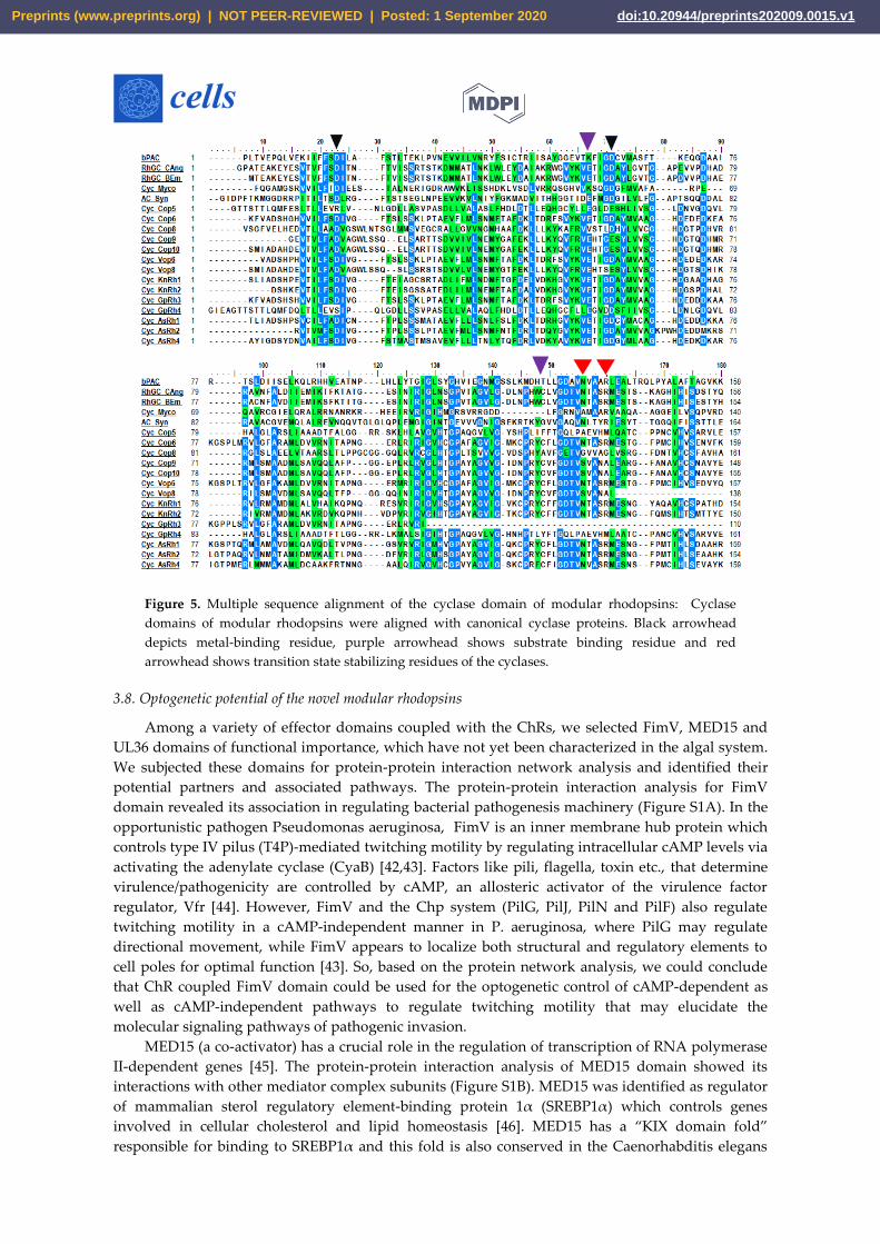
Figure 5. Multiple sequence alignment of the cyclase domain of modular rhodopsins: Cyclase
domains of modular rhodopsins were aligned with canonical cyclase proteins. Black arrowhead
depicts metal-binding residue, purple arrowhead shows substrate binding residue and red
arrowhead shows transition state stabilizing residues of the cyclases.
3.8. Optogenetic potential of the novel modular rhodopsins
Among a variety of effector domains coupled with the ChRs, we selected FimV, MED15 and
UL36 domains of functional importance, which have not yet been characterized in the algal system.
We subjected these domains for protein-protein interaction network analysis and identified their
potential partners and associated pathways. The protein-protein interaction analysis for FimV
domain revealed its association in regulating bacterial pathogenesis machinery (Figure S1A). In the
opportunistic pathogen Pseudomonas aeruginosa, FimV is an inner membrane hub protein which
controls type IV pilus (T4P)-mediated twitching motility by regulating intracellular cAMP levels via
activating the adenylate cyclase (CyaB) [42,43]. Factors like pili, flagella, toxin etc., that determine
virulence/pathogenicity are controlled by cAMP, an allosteric activator of the virulence factor
regulator, Vfr [44]. However, FimV and the Chp system (PilG, PilJ, PilN and PilF) also regulate
twitching motility in a cAMP-independent manner in P. aeruginosa, where PilG may regulate
directional movement, while FimV appears to localize both structural and regulatory elements to
cell poles for optimal function [43]. So, based on the protein network analysis, we could conclude
that ChR coupled FimV domain could be used for the optogenetic control of cAMP-dependent as
well as cAMP-independent pathways to regulate twitching motility that may elucidate the
molecular signaling pathways of pathogenic invasion.
MED15 (a co-activator) has a crucial role in the regulation of transcription of RNA polymerase
II-dependent genes [45]. The protein-protein interaction analysis of MED15 domain showed its
interactions with other mediator complex subunits (Figure S1B). MED15 was identified as regulator
of mammalian sterol regulatory element-binding protein 1α (SREBP1α) which controls genes
involved in cellular cholesterol and lipid homeostasis [46]. MED15 has a “KIX domain fold”
responsible for binding to SREBP1α and this fold is also conserved in the Caenorhabditis elegans
Preprints (www.preprints.org) | NOT PEER-REVIEWED | Posted: 1 September 2020 doi:10.20944/preprints202009.0015.v1
orthologue, MDT15 and Yeast orthologue GAL11p [46,47]. It has also been reported that
dysregulation of MED15 expression promotes human malignancies and inactivation of MED15 may
inhibit the progression of several types of cancers [45,48]. Several studies found MED15 as an
important prognostic biomarker for patients with various types of carcinomas [45,48] . In breast
cancer and few epithelial cancers, inactivation of MED15 inhibits aberrant transforming growth
factor β (TGFβ)-induced epithelial-mesenchymal transition (EMT), as it acts as a crucial cofactor for
TGFβ signaling [49]. Localized tumor specific expression of ChR coupled MED15 could be used to
target tumor cell signaling and eventually induce the tumour for autophagy or growth arrest in
conjunction with other engineered proteins, in a light dependent manner.
The UL36 domain, associated with modular ChR, GpRh1 from G. pectorale is a the largest
tegument viral protein found in herpes simplex virus 1 (HSV-1) and its homologues are well
distributed across the members of Herpes viridae [50]. UL36 protein is an ubiquitin-specific
protease [51] which is evident from our protein-protein interaction analysis of UL36 protein (Figure
S2A). Most of the interacting partners like Ubiquitin, 26S proteasome regulatory subunit S5A,
proteasome regulatory particle subunit (RpnC) and DSS1/SEM1 family protein belongs to the
ubiquitin-dependent proteolysis machinery [52–54]. Proteasome subunit S5a (the human
homologue of Rpn10) functions in conjunction with hHR23a/b (the two human homologues of
Rad23) to recruit ubiquitylated substrates to the proteasome for their degradation [55]. In humans,
DSS1/SEM1 is related to a tumour suppressor protein (BRCA2), which has a crucial role in the
recombinational DNA repair in association with RAD51 [56,57]. UL36 deubiquitinating activity has
a role in inhibiting the interferon-mediated immune defense upon viral invasion in the host [51].
Interestingly, the UL36 domain coupled to GpRh1 showed similarity to the C-terminal segment of
HSV-1 UL36 protein (Figure S2B). Böttcher et al. (2005), in a mutation analysis with UL36
homologues from Pseudorabies Virus, constructed several truncations and showed that the extreme
C terminus of UL36 having proline/alanine rich region is crucial for viral replication [58]. Overall, as
observed from the protein-protein interaction analysis, it may be assumed that, ChRs coupled
effector domain can be utilized as the next generation optogenetic tools, which might help in
controlling processes ranging from lipid metabolism, ubiquitin-mediating proteolysis, and
pathogenesis to carcinogenesis. Apart from the natural variant, the modular rhodopsins could also
be genetically engineered for enhanced kinetics, better spectral tuning and modulation to precisely
controlled diverse cellular physiological responses.
Acknowledgments: KS fellowship was supported by DBT, India. MSK is financially supported by UGC-
DSKPDF, India. The SERB-India [ECR/2017/000354] and DBT, Government of India (BT/010/IYBA/2016) are
highly acknowledged for the support of research grants to SK. Adivitiya is kindly acknowledged for editing of
the manuscript.
Conflicts of Interest: All authors declare no conflicts of interest.
References
[1] D. Oesterhelt, S. Walther, Functions of a new photoreceptor membrane, PNAS. 70 (1973) 2853–2857.
[2] B. Schobert, J.K. Lanyi, Halorhodopsin is a light-driven chloride pump*, J. Biol. Chem. 257 (1982)
10306–10313.
[3] G. Nagel, D. Ollig, M. Fuhrmann, S. Kateriya, A.M. Musti, E. Bamberg, P. Hegemann,
Channelrhodopsin-1: A light-gated proton channel in green algae, Science (80-. ). 296 (2002) 2395–2398.
[4] G. Nagel, T. Szellas, W. Huhn, S. Kateriya, N. Adeishvili, P. Berthold, D. Ollig, P. Hegemann, E.
Bamberg, Channelrhodopsin-2, a directly light-gated cation-selective membrane channel, PNAS. 100
(2003) 13940–13945. https://doi.org/10.1073/pnas.1936192100.
[5] W.D. Hoff, K. Jung, J.L. Spudich, Molecular mechanism of photosignaling by archaeal sensory
rhodopsins, Annu. Rev. Biophys. Biomol. Struct. 26 (1997) 223–258.
Preprints (www.preprints.org) | NOT PEER-REVIEWED | Posted: 1 September 2020 doi:10.20944/preprints202009.0015.v1
[6] M. Nakagawa, T. Iwasa, S. Kikkawa, M. Tsuda, T.G. Ebrey, How vertebrate and invertebrate visual
pigments differ in their mechanism of photoactivation, PNAS. 96 (1999) 6189–6192.
[7] M. Awasthi, P. Ranjan, K. Sharma, S.K. Veetil, S. Kateriya, The trafficking of bacterial type rhodopsins
into the Chlamydomonas eyespot and flagella is IFT mediated, Sci. Rep. 6 (2016) 34646.
https://doi.org/10.1038/srep34646.
[8] F. Zhang, J. Vierock, O. Yizhar, L.E. Fenno, S. Tsunoda, A. Kianianmomeni, M. Prigge, A. Berndt, J.
Cushman, J. Polle, J. Magnuson, P. Hegemann, K. Deisseroth, The microbial opsin family of
optogenetic tools, Cell. 147 (2011) 1446–1457. https://doi.org/10.1016/j.cell.2011.12.004.
[9] K. Yoshida, S.P. Tsunoda, L.S. Brown, H. Kandori, A unique choanoflagellate enzyme rhodopsin
exhibits light- dependent cyclic nucleotide phosphodiesterase activity, J. Biol. Chem. 292 (2017) 7531–
7541. https://doi.org/10.1074/jbc.M117.775569.
[10] Y. Tian, S. Gao, S. Yang, G. Nagel, A novel rhodopsin phosphodiesterase from Salpingoeca rosetta
shows light-enhanced substrate affinity, Biochem. J. 475 (2018) 1121–1128.
[11] L.B. Lamarche, R.P. Kumar, M.M. Trieu, E.L. Devine, L.E. Cohen-abeles, D.L. Theobald, D.D. Oprian,
Purification and characterization of RhoPDE, a retinylidene/phosphodiesterase fusion protein and
potential optogenetic tool from the choanoflagellate Salpingoeca rosetta, Biochemistry. 56 (2017) 5812–
5822. https://doi.org/10.1021/acs.biochem.7b00519.
[12] U. Scheib, K. Stehfest, C.E. Gee, H.G. Körschen, R. Fudim, T.G. Oertner, P. Hegemann, The rhodopsin
– guanylyl cyclase of the aquatic fungus Blastocladiella emersonii enables fast optical control of cGMP
signaling, Optogenetics. 8 (2015) rs8.
[13] U. Scheib, M. Broser, O.M. Constantin, S. Yang, S. Gao, S. Mukherjee, K. Stehfest, G. Nagel, C.E. Gee, P.
Hegemann, Rhodopsin-cyclases for photocontrol of cGMP/cAMP and 2.3 Ǻ structure of the adenylyl
cyclase domain, Nat. Commun. 9 (2018) 2046. https://doi.org/10.1038/s41467-018-04428-w.
[14] A. Butryn, H. Raza, H. Rada, I. Moraes, R.J. Owens, A.M. Orville, Molecular basis for GTP recognition
by light-activated guanylate cyclase RhGC, FEBS J. (2019). https://doi.org/10.1111/febs.15167.
[15] M.M. Trieu, E.L. Devine, L.B. Lamarche, A.E. Ammerman, J.A. Greco, R.R. Birge, D.L. Theobald, D.D.
Oprian, Expression, purification, and spectral tuning of RhoGC, a retinylidene/guanylyl cyclase fusion
protein and optogenetics tool from the aquatic fungus Blastocladiella emersonii, 292 (2017) 10379–
10389. https://doi.org/10.1074/jbc.M117.789636.
[16] S. Mukherjee, P. Hegemann, M. Broser, Enzymerhodopsins: novel photoregulated catalysts for
optogenetics, Curr. Opin. Struct. Biol. 57 (2019) 118–126. https://doi.org/10.1016/j.sbi.2019.02.003.
[17] A. Greiner, S. Kelterborn, H. Evers, G. Kreimer, I. Sizova, P. Hegemann, Targeting of photoreceptor
genes in Chlamydomonas reinhardtii via zinc-finger nucleases and CRISPR/Cas9, Plant Cell. 29 (2017)
2498–2518. https://doi.org/10.1105/tpc.17.00659.
[18] J.D. Thompson, T.J. Gibson, F. Plewniak, F. Jeanmougin, D.G. Higgins, The CLUSTAL_X windows
interface: flexible strategies for multiple sequence alignment aided by quality analysis tools, Nucleic
Acids Res. 25 (1997) 4876–4882.
[19] L.Y. Geer, M. Domrachev, D.J. Lipman, S.H. Bryant, CDART : Protein homology by domain
architecture, Genome Res. 12 (2002) 1619–1623. https://doi.org/10.1101/gr.278202.CDART.
[20] A. Marchler-bauer, Y. Bo, L. Han, J. He, C.J. Lanczycki, S. Lu, F. Chitsaz, M.K. Derbyshire, R.C. Geer,
N.R. Gonzales, M. Gwadz, D.I. Hurwitz, F. Lu, G.H. Marchler, J.S. Song, N. Thanki, Z. Wang, R.A.
Yamashita, D. Zhang, C. Zheng, L.Y. Geer, S.H. Bryant, CDD/SPARCLE : functional classification of
proteins via subfamily domain architectures, Nucleic Acids Res. 45 (2017) D200–D203.
https://doi.org/10.1093/nar/gkw1129.
[21] K. Tamura, D. Peterson, N. Peterson, G. Stecher, M. Nei, S. Kumar, MEGA : Molecular evolutionary
Preprints (www.preprints.org) | NOT PEER-REVIEWED | Posted: 1 September 2020 doi:10.20944/preprints202009.0015.v1
genetics analysis using maximum likelihood, evolutionary distance, and maximum parsimony
methods, Mol. Biol. Evol. 28 (2011) 2731–2739. https://doi.org/10.1093/molbev/msr121.
[22] G. Perriere, M. Gouy, WWW-Query: An on-line retrieval system for biological sequence banks,
Biochimie. 78 (1996) 364–369.
[23] D. Szklarczyk, J.H. Morris, H. Cook, M. Kuhn, S. Wyder, M. Simonovic, A. Santos, N.T. Doncheva, A.
Roth, P. Bork, L.J. Jensen, C. Von Mering, The STRING database in 2017: quality-controlled protein –
protein association networks, made broadly accessible, Nucleic Acids Res. 45 (2017) D362–D368.
https://doi.org/10.1093/nar/gkw937.
[24] P. Shannon, A. Markiel, O. Ozier, N.S. Baliga, J.T. Wang, D. Ramage, N. Amin, B. Schwikowski, T.
Ideker, Cytoscape: A software environment for integrated models of biomolecular interaction
networks, Genome Res. 13 (2003) 2498–2504. https://doi.org/10.1101/gr.1239303.
[25] P.A. Salome, S.S. Merchant, A series of fortunate events: introducing Chlamydomonas as a reference
organism, Plant Cell. 31 (2019) 1682–1707. https://doi.org/10.1105/tpc.18.00952.
[26] P. Ranjan, M. Awasthi, W.J. Snell, Transient internalization and microtubule-dependent trafficking of a
ciliary signaling receptor from the plasma membrane to the cilium, Curr. Biol. 29 (2019) 2942–2947.
https://doi.org/10.1016/j.cub.2019.07.022.
[27] E.G. Govorunova, O.A. Sineshchekov, H. Li, R. Janz, J.L. Spudich, Characterization of a highly efficient
blue-shifted Channelrhodopsin from the marine Alga Platymonas subcordiformis, J. Biol. Chem. 288
(2013) 29911–29922. https://doi.org/10.1074/jbc.M113.505495.
[28] M. Nack, I. Radu, M. Gossing, C. Bamann, E. Bamberg, G.F. Von Mollard, J. Heberle, The DC gate in
channelrhodopsin-2: crucial hydrogen bonding interaction between C128 and D156 †, Photochem.
Photobiol. Sci. 9 (2010) 194–198. https://doi.org/10.1039/b9pp00157c.
[29] K. Stehfest, E. Ritter, A. Berndt, F. Bartl, P. Hegemann, The branched photocycle of the slow-cycling
channelrhodopsin-2 mutant C128T, J. Mol. Biol. 398 (2010) 690–702.
https://doi.org/10.1016/j.jmb.2010.03.031.
[30] M. Luck, T. Mathes, S. Bruun, R. Fudim, R. Hagedorn, T.M.T. Nguyen, S. Kateriya, J.T.M. Kennis, P.
Hildebrandt, P. Hegemann, A photochromic histidine kinase rhodopsin (HKR1) that is bimodally
switched by ultraviolet and blue light, J. Biol. Chem. 287 (2012) 40083–40090.
https://doi.org/10.1074/jbc.M112.401604.
[31] Y. Tian, S. Gao, E.L. Von Der Heyde, A. Hallmann, G. Nagel, Two-component cyclase opsins of green
algae are ATP-dependent and light-inhibited guanylyl cyclases, BMC Biol. 16 (2018) 144.
[32] M. Luck, F.V. Escobar, K. Glass, M.-I. Sabotke, R. Hagedorn, F. Corellou, F. Siebert, P. Hildebrandt, P.
Hegemann, Photoreactions of the histidine kinase rhodopsin Ot-HKR from the marine picoalga
Ostreococcus tauri, Biochemistry. 58 (2019) 1878–1891. https://doi.org/10.1021/acs.biochem.8b01200.
[33] D. Man, W. Wang, G. Sabehi, L. Aravind, A.F. Post, R. Massana, E.N. Spudich, J.L. Spudich, O. Beja,
Diversification and spectral tuning in marine proteorhodopsins, EMBO J. 22 (2003) 1725–1731.
[34] K. Shimono, M. Iwamoto, M. Sumi, N. Kamo, Effects of three characteristic amino acid residues of
pharaonis phoborhodopsin on the absorption maximum, Photochem. Photobiol. 72 (2000) 141–145.
[35] K. Shimono, Y. Ikeura, Y. Sudo, M. Iwamoto, N. Kamo, Environment around the chromophore in
pharaonis phoborhodopsin : mutation analysis of the retinal binding site, Biochim. Biophys. Acta. 1515
(2001) 92–100.
[36] M. Luck, S. Bruun, A. Keidel, P. Hegemann, P. Hildebrandt, Photochemical chromophore
isomerization in histidine kinase rhodopsin, FEBS Lett. 589 (2015) 1067–1071.
https://doi.org/10.1016/j.febslet.2015.03.024.
Preprints (www.preprints.org) | NOT PEER-REVIEWED | Posted: 1 September 2020 doi:10.20944/preprints202009.0015.v1
[37] M. Luck, P. Hegemann, The two parallel photocycles of the Chlamydomonas sensory photoreceptor
histidine kinase rhodopsin 1☆, J. Plant Physiol. 217 (2017) 77–84.
https://doi.org/10.1016/j.jplph.2017.07.008.
[38] L.S. Brown, Fungal rhodopsins and opsin-related proteins : eukaryotic homologues of
bacteriorhodopsin with unknown functions, Photochem. Photobiol. Sci. 3 (2004) 555–565.
[39] I. Marin, M.X. Ruiz-Gonzalez, New insights into the evolutionary history of type 1 rhodopsins, J. Mol.
Evol. 58 (2004) 348–358. https://doi.org/10.1007/s00239-003-2557-8.
[40] S.M. Pasquale, U.W. Goodenough, Cyclic AMP functions as a primary sexual signal in gametes of
Chlamydomonas reinhardtii, J. Cell Biol. 105 (1987) 2279–2292.
[41] M. Boonyareth, J. Saranak, D. Pinthong, Y. Sanvarinda, K.W. Foster, Roles of cyclic AMP in regulation
of phototaxis in Chlamydomonas reinhardtii, Biologia (Bratisl). 64 (2009) 1058–1065.
https://doi.org/10.2478/s11756-009-0194-4.
[42] R.N.C. Buensuceso, Y. Nguyen, K. Zhang, M. Daniel-ivad, S.N. Sugiman-marangos, A.D. Fleetwood,
I.B. Zhulin, M.S. Junop, P.L. Howell, L.L. Burrows, The conserved tetracopeptide repeat-containing C-
terminal domain of Pseudomonas aeruginosa FimV is required for its cyclic AMP-dependent and -
independent Functions, J. Bacteriol. 198 (2016) 2263–2274. https://doi.org/10.1128/JB.00322-16.Editor.
[43] R.N.C. Buensuceso, M. Daniel-Ivad, S.L.N. Kilmury, T.L. Leighton, H. Harvey, L.P. Howell, L.L.
Burrows, Cyclic AMP-independent control of twitching motility in Pseudomonas aeruginosa, J.
Bacteriol. 199 (2017) e00188-17.
[44] A. Berry, K. Han, J. Trouillon, M. Robert-genthon, M. Ragno, S. Lory, I. Attrée, S. Elsen, cAMP and Vfr
control exolysin expression and cytotoxicity of Pseudomonas aeruginosa taxonomic outliers, J.
Bacteriol. 200 (2018) e00135-18.
[45] K. Wang, C. Duan, X. Zou, Y. Song, W. Li, L. Xiao, J. Peng, L. Yao, Q. Long, L. Liu, Increased mediator
complex subunit 15 expression is associated with poor prognosis in hepatocellular carcinoma, Oncol.
Lett. 15 (2018) 4303–4313. https://doi.org/10.3892/ol.2018.7820.
[46] F. Yang, B.W. Vought, J.S. Satterlee, A.K. Walker, Z.-Y.J. Sun, J.L. Watts, R. Debeaumont, R.M. Saito,
S.G. Hyberts, S. Yang, C. Macol, L. Iyer, R. Tjian, S. Van Den Heuvel, A.C. Hart, G. Wagner, A.M. Naar,
An ARC/Mediator subunit required for SREBP control of cholesterol and lipid homeostasis, Nature.
442 (2006) 700–704. https://doi.org/10.1038/nature04942.
[47] J.K. Thakur, H. Arthanari, F. Yang, K.H. Chau, G. Wagner, A.M. Naar, Mediator subunit
Gal11p/MED15 is required for fatty acid-dependent gene activation by yeast transcription factor
Oaf1p, J. Biol. Chem. 284 (2009) 4422–4428. https://doi.org/10.1074/jbc.M808263200.
[48] I. Syring, R. Weiten, T. Müller, D. Schmidt, S. Steiner, G. Kristiansen, S.C. Müller, J. Ellinger, The
knockdown of the Mediator complex subunit MED15 restrains urothelial bladder cancer cells
malignancy, Oncol. Lett. 16 (2018) 3013–3021. https://doi.org/10.3892/ol.2018.9014.
[49] M. Zhao, X. Yang, Y. Fu, H. Wang, Y. Ning, J. Yan, Y.-G. Chen, G. Wang, Mediator MED15 modulates
transforming growth factor beta (TGFβ)/Smad signaling and breast cancer cell metastasis, J. Mol. Cell
Biol. 5 (2013) 57–60.
[50] C. Schlieker, G.A. Korbel, L.M. Kattenhorn, H.L. Ploegh, A deubiquitinating activity is conserved in
the large tegument protein of the Herpesviridae, J. Virol. 79 (2005) 15582–15585.
https://doi.org/10.1128/JVI.79.24.15582.
[51] S. Wang, K. Wang, J. Li, C. Zheng, Herpes simplex virus 1 ubiquitin-specific protease UL36 inhibits
beta interferon production by deubiquitinating TRAF3, J. Virol. 87 (2013) 11851–11860.
https://doi.org/10.1128/JVI.01211-13.
[52] G.C. Lander, E. Estrin, M.E. Matyskiela, C. Bashore, E. Nogales, A. Martin, Complete subunit
Preprints (www.preprints.org) | NOT PEER-REVIEWED | Posted: 1 September 2020 doi:10.20944/preprints202009.0015.v1
architecture of the proteasome regulatory particle, Nature. 482 (2012) 186–191.
https://doi.org/10.1038/nature10774.
[53] L. Josse, M.E. Harley, I.M.S. Pires, D.A. Hughes, Fission yeast Dss1 associates with the proteasome and
is required for efficient ubiquitin-dependent proteolysis, Biochem. J. 393 (2006) 303–309.
https://doi.org/10.1042/BJ20051238.
[54] Q. Wang, P. Young, K.J. Walters, Structure of S5a bound to monoubiquitin provides a model for
polyubiquitin recognition, J. Mol. Biol. 348 (2005) 727–739. https://doi.org/10.1016/j.jmb.2005.03.007.
[55] K.J. Walters, A.M. Goh, Q. Wang, G. Wagner, P.M. Howley, Ubiquitin family proteins and their
relationship to the proteasome: a structural perspective, Biochim. Biophys. Acta. 1695 (2004) 73–87.
https://doi.org/10.1016/j.bbamcr.2004.10.005.
[56] A.R. Venkitaraman, Cancer susceptibility and the functions of BRCA1 and BRCA2, Cell. 108 (2002)
171–182.
[57] N.J. Marston, W.J. Richards, D. Hughes, D. Bertwistle, C.J. Marshall, A. Ashworth, Interaction between
the product of the breast cancer susceptibility gene BRCA2 and DSS1, a protein functionally conserved
from yeast to mammals, Mol. Cell. Biol. 19 (1999) 4633–4642.
[58] S. Bottcher, B.G. Klupp, H. Granzow, W. Fuchs, K. Michael, T.C. Mettenleiter, Identification of a 709-
amino-acid internal nonessential region within the essential conserved tegument protein (p) UL36 of
Pseudorabies Virus, J. Virol. 80 (2006) 9910–9915. https://doi.org/10.1128/JVI.01247-06.
© 2020 by the authors. Submitted for possible open access publication under the terms
and conditions of the Creative Commons Attribution (CC BY) license
(http://creativecommons.org/licenses/by/4.0/).
Preprints (www.preprints.org) | NOT PEER-REVIEWED | Posted: 1 September 2020 doi:10.20944/preprints202009.0015.v1