Embed Size (px)
Citation preview
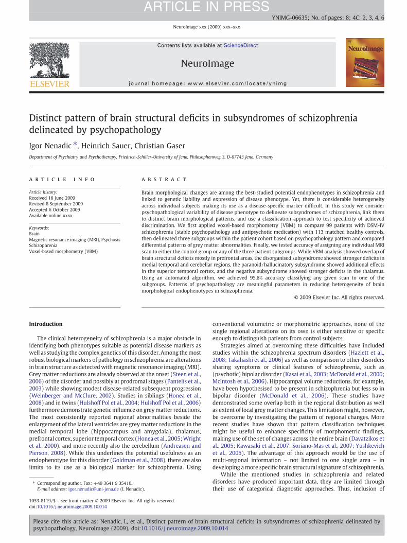
Distinct pattern of brain structural de!cits in subsyndromes of schizophreniadelineated by psychopathology
Igor Nenadic !, Heinrich Sauer, Christian GaserDepartment of Psychiatry and Psychotherapy, Friedrich-Schiller-University of Jena, Philosophenweg 3, D-07743 Jena, Germany
a b s t r a c ta r t i c l e i n f o
Article history:Received 18 June 2009Revised 8 September 2009Accepted 6 October 2009Available online xxxx
Keywords:BrainMagnetic resonance imaging (MRI), PsychosisSchizophreniaVoxel-based morphometry (VBM)
Brain morphological changes are among the best-studied potential endophenotypes in schizophrenia andlinked to genetic liability and expression of disease phenotype. Yet, there is considerable heterogeneityacross individual subjects making its use as a disease-speci!c marker dif!cult. In this study we considerpsychopathological variability of disease phenotype to delineate subsyndromes of schizophrenia, link themto distinct brain morphological patterns, and use a classi!cation approach to test speci!city of achieveddiscrimination. We !rst applied voxel-based morphometry (VBM) to compare 99 patients with DSM-IVschizophrenia (stable psychopathology and antipsychotic medication) with 113 matched healthy controls,then delineated three subgroups within the patient cohort based on psychopathology pattern and compareddifferential patterns of grey matter abnormalities. Finally, we tested accuracy of assigning any individual MRIscan to either the control group or any of the three patient subgroups. While VBM analysis showed overlap ofbrain structural de!cits mostly in prefrontal areas, the disorganised subsyndrome showed stronger de!cits inmedial temporal and cerebellar regions, the paranoid/hallucinatory subsyndrome showed additional effectsin the superior temporal cortex, and the negative subsyndrome showed stronger de!cits in the thalamus.Using an automated algorithm, we achieved 95.8% accuracy classifying any given scan to one of thesubgroups. Patterns of psychopathology are meaningful parameters in reducing heterogeneity of brainmorphological endophenotypes in schizophrenia.
© 2009 Elsevier Inc. All rights reserved.
Introduction
The clinical heterogeneity of schizophrenia is a major obstacle inidentifying both phenotypes suitable as potential disease markers aswell as studying the complex genetics of this disorder. Among themostrobust biologicalmarkers of pathology in schizophrenia are alterationsin brain structure as detectedwithmagnetic resonance imaging (MRI).Grey matter reductions are already observed at the onset (Steen et al.,2006) of the disorder and possibly at prodromal stages (Pantelis et al.,2003) while showing modest disease-related subsequent progression(Weinberger and McClure, 2002). Studies in siblings (Honea et al.,2008) and in twins (Hulshoff Pol et al., 2004; Hulshoff Pol et al., 2006)furthermore demonstrate genetic in"uence on greymatter reductions.The most consistently reported regional abnormalities beside theenlargement of the lateral ventricles are grey matter reductions in themedial temporal lobe (hippocampus and amygdala), thalamus,prefrontal cortex, superior temporal cortex (Honea et al., 2005;Wrightet al., 2000), and more recently also the cerebellum (Andreasen andPierson, 2008). While this underlines the potential usefulness as anendophenotype for this disorder (Goldman et al., 2008), there are alsolimits to its use as a biological marker for schizophrenia. Using
conventional volumetric or morphometric approaches, none of thesingle regional alterations on its own is either sensitive or speci!cenough to distinguish patients from control subjects.
Strategies aimed at overcoming these dif!culties have includedstudies within the schizophrenia spectrum disorders (Hazlett et al.,2008; Takahashi et al., 2006) as well as comparison to other disorderssharing symptoms or clinical features of schizophrenia, such as(psychotic) bipolar disorder (Kasai et al., 2003; McDonald et al., 2006;McIntosh et al., 2006). Hippocampal volume reductions, for example,have been hypothesised to be present in schizophrenia but less so inbipolar disorder (McDonald et al., 2006). These studies havedemonstrated some overlap both in the regional distribution as wellas extent of local greymatter changes. This limitationmight, however,be overcome by investigating the pattern of regional changes. Morerecent studies have shown that pattern classi!cation techniquesmight be useful to enhance speci!city of morphometric !ndings,making use of the set of changes across the entire brain (Davatzikos etal., 2005; Kawasaki et al., 2007; Soriano-Mas et al., 2007; Yushkevichet al., 2005). The advantage of this approach would be the use ofmulti-regional information – not limited to one single area – indeveloping amore speci!c brain structural signature of schizophrenia.
While the mentioned studies in schizophrenia and relateddisorders have produced important data, they are limited throughtheir use of categorical diagnostic approaches. Thus, inclusion of
NeuroImage xxx (2009) xxx–xxx
! Corresponding author. Fax: +49 3641 9 35410.E-mail address: [email protected] (I. Nenadic).
YNIMG-06635; No. of pages: 8; 4C: 2, 3, 4, 6
1053-8119/$ – see front matter © 2009 Elsevier Inc. All rights reserved.doi:10.1016/j.neuroimage.2009.10.014
Contents lists available at ScienceDirect
NeuroImage
j ourna l homepage: www.e lsev ie r.com/ locate /yn img
ARTICLE IN PRESS
Please cite this article as: Nenadic, I., et al., Distinct pattern of brain structural de!cits in subsyndromes of schizophrenia delineated bypsychopathology, NeuroImage (2009), doi:10.1016/j.neuroimage.2009.10.014
additional information related to phenotypic variationwithin a samplemight be useful to form subgroups, which aremore homogeneous andcould then be subject to classi!cation analysis to prove or disprovesuf!cient speci!city for brain structure as a biological marker.
Only a few structural MRI studies have addressed the issue ofsubgroups or subsyndromes within schizophrenia, even despite theavailability of the subtyping according to DSM-IIIR or DSM-IVdiagnostic criteria. For example, abnormalities in cortical foldinghave been shown to be more pronounced in the disorganised subtypeof schizophrenia (Sallet et al., 2003a), although there was noindication of subgroups within patients in another study on corticalthickness (Lawyer et al., 2008), while yet another study dividingpatients in paranoid vs. non-paranoid schizophrenia has givenevidence for the negative symptom dimension to be related toincreased rightward structural asymmetry (Sallet et al., 2003b).
In this study, we implemented such an approach by applying aclassi!cation analysis (based on voxel-based morphometric analysisof whole-brain MRI) on both a comparison of schizophrenia patientsand healthy controls as well as subdividing the schizophrenia sampleinto subgroups based on symptoms. We tested whether the pattern ofbrain structural changes (i.e. combination of regionally distributedchanges) would provide suf!ciently accurate classi!cation of a givenbrain scan from the cohort to be assigned to either schizophrenia orhealthy control groups, and (for patients) to one of three schizophre-nia subgroups. These three subgroups were formed on the basis offactor analysis applied to cross-sectional psychopathology. Therationale for this approach to forming subgroups was based onprevious studies on robustness of three-factor models in schizophre-nia (Cuesta and Peralta, 1995b; Peralta et al., 1997) as well as the factthat this grouping most closely matches the already establishedclinical subtypes within the DSM-IV and ICD-10 diagnostic systems.Secondly, we challenged the notion that there is a core pattern ofchanges independent of clinical or phenotypic variability, assumingthat the most consistently reported structures in schizophreniapathophysiology (hippocampus, thalamus, and dorsolateral prefron-tal cortex) would be altered in all subgroups, while other structuralchanges (e.g. in the superior temporal cortex, cerebellum), whichhave been linked to particular psychopathological phenomena (Gaseret al., 2004), might be altered in only one of the subgroups expressingthis particular disease phenotype.
Materials and methods
Subjects
We studied 99 patients with a DSM-IV diagnosis of schizophreniaand 113 healthy controls. The patients (n=99; 57 male/42 female;mean age=36.2 years, SD=11.2) were recruited from the Depart-
ment of Psychiatry in Jena and !rst screened with a semi-structuredinterview before being assessed by two psychiatrists establishing theDSM-IV diagnosis. None of the patients had a second psychiatric, aneurological or major medical condition. All patients were inpatientsand none of them was in an acute episode of illness, all were inremitted clinical state, showed stable psychopathology, and were onstable antipsychotic medication. Current psychopathology in patientswas assessed using the Scales for Assessment of Positive Symptoms(SAPS), and Scales for Assessment of Negative Symptoms (SANS),which was administered by an experienced and trained clinicalpsychiatrist. Healthy controls (n= 113; 70 male/43 female; meanage=32.38 years, SD=10.26) were recruited from the city of Jenaand surrounding counties andmatched to the patients with regards togender, age, and overall school/academic achievement. They werescreened to exclude a concurrent or past history of psychiatric,neurological, and major medical conditions using a semi-structured
Table 1Demographics and psychopathology scores (mean global items and their standarddeviation) of the schizophrenia subgroups.
1— Negative 2— Disorganised 3—Hallucinatory/paranoid
Gender (M/F) 18/17 15/14 24/11Age 35.07 ( 9.3) 35.95 (12.0) 37.64 (12.3)Duration of illness 7.77 (7.5) 8.69 (8.5) 10.25 (8.7)Age of onset 27.36 (7.9) 26.27 (8.8) 27.72 (8.6)Hallucinations 1.66 (2.74) 1.79 (1.82) 6.63 (5.99)Delusions 3.49 (4.57) 6.24 (6.21) 8.31 (7.33)Bizarre behaviour 2.06 (2.39) 5.45 (3.67) 3.4 (3.54)Positive formalthought disorder
3.57 (4.49) 11.55 (6.16) 4.17 (4.82)
Affective "attening 17.49 (5.03) 11.0 (6.73) 8.57 (7.42)Alogia 7.2 (4.06) 5.97 (3.99) 3.23 (3.6)Abulia 6.34 (3.11) 5.55 (2.38) 5.71 (4.0)Avolition 3.31 (2.12) 3.59 (2.41) 1.77 (1.99)Anhedonia 10.86 (3.58) 9.31 (3.61) 7.85 (4.55)
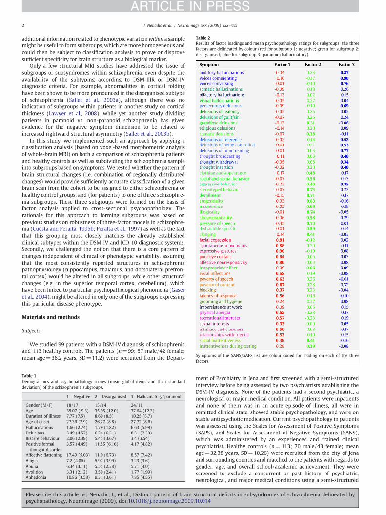
Table 2Results of factor loadings and mean psychopathology ratings for subgroups: the threefactors are delineated by colour (red for subgroup 1: negative; green for subgroup 2:disorganised; blue for subgroup 3: paranoid/hallucinatory).
Symptoms of the SANS/SAPS list are colour coded for loading on each of the threefactors.
2 I. Nenadic et al. / NeuroImage xxx (2009) xxx–xxx
ARTICLE IN PRESS
Please cite this article as: Nenadic, I., et al., Distinct pattern of brain structural de!cits in subsyndromes of schizophrenia delineated bypsychopathology, NeuroImage (2009), doi:10.1016/j.neuroimage.2009.10.014
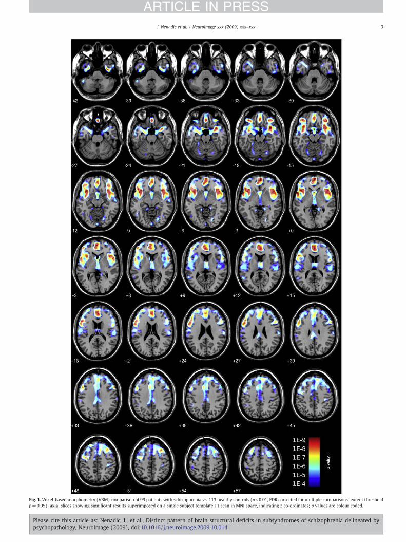
Fig. 1. Voxel-based morphometry (VBM) comparison of 99 patients with schizophrenia vs. 113 healthy controls (pb 0.01, FDR corrected for multiple comparisons; extent thresholdp=0.05): axial slices showing signi!cant results superimposed on a single subject template T1 scan in MNI space, indicating z co-ordinates; p values are colour coded.
3I. Nenadic et al. / NeuroImage xxx (2009) xxx–xxx
ARTICLE IN PRESS
Please cite this article as: Nenadic, I., et al., Distinct pattern of brain structural de!cits in subsyndromes of schizophrenia delineated bypsychopathology, NeuroImage (2009), doi:10.1016/j.neuroimage.2009.10.014
4 I. Nenadic et al. / NeuroImage xxx (2009) xxx–xxx
ARTICLE IN PRESS
Please cite this article as: Nenadic, I., et al., Distinct pattern of brain structural de!cits in subsyndromes of schizophrenia delineated bypsychopathology, NeuroImage (2009), doi:10.1016/j.neuroimage.2009.10.014
interview. Further exclusion criteria for both samples were a historyof head trauma, concurrent or previous substance dependence oralcoholism, and learning disability (mental retardation). All subjectswere right-handed as scored from the short version of the EdinburghHandedness Scale (Old!eld, 1971). All participants gave writteninformed consent to a study protocol approved by the EthicsCommittee of the Friedrich-Schiller-University of Jena.
Subgroup formation
The delineation of subgroups of schizophrenia patients was basedon cross-sectional psychopathology and factor analysis, dividing thepatients into three groups with a similar pro!le of symptoms. Manyprevious studies on the factor structure of schizophrenic symptomshave derived a three-factor solution (Andreasen et al., 1995), and thisthree-factor solution appears to be most stable, although alternativefour-factor or even !ve-factor models have been proposed. The three-factor solutions have normally produced groups dominated bynegative symptoms/psychomotor poverty, by psychotic/paranoid/reality distortion symptoms, and by disorganisation. In order to avoidconfounds of longitudinal symptom change (Marengo et al., 2000), allpatients included were chronic schizophrenia patients.
In order to form the subgroups, we entered all SANS and SAPSsingle items into a factor analysis de!ning a three-factor solution andapplying a Promax rotation. The analysis yielded three approximatelyequally sized groups with 35 patients in the negative subgroup (18male/17 female; mean age=35.1, SD=9.3), 29 in the disorganisedsubgroup (15 male, 14 female; mean age=36.0, SD=12.0), and 35 inthe paranoid subgroup (24 male/11 female; mean age=37.6,SD=12.3). Demographics and raw psychopathology scores (SAPSand SANS mean global items) of these subgroups are given in Table 1.Results of the factor loadings and mean psychopathology ratings forthe subgroups are given in Table 2. This analysis was compared toprevious studies to con!rm that the subsamples match the negative,paranoid, and disorganised subgroups described in previous studies.
Imaging protocol
We obtained a high-resolution structural MRI for each subject on a1.5-T Phillips Gyroscan ASCII system using a T1-weighted sequenceobtaining 256 sagittal slices covering the entire brain (TR=13 ms,TE= 5 ms, a 25°, !eld of view [FOV]=256 mm, voxeldimensions=1!1!1 mm3) for all subjects. Foam pads were usedwhere appropriate to further restrict head movement of subjects.Prior to image processing, each image was checked manually forartefacts. In addition, we used an automated tool for detection ofoutliers implemented in the VBM2 package (see below). All scanspassed both the manual and automated quality checks.
Image analysis and classi!cation analysis
We !rst applied voxel-based morphometry (VBM) using VBM2software. VBM2 is a toolbox (available on http://dbm.neuro.uni-jena.de/vbm) implemented in SPM2 (Statistical Parametric Mapping,Institute of Neurology, London, UK) and uses an optimised VBMprotocol (Ashburner and Friston, 2000; Good et al., 2001) as well as amodel based on hidden Markov random !elds (HMRF) developed toincrease signal-to-noise ratio (Cuadra et al., 2005).
First, we created a customised template image using a two-stepsegmentation approach. Segmentation in this process assigns eachvoxel to a tissue class (i.e. grey matter, white matter or CSF) and isoptimised using a modi!ed Gaussian mixture model (Ashburner and
Friston, 2000) incorporating prior knowledge into the assignment ofeach voxel. The two-step segmentation !rst applies segmentation tothe grey matter image derived from each individual scan, thennormalising the segmented image to a standard template (MNItemplate) and averaging these normalised images to obtain acustomised template for this study population. Using a customisedtemplate has advantages of accounting for differences between thestudy populations and the population used for standard templates aswell as taking into account scanner-speci!c non-uniformities in imageintensity and inhomogeneities of the B0 !eld. Secondly, we then usedthis customised template for normalisation of each study subject'sscan. These grey matter images are then compared across the groupapplying a general linear model within each voxel to test fordifferences in grey matter “concentration” or density. The VBM2algorithm also includes an automated quality control for images basedon their homogeneity, calculating the standard deviation as thesquared distance of each image (sum of intensity values) from thesample mean image.
Statistical analyses of VBM included !rst a comparison of the groupof schizophrenia patients with healthy controls, and secondly acomparison of each subgroup with the healthy controls. Age andgender were entered as confound variables to remove effects relatedto minor differences between groups. For all comparisons, we applieda threshold of pb0.01 corrected for multiple comparisons (FDR) aswell as an extent threshold of pb 0.05 to exclude smaller clustersfailing to reach the statistical extent criterion.
For classi!cation analysis, we applied a method based on anorthonormalised partial least square (OPLS) approach based on amultivariate statistical model, which we have described in detail in aprevious paper (Soriano-Mas et al., 2007). The OPLS is based on asingular value decomposition (SVD) of the orthonormalised predictorvalues (equals the design matrix) X and the data Y and results inEigenimages V, Eigenvalues ! , and Eigenvectors U". The Eigenimagesand the grey matter images for each subject can be used to calculate aso-called expression value for each subject. This single valueexpresses the extent to which this subject represents the pattern ofthe Eigenimage. Using the expression values for each subject, we cancompute the probability for belonging to a particular group
To further test the !ndings, we used a jackknife (“leave-one-out”)approach, in which the whole computation was performed leavingout one subject, resulting in a new ordering of group differences and anew individual expression values and probabilities for a total of 212computations.
Results
Group-wise voxel-based morphometry
The !rst comparison, contrasting the schizophrenia patient sampleand healthy controls showed signi!cant reductions of grey matter inpatients in several cortical and subcortical areas, as shown in Fig. 1.This included the prefrontal cortices (dorsolateral, medial, andorbitofrontal areas), temporal lobe (including amygdala, hippocam-pus, lateral temporal pole, and superior temporal cortex), andthalamus. Within the prefrontal cortex, the dorsolateral and medialareas were most widely affected, whereas the orbitofrontal cortexshowed reductions in its lateral parts with relative sparing of medial(posterior) areas. Within the temporal lobe, the medial temporalstructures (including amygdala and hippocampus) were reduced ingrey matter as well as the temporal pole (both lateral and medialaspects) and superior temporal gyrus, with sparing of the mid andposterior middle and inferior temporal gyri.
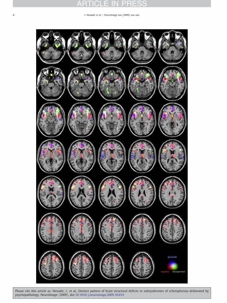
Fig. 2. Voxel-based morphometry (VBM) comparison of schizophrenia subgroups healthy controls (pb 0.01, FDR corrected for multiple comparisons; extent threshold p= 0.05):Composite image of group-wise comparisons with negative subgroup (red), disorganised subgroups (green), and hallucinatory subgroup (blue); intermediate colours re"ect overlap(axial slices superimposed on single subject T1 scan).
5I. Nenadic et al. / NeuroImage xxx (2009) xxx–xxx
ARTICLE IN PRESS
Please cite this article as: Nenadic, I., et al., Distinct pattern of brain structural de!cits in subsyndromes of schizophrenia delineated bypsychopathology, NeuroImage (2009), doi:10.1016/j.neuroimage.2009.10.014
Comparing each subgroup separately against the healthy controls,we identi!ed diverging as well as overlapping areas of alteration. Acomposite image illustrating the areas of overlap and difference isshown in Fig. 2.
All groups showed grey matter reductions in the prefrontalcortices, although to a different magnitude and extent and also indiffering locations. Most extensive prefrontal cortical de!cits wereseen in the negative patient group, whereas the medial temporalchanges where mostly present in the disorganised subgroup. Reduc-tions in the thalamus were most prominent in the negative subgroup.
Classi!cation analysis
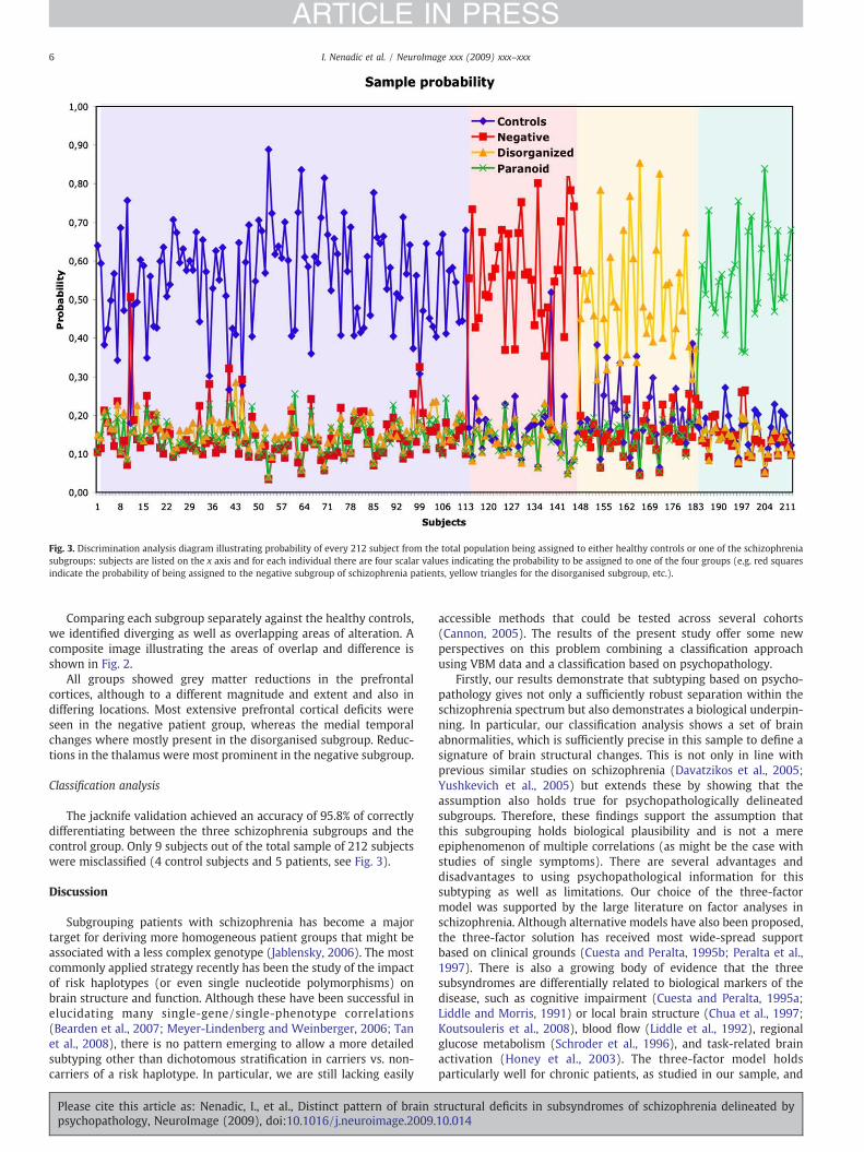
The jacknife validation achieved an accuracy of 95.8% of correctlydifferentiating between the three schizophrenia subgroups and thecontrol group. Only 9 subjects out of the total sample of 212 subjectswere misclassi!ed (4 control subjects and 5 patients, see Fig. 3).
Discussion
Subgrouping patients with schizophrenia has become a majortarget for deriving more homogeneous patient groups that might beassociated with a less complex genotype (Jablensky, 2006). The mostcommonly applied strategy recently has been the study of the impactof risk haplotypes (or even single nucleotide polymorphisms) onbrain structure and function. Although these have been successful inelucidating many single-gene/single-phenotype correlations(Bearden et al., 2007; Meyer-Lindenberg and Weinberger, 2006; Tanet al., 2008), there is no pattern emerging to allow a more detailedsubtyping other than dichotomous strati!cation in carriers vs. non-carriers of a risk haplotype. In particular, we are still lacking easily
accessible methods that could be tested across several cohorts(Cannon, 2005). The results of the present study offer some newperspectives on this problem combining a classi!cation approachusing VBM data and a classi!cation based on psychopathology.
Firstly, our results demonstrate that subtyping based on psycho-pathology gives not only a suf!ciently robust separation within theschizophrenia spectrum but also demonstrates a biological underpin-ning. In particular, our classi!cation analysis shows a set of brainabnormalities, which is suf!ciently precise in this sample to de!ne asignature of brain structural changes. This is not only in line withprevious similar studies on schizophrenia (Davatzikos et al., 2005;Yushkevich et al., 2005) but extends these by showing that theassumption also holds true for psychopathologically delineatedsubgroups. Therefore, these !ndings support the assumption thatthis subgrouping holds biological plausibility and is not a mereepiphenomenon of multiple correlations (as might be the case withstudies of single symptoms). There are several advantages anddisadvantages to using psychopathological information for thissubtyping as well as limitations. Our choice of the three-factormodel was supported by the large literature on factor analyses inschizophrenia. Although alternative models have also been proposed,the three-factor solution has received most wide-spread supportbased on clinical grounds (Cuesta and Peralta, 1995b; Peralta et al.,1997). There is also a growing body of evidence that the threesubsyndromes are differentially related to biological markers of thedisease, such as cognitive impairment (Cuesta and Peralta, 1995a;Liddle and Morris, 1991) or local brain structure (Chua et al., 1997;Koutsouleris et al., 2008), blood "ow (Liddle et al., 1992), regionalglucose metabolism (Schroder et al., 1996), and task-related brainactivation (Honey et al., 2003). The three-factor model holdsparticularly well for chronic patients, as studied in our sample, and
Fig. 3. Discrimination analysis diagram illustrating probability of every 212 subject from the total population being assigned to either healthy controls or one of the schizophreniasubgroups: subjects are listed on the x axis and for each individual there are four scalar values indicating the probability to be assigned to one of the four groups (e.g. red squaresindicate the probability of being assigned to the negative subgroup of schizophrenia patients, yellow triangles for the disorganised subgroup, etc.).
6 I. Nenadic et al. / NeuroImage xxx (2009) xxx–xxx
ARTICLE IN PRESS
Please cite this article as: Nenadic, I., et al., Distinct pattern of brain structural de!cits in subsyndromes of schizophrenia delineated bypsychopathology, NeuroImage (2009), doi:10.1016/j.neuroimage.2009.10.014
can be applied reliably also in geriatric populations (Sauer et al., 1999).Some more recent data imply that the three-factor solution might belinked to underlying genetical predisposition, as shown in twin studies(Cardno et al., 2001). This lends additional validity to this approach ofsubtyping a complex disorder. Yet, delineation of subgroups based oncross-sectional psychopathological data has inherent limitations forseveral reasons. Even though we chose chronic patients, for which thethree-factor model holds best, and limited ourselves to stabilisedpatients, we cannot exclude effects of medication, especially in thebasal ganglia,which are known tobeprone to these, but also for corticalreasons. As psychopathology changes over time (especially whenpsychosis "ares up) one might therefore argue that correlating amostly state-relatedparameterwith a presumably trait-relatedmarkersuch as brain structure might could lead to an excess of both false-positive and false-negative results. However, we should argue thatneither of theseparameters is purely state or trait related. In ourpatientcohort, for example, we chose chronic patients, well past their !rstyears of illness onset, who had developed a stable psychopathology. Allof them were well outside an acute episode of their psychosis and at astage where psychopathology enters a residual phase with moderate"uctuations in bothpositive andnegative symptoms. Itmight thereforebe assumed that we identi!ed time points that are most stable (andthus possibly most characteristic) for each individual patient's diseasecourse. There is limited data on the time course of symptoms inthe different three subsyndromes. Lack of longitudinal data did notallow us to assign patients to either Kraepelinian (de!cit) or non-Kraepelinian subtypes—an alternative recently proposed subtyping, forwhich differing patterns of brain pathology have been shown (Heckerset al., 1999; Mitelman et al., 2003). Brain structure, on the other hand,cannot be assumed to be a purely trait-related marker. Although thereis good evidence that some structural changes occur at the point ofdisease onset or even before (Pantelis et al., 2003), there is now goodevidence for disease progression and even changes with acute phasesof psychosis. Hence, despite these limitations, the study can serve as aparadigm to disentangle the effects of phenotypic variation on brainstructure as a putative endophenotype.
Regarding our second hypothesis of this study, we could notcon!rm the proposed pattern of hippocampus, thalamus, andprefrontal cortex as a set of core abnormalities characterising allschizophrenia patients irrespective of subgrouping. We identi!edgreymatter reduction in all of these structures (as well as the superiortemporal gyrus and the cerebellum) in the schizophrenia vs. healthycontrols comparison, thus replicating the most stable !ndings in oneof the largest VBM samples so far (Honea et al., 2005). Overall, themorphometric results in the subgroups are consistent with theprevious classical imaging studies by Liddle et al. (1992) on thethree-subgroup model of schizophrenia, which closely resembles ourfactor solution as well as more recent VBM data (Koutsouleris et al.,2008). However, there was marked heterogeneity among the threesubgroups in these regions. For the hippocampus, it was thedisorganised subgroup and (to lesser extent) also the paranoidsubgroup (esp. in the anterior hippocampus and amygdala) thatshowed prominent changes. For the thalamus, the reductionswere most prominent in the negative symptoms subgroup. For thecerebellum, this is the !rst study to identify the disorganised (and tolesser extent the negative) subsyndromes of schizophrenia to showvolume de!cits in this region. The former is of particular interest inview of the concept of formal thought disorder linked to cognitivedysmetria, which has been proposed as a general model forschizophrenia (Andreasen and Pierson, 2008). On the other hand,the medial temporal lobe structures have often been linked todelusions, which makes the lack of more wide-spread !ndings in thisregion in the paranoid subgroup somewhat surprising. Interestingly,the area of greatest convergence or spatial overlap is the prefrontalcortex, and in particular the right ventrolateral and lower dorsolateralcortices and the medial prefrontal areas bilaterally. For our results, it
appears that alterations of all subtypes converge in these areas, withnegative patients showing greatest spatial extent of changes.
We should also point out that several of the spatial components ofthe brain structural patterns resemble previous !ndings of correla-tions with single symptoms or groups of single psychopathologicalitems. For example, negative symptoms have repeatedly been linkedto grey matter reductions in prefrontal cortex (San!lipo et al., 2000),yet another study found a positive correlation in the thalamus in anearly onset sample (Yoshihara et al., 2008), whichwould not be in linewith our thalamus !ndings in chronic patients. A more recent studyfound delusions to be correlated to medial prefrontal cortex, close tothe area of the clusters seen in our paranoid subgroup (Whitford et al.,2009). However, there is also considerable heterogeneity in studies onstructure–symptom correlations, owing to a number of confoundingfactors such as patient group studied (symptom distribution, !rst-episode vs. chronic patients), medication status, etc.
Finally, we need to consider limitations of the study such aspotential effects of antipsychotic medication (past and current). Allour patients were on stable antipsychotic medication with wither a!rst- or second-generation antipsychotic. Experiments in monkeyshave demonstrated that chronic exposure to antipsychotics leads tohistologically evident volume reductions in brain grey and whitematter, which might have both a generalised component as well asregional variation of effects (Dorph-Petersen et al., 2005). Inpatientsamples, changes of brain volumes related to antipsychotic exposurehave been shown especially for basal ganglia, where !rst-generationantipsychotics lead to volume increases (Navari and Dazzan, 2009),which might be reversible by atypical agents such as clozapine. Morerecent studies, however, have also shown !rst-generation antipsy-chotic haloperidol to lead to cortical volume loss after one year oftreatment, but not olanzapine, which is a second-generation antipsy-chotic (SGA) (Lieberman et al., 2005). Also, SGAs might lead to partialreversal of cortical atrophy by inducing subtle expansion of greymatter (Garver et al., 2005), possibly an effect closely related to thefunctional properties regarding blood "ow and metabolism (Molinaet al., 2005). A major conclusion from the available literature (Navariand Dazzan, 2009) is therefore that medication effects (especially ofSGAs) might be quite variable across different cortical areas. Even ifmedication dosage is converted to a comparable standard such aschlorpromazine equivalents, we still need to consider the fact thatthere is considerable non-adherence to antipsychotic medication inpatients (Lindenmayer et al., 2009), which limits the value of usingthese indicators of antipsychotic exposure.
Taken together, this study provides biological evidence on theplausibility and face validity of a combination of overlapping anddiffering brain structural pathology in psychopathologically delineat-ed subgroups of schizophrenia. It also rejects the notion that allpatients might share pathology of a core network.
Acknowledgments
I.N. was supported by a Young Scientist Grant of the Friedrich-Schiller-University Jena and a Junior Scientist Grant of the IZKF,Friedrich-Schiller-University Medical School, Jena, C.G. was supportedby BMBF 01EV0709 and 01GW0740.
Con"icts of interest and !nancial disclosure: The authors declarethat they have no con"ict of interest other than those potentiallyarising through above mentioned funding and that they have no!nancial relationships or af!liations that could inappropriatelyin"uence or bias the author's decision, work, or manuscript.
References
Andreasen, N.C., Pierson, R., 2008. The role of the cerebellum in schizophrenia. Biol.Psychiatry 64, 81–88.
Andreasen, N.C., Arndt, S., Alliger, R., Miller, D., Flaum, M., 1995. Symptoms ofschizophrenia. Methods, meanings, and mechanisms. Arch. Gen. Psychiatry 52,341–351.
7I. Nenadic et al. / NeuroImage xxx (2009) xxx–xxx
ARTICLE IN PRESS
Please cite this article as: Nenadic, I., et al., Distinct pattern of brain structural de!cits in subsyndromes of schizophrenia delineated bypsychopathology, NeuroImage (2009), doi:10.1016/j.neuroimage.2009.10.014
Ashburner, J., Friston, K.J., 2000. Voxel-based morphometry—the methods. NeuroImage11, 805–821.
Bearden, C.E., van Erp, T.G., Thompson, P.M., Toga, A.W., Cannon, T.D., 2007. Corticalmapping of genotype–phenotype relationships in schizophrenia. Hum. BrainMapp.28, 519–532.
Cannon, T.D., 2005. The inheritance of intermediate phenotypes for schizophrenia. Curr.Opin. Psychiatry 18, 135–140.
Cardno, A.G., Sham, P.C., Murray, R.M., McGuf!n, P., 2001. Twin study of symptomdimensions in psychoses. Br. J. Psychiatry 179, 39–45.
Chua, S.E., Wright, I.C., Poline, J.B., Liddle, P.F., Murray, R.M., Frackowiak, R.S., Friston,K.J., McGuire, P.K., 1997. Grey matter correlates of syndromes in schizophrenia. Asemi-automated analysis of structural magnetic resonance images. Br. J. Psychiatry170, 406–410.
Cuadra, M.B., Cammoun, L., Butz, T., Cuisenaire, O., Thiran, J.P., 2005. Comparison andvalidation of tissue modelization and statistical classi!cation methods in T1-weighted MR brain images. IEEE Trans. Med. Imaging 24, 1548–1565.
Cuesta, M.J., Peralta, V., 1995a. Cognitive disorders in the positive, negative, anddisorganization syndromes of schizophrenia. Psychiatry Res. 58, 227–235.
Cuesta, M.J., Peralta, V., 1995b. Psychopathological dimensions in schizophrenia.Schizophr. Bull. 21, 473–482.
Davatzikos, C., Shen, D., Gur, R.C., Wu, X., Liu, D., Fan, Y., Hughett, P., Turetsky, B.I., Gur,R.E., 2005. Whole-brain morphometric study of schizophrenia revealing a spatiallycomplex set of focal abnormalities. Arch. Gen. Psychiatry 62, 1218–1227.
Dorph-Petersen, K.A., Pierri, J.N., Perel, J.M., Sun, Z., Sampson, A.R., Lewis, D.A., 2005. Thein"uence of chronic exposure to antipsychotic medications on brain size before andafter tissue !xation: a comparison of haloperidol and olanzapine in macaquemonkeys. Neuropsychopharmacology 30, 1649–1661.
Garver, D.L., Holcomb, J.A., Christensen, J.D., 2005. Cerebral cortical gray expansionassociated with two second-generation antipsychotics. Biol. Psychiatry 58, 62–66.
Gaser, C., Nenadic, I., Volz, H.P., Buchel, C., Sauer, H., 2004. Neuroanatomy of “hearingvoices”: a frontotemporal brain structural abnormality associated with auditoryhallucinations in schizophrenia. Cereb. Cortex 14, 91–96.
Goldman, A.L., Pezawas, L., Mattay, V.S., Fischl, B., Verchinski, B.A., Zoltick, B.,Weinberger, D.R., Meyer-Lindenberg, A., 2008. Heritability of brain morphologyrelated to schizophrenia: a large-scale automated magnetic resonance imagingsegmentation study. Biol. Psychiatry 63, 475–483.
Good, C.D., Johnsrude, I.S., Ashburner, J., Henson, R.N., Friston, K.J., Frackowiak, R.S.,2001. A voxel-based morphometric study of ageing in 465 normal adult humanbrains. Neuroimage 14, 21–36.
Hazlett, E.A., Buchsbaum, M.S., Haznedar, M.M., Newmark, R., Goldstein, K.E.,Zelmanova, Y., Glanton, C.F., Torosjan, Y., New, A.S., Lo, J.N., Mitropoulou, V., Siever,L.J., 2008. Cortical gray and white matter volume in unmedicated schizotypal andschizophrenia patients. Schizophr. Res. 101, 111–123.
Heckers, S., Goff, D., Schacter, D.L., Savage, C.R., Fischman, A.J., Alpert, N.M., Rauch, S.L.,1999. Functional imaging of memory retrieval in de!cit vs nonde!cit schizophre-nia. Arch. Gen. Psychiatry 56, 1117–1123.
Honea, R., Crow, T.J., Passingham, D., Mackay, C.E., 2005. Regional de!cits in brainvolume in schizophrenia: a meta-analysis of voxel-based morphometry studies.Am. J. Psychiatry 162, 2233–2245.
Honea, R.A., Meyer-Lindenberg, A., Hobbs, K.B., Pezawas, L., Mattay, V.S., Egan, M.F.,Verchinski, B., Passingham, R.E., Weinberger, D.R., Callicott, J.H., 2008. Is graymatter volume an intermediate phenotype for schizophrenia? A voxel-basedmorphometry study of patients with schizophrenia and their healthy siblings. Biol.Psychiatry 63, 465–474.
Honey, G.D., Sharma, T., Suckling, J., Giampietro, V., Soni, W., Williams, S.C., Bullmore,E.T., 2003. The functional neuroanatomy of schizophrenic subsyndromes. Psychol.Med. 33, 1007–1018.
Hulshoff Pol, H.E., Brans, R.G., van Haren, N.E., Schnack, H.G., Langen, M., Baare, W.F.,van Oel, C.J., Kahn, R.S., 2004. Gray and white matter volume abnormalities inmonozygotic and same-gender dizygotic twins discordant for schizophrenia. Biol.Psychiatry 55, 126–130.
Hulshoff Pol, H.E., Schnack, H.G., Mandl, R.C., Brans, R.G., van Haren, N.E., Baare, W.F.,van Oel, C.J., Collins, D.L., Evans, A.C., Kahn, R.S., 2006. Gray and white matterdensity changes in monozygotic and same-sex dizygotic twins discordant forschizophrenia using voxel-based morphometry. Neuroimage 31, 482–488.
Jablensky, A., 2006. Subtyping schizophrenia: implications for genetic research. Mol.Psychiatry 11, 815–836.
Kasai, K., Shenton, M.E., Salisbury, D.F., Onitsuka, T., Toner, S.K., Yurgelun-Todd, D.,Kikinis, R., Jolesz, F.A., McCarley, R.W., 2003. Differences and similarities in insularand temporal pole MRI gray matter volume abnormalities in !rst-episodeschizophrenia and affective psychosis. Arch. Gen. Psychiatry 60, 1069–1077.
Kawasaki, Y., Suzuki, M., Kherif, F., Takahashi, T., Zhou, S.Y., Nakamura, K., Matsui, M.,Sumiyoshi, T., Seto, H., Kurachi, M., 2007. Multivariate voxel-based morphometrysuccessfully differentiates schizophrenia patients fromhealthy controls. Neuroimage34, 235–242.
Koutsouleris, N., Gaser, C., Jager, M., Bottlender, R., Frodl, T., Holzinger, S., Schmitt, G.J.,Zetzsche, T., Burgermeister, B., Scheuerecker, J., Born, C., Reiser, M., Moller, H.J.,Meisenzahl, E.M., 2008. Structural correlates of psychopathological symptomdimensions in schizophrenia: a voxel-based morphometric study. Neuroimage 39,1600–1612.
Lawyer, G., Nesvag, R., Varnas, K., Frigessi, A., Agartz, I., 2008. Investigating possiblesubtypes of schizophrenia patients and controls based on brain cortical thickness.Psychiatry Res. 164, 254–264.
Liddle, P.F., Morris, D.L., 1991. Schizophrenic syndromes and frontal lobe performance.Br. J. Psychiatry 158, 340–345.
Liddle, P.F., Friston, K.J., Frith, C.D., Hirsch, S.R., Jones, T., Frackowiak, R.S., 1992. Patternsof cerebral blood "ow in schizophrenia. Br. J. Psychiatry 160, 179–186.
Lieberman, J.A., Tollefson, G.D., Charles, C., Zipursky, R., Sharma, T., Kahn, R.S., Keefe,R.S., Green, A.I., Gur, R.E., McEvoy, J., Perkins, D., Hamer, R.M., Gu, H., Tohen, M.,2005. Antipsychotic drug effects on brain morphology in !rst-episode psychosis.Arch. Gen. Psychiatry 62, 361–370.
Lindenmayer, J.P., Liu-Seifert, H., Kulkarni, P.M., Kinon, B.J., Stauffer, V., Edwards, S.E.,Chen, L., Adams, D.H., Ascher-Svanum, H., Buckley, P.F., Citrome, L., Volavka, J.,2009. Medication nonadherence and treatment outcome in patients withschizophrenia or schizoaffective disorder with suboptimal prior response. J. Clin.Psychiatry 70, 990–996.
Marengo, J., Harrow, M., Herbener, E.S., Sands, J., 2000. A prospective longitudinal 10-year study of schizophrenia's three major factors and depression. Psychiatry Res.97, 61–77.
McDonald, C., Marshall, N., Sham, P.C., Bullmore, E.T., Schulze, K., Chapple, B., Bramon,E., Filbey, F., Quraishi, S., Walshe, M., Murray, R.M., 2006. Regional brainmorphometry in patients with schizophrenia or bipolar disorder and theirunaffected relatives. Am. J. Psychiatry 163, 478–487.
McIntosh, A.M., Job, D.E., Moorhead, W.J., Harrison, L.K., Whalley, H.C., Johnstone, E.C.,Lawrie, S.M., 2006. Genetic liability to schizophrenia or bipolar disorder and itsrelationship to brain structure. Am. J. Med. Genet. B. Neuropsychiatr. Genet. 141B,76–83.
Meyer-Lindenberg, A., Weinberger, D.R., 2006. Intermediate phenotypes and geneticmechanisms of psychiatric disorders. Nat. Rev. Neurosci. 7, 818–827.
Mitelman, S.A., Shihabuddin, L., Brickman, A.M., Hazlett, E.A., Buchsbaum, M.S., 2003.MRI assessment of gray and white matter distribution in Brodmann's areas of thecortex in patients with schizophrenia with good and poor outcomes. Am. J.Psychiatry 160, 2154–2168.
Molina, V., Gispert, J.D., Reig, S., Sanz, J., Pascau, J., Santos, A., Desco, M., Palomo, T., 2005.Cerebral metabolic changes induced by clozapine in schizophrenia and related toclinical improvement. Psychopharmacology (Berl). 178, 17–26.
Navari, S., Dazzan, P., 2009. Do antipsychotic drugs affect brain structure? A systematicand critical review of MRI !ndings. Psychol. Med. 1–15.
Old!eld, R.C., 1971. The assessment and analysis of handedness: the Edinburghinventory. Neuropsychologia 9, 97–113.
Pantelis, C., Velakoulis, D., McGorry, P.D., Wood, S.J., Suckling, J., Phillips, L.J., Yung, A.R.,Bullmore, E.T., Brewer, W., Soulsby, B., Desmond, P., McGuire, P.K., 2003.Neuroanatomical abnormalities before and after onset of psychosis: a cross-sectional and longitudinal MRI comparison. Lancet 361, 281–288.
Peralta, V., Cuesta, M.J., Farre, C., 1997. Factor structure of symptoms in functionalpsychoses. Biol. Psychiatry 42, 806–815.
Sallet, P.C., Elkis, H., Alves, T.M., Oliveira, J.R., Sassi, E., Campi de Castro, C., Busatto, G.F.,Gattaz, W.F., 2003a. Reduced cortical folding in schizophrenia: an MRI morpho-metric study. Am. J. Psychiatry 160, 1606–1613.
Sallet, P.C., Elkis, H., Alves, T.M., Oliveira, J.R., Sassi, E., de Castro, C.C., Busatto, G.F.,Gattaz, W.F., 2003b. Rightward cerebral asymmetry in subtypes of schizophreniaaccording to Leonhard's classi!cation and to DSM-IV: a structural MRI study.Psychiatry Res. 123, 65–79.
San!lipo, M., Lafargue, T., Rusinek, H., Arena, L., Loneragan, C., Lautin, A., Feiner, D.,Rotrosen, J., Wolkin, A., 2000. Volumetric measure of the frontal and temporal loberegions in schizophrenia: relationship to negative symptoms. Arch. Gen. Psychiatry57, 471–480.
Sauer, H., Hornstein, C., Richter, P., Mortimer, A., Hirsch, S.R., 1999. Symptomdimensions in old-age schizophrenics. Relationship to neuropsychological andmotor abnormalities. Schizophr. Res. 39, 31–38.
Schroder, J., Buchsbaum, M.S., Siegel, B.V., Geider, F.J., Lohr, J., Tang, C., Wu, J., Potkin,S.G., 1996. Cerebral metabolic activity correlates of subsyndromes in chronicschizophrenia. Schizophr. Res. 19, 41–53.
Soriano-Mas, C., Pujol, J., Alonso, P., Cardoner, N., Menchon, J.M., Harrison, B.J., Deus, J.,Vallejo, J., Gaser, C., 2007. Identifying patients with obsessive-compulsive disorderusing whole-brain anatomy. Neuroimage 35, 1028–1037.
Steen, R.G., Mull, C., McClure, R., Hamer, R.M., Lieberman, J.A., 2006. Brain volume in!rst-episode schizophrenia: systematic review and meta-analysis of magneticresonance imaging studies. Br. J. Psychiatry 188, 510–518.
Takahashi, T., Suzuki, M., Zhou, S.Y., Tanino, R., Hagino, H., Kawasaki, Y., Matsui, M., Seto,H., Kurachi, M., 2006. Morphologic alterations of the parcellated superior temporalgyrus in schizophrenia spectrum. Schizophr. Res. 83, 131–143.
Tan, H.Y., Callicott, J.H., Weinberger, D.R., 2008. Intermediate phenotypes inschizophrenia genetics redux: is it a no brainer? Mol. Psychiatry 13, 233–238.
Weinberger, D.R., McClure, R.K., 2002. Neurotoxicity, neuroplasticity, and magneticresonance imaging morphometry: what is happening in the schizophrenic brain?Arch. Gen. Psychiatry 59, 553–558.
Whitford, T.J., Farrow, T.F., Williams, L.M., Gomes, L., Brennan, J., Harris, A.W., 2009.Delusions and dorso-medial frontal cortex volume in !rst-episode schizophrenia: avoxel-based morphometry study. Psychiatry Res. 172, 175–179.
Wright, I.C., Rabe-Hesketh, S., Woodruff, P.W., David, A.S., Murray, R.M., Bullmore, E.T.,2000. Meta-analysis of regional brain volumes in schizophrenia. Am. J. Psychiatry157, 16–25.
Yoshihara, Y., Sugihara, G., Matsumoto, H., Suckling, J., Nishimura, K., Toyoda, T., Isoda,H., Tsuchiya, K.J., Takebayashi, K., Suzuki, K., Sakahara, H., Nakamura, K., Mori, N.,Takei, N., 2008. Voxel-based structural magnetic resonance imaging (MRI) study ofpatients with early onset schizophrenia. Ann. Gen. Psychiatry 7, 25.
Yushkevich, P., Dubb, A., Xie, Z., Gur, R., Gur, R., Gee, J., 2005. Regional structuralcharacterization of the brain of schizophrenia patients. Acad. Radiol. 12,1250–1261.
8 I. Nenadic et al. / NeuroImage xxx (2009) xxx–xxx
ARTICLE IN PRESS
Please cite this article as: Nenadic, I., et al., Distinct pattern of brain structural de!cits in subsyndromes of schizophrenia delineated bypsychopathology, NeuroImage (2009), doi:10.1016/j.neuroimage.2009.10.014