Embed Size (px)
Citation preview

Csaba Hankó, sales director Valencia, the 24. november 2011
Are we ready for Digital Pathology?

1. Hungary
2. Our company
3. Our product portfolio
4. Why we decided to use area scanning, why not the linear one?
5. The comparison of the 2 scanner technologies
6. The role of digital slides in education
7. Our new flash scan technology
8. Our „CASE CENTER” for slide storage and networking
9. Our future developments
You will hear now about:

A little bit about Hungary
What you may not know…
This video clip shows the most famous inventions!

Hungary is famous not only about „GULASH”,
but also about science:
We have 14 Nobel laureates for a population of 10 millions
1905 – Fülöp Lénárd in physics
1914 – Robert Bárány in medicine for „pathology of the vestibular apparatus”
1925 – Richard Zsigmondy in chemistry
1937 – Albert Szent-Györgyi in medicine for „vitamine C”
1943 – György Hevesy in chemistry
1961 – György Békésy in medicine for „discovery of the physical mechanism of
stimulation within the cochlea”
1963 – Jenő Wigner in physics
1971 – Dénes Gábor in physics
1976 – Milton Friedman in economics
1986 – János Polányi in chemistry
1994 – György Oláh in chemistry
1994 – János Harsányi in economics
2002 – Imre Kertész in literature for his roman „Fateless” written in 1992
2004 – Ferenc Herskó in chemistry
(Spain has a population of 46 millions and 8 Nobel laureates, 2 of them in medicine)

• private owned spin-off company born at the
Semmelweis Medical University in Budapest
• Sole owner: Dr. med. PhD. Béla Molnár, CEO
• 1998 – START with development of Whole Slide Imaging
systems
• 2004-2009 exclusive distribution with Zeiss Germany:
sales of scanners under brandname MIRAX
• 2010 – new distribution channels with local distributors
• Today: nearly 500 scanners installed worldwide => >
• market leader in Europe (over 350 installed scanners)
• globally 2nd place after Aperio
• 70 employees (50 of them are in development!)
Introduction of 3DHISTECH Ltd.

Our reference sites worldwide
Only the most important ones! Sanofi-Aventis, Frankfurt Novartis Pharma, Basel Bayer Schering (US and Germany) Roche (US, Germany and Swiss) Merck Pharma, Darmstadt Abbott Pharma, Ludwigshafen Beiersdorf AG, Hamburg Astra Zeneca, Macclesfield (UK) Histogenex, Antwerpen Semmelweis University Clinic, Budapest Memorial Sloan Kettering, New York University Hospital for Sick Children, Toronto
McGill University Health Centre, Montreal Harvard Medical School, Cambridge Massachusetts General Hospital, Boston University of Pittsburgh Medical Center Vanderbilt University Medical Center, Nashville University Hospital Charité, Berlin Deutsches Herzzentrum, Berlin Asklepios Kliniken (5x) + Helios Kliniken (3x) Unikliniken (12x) in Germany Sasebo General Hospital, Nagasaki Kuwait Cancer Center Wales Cancer Bank, Cardiff

Product portfolio 1.: Pannoramic Scanners
DESK 1 slide
Frozen & BF
MIDI 12 Slides BF & FL
P250 FLASH 250 slides
BF & FL
SCAN 150 Slides
BF & FL

Product portfolio 2.: Tissue Microarray
TMA Master for 5 blocks
TMA Grand Master for 72 blocks
SCAN - 150 Slides Brightfield & Fluorescence

Product portfolio 3.: Software applications
We offer SW for almost everything what pathologists need?
– Workflow management: CaseCenter
– Remote slide access: CaseCenter
– Teleconsultation: CaseCenter
– Advanced diagnostic tools: TumorBoard,
TMA
– Automated image analysis: HistoQuant MembraneQuant (HER2, EGFR)
NuclearQuant (ER, PR, Ki67)
DensitoQuant
– Fluorescent slide analysis: FISH-Quant
– Advanced education: E-School
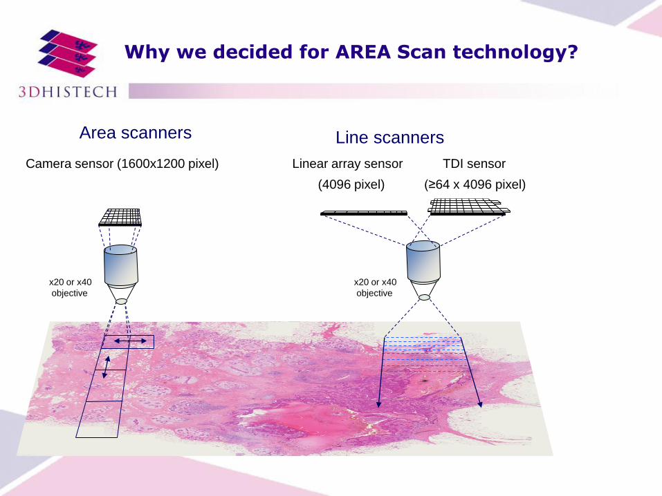
Area scanners
Camera sensor (1600x1200 pixel)
x20 or x40
objective
Line scanners
Linear array sensor TDI sensor
x20 or x40
objective
(4096 pixel) (≥64 x 4096 pixel)
Why we decided for AREA Scan technology?

• similar quality can be achieved, both technologies have minor advantages
• Advantages: • Area: Extended Focus is possible • Line: Sample topography can be followed better
• Speed:
• „MIRAX” technology of 3DH @ 43x (0.23µm / pixel resolution) is similar to Hamamatsu and Leica but faster, than Aperio!
• New „FLASH” technology is already faster than all current competition!
Area vs. Line scanning in BF
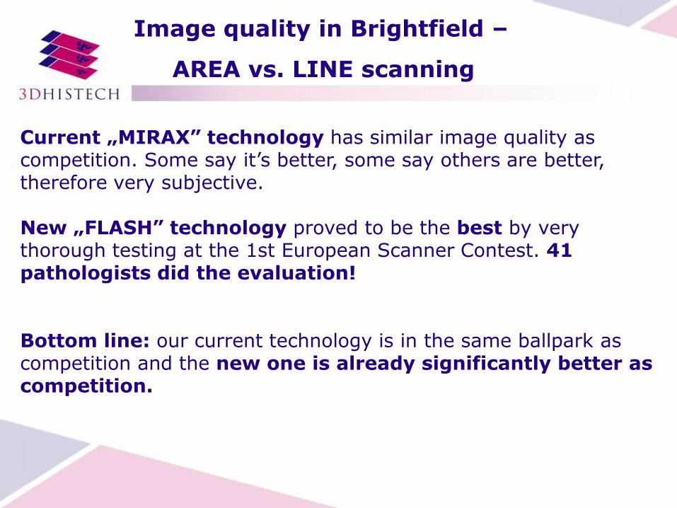
Current „MIRAX” technology has similar image quality as competition. Some say it’s better, some say others are better, therefore very subjective.
New „FLASH” technology proved to be the best by very thorough testing at the 1st European Scanner Contest. 41 pathologists did the evaluation!
Bottom line: our current technology is in the same ballpark as competition and the new one is already significantly better as competition.
Image quality in Brightfield –
AREA vs. LINE scanning

Area scanning gives us
the ability to extended
depth of field
Advantages of AREA vs. LINE scanning
in brightfield
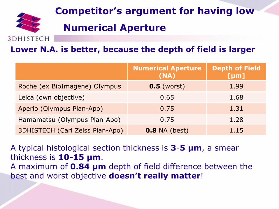
Lower N.A. is better, because the depth of field is larger
A typical histological section thickness is 3-5 µm, a smear thickness is 10-15 µm. A maximum of 0.84 µm depth of field difference between the best and worst objective doesn’t really matter!
Competitor’s argument for having low
Numerical Aperture
Numerical Aperture (NA)
Depth of Field [µm]
Roche (ex BioImagene) Olympus 0.5 (worst) 1.99
Leica (own objective) 0.65 1.68
Aperio (Olympus Plan-Apo) 0.75 1.31
Hamamatsu (Olympus Plan-Apo) 0.75 1.28
3DHISTECH (Carl Zeiss Plan-Apo) 0.8 NA (best) 1.15

• Line camera sensor needs the stage move continuously!
• CCD camera (area) sensor doesn’t need this: we can stop the stage!
• Why is this so important?
• Area sensor makes possible: • Extended Focus - FISH! • Z-Stack, perfect co-localization – FISH! • Change the filter as many time as we want • Have any exposure time we need • Live image
Advantages of AREA vs. LINE scanning
in fluorescence

• In fluorescence the amount of light is the main limitation
• Area sensor (3DHISTECH) collects much more light than TDI sensor (Aperio, Hamamatsu) because the sensor area is larger.
Imaged area / Magnification AxioCam %
Aperio 5 383 µm2 / 43x 4 %
Aperio 21 531 µm2 / 21x 14 %
Hamamatsu 13 867 µm2 / 21x and 43x 9 %
3DHISTECH old FL camera
150 135 µm2 / 31x 100 %
3DHISTECH new FL camera
270 400 µm2 / 31x 180 %
Advantages of AREA vs. LINE scanning
in fluorescence
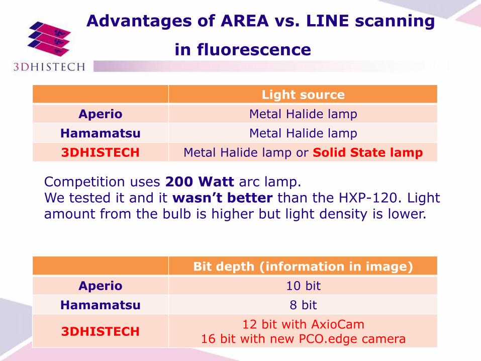
Competition uses 200 Watt arc lamp. We tested it and it wasn’t better than the HXP-120. Light amount from the bulb is higher but light density is lower.
Light source
Aperio Metal Halide lamp
Hamamatsu Metal Halide lamp
3DHISTECH Metal Halide lamp or Solid State lamp
Bit depth (information in image)
Aperio 10 bit
Hamamatsu 8 bit
3DHISTECH 12 bit with AxioCam
16 bit with new PCO.edge camera
Advantages of AREA vs. LINE scanning
in fluorescence

Brightness Index = NA4 / Magnification2
• 3DH = 0.804 / 202
• Aperio = 0.754 / 202 • Hamamatsu = 0.754 / 202
• Leica = 0.654 / 202
Conclusion: Our objective collects 30% more light, than Aperio or Hamamatsu and 230% more light, than Leica
Advantages of AREA vs. LINE scanning
in fluorescence

Using digital slides in education
standardization
same opportunities
same circumstances
same quality

More than 1500 hours of histology! practice!
Digital slide based education – 5 years experience at Semmelweis Budapest
•Histology lab
•Remote access
•Consultaton
•Exams
• 40 x PCs
• 2 x slide servers
• intranet connects student’s PC’s with teacher’s laptop & local server
• 200 slides digitized for teaching

• Fastest scanner of the world @ 45x magn.
• 250 slides capacity = >> overnight scanner
• Superior image quality
• Relatively small footprint (less than Hamamatsu
and Leica)
• Smaller weight than Hamamatsu or Leica
• 2 objectives (Carl Zeiss Plan-Apo 20x + 40x)
• 2 cameras (a 3CCD for BF + a CMOS for FL)
Main features of Pannoramic 250 Flash

How fast is it?
On the 1st European Scanner Contest (2010) each vendor scanned
the same 35 slides total area of 42,7 cm².
P250 FLASH is 3 times faster, than SCAN 150 & at least 2 times faster than Leica, the winner in 2010
Speed of the Pannoramic 250 FLASH
Vendor model
Resolution µm / pixel
Total scan time [min]
Scan time / slide [min]
Scan time cm²/min
Leica (the winner) SCN400 + SL801
0.25 (77%), 40x 160 4,5 0,26
3DHISTECH SCAN 150
0.22 (100%), 45x 190 5,6 0,23
3DHISTECH P250 flash
0.22 (100%), 45x 2,8 0,6-1,0

Pannoramic 250 FLASH
Movie:

• New focus algorithm: Finds a visually more appealing
focus level
• Sharpening
• Interpolated focus
• We were the winner of the 1st European Scanner
Contest in „40x image quality” category.
http://scanner-contest.charite.de/en/results/1st_isc
Improved image quality
Improved image quality

Improved image quality
Improved image quality

Single band pass filter
Video:
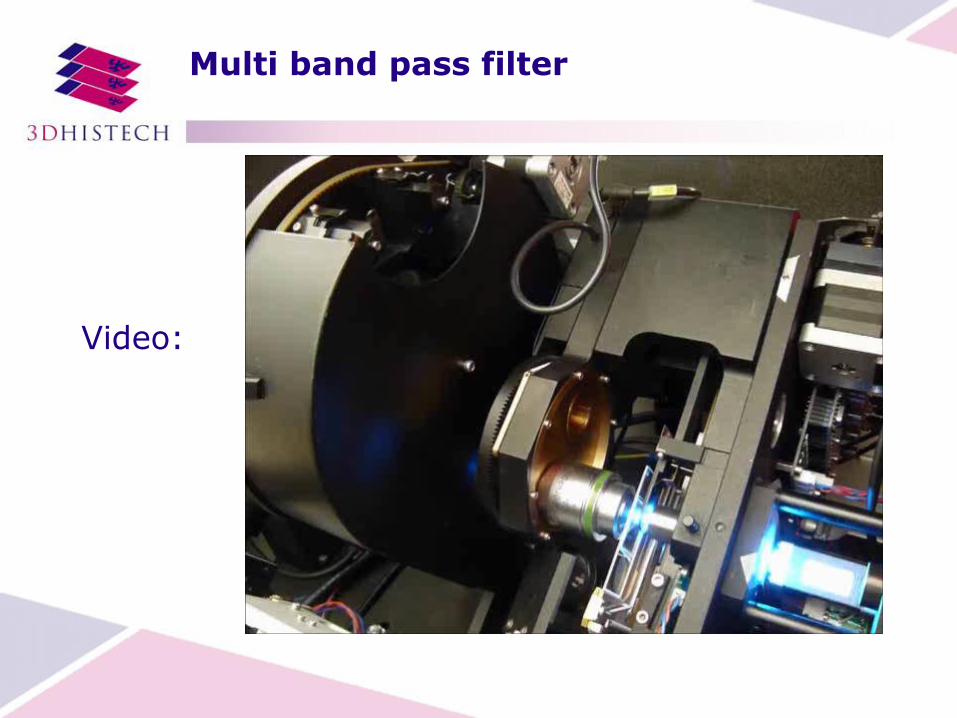
Multi band pass filter
Video:

Advantages of scientific CMOS camera vs.
CCD camera in fluorescent scanning
Key data: PCO.edge NEW Zeiss AxioCam MRm
Resolution Used: 1600x1600 (2.5 megapixel)
1388 x 1040 pixel (1.4 megapixel)
Dynamic range 22 000 : 1 2 200 : 1
Bit depth 16 bit 12 bit
Frame rate 33 frames / sec 5 frames / sec
Readout noise < 1.4 e- Typical < 7.7 e-
Size of pixels 6.5 µm x 6.5 µm 6.45 μm x 6.45 μm
Spectral range 370 nm - 1100 nm 350 nm - 1000 nm

Z-stacking
Extended focus
1
2
3
Single focus Extended focus
FISH scanning and extended focus
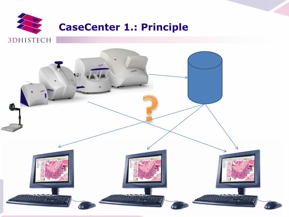
CaseCenter 1.: Principle
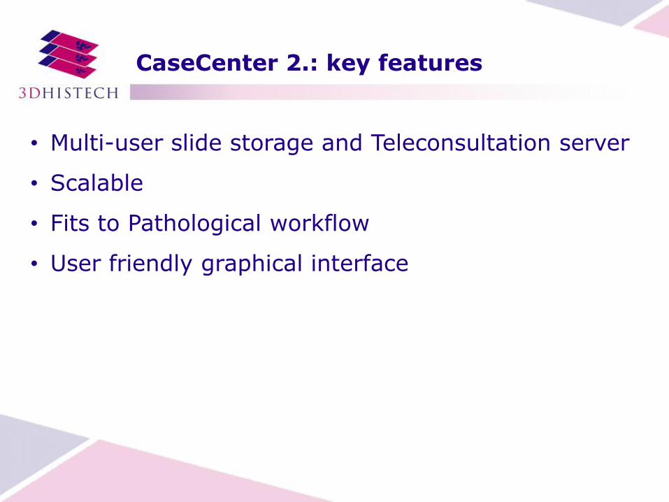
CaseCenter 2.: key features
• Multi-user slide storage and Teleconsultation server
• Scalable
• Fits to Pathological workflow
• User friendly graphical interface

CaseCenter 3.: Scan to server

Scanners
CaseCenter
Client (Pannoramic
Viewer)
auth
entic
atio
n
slides
CaseCenter 4.: Scalable solution
Client (remote)
Remote location

Client (Pannoramic
Viewer)
auth
entic
atio
n
slides SU part
HUB part SU part
CaseCenter 4.: Scalable solution 2
CaseCenter HUB + SU
Ext. Storage
slides
slides

CaseCenter 5.: Teleconsultation

CaseCenter 5.: Teleconsultation 2
CaseCenter
Client (Pannoramic
Viewer)
Client (Pannoramic
Viewer)
Client (Pannoramic
Viewer)
Client HOST
Open slide
Share Slide

Ways of Client connection
• Clinical workstation
• iPAD
• Any PC or laptop with a web browser

Clinical workstation
Case data
Measurement data
An optimal environment for high throughput
daily work with 30 inch HD med displays (Barco)

Tablet
• Accessing important information ANYWHERE!
• Frozen section
• Emergency
• Convenience

Future developments:
We continue the improvement of
• the workflow
• the scanning speed
• the slide storage
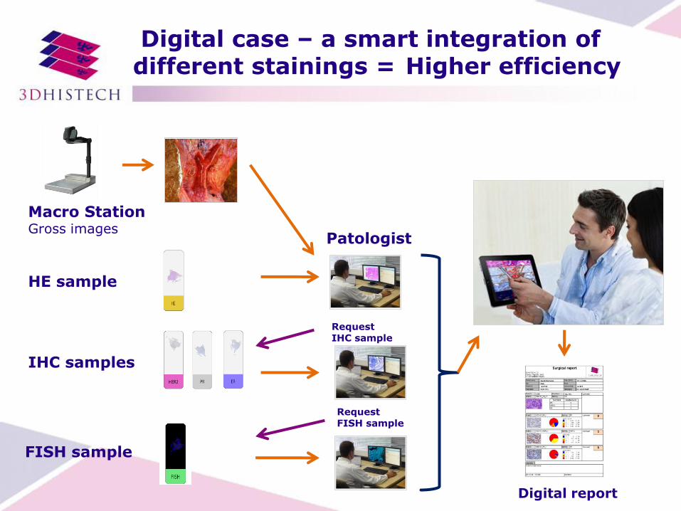
Digital case – a smart integration of different stainings = Higher efficiency
Macro Station Gross images
Request FISH sample
HE sample
IHC samples
FISH sample
Request IHC sample
Patologist
Digital report

Annotation handling on CaseCenter
Making annotation on the first slide
Following the first annotation measurement is done automatically on each slide of the case

Aligned parallel view of the whole case
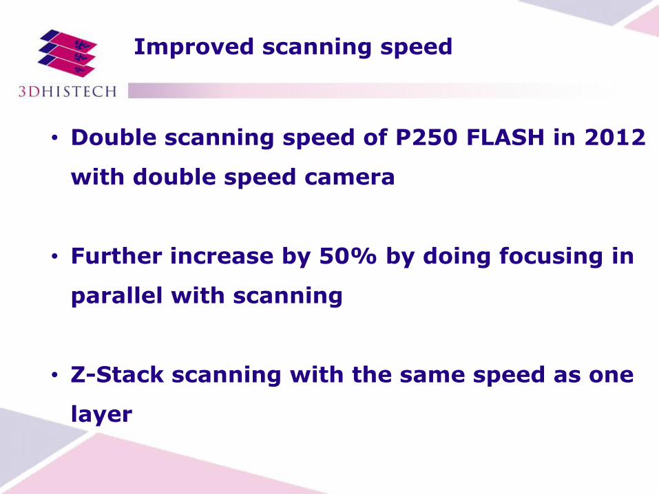
Improved scanning speed
• Double scanning speed of P250 FLASH in 2012
with double speed camera
• Further increase by 50% by doing focusing in
parallel with scanning
• Z-Stack scanning with the same speed as one
layer

Improved slide storage
Source: Panasonic Technology overview AVC-Intra (H.264) Compression
Farewell
I hope, you believe now, we are
ready for digital pathology!
….and what about you?
Thank you for your attention!
Any Question?