Embed Size (px)
Citation preview

8/7/2019 Approach to managing diabetic foot ulcers
http://slidepdf.com/reader/full/approach-to-managing-diabetic-foot-ulcers 1/7
VOL 50: APRIL • AVRIL 2004 d Canadian Family Physician • Le Médecin de famille canadien 561
Approach to managingdiabetic foot ulcersJohn A.A. Nesbitt, MD, MBA
ABSTRACT
INTRODUCTION Of an estimated 1.7 to 2 million Canadians with diabetes, approximately 10% will present each year
to their family doctors with plantar ulcers. Nearly 3500 will require major lower extremity amputations.
SOURCES OF INFORMATION Most of the recommendations outlined in this paper are based on level I evidence from
excellent bench research and epidemiologic studies.
MAIN MESSAGE Both insulin-dependent and non–insulin-dependent diabetics develop foot infections. These
patients are on average 60 years old and have had diabetes for more than 10 years. Physicians who insist on excellent
blood sugar control, provide ongoing patient education on diabetic foot care, prescribe appropriate shoes, and
practise an aggressive multidisciplinary approach to wound care can reduce the rate of lower extremity amputations
by more than 50%.
CONCLUSION Foot problems remain one of the main challenges associated with diabetes, but family physicians can
manage them successfully.
RÉSUMÉ
INTRODUCTION Chaque année au Canada, environ 10% des 1,7 à 2 millions de diabétiques consultent leur médecin
de famille pour des ulcères plantaires. Près de 3 500 d’entre eux nécessiteront des amputations majeures des
membres inférieurs.
SOURCE DE L’INFORMATION La plupart des recommandations dans cet article sont fondées sur des preuves de
niveau I provenant d’études pilotes et épidémiologiques d’excellente qualité.
PRINCIPAL MESSAGE Les diabétiques, qu’ils soient insulino-dépendants ou non, risquent des infections aux pieds.
Ils ont en moyenne 60 ans et soufrent de diabète depuis plus de 10 ans. En insistant pour un excellent contrôle de la
glycémie, renseignant régulièrement le patient sur les soins des pieds des diabétiques, prescrivant des chaussures
adéquates, et en utilisant une approche multidisciplinaire agressive pour le traitement des blessures, le médecin peut
réduire de plus de 50% le taux d’amputation des membres inférieurs.CONCLUSION Les lésions des pieds demeurent l’un des principaux défis associés au diabète, mais le médecin de
famille peut s’en occuper adéquatement.
This article has been peer reviewed.
Cet article a fait l’objet d’une évaluation externe.
Can Fam Physician 2004;50:561-567.

8/7/2019 Approach to managing diabetic foot ulcers
http://slidepdf.com/reader/full/approach-to-managing-diabetic-foot-ulcers 2/7
562 Canadian Family Physician • Le Médecin de famille canadien d VOL 50: APRIL • AVRIL 2004
ME Approach to managing diabetic foot ulcers
CaseMr H.S., a 47-year-old construction worker who
has type 2 diabetes, has had a plantar ulcer under
his right big toe for 6 weeks. Approximately 2 months ago, Mr H.S. purchased a new pair of
steel-toed work boots and shortly thereafter devel-
oped blisters on the medial aspect of both big toes.
The blister on the left big toe quickly resolved
when he acquired a new extra-wide pair of boots
with an over-the-counter cushioned insert, but the
lesion on the right big toe continued to grow.
This prompted him to visit his family doctor
who removed the loose skin surrounding the small,
full-thickness lesion, and prescribed cephalexin
(250 mg four times daily for 10 days), daily sodium
fucidate dressings, and evening foot soaks. Mr H.S.
reported that this treatment appeared beneficial
for the first 5 days, but slowly over the subsequent
7 days, his right big toe became inflamed and swol-
len again. He returned to his family doctor who
prescribed ciprofloxacin (500 mg twice daily) and
called my offi ce for an urgent diabetic foot ulcer
assessment. I saw Mr H.S. the following day.
A review of Mr H.S.’s medical history revealed
that, despite being a type 2 diabetic for 4 years, hehad not been checking his blood sugar because the
chemical strips were too expensive. He said he had
had no emergency hospital visits for glycemic com-
plications, previous foot injuries, plantar ulcers, or
lower extremity infections. He was not aware of any
cardiac (including hypertension and dyslipidemia),
renal, gastrointestinal, or neurologic problems, and
he did not smoke. His medications included met-
formin (850 mg three times daily), gliclazide (80 mg
twice daily), and ciprofloxacin (500 mg twice daily).
He was still applying sodium fucidate dressings
each day to his toe ulcer and soaking his feet each
evening for 10 to 15 minutes in warm water. He had
no known medical allergies.
On examination, Mr H.S. was normotensive
and afebrile. A 1.8 x 1.0 cm full-thickness lesion
was on the plantar aspect of his right big toe; the
lesion’s base extended to bone (Figure 1). e toe
was markedly swollen, and a purulent, foul-smell-
ing discharge seeped from the ulcer. ere was no
associated forefoot swelling, erythema, fluctuation,
or articuler crepitus, and no groin lymphadenopa-
thy. No foot or ankle deformities were observed.
Mr H.S.’s posterior pulses were easily palpable, but
neurologic assessment found he had no sensationon monofilament testing in a symmetrical stock-
ing distribution (Figure 2), and both ankle reflexes
were absent.
Results of screening blood tests are shown in
Table 1. A culture of the debrided wound base grew
Staphlococcus aureus and Gram-negative bacilli.
Plain x-ray film of his right big toe showed a cortical
break involving the head of the proximal phalanx and
a surrounding periosteal reaction (Figure 3). A three-
phase bone scan showed marked isotope uptake in
this area confirming the clinical and radiologic pic-
ture of osteomyelitis. Formal vascular testing was
not done because Mr H.S.’s peripheral pulses were
easily palpable, and there was no evidence of an isch-
emic foot (ie, absence of posterior tibial or dorsalis
pedis pulses, dependent rubor, delayed capillary fill-
ing, shiny skin over the dorsal aspect of the foot with
loss of hair, or thickened brittle nails).
Of an estimated 1.7 to 2 million Canadians
with diabetes, approximately 10% will present
Dr Nesbitt is Medical Director of the Calgary Foot
and Ulcer Care Clinic and a wound consultant at the
Rockyview General Hospital in Calgary, Alta.
Figure 1. Lesion on right big toe

8/7/2019 Approach to managing diabetic foot ulcers
http://slidepdf.com/reader/full/approach-to-managing-diabetic-foot-ulcers 3/7

8/7/2019 Approach to managing diabetic foot ulcers
http://slidepdf.com/reader/full/approach-to-managing-diabetic-foot-ulcers 4/7
564 Canadian Family Physician • Le Médecin de famille canadien d VOL 50: APRIL • AVRIL 2004
ME Approach to managing diabetic foot ulcers
ointments, antibiotics, and off-loading devices. e
recommendations outlined in this paper are based
on level I evidence from excellent bench research
and epidemiologic studies.2-4,7,8
Diabetes and foot ulcersBoth insulin-dependent and non–insulin-dependent
diabetics develop foot infections. Most are about
60 years old and have had diabetes for more than
10 years. Research has shown a direct correlation
between number of diabetic complications (retinopa-
thy, cardiovascular disease, nephropathy, gastropare-
sis, peripheral neuropathy, vasculopathy, and previous
foot infections) and incidence of foot ulcers.2
Patients are usually acutely ill and have erythema
and drainage from the ulcers. ey do not usually
have fever or rigours. Occasionally, they present in
ketoacidosis and are unaware of the infection.
TreatmentPhysicians who provide ongoing diabetic foot care
education (Table 2), prescribe appropriate shoes,
and take an aggressive multidisciplinary approachto wound care (Figure 43,10) can reduce the lower
extremity amputation rate in their patient popula-
tions by more than 50%.2,3
In assessing patients with diabetic foot ulcers, the
most important initial decision is whether patients
have neuropathic or neuro-ischemic foot lesions.
ey must be treated accordingly. If Mr H.S.’s lower
extremity vascular status was deemed compro-
mised (ie, he had signs of an ischemic lesion) an
urgent peripheral vascular surgical consultation
would have been imperative.
Mr H.S.’s treatment regimenree-times-a-day blood-sugar monitoring was initi-
ated with subsequent oral hypoglycemic agent adjust-
ment. Good diabetic control improves fibroblast
activity, capillary blood flow, and leukocyte function,
thereby enhancing wound healing. He received paren-
teral clindamycin and systemic ciprofloxacin combined
therapy for 12 weeks. It is essential to treat diabetic
infections aggressively since they are deceptively subtle
and polymicrobic, tend to spread quickly, and are often
associated with underlying osteomyelitis.
He began to use an Air Cast™ walking bracewith total contact inserts (Figure 5). Pressure relief
Table 2. General principles of diabetic foot-care education:Target the level of information at the specific needs of patients. Those not at
risk require only general advice about foot hygiene and footwear. Use “do”
rather than “don’t” in your teaching approach to convey foot care in a positive
light that might be more acceptable to patients.
DO inspect feet daily.
DO report any problems immediately (ie, all skin lesions, including fissures,
abrasions, calluses, hot or red spots, and web-space maceration).
DO buy shoes with extra-depth toe boxes and molded rocker soles.
DO inspect the inside of shoes for foreign objects every day before putting
them on.
DO visit a skilled skin and nail specialist on a regular basis.
DO cut your nails straight across, not rounded.
DO keep your feet away from heat (fires, radiators, hot water bottles) and check
the bath water with a thermometer or your elbow before stepping into it.
DO wear something on your feet at all times to protect them, and never walk
barefoot.
Repeat this advice at regular intervals. You or your nurse should
check that it is being followed at each offi ce visit.
Disseminate advice to other family members and health care professionals
involved in the care of your patient.
Figure 5. Air Cast™ walking splint with total contact insert

8/7/2019 Approach to managing diabetic foot ulcers
http://slidepdf.com/reader/full/approach-to-managing-diabetic-foot-ulcers 5/7
VOL 50: APRIL • AVRIL 2004 d Canadian Family Physician • Le Médecin de famille canadien 565
Approach to managing diabetic foot ulcers ME
Figure 4. Approach to treatment of a patient with an infected diabetic ulcer: Decision tree adapted from Levin3 and Thompson.10
HISTORY Previous infections, foreign bodies in the ulcer area
Diabetic complications that would impede wound healing
Pulseless foot
Immediate vascular consultation for possible bypass
(ankle-brachial index < .05 i ndicates lowprobability of spontaneous wound healing)
Soft tissue gas,
plantar lesion with dorsal foot
fluctuation, erythema
Admit for plastic surgery or
orthopedic consultation and
débridementADMIT TO HOSPITAL
INVESTIGATIONSAerobic and anaerobic c ultures:
• Ulcer bed biopsy: after cleansing the wound with gauze soaked in normal saline
and before formal débridement
• Blood: if febrile or septic (at time of débridement if possible)
X-ray examination to rule out presence of foreign body or soft tissue gas; can at times
show bone destruction underlying ulcer indicating osteomyelitis
Three-phase bone scan or white blood cell scan can indicate early osteomyelitis before
changes are seen on x-ray image
TREATMENT
ANTIBIOTICS Ulcer management
Empiric aerobic or anaerobic coverage
Taped according to culture results
and patient’s renal function
1. Manage concomitant medical problems (ie, blood sugar control,
hypothyroidism, anemia, etc)
2. Pressure relief is the mainstay of treatment of neuropathic but
adequately perfused foot ulcers (Air Cast™ walking brace or total
contact cast)3. Routine sharp débridement to release intrinsic growth factors
and reduce bacteria in the wound
4. Prohibit foot soaking
5. Dressing changes: frequency is determined by amount of exudate
from the ulcer (objective: to keep ulcer clean and surrounding skin dry)
6. Proper footwear and inserts for the other biomechanically loaded foot
7. Patient education: daily foot inspections have been shown to be the
primary factor in reducing the occurrence of foot ulcers
8. Orthopedic consultation for removal of ulcerogenic bony deformities
or possible Achilles tendon lengthening to reduce patient’s midfoot
or forefoot ulcer recurrence rate
If no
osteomyelitis,
then 2-3 weeks’
duration
If
osteomyelitis,
then minimum 10-15 weeks
combined intravenous
and oral therapy
Sepsis, leukocytosis, marked
peripheral arterial disease,
uncontrolled diabetes, lack of
home support system
PHYSICAL EXAMINATIONEvidence of shock, systemic infection, poor vascular supply, loss of protective sensation
If ulcer can be probed to bone, osteomyelitis is present
8/7/2019 Approach to managing diabetic foot ulcers
http://slidepdf.com/reader/full/approach-to-managing-diabetic-foot-ulcers 6/7
566 Canadian Family Physician • Le Médecin de famille canadien d VOL 50: APRIL • AVRIL 2004
ME Approach to managing diabetic foot ulcers
is the mainstay of treatment for neuropathic but
adequately perfused foot lesions. Mr H.S. was told
he must use the walking brace every step of the day and night because any biomechanical friction on
the ulcer would markedly impede healing. He was
also fitted with an extra-depth left walking shoe
with total contact inserts because his left foot was
at risk of developing a plantar ulcer as a result of
the off-loading device on the right foot. Mr H.S.
was placed on short-term disability, as he could not
continue his construction job (and did not qualify
for a desk job) until the wound healed.
At each follow-up visit, sharp débridement of the
wound was done since complete removal of callus,
fibrin, and necrotic tissue activates wound heal-
ing by releasing intrinsic growth factors. Debriding
ulcers to the point where they are bleeding freely
is not necessary, but thorough removal of necrotic
tissue is essential. Small areas of active bleeding
should be cauterized.
Daily sodium fucidate dressings (Telfa™ top-
per held in place with Mefix™ tape) were contin-
ued after the ulcer had been cleaned with normal
saline and dried with sterile gauze. The eveningfoot soaks were discontinued immediately because,
with lower extremity infection, soaking can pro-
mote maceration of the skin surrounding the ulcers,
facilitate spread of soft-tissue infection, and lead to
abscess formation.
Mr H.S.’s lesion resolved within 4 weeks with
outpatient therapy. Some conditions require hospi-
talization and treatment with parenteral antibiotics.
Criteria for hospitalization are sepsis, leukocytosis,
marked peripheral arterial disease, uncontrolled
diabetes, a minor plantar infection with erythema
and edema on the dorsal aspect of the foot (high
probability of a deep-space infection requiring
immediate aggressive surgical débridement), and
inadequate home support.
Recent substantivechanges in treatmentIncreased knowledge of foot biomechanics has led
to improved ulcer off-loading materials and devices.
Referral to a certified orthotist or pedorthist is
highly recommended so that patients can be prop-
erly fitted with these devices.
e increased availability of magnetic resonance
angiography allows peripheral vascular surgeons to
plan bypass procedures without risk of inducing fur-
ther renal impairment, which sometimes happened
with use of traditional angiography dyes. ese sur-
geons are attempting more distal bypass procedures
with grafts extending into the small vessels of the
foot. Incidence of postoperative wound dehiscence
appears to be slightly higher with this procedure,
but improved blood flow to the foot and subsequent
wound healing can be dramatic. Magnetic resonance
images are now indicated to rule out osteomyelitis
when clinical suspicion of osteomyelitis is high but
patients’ nuclear studies are equivocal.
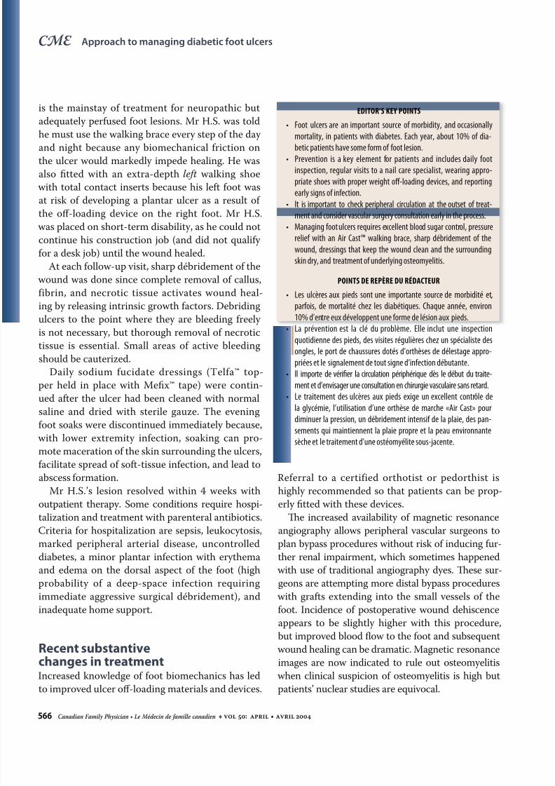
EDITOR’S KEY POINTS
• Foot ulcers are an important source of morbidity, and occasionally
mortality, in patients with diabetes. Each year, about 10% of dia-betic patients have some form of foot lesion.
• Prevention is a key element for patients and includes daily foot
inspection, regular visits to a nail care specialist, wearing appro-
priate shoes with proper weight off-loading devices, and reporting
early signs of infection.
• It is important to check peripheral circulation at the outset of treat-
ment and consider vascular surgery consultation early in the process.
• Managing foot ulcers requires excellent blood sugar control, pressure
relief with an Air Cast™ walking brace, sharp débridement of the
wound, dressings that keep the wound clean and the surrounding
skin dry, and treatment of underlying osteomyelitis.
POINTS DE REPÈRE DU RÉDACTEUR
• Les ulcères aux pieds sont une importante source de morbidité et,
parfois, de mortalité chez les diabétiques. Chaque année, environ
10% d’entre eux développent une forme de lésion aux pieds.
• La prévention est la clé du problème. Elle inclut une inspection
quotidienne des pieds, des visites régulières chez un spécialiste des
ongles, le port de chaussures dotés d’orthèses de délestage appro-
priées et le signalement de tout signe d’infection débutante.
• Il importe de vérifier la circulation périphérique dès le début du traite-
ment et d’envisager une consultation en chirurgie vasculaire sans retard.
• Le traitement des ulcères aux pieds exige un excellent contrôle de
la glycémie, l’utilisation d’une orthèse de marche «Air Cast» pour
diminuer la pression, un débridement intensif de la plaie, des pan-
sements qui maintiennent la plaie propre et la peau environnante
sèche et le traitement d’une ostéomyélite sous-jacente.

8/7/2019 Approach to managing diabetic foot ulcers
http://slidepdf.com/reader/full/approach-to-managing-diabetic-foot-ulcers 7/7