Embed Size (px)
Citation preview
1
Tzu-Fei Wang, MDAssistant Professor
Department of Internal MedicineDivision of Hematology
The Ohio State University Wexner Medical Center
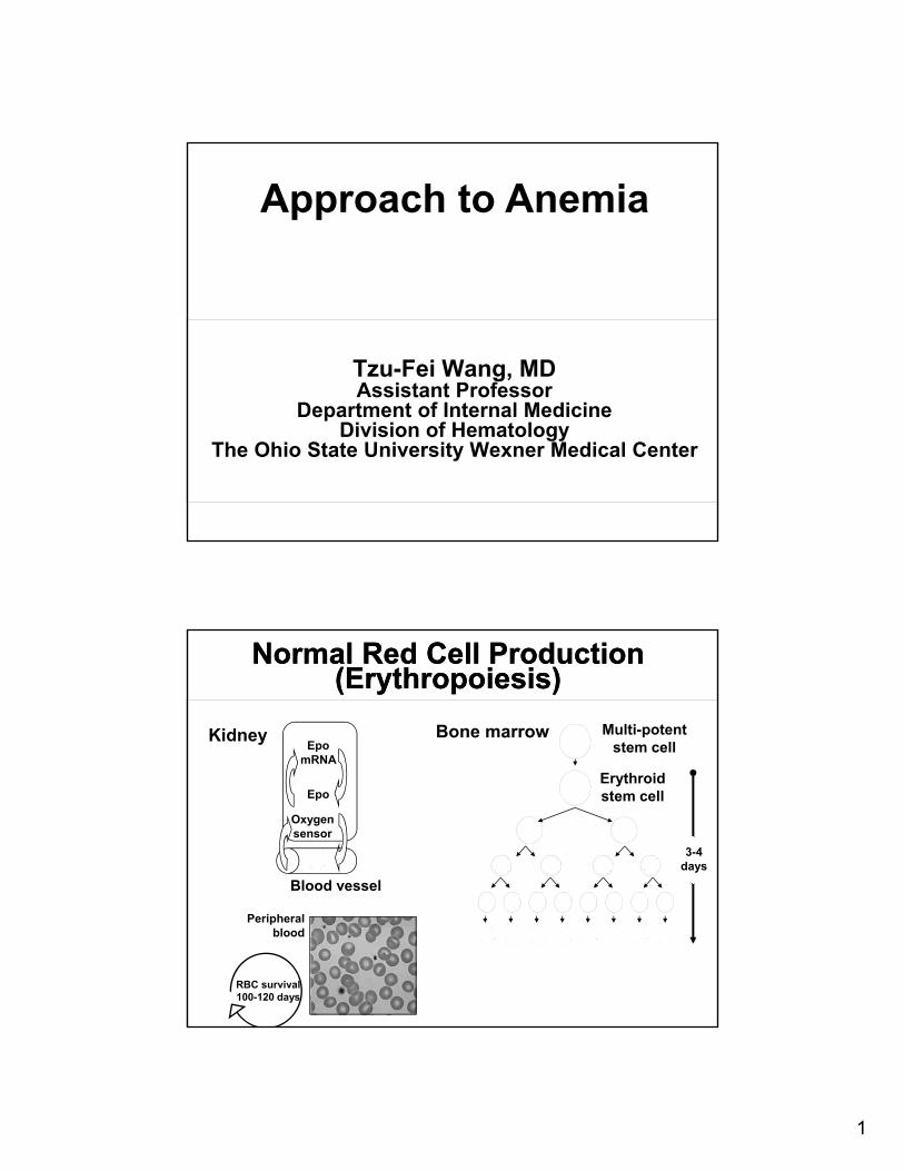
Approach to Anemia
Normal Red Cell Production (Erythropoiesis)
Normal Red Cell Production (Erythropoiesis)
Kidney
Oxygensensor
EpomRNA
Epo
Blood vessel
Bone marrow Multi-potentstem cell
Erythroidstem cell
3-4days
Peripheralblood
RBC survival100-120 days
2
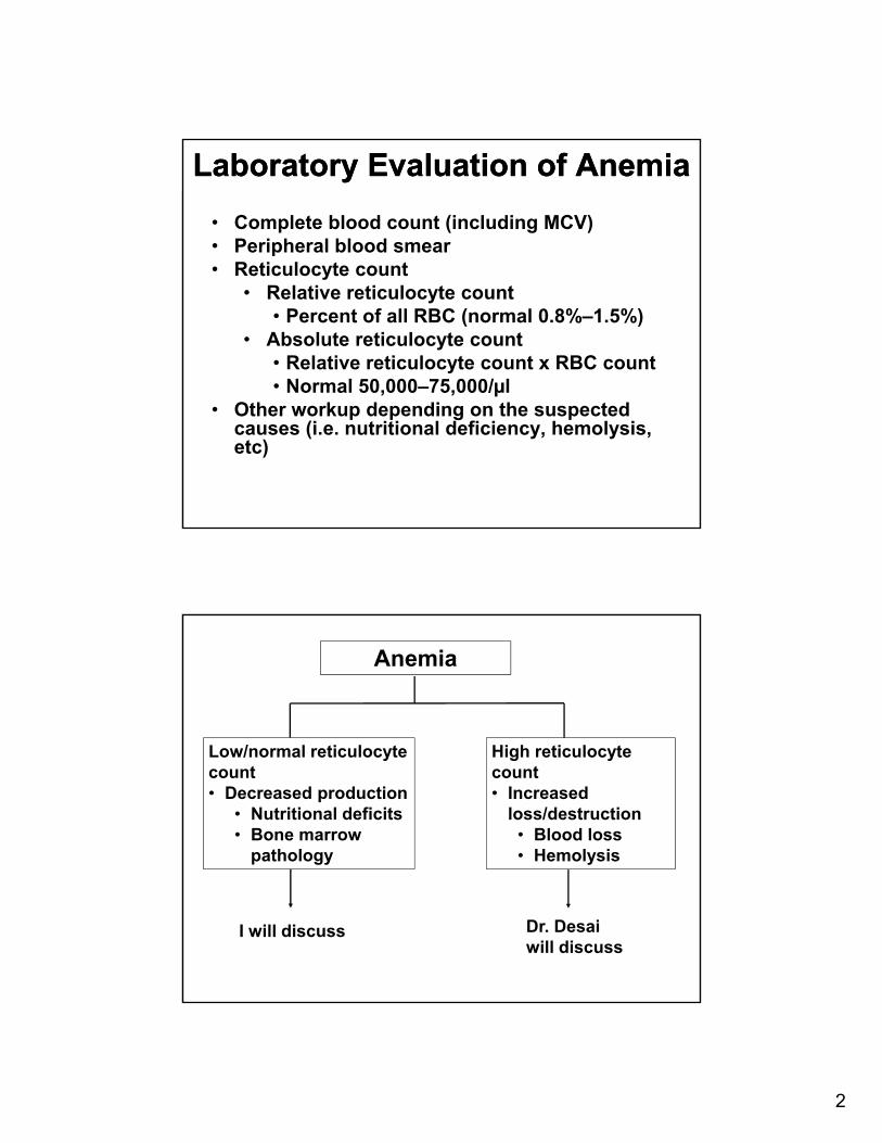
Laboratory Evaluation of AnemiaLaboratory Evaluation of Anemia
• Complete blood count (including MCV)• Peripheral blood smear• Reticulocyte count
• Relative reticulocyte count• Percent of all RBC (normal 0.8%–1.5%)
• Absolute reticulocyte count• Relative reticulocyte count x RBC count• Normal 50,000–75,000/µl
• Other workup depending on the suspected causes (i.e. nutritional deficiency, hemolysis, etc)
Anemia
Low/normal reticulocyte count• Decreased production
• Nutritional deficits• Bone marrow
pathology
High reticulocyte count• Increased
loss/destruction• Blood loss • Hemolysis
Dr. Desai will discuss
I will discuss
3
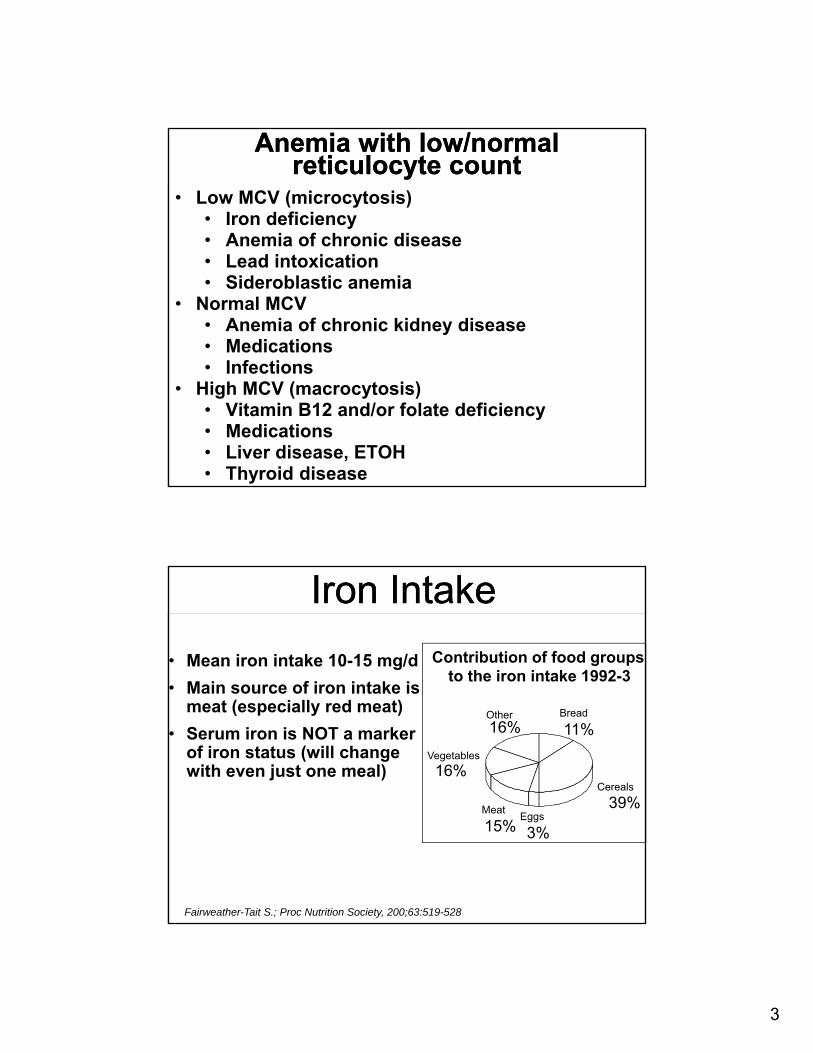
Anemia with low/normal reticulocyte count
Anemia with low/normal reticulocyte count
• Low MCV (microcytosis)• Iron deficiency• Anemia of chronic disease• Lead intoxication• Sideroblastic anemia
• Normal MCV• Anemia of chronic kidney disease• Medications• Infections
• High MCV (macrocytosis)• Vitamin B12 and/or folate deficiency• Medications• Liver disease, ETOH• Thyroid disease
Iron IntakeIron Intake
Fairweather-Tait S.; Proc Nutrition Society, 200;63:519-528
to the iron intake 1992-3
Bread11%
Cereals39%
Eggs3%
Meat15%
Vegetables16%
Other16%
Contribution of food groupsto the iron intake 1992-3
Bread
11%
Cereals
39%Eggs
3%
Meat
15%
Vegetables
16%
Other16%
• Mean iron intake 10-15 mg/d
• Main source of iron intake is meat (especially red meat)
• Serum iron is NOT a marker of iron status (will change with even just one meal)
4
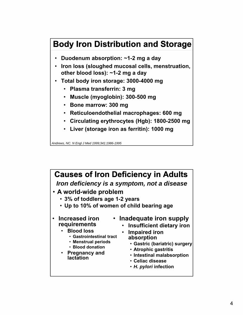
Body Iron Distribution and StorageBody Iron Distribution and Storage
• Duodenum absorption: ~1-2 mg a day
• Iron loss (sloughed mucosal cells, menstruation, other blood loss): ~1-2 mg a day
• Total body iron storage: 3000-4000 mg
• Plasma transferrin: 3 mg
• Muscle (myoglobin): 300-500 mg
• Bone marrow: 300 mg
• Reticuloendothelial macrophages: 600 mg
• Circulating erythrocytes (Hgb): 1800-2500 mg
• Liver (storage iron as ferritin): 1000 mg
Andrews, NC. N Engl J Med 1999;341:1986-1995
Causes of Iron Deficiency in AdultsCauses of Iron Deficiency in Adults
• Increased iron requirements• Blood loss
• Gastrointestinal tract• Menstrual periods• Blood donation
• Pregnancy and lactation
• Inadequate iron supply• Insufficient dietary iron• Impaired iron
absorption• Gastric (bariatric) surgery• Atrophic gastritis• Intestinal malabsorption• Celiac disease• H. pylori infection
Iron deficiency is a symptom, not a disease• A world-wide problem
• 3% of toddlers age 1-2 years• Up to 10% of women of child bearing age
5
Neurologic syndromes associated with iron deficiency
Neurologic syndromes associated with iron deficiency
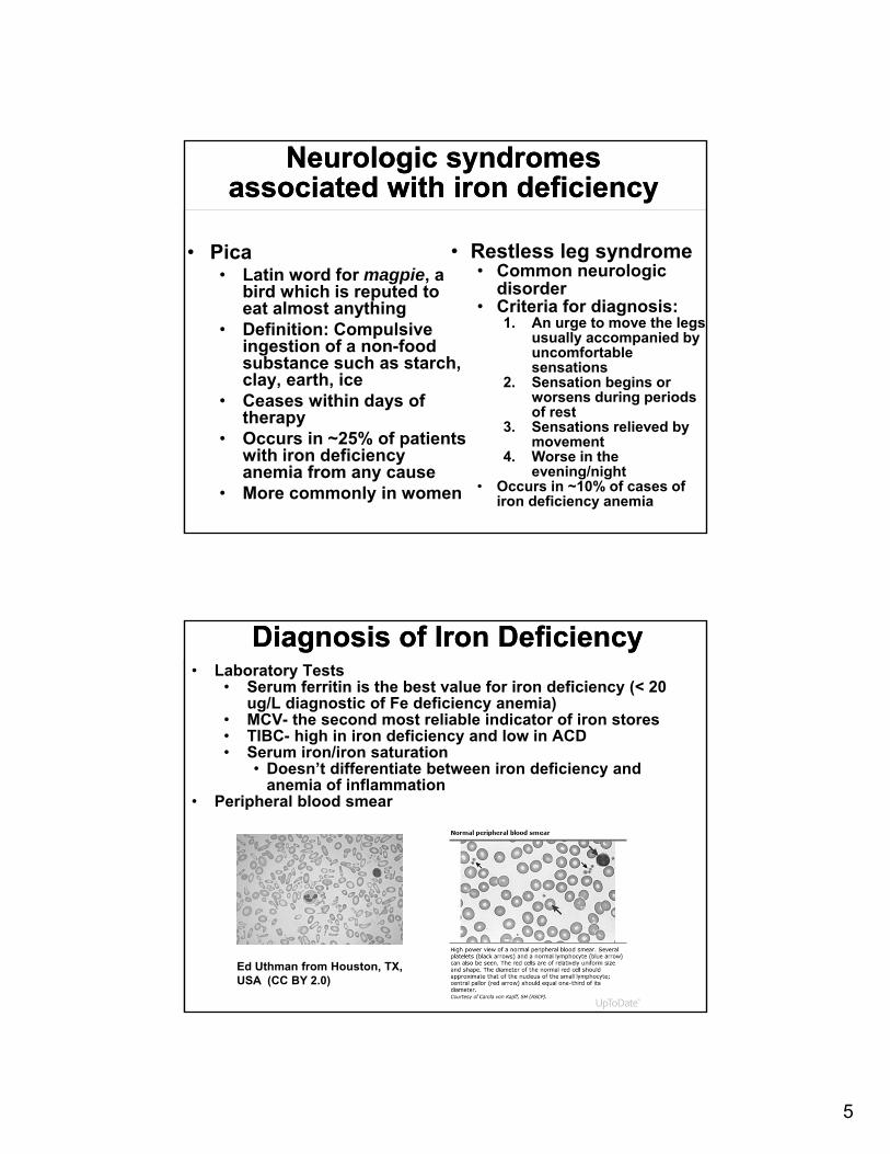
• Pica• Latin word for magpie, a
bird which is reputed to eat almost anything
• Definition: Compulsive ingestion of a non-food substance such as starch, clay, earth, ice
• Ceases within days of therapy
• Occurs in ~25% of patients with iron deficiency anemia from any cause
• More commonly in women
• Restless leg syndrome• Common neurologic
disorder• Criteria for diagnosis:
1. An urge to move the legs usually accompanied by uncomfortable sensations
2. Sensation begins or worsens during periods of rest
3. Sensations relieved by movement
4. Worse in the evening/night
• Occurs in ~10% of cases of iron deficiency anemia
Diagnosis of Iron DeficiencyDiagnosis of Iron Deficiency• Laboratory Tests
• Serum ferritin is the best value for iron deficiency (< 20 ug/L diagnostic of Fe deficiency anemia)
• MCV- the second most reliable indicator of iron stores• TIBC- high in iron deficiency and low in ACD• Serum iron/iron saturation
• Doesn’t differentiate between iron deficiency and anemia of inflammation
• Peripheral blood smear
Ed Uthman from Houston, TX, USA (CC BY 2.0)
6
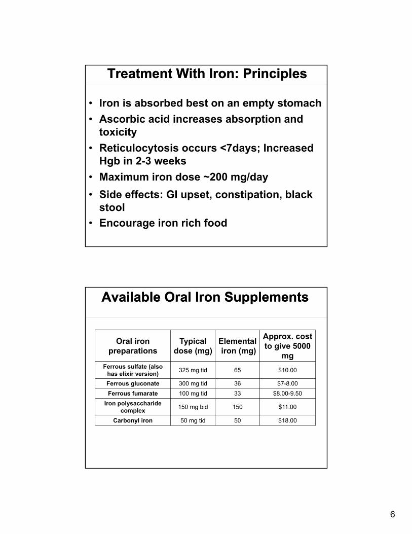
Treatment With Iron: PrinciplesTreatment With Iron: Principles
• Iron is absorbed best on an empty stomach
• Ascorbic acid increases absorption and toxicity
• Reticulocytosis occurs <7days; Increased Hgb in 2-3 weeks
• Maximum iron dose ~200 mg/day
• Side effects: GI upset, constipation, black stool
• Encourage iron rich food
Available Oral Iron SupplementsAvailable Oral Iron Supplements
Oral iron preparations
Typical dose (mg)
Elementaliron (mg)
Approx. cost to give 5000
mgFerrous sulfate (also
has elixir version)325 mg tid 65 $10.00
Ferrous gluconate 300 mg tid 36 $7-8.00
Ferrous fumarate 100 mg tid 33 $8.00-9.50
Iron polysaccharide complex
150 mg bid 150 $11.00
Carbonyl iron 50 mg tid 50 $18.00
7
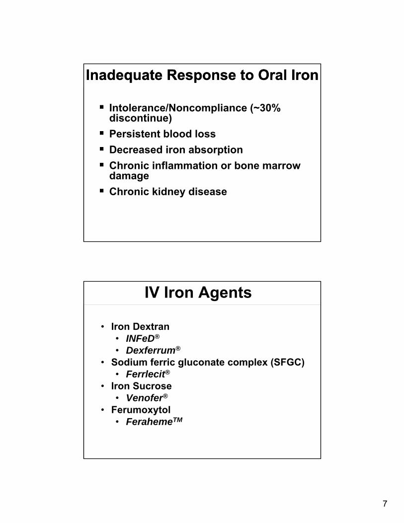
Inadequate Response to Oral IronInadequate Response to Oral Iron
Intolerance/Noncompliance (~30% discontinue)
Persistent blood loss
Decreased iron absorption
Chronic inflammation or bone marrow damage
Chronic kidney disease
IV Iron Agents
• Iron Dextran• INFeD®
• Dexferrum®
• Sodium ferric gluconate complex (SFGC)• Ferrlecit®
• Iron Sucrose• Venofer®
• Ferumoxytol• FerahemeTM
8
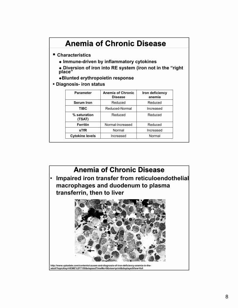
Anemia of Chronic DiseaseAnemia of Chronic Disease Characteristics
Immune-driven by inflammatory cytokines Diversion of iron into RE system (iron not in the “right place”Blunted erythropoietin response
Diagnosis- iron status
Parameter Anemia of Chronic Disease
Iron deficiency anemia
Serum Iron Reduced Reduced
TIBC Reduced-Normal Increased
% saturation (TSAT)
Reduced Reduced
Ferritin Normal-increased Reduced
sTfR Normal Increased
Cytokine levels Increased Normal
Anemia of Chronic DiseaseAnemia of Chronic Disease• Impaired iron transfer from reticuloendothelial
macrophages and duodenum to plasma transferrin, then to liver
http://www.uptodate.com/contents/causes-and-diagnosis-of-iron-deficiency-anemia-in-the-adult?topicKey=HEME%2F7150&elapsedTimeMs=0&view=print&displayedView=full
9
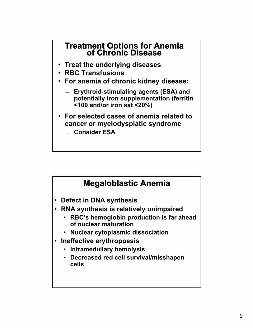
Treatment Options for Anemia of Chronic Disease
Treatment Options for Anemia of Chronic Disease
• Treat the underlying diseases• RBC Transfusions• For anemia of chronic kidney disease:
Erythroid-stimulating agents (ESA) and potentially iron supplementation (ferritin <100 and/or iron sat <20%)
• For selected cases of anemia related to cancer or myelodysplatic syndrome Consider ESA
Megaloblastic AnemiaMegaloblastic Anemia
• Defect in DNA synthesis• RNA synthesis is relatively unimpaired
• RBC’s hemoglobin production is far ahead of nuclear maturation
• Nuclear cytoplasmic dissociation• Ineffective erythropoesis
• Intramedullary hemolysis• Decreased red cell survival/misshapen
cells
10
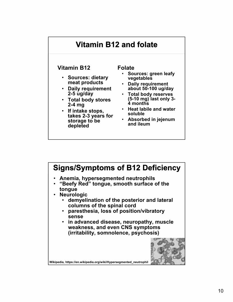
Vitamin B12 and folateVitamin B12 and folate
Vitamin B12
• Sources: dietary meat products
• Daily requirement 2-5 ug/day
• Total body stores 2-4 mg
• If intake stops, takes 2-3 years for storage to be depleted
Folate• Sources: green leafy
vegetables• Daily requirement
about 50-100 ug/day• Total body reserves
(5-10 mg) last only 3-4 months
• Heat labile and water soluble
• Absorbed in jejenum and ileum
Signs/Symptoms of B12 Deficiency Signs/Symptoms of B12 Deficiency • Anemia, hypersegmented neutrophils• “Beefy Red” tongue, smooth surface of the
tongue• Neurologic
• demyelination of the posterior and lateral columns of the spinal cord
• paresthesia, loss of position/vibratory sense
• in advanced disease, neuropathy, muscle weakness, and even CNS symptoms (irritability, somnolence, psychosis)
Wikipedia, https://en.wikipedia.org/wiki/Hypersegmented_neutrophil
11
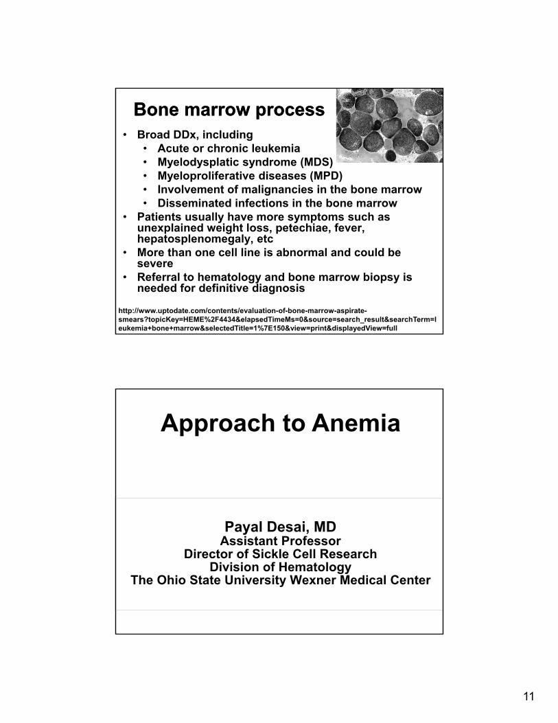
Bone marrow processBone marrow process• Broad DDx, including
• Acute or chronic leukemia• Myelodysplatic syndrome (MDS)• Myeloproliferative diseases (MPD)• Involvement of malignancies in the bone marrow• Disseminated infections in the bone marrow
• Patients usually have more symptoms such as unexplained weight loss, petechiae, fever, hepatosplenomegaly, etc
• More than one cell line is abnormal and could be severe
• Referral to hematology and bone marrow biopsy is needed for definitive diagnosis
http://www.uptodate.com/contents/evaluation-of-bone-marrow-aspirate-smears?topicKey=HEME%2F4434&elapsedTimeMs=0&source=search_result&searchTerm=leukemia+bone+marrow&selectedTitle=1%7E150&view=print&displayedView=full
Payal Desai, MDAssistant Professor
Director of Sickle Cell ResearchDivision of Hematology
The Ohio State University Wexner Medical Center
Approach to Anemia
12
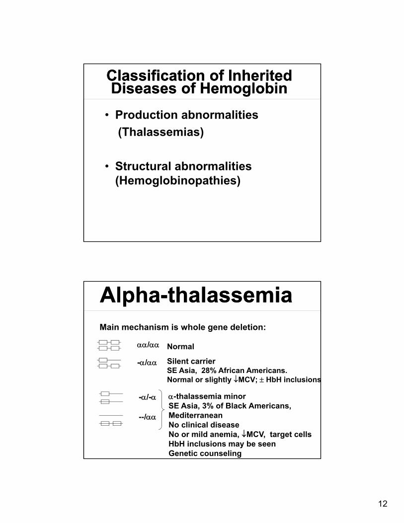
Classification of Inherited Diseases of Hemoglobin
Classification of Inherited Diseases of Hemoglobin
• Production abnormalities
(Thalassemias)
• Structural abnormalities (Hemoglobinopathies)
Alpha-thalassemiaAlpha-thalassemiaMain mechanism is whole gene deletion:
/ Normal
-/ Silent carrier SE Asia, 28% African Americans. Normal or slightly MCV; HbH inclusions
-/-
--/
-thalassemia minorSE Asia, 3% of Black Americans, MediterraneanNo clinical diseaseNo or mild anemia, MCV, target cellsHbH inclusions may be seenGenetic counseling
13
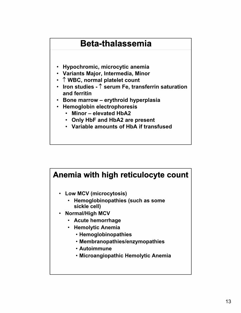
Beta-thalassemiaBeta-thalassemia
• Hypochromic, microcytic anemia• Variants Major, Intermedia, Minor• WBC, normal platelet count • Iron studies - serum Fe, transferrin saturation
and ferritin• Bone marrow – erythroid hyperplasia• Hemoglobin electrophoresis
• Minor – elevated HbA2• Only HbF and HbA2 are present• Variable amounts of HbA if transfused
Anemia with high reticulocyte countAnemia with high reticulocyte count
• Low MCV (microcytosis)• Hemoglobinopathies (such as some
sickle cell) • Normal/High MCV
• Acute hemorrhage• Hemolytic Anemia
• Hemoglobinopathies• Membranopathies/enzymopathies• Autoimmune• Microangiopathic Hemolytic Anemia
14
Unstable HemoglobinsUnstable Hemoglobins
• Rare disorders. Many variants described• Autosomal dominant only heterozygotes
exist (homozygous do not survive)
• Disrupt contact between heme and globin
• Alter amino acids at interface between and chains
• Alter the shape or structure of the globin molecule
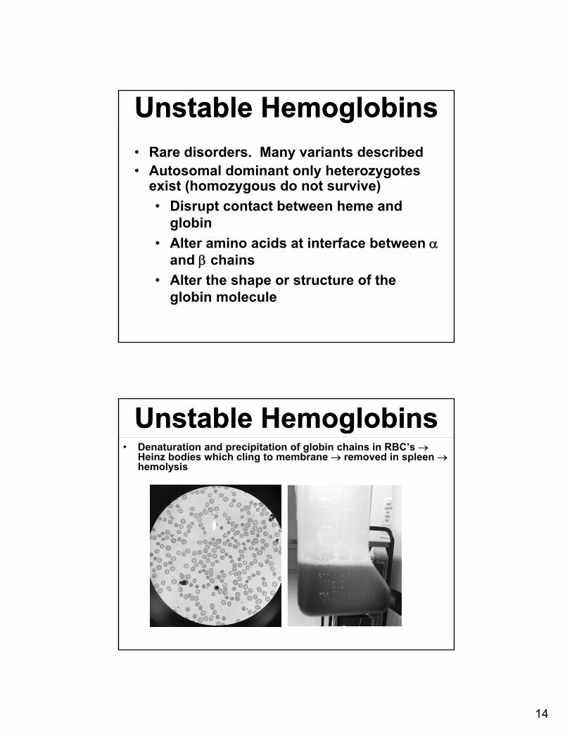
Unstable HemoglobinsUnstable Hemoglobins• Denaturation and precipitation of globin chains in RBC’s
Heinz bodies which cling to membrane removed in spleen hemolysis
15
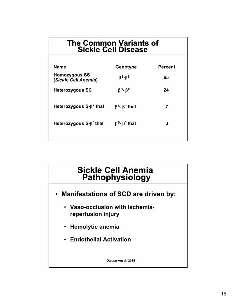
Name Genotype Percent
Homozygous SS(Sickle Cell Anemia) S-S 65
Heterozygous SC S- C 24
Heterozygous S-+ thal S- + thal 7
Heterozygous S-° thal S- ° thal 3
The Common Variants of Sickle Cell Disease
The Common Variants of Sickle Cell Disease
Sickle Cell Anemia Pathophysiology
Sickle Cell Anemia Pathophysiology
• Manifestations of SCD are driven by:
• Vaso-occlusion with ischemia-reperfusion injury
• Hemolytic anemia
• Endothelial Activation
Owusu-Ansah 2015
16
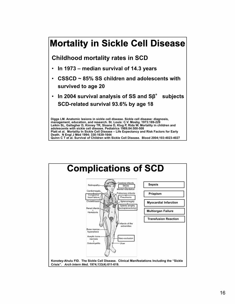
Mortality in Sickle Cell DiseaseMortality in Sickle Cell Disease
Childhood mortality rates in SCD
• In 1973 – median survival of 14.3 years
• CSSCD ~ 85% SS children and adolescents with
survived to age 20
• In 2004 survival analysis of SS and Sβ° subjects
SCD-related survival 93.6% by age 18
Diggs LM. Anatomic lesions in sickle cell disease. Sickle cell disease: diagnosis, management, education, and research. St. Louis: C.V. Mosby, 1973:189-229Leikin SL, Gallagher D, Kinney TR, Sloane D, Klug P, Rida W. Mortality in children and adolescents with sickle cell disease. Pediatrics 1989;84:500-508Platt et al. Mortality In Sickle Cell Disease – Life Expectancy and Risk Factors for Early Death. N Engl J Med 1994; 330:1639-1644Quinn C T et al. Survival of Children with Sickle Cell Disease. Blood 2004;103:4023-4027
Complications of SCDComplications of SCD
Konotey-Ahulu FID. The Sickle Cell Disease. Clinical Manifestations Including the “Sickle Crisis”. Arch Intern Med. 1974;133(4):611-619.
Sepsis
Multiorgan Failure
Myocardial Infarction
Priapism
Transfusion Reaction
17
Immune Hemolytic AnemiasImmune Hemolytic Anemias
• Autoimmune• Warm antibody-mediated• Cold antibody-mediated• Paroxysmal Cold Hemoglobinuria
• Drug-related hemolysis • Hemolytic transfusion reactions• Hemolytic disease of the newborn• Paroxysmal Nocturnal
Hemoglobinuria
Auto-Immune Hemolytic AnemiasAuto-Immune Hemolytic Anemias
• Antibodies causing hemolysis can be broken down into 2 general categories: warm and cold
• Warm antibodies react with RBCs best at 37° and typically do not agglutinate red cells
• Cold antibodies typically react best at <32°and do cause RBC agglutination
18
Coomb’s TestCoomb’s Test• The Direct Coomb’s = DAT (Direct
Antiglobulin Test) - tests for IgG or C3 DIRECTLY ON THE RED CELLS.
• The Indirect Coomb’s - tests for IgG or C3 in the serum which react with generic normal red cells. This is also known as the antibody screen in blood-banking.
Warm-Antibody Hemolytic Anemias Etiology
Warm-Antibody Hemolytic Anemias Etiology
• Primary or Secondary
• Drugs
• Solid or hematologic malignancy
• Infection
• Collagen Disease
• Pregnancy
• Can be associated with immune platelet destruction = Evan’s syndrome
19
Warm-Antibody Hemolytic AnemiasClinical Features
Warm-Antibody Hemolytic AnemiasClinical Features
• Splenomegaly, jaundice is usually present
• Depending on degree of anemia and rate of fall in hemoglobin, patients can have VERY symptomatic anemia
• Lab Dx -
• reticulocytes, bili, LDH, ↓haptoglobin
• Positive Coomb’s test - both direct and indirect
• Spherocytes are seen on the peripheral smear
Warm-Antibody Hemolytic Anemias Treatment
Warm-Antibody Hemolytic Anemias Treatment
• Patients may require red cell transfusions, if they are symptomatic with their anemia
• However, immunosuppression is the mainstay of therapy
• First Line – Steroids• 1mg/kg/d prednisone oral or
methlyprednisolone IV• Continue until Hb > 10g/dL then taper• Continue with Vitamin D, Ca, +
bisphosphonates• Consider PCP prophylaxis • Supplement with Folic Acid
Lechner et al. How I Treat Autoimmne Hemolytic Anemia. Blood 2010.Crowther et al. Evidence-based focused review of the treatment of idiopathic warm immune hemolytic anemia in adults. Blood 2011

20
WAIHA: TreatmentWAIHA: Treatment• Second Line Therapy
• 80% patient with CR or PR with prednisone
• 15-20% need higher than maintenance dose to stay in remission
• <20% of patients stay in remission after withdrawal of steroids
Lechner et al. How I Treat Autoimmne Hemolytic Anemia. Blood 2010.Crowther et al. Evidence-based focused review of the treatment of idiopathic warm immune hemolytic anemia in adults. Blood 2011
WAIHA: TreatmentWAIHA: Treatment• 2nd Line Therapy
• Splenectomy• Rituxan
• Other Therapies• Danazol• Cyclophosphamide• Cyclophosphamide• Mycophenolate Mofetil• Cyclosporine• Vincristine• Alemtuzumab• Ofatumumab
• Ineffective therapies
• Azathioprine
• BMT
• IVIG
• Plasma Exchange
Lechner et al. How I Treat Autoimmne Hemolytic Anemia. Blood 2010.Crowther et al. Evidence-based focused review of the treatment of idiopathic warm immune hemolytic anemia in adults. Blood 2011
21
Drug-Induced Immune HemolysisThree general mechanisms
Drug-Induced Immune HemolysisThree general mechanisms
• Innocent bystander
• Quinine, Quinidine, Isoniazide
• Hapten
• Penicillins, Cephalosporins
• True autoimmune
• Alpha-methyldopa, L-DOPA, Procainamide
Drug-Induced Positive Antiglobulin TestsDrug-Induced Positive Antiglobulin Tests
Mechanism DATSerum and
EluateNeoantigen
-Drug +RBC complex
C3 (sometimes IgG also) Serum reacts with rbcs only in the presence of drug; eluate non-reactive
Drug Adsorption (DA)
-Drug binds to RBC
IgG (sometimes C3 also) React with drug-coated RBCs but not untreated RBCs- Ab to drug
Autoantibodies
-WAIHA
IgG (rarely C3 also)
11-36% of pts
React with normal RBCs in absence of drug
22
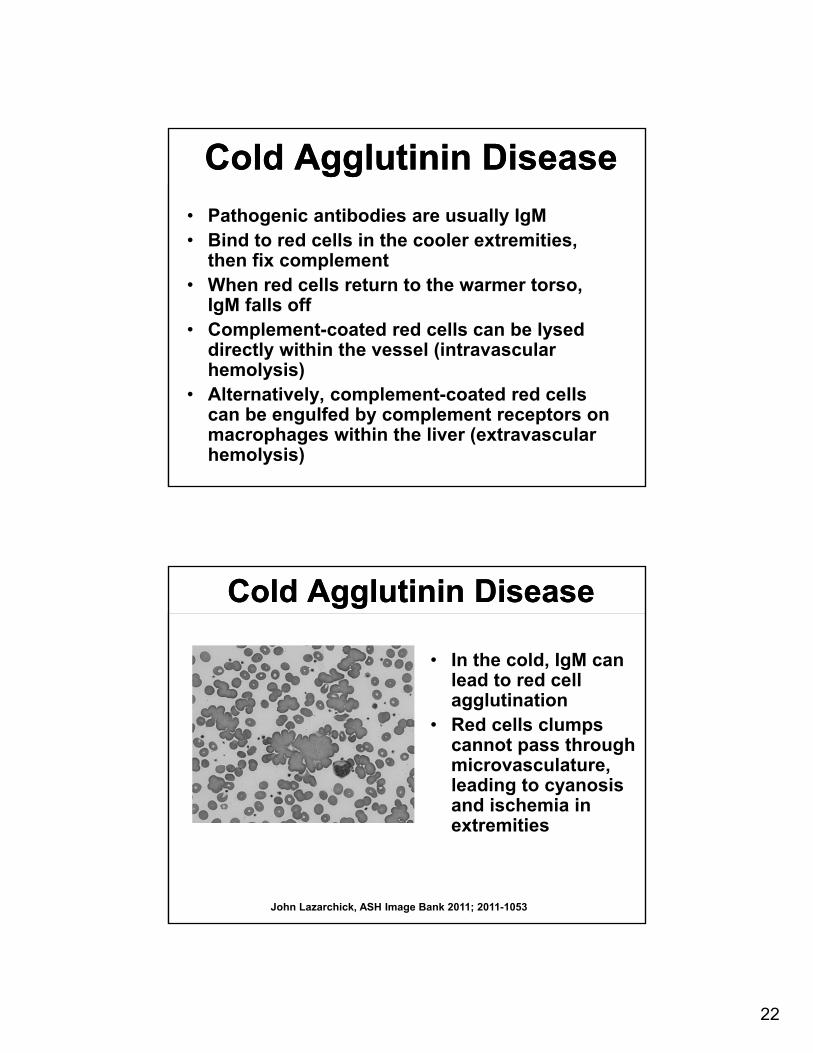
Cold Agglutinin DiseaseCold Agglutinin Disease
• Pathogenic antibodies are usually IgM• Bind to red cells in the cooler extremities,
then fix complement• When red cells return to the warmer torso,
IgM falls off• Complement-coated red cells can be lysed
directly within the vessel (intravascular hemolysis)
• Alternatively, complement-coated red cells can be engulfed by complement receptors on macrophages within the liver (extravascular hemolysis)
Cold Agglutinin DiseaseCold Agglutinin Disease
• In the cold, IgM can lead to red cell agglutination
• Red cells clumps cannot pass through microvasculature, leading to cyanosis and ischemia in extremities
John Lazarchick, ASH Image Bank 2011; 2011-1053
23
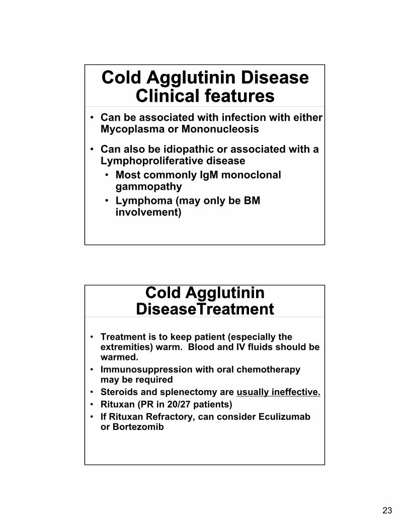
Cold Agglutinin DiseaseClinical features
Cold Agglutinin DiseaseClinical features
• Can be associated with infection with either Mycoplasma or Mononucleosis
• Can also be idiopathic or associated with a Lymphoproliferative disease• Most commonly IgM monoclonal
gammopathy• Lymphoma (may only be BM
involvement)
Cold Agglutinin DiseaseTreatment
Cold Agglutinin DiseaseTreatment
• Treatment is to keep patient (especially the extremities) warm. Blood and IV fluids should be warmed.
• Immunosuppression with oral chemotherapy may be required
• Steroids and splenectomy are usually ineffective.• Rituxan (PR in 20/27 patients)• If Rituxan Refractory, can consider Eculizumab
or Bortezomib
24
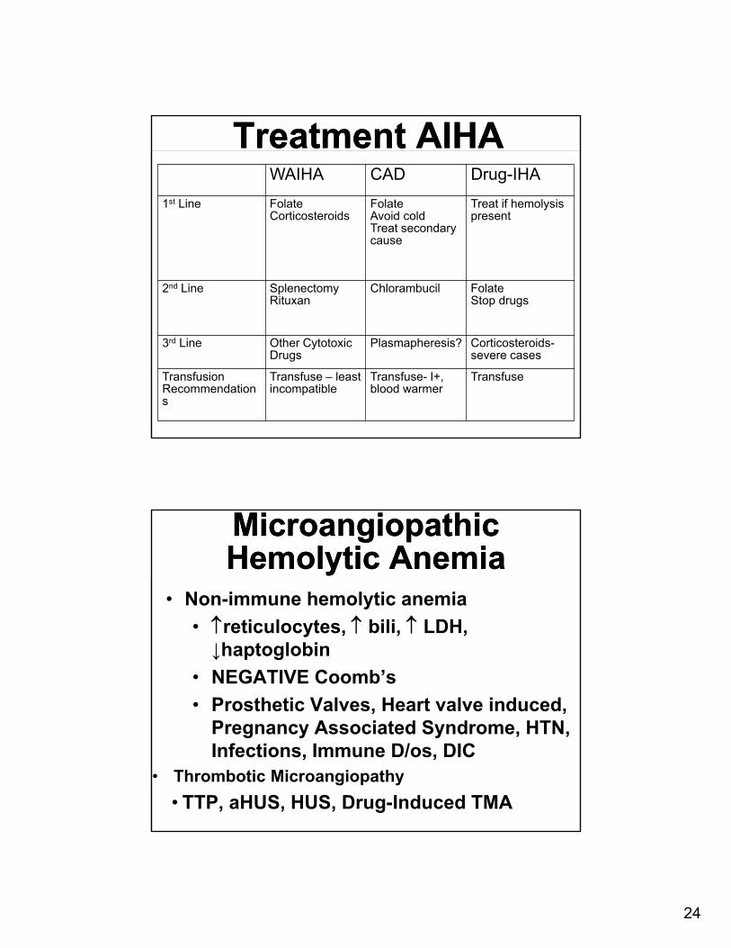
Treatment AIHATreatment AIHAWAIHA CAD Drug-IHA
1st Line FolateCorticosteroids
FolateAvoid coldTreat secondary cause
Treat if hemolysis present
2nd Line SplenectomyRituxan
Chlorambucil FolateStop drugs
3rd Line Other Cytotoxic Drugs
Plasmapheresis? Corticosteroids-severe cases
TransfusionRecommendations
Transfuse – least incompatible
Transfuse- I+, blood warmer
Transfuse
Microangiopathic Hemolytic AnemiaMicroangiopathic Hemolytic Anemia
• Non-immune hemolytic anemia
• reticulocytes, bili, LDH, ↓haptoglobin
• NEGATIVE Coomb’s
• Prosthetic Valves, Heart valve induced, Pregnancy Associated Syndrome, HTN, Infections, Immune D/os, DIC
• Thrombotic Microangiopathy
• TTP, aHUS, HUS, Drug-Induced TMA