Embed Size (px)
DESCRIPTION
CONSTITUTIONAL APLASTIC ANEMIA (FANCONİ ANEMIA) Characterized by defective DNA repair that is caused by a variety of genetic mutations Autosomal recessive Hematologic manifestations usually begin with thrombocytopenia or neutropenia Diagnosis is made between ages 2-10 years. - PowerPoint PPT Presentation
Citation preview
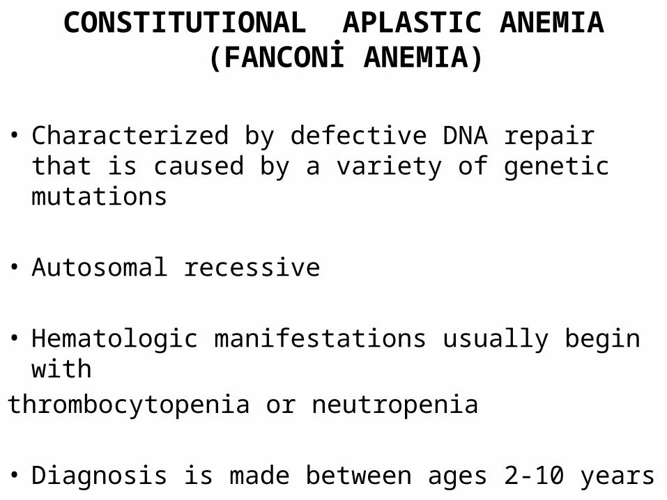
CONSTITUTIONAL APLASTIC ANEMIA(FANCONİ ANEMIA)
• Characterized by defective DNA repair that is caused by a variety of genetic mutations
• Autosomal recessive
• Hematologic manifestations usually begin withthrombocytopenia or neutropenia
• Diagnosis is made between ages 2-10 years
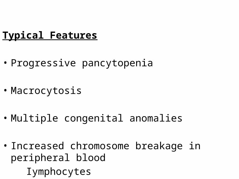
Typical Features
• Progressive pancytopenia
• Macrocytosis
• Multiple congenital anomalies
• Increased chromosome breakage in peripheral blood Iymphocytes
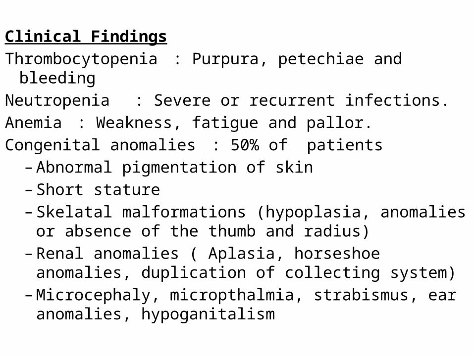
Clinical FindingsThrombocytopenia : Purpura, petechiae and bleedingNeutropenia : Severe or recurrent infections.Anemia : Weakness, fatigue and pallor.Congenital anomalies : 50% of patients
– Abnormal pigmentation of skin – Short stature– Skelatal malformations (hypoplasia, anomalies or
absence of the thumb and radius)– Renal anomalies ( Aplasia, horseshoe anomalies,
duplication of collecting system)– Microcephaly, micropthalmia, strabismus, ear
anomalies, hypoganitalism
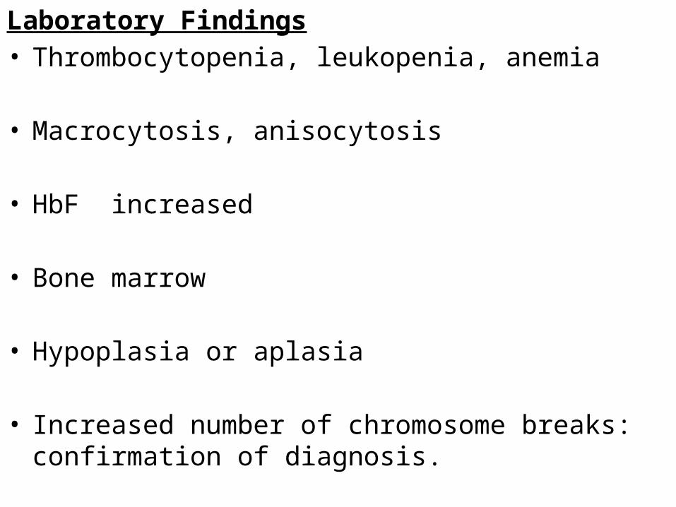
Laboratory Findings• Thrombocytopenia, leukopenia, anemia
• Macrocytosis, anisocytosis
• HbF increased
• Bone marrow
• Hypoplasia or aplasia
• Increased number of chromosome breaks: confirmation of diagnosis.
Differential diagnossis• ITP
• Acquired aplastic anemia
• Acute leukemia
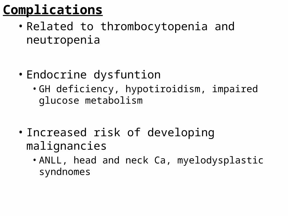
Complications• Related to thrombocytopenia and neutropenia
• Endocrine dysfuntion• GH deficiency, hypotiroidism, impaired glucose metabolism
• Increased risk of developing malignancies• ANLL, head and neck Ca, myelodysplastic syndnomes
• Treatment– Supportive care ( antibiotics, transfusion)– Oxymethalone (synthetic anabolic steroid),androgen
therapy: in complete response– Bone marrow transplantation: an important treatment
option for chilren
ACQUIRED APLASTIC ANEMIA• Characterized by peripheral pancytopenia with hypocelluar bone marrow• 50% of cases in childhood are idiopathic• Other cases : Secondary to idiosyncratic reactions to
– Drugs • Phenylbutazone• Sulfonamides• Nonsteroidol antiinflammatory drugs• Anticonvulsants
– Toxic causes• Benzene• Insecticides• Heavy metals
– Infectious Causes• Viral hapatitis• IM• HIV• Parvovirus B(19) Especially in immunocompromised children
Clinical Findings• Weakness, fatigue, pallor
– Anemia
• Petechiae,purpura,bleeding– Thrombocytopenia
• Fever, generalized,localized infection– Neutropenia– Hepatosplenomegaly and significant lymphadenopathy
are unusual
Laboratory Finding • Normocytic anemia
• Low reticulocyte count
• WBC count is low
• Platelet count below 50.000/mm3
• Bm aspiration and biopsy– Hypocellularity
Differential diagnosis– Acute leukemia– Storage disease Examination of BM– Myelofibrosis Presence of hepatosplenomegaly
*Newly diagnosed aplastic anemia should be studiedfor chromosome breaks for the diagnosis of Fanconi
anemia
Treatment• Supportive care ( antibiotics, RBC transfusion
• Platelet transfusion :many patients develop platelet alloantibodies and become refractory
• BM transplantation
• Antithymocyte globulin
• Cyclosporine
• Children receiving early BM transplantation from an
– HLA identical sibling have a long term survival rate of greater than 80%
– Complete remissions may be seen in 65-80 % of patients receiving immunosuppressive therapy
– Both therapies are associated with an increased risk of myelodysplastic syndromes, acute leukemia and other malignancies in long term survivors
CONGENITAL HYPOPLASTIC ANEMIA(Diamond Blackfan Anemia)
• Rare cause of anemia that usually presents in infancy or early childhood (birth to 1 year)
• Treatment with CST results in increased erythropoiesis in about 2/3 of patients ( early diagnosis is important )
• The cause is unclear, both autosomal dominant and autosomal recessive modes of inheritance occur
Clinical Findings
• Pallor, fatigue
• Congestive heart failure
• Short stature or other congenital anomalies are present in 1/3 of patients ( affecting head, face and thumbs)
Laboratory Findings• Macrocytic anemia
• Marked reticulocytopenia
• Platelet count is N/ ↑/↓
• Neutrophil count is usually N/ slightly ↓
• BM → marked decrease in erythroid precursors but is otherwise normal
• HbF is usually increased → (persistent of fetal erythropoesis )
• Adenosine deaminase in RBC is elevated
Differential Diagnosis
• Transient erythroblastopenia
• Renal failure
• Hypothyroidism
• Anemia of chronic disease
Treatment• Oral CST: ⅔ of patients respond to prednisone 2 mg/kg/d
• Transfusion:unresponsive patients
• BM transplantation:for transfusion dependent patients
• Spontaneous remissions occur in up to20% of patients
TRANSIENT ERYTHROBLASTOPENİA of CHILDHOOD• Common cause of acquired anemia in early
childhood
• Age : 6 months to 4 years
• Normocytic anemia with reticulocytopenia
• Erythroid precursors initially absent from bone marrow Anemia develops slowly, the cardiovascular system has time to compensate
• The disorder is thought to be autoimmune in most cases, because IgG from some patients has been shown to suppress erythropoiesis in vitro.
• Usually resolves within 6-8 weeks of diagnosis – Resolution of the anemia with reticulocytosis
• Not treated with CST because of its short course
Although there is an overlap in the age of presentation, Diamond-Blackfan syndrome commonly causes anemia during the first 6 months of life, whereas TEC occurs more frequently after age of 1 year.
The RBCs in patients with Diamond-Blackfan syndrome have fetal characteristics that are useful for distinguishing this disorder from TEC, including increased mean cell volume, elevated level of hemoglobin F, and presence of i antigen. The level of adenosine deaminase may be elevated in patients with Diamond-Blackfan syndrome but normal in children with TEC.
Twenty-five percent of white patients with Diamond-Blackfan anemia have been found to have mutations in the gene for ribosomal protein S19, and molecular diagnosis for these mutations is helpful when positive. Recently, additional gene mutations have been identified in Diamond Blackfan anemia.. In total, about three fourths of Diamond-Blackfan patients can be identified by mutational analysis
IRON DEFICIENCY ANEMIA• The most common cause of anemia in pediatrics• Iron deficiency has decreased substantially in incidence
due to improved nutrition and the increased availability of iron fortified infant formulas and cereals.
• Normal term infants are born with sufficient iron stores to prevent iron deficiency is most common between 6 and 24 months of life
• Deficiency earlier than 6 months of age may occur if iron stores at birth are reduced by prematurily, small birth weight, neonatal anemia, perinatal blood loss, hemorrhage
• Iron deficient children older than 24 months of age should be evaluated for the blood loss.
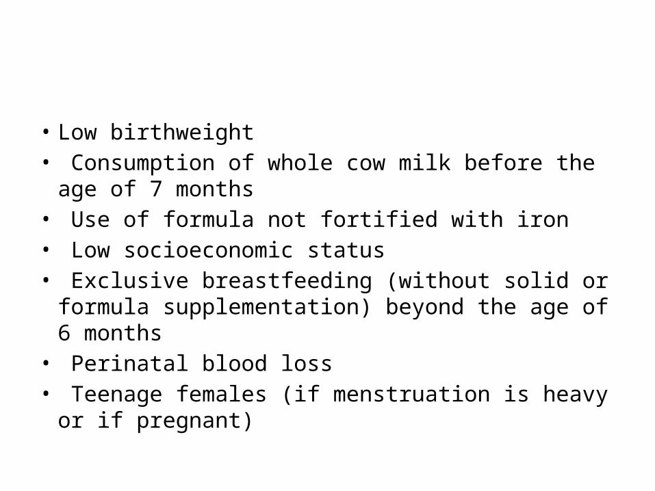
• Low birthweight• Consumption of whole cow milk before the age of 7
months• Use of formula not fortified with iron• Low socioeconomic status• Exclusive breastfeeding (without solid or formula
supplementation) beyond the age of 6 months• Perinatal blood loss• Teenage females (if menstruation is heavy or if
pregnant)
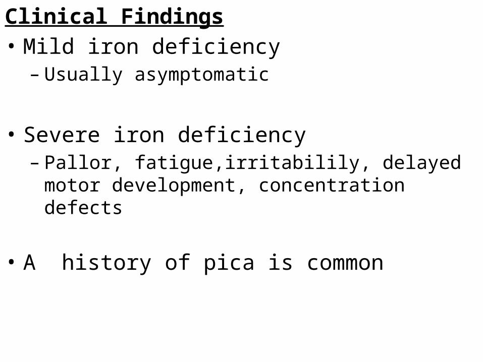
Clinical Findings• Mild iron deficiency
– Usually asymptomatic
• Severe iron deficiency – Pallor, fatigue,irritabilily, delayed motor development,
concentration defects
• A history of pica is common
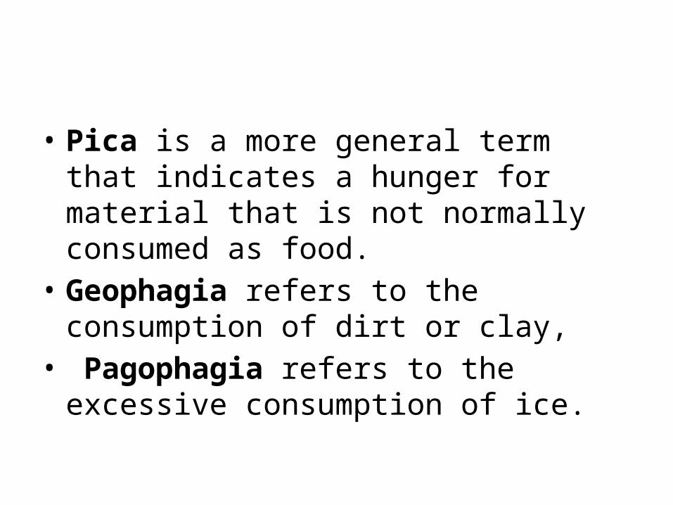
• Pica is a more general term that indicates a hunger for material that is not normally consumed as food.
• Geophagia refers to the consumption of dirt or clay,
• Pagophagia refers to the excessive consumption of ice.
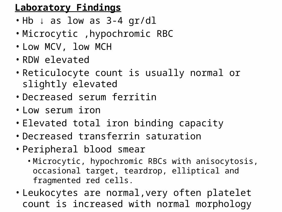
Laboratory Findings• Hb ↓ as low as 3-4 gr/dl• Microcytic ,hypochromic RBC• Low MCV, low MCH• RDW elevated• Reticulocyte count is usually normal or slightly elevated• Decreased serum ferritin• Low serum iron• Elevated total iron binding capacity• Decreased transferrin saturation• Peripheral blood smear
• Microcytic, hypochromic RBCs with anisocytosis, occasional target, teardrop, elliptical and fragmented red cells.
• Leukocytes are normal,very often platelet count is increased with normal morphology
Differential Diagnosis• Thalassemia
• Elevated number of RBC so Mentzer index < 13 (β th.minor) high HbA2
• Lead poisoning
• The anemia of chronic inflammation or infection in late stages
• Mild infections during infancy → ideally screening tests for anemia should not be obtained within 3-4 wks of such infections
Treatment
• Oral dose of elemental iron 6 mg/kg/d in 3 divided doses
– Results in an increased reticulocyte count within 3-5 days when the iron deficiency is the only cause of anemia, adequate treatment usually results in a resolution of anemia within 4-6 wks. Treatment is generally continued for a few additional months to replenish iron stores.
MEGALOBLASTIC ANEMIAS
• Megaloblastic anemia is a macrocytic anemia that is characterized by large RBC precursors(megaloblasts) in the bone marrow and that is usually caused by nutritional deficiencies of either folic acid or vitamin B12
• RBCs: Elevated MCH and mean cell volume (often 106 fl or more), with normal MCHC;marked variability in cell size (anisocytosis) and shape (poikilocytosis)
• Neutrophils: Hypersegmentation (>5% of neutrophils with five lobes or a single neutrophilwith six lobes)
• Platelets: Usually normal; thrombocytopenia in more severe anemia
MEGALOBLASTIC ANEMIAS• Macrocytic anemia caused by deficiency of B12, folicacid or
both• B12 deficiency due to dietary deficiency may occur in infants
who are breast fed by mothers who are strict vegeterians or who have pernisious anemia
• B12 deficiency due to intestinal malabsorbtion may occur in children with Crohn disease, chronic pancreatitis, bacterial ovengrowth of the small bowel,infection with the fish tapeworm ( Diphylobothrium latum) or after surgical resection of the terminal ileum
• B12 defeciency due to inborn errors of metabolism (transcobalamin deficiency and methylmalonic aciduria)
• Malabsorbtion of cobalamin due to deficiency of IF (pernicious anemia) is rare in chilhood.
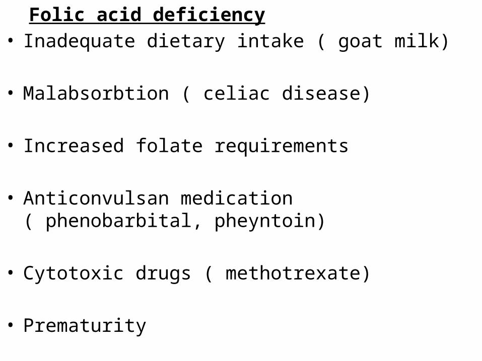
Folic acid deficiency• Inadequate dietary intake ( goat milk)
• Malabsorbtion ( celiac disease)
• Increased folate requirements
• Anticonvulsan medication ( phenobarbital, pheyntoin)
• Cytotoxic drugs ( methotrexate)
• Prematurity
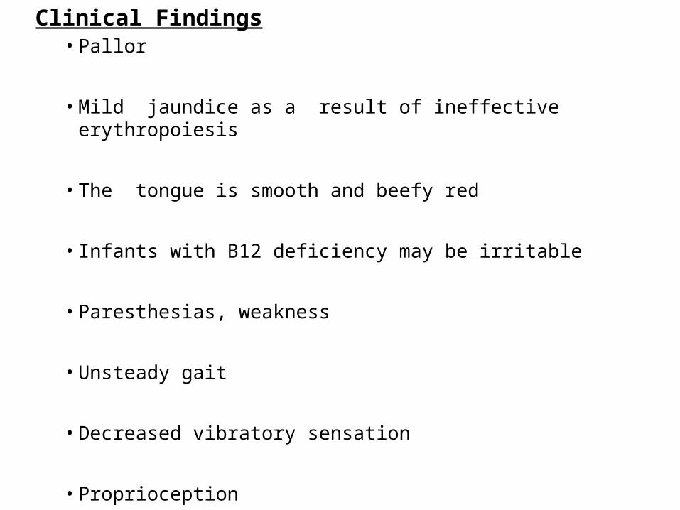
Clinical Findings• Pallor
• Mild jaundice as a result of ineffective erythropoiesis
• The tongue is smooth and beefy red
• Infants with B12 deficiency may be irritable
• Paresthesias, weakness
• Unsteady gait
• Decreased vibratory sensation
• Proprioception
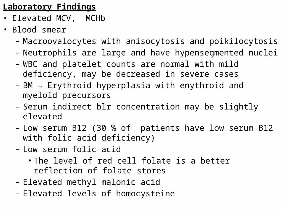
Laboratory Findings• Elevated MCV, MCHb• Blood smear
– Macroovalocytes with anisocytosis and poikilocytosis– Neutrophils are large and have hypensegmented nuclei– WBC and platelet counts are normal with mild deficiency, may be
decreased in severe cases– BM → Erythroid hyperplasia with enythroid and myeloid
precursors– Serum indirect blr concentration may be slightly elevated– Low serum B12 (30 % of patients have low serum B12 with folic
acid deficiency)– Low serum folic acid
• The level of red cell folate is a better reflection of folate stores– Elevated methyl malonic acid– Elevated levels of homocysteine
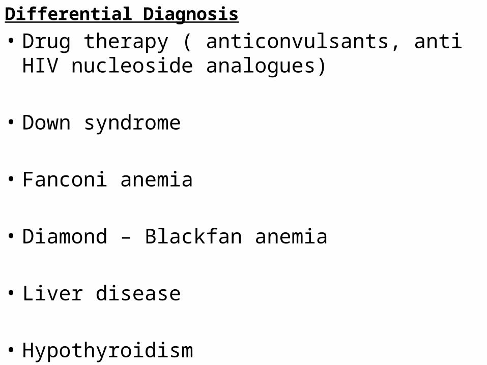
Differential Diagnosis• Drug therapy ( anticonvulsants, anti HIV nucleoside
analogues)
• Down syndrome
• Fanconi anemia
• Diamond – Blackfan anemia
• Liver disease
• Hypothyroidism
Treatment• Oral supplementation
• Parentenal treatment
* Children at risk for the development of folic acid deficiencies such as premature infants and those with chronic hemolysis are often given folic acid prophylactically
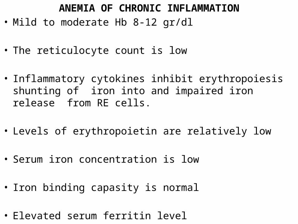
ANEMIA OF CHRONIC INFLAMMATION• Mild to moderate Hb 8-12 gr/dl
• The reticulocyte count is low
• Inflammatory cytokines inhibit erythropoiesis shunting of iron into and impaired iron release from RE cells.
• Levels of erythropoietin are relatively low
• Serum iron concentration is low
• Iron binding capasity is normal
• Elevated serum ferritin level
• Chronic infection and other inflammatory states impair the release of iron from reticuloendothelialcells, thereby decreasing the amount of this necessary ingredient that are available for RBCproduction.
• The lack of mobilizable iron may be the result of the action of proinflammatorycytokines (e.g., interleukin-1, tumor necrosis factor-a).
• Giving additional iron under these circumstances further increases reticuloendothelial iron stores and does little to help the anemia.
• Acute infection may cause anemia through a variety of mechanisms, including bone marrow suppression, shortened RBC life span, RBC fragmentation, and immune-mediated RBC destruction
ANEMIA of CHRONIC RENAL FAILURE• Normocytic anemia
• BM → significant hypoplasia of the erythroid series
• Reticulocyte count is low
• Deficiency of erytropoetin– Recombinant human erythropoietin corrects the anemia
ANEMIA of HYPOTHYROIDISM
• Normocytic / macrocytic ( not megaloblastic)
• Replacement therapy with thyroid hormone is usually efective in correcting the anemia