Embed Size (px)
Citation preview

1
Applications of nanoparticles in topical drug
delivery and in cosmetics
Xiao Wu1,2 and Richard H. Guy1,3
1Department of Pharmacy & Pharmacology
University of Bath
Claverton Down
Bath, BA2 7AY, UK
2Present address: University of Kentucky, College of Pharmacy, Lexington, KY 40536-0082,
U.S.A.
3Correspondence: Address as above. Email: [email protected] Tel. +44-1225-384901
Abstract
The delivery of drugs and active agents to the skin by formulations containing
nanoparticles is a topic of considerable current interest. A number of studies have shown
important advantages of these nanostructure-based delivery systems over conventional
formulations. This review describes the composition, preparation, and characteristics of a
wide range of novel vectors, including nanoemulsions, liposomes, transfersomes, solid lipid
nanoparticles, polymeric nanoparticles, ethosomes and niosomes.
Keywords: Nanoparticles, skin, stratum corneum, cosmetics, pharmaceutics
Acknowledgements: Supported by the European Commission 6th Research and
Technological Development Framework Programme (NAPOLEON: NAnostructured
waterborne POLymEr films with OutstaNding properties) and a University Research
Scholarship for Xiao Wu.

2
1 Introduction
The skin is the largest organ in the human body by weight, contributing about 10% of total
weight, and covering an average area of 1.7 m2. It regulates water and heat loss, and prevents
the invasion of noxious chemicals and microorganisms. Because skin is an easily accessible
organ, its potential as an alternative route for administering drugs for both systemic and local
effect has attracted considerable interest [1]. Equally, a large segment of the cosmetic industry
is focused on the delivery of “actives” to and into the skin. However, molecules do not easily
penetrate the skin because of its excellent barrier function. As a result, various nano-carriers
have been developed in an attempt to reversibly modulate the skin barrier and/or to provide
novel delivery systems for the active of interest. These particulate carriers include
nanoemulsions, liposomes, transfersomes, solid lipid nanoparticles, polymeric nanoparticles,
ethosomes and niosomes. A schematic representation of their structures is shown in Figure 1.
3
2 Skin
2.1 The structure of the skin
The skin consists of the epidermis and the dermis, which sits on a layer of subcutaneous
fat.
The epidermis contains four histologically distinct layers, from the innermost stratum
basale via the stratum spinosum and stratum granulosum (SG) to the superficial stratum
corneum (SC). The SC has been represented as a “brick and mortar” structure [2] in which the
corneocytes are embedded in an intercellular lipid matrix. The corneocytes comprise insoluble
keratins enveloped by cross-linked proteins, and are arranged in parallel, overlapping,
multicellular stacks perpendicular to the skin surface [1]. The inter-corneocyte space is filled
with lipids, usually present in the crystalline phase [3]. Most SC lipids are synthesized in the
viable epidermis during differentiation [4], they are released into the intercellular spaces at the
SG-SC interface from lamellar bodies. The major SC lipids are ceramides, fatty acids and
cholesterol. Eight classes of ceramides have been identified. The lipids are arranged in
multiple bilayers with a periodicity of about 13 nm. Unlike almost all other membranes in the
body, the SC does not contain phospholipid [2]. This “brick and mortar” structure is now
accepted as the location of the skin’s excellent permeability barrier, and the SC is the
rate-limiting barrier to the transcutaneous penetration and absorption of most chemicals
following topical administration [5].
There are three possible pathways of molecular penetration across the SC: (i) intercellular
via the lipids between the corneocytes; (ii) transcellular crossing through the corneocytes and
the surrounding lipids; (iii) appendagal via follicles and sweat ducts (Figure 2) [6]. The
principal route is generally believed to be intercellular. The principal route is generally
believed to be intercellular, although the appendageal route, in particular that encompassing
the hair follicle and associated sebaceous gland, is also important in certain circumstances
[7-12] and may offer an opportunity for drug targeting in the treatment of hair loss or acne,
for example [13-16]. Some permeation enhancers (e.g., oleic acid) and vesicular carriers are
thought to disorder the SC lipids and facilitate transport across the skin [17-19].
Underlying the SC is the viable epidermis, the thickness of which is typically ~100 µm,
4
ranging from as little as 50 µm to around 800 µm on the load-bearing palms and soles of the
feet [1]. The principal cells of the viable epidermis are keratinocytes, but there are also
melanocytes, Langerhans cells, migrant macrophages and lymphocytes [20]. However, there
are no blood vessels in the epidermis.
The dermis is typically 3–5 mm thick and is the major component of human skin. It is rich
in blood vessels, lymphatic vessels and nerve endings. The skin appendages, which include
hair follicles, sebaceous glands and sweat glands, also originate in the dermis. This layer
resembles an aqueous gel and is a minimal barrier to drug transport. As mentioned above, the
hair follicles and associated sebaceous glands are considered to play a role with respect to
transport across the skin; given that sebum consists mostly of neutral, non-polar lipids, it may
be anticipated that this route favours more lipophilic permeants.
The subcutaneous fat layer bridges between the dermis to the underlying tissue, and plays
a negligible role in the percutaneous absorption of topically applied substances.
2.2 Techniques to evaluate nanoparticle disposition on and within the skin
To evaluate nanoparticle penetration disposition on/ and/or within the skin, several
methods have been employed, including Franz-type diffusion cell experiments, differential
tape-stripping of the stratum corneum [21,22], laser scanning confocal microscopy (LSCM)
[23-27], multi-photon fluorescence microscopy (MFM) [28], and transmission electron
microscopy (TEM) [29]. These techniques have permitted qualitative and semi-quantitative
deductions about the extent and mechanisms of nanoparticle uptake into the stratum corneum
to be deduced.
5
3 Nanoemulsions
3.1 Composition and preparation
Nanoemulsions are stable dispersions with mean droplet diameters of a few hundred
nanometers, and are sometimes called sub-micron or mini-emulsions. These systems are
composed of oil, water, and one or more surface-active agents, and may be oil-in-water (o/w)
or water-in-oil (w/o) dispersions [30]. In some circumstances, nanoemulsions may be formed
using phospholipids as one of the surface-active constituents; if the level of lipid is high, the
concurrent formulation of liposomes is possible [31]. The aqueous phase may contain
hydrophilic, pharmaceutical or cosmetic active ingredients and preservatives, while the oil
phase is typically composed of mineral oil, silicone oil, vegetable oil, esters of fatty acids,
and/or lipophilic active ingredients. Surfactants, such as disodium stearoyl glutamate, sucrose
alkyl ester, sorbitan alkyl ester and dimethicone copolyol, are added to the formulation to
allow formation of a stable dispersion and guarantee an appropriate shelf life of the product.
Both o/w and w/o nanoemulsions can be used in pharmaceutical preparations for topical
administration. In the former case, the common oil core constituents are triglycerides,
propylene glycol mono caprylic ester, cholesteryl esters and cholesterol [32]. Most
nanoemulsions in cosmetics are oil-in-water and contain 10-20% oil stabilized with 0.5-2%
emulsifying agent. Popular oils are triglycerides, silicones, isopropyl myristate, isocetyl
isostearate and isododecane. As well as conventional surfactants, polymeric emulsifiers, such
as carbomers and hydroxypropyl methylcellulose (HPMC), are also being used to produce
stable products with a pleasant appearance [33, 34]. Carbomers are crosslinked polyacrylic
acid polymers; and represent the most widely used thickening agents in skincare products [35].
Once introduced into a nanoemulsion, irregular structures on the order of a micron in size are
observed (Figure 3), in proportion to the amount of carbomer employed. Carbomers can form
a thick protective gel layer around each oil droplet and increase the viscosity of the external
phase. Following contact with the skin, electrolytes from the skin surface cause the protective
gel layer to deswell instantly. The oil phase is released and a thin film is deposited on the skin.
This mechanism permits the convenient formulation of sun-care products which are
6
ultimately waterproof despite their predominantly hydrophilic properties prior to application.
The emulsification mechanism of HPMC is similar to that of carbomer, although the former is
less sensitive to the presence of electrolytes. It is believed that the mechanical stress, imparted
on application of these emulsions, causes a partial breakdown of their structures such that a
thin film of oil spreads over the skin surface, reducing its wettability. After the water has
evaporated, a flexible film remains consisting of oil droplets embedded into the polymer
matrix [33].
Nanoemulsions are easily produced in large quantities by mixing a water-immiscible oil
phase into an aqueous phase using a high-stress, mechanical extrusion process [36, 37].
3.2 Applications of nanoemulsions in pharmaceutics
An o/w nanoemulsion containing 10% (m/m) oil (propylene glycol mono-caprylic ester
and glycerol triacetate), 50% (m/m) surfactant (diethylene glycol monoethyl ether and
Tween-80) and 40% (m/m) water has been suggested as a vehicle for the improved
transdermal delivery of celecoxib [38]. In vitro skin permeation studies showed enhanced
percutaneous uptake of the drug from the nanoemulsion relative to a simple gel [38]. In vivo,
inhibition of carrageenan-induced paw edema in rats was observed when celecoxib
nanoemulsion was used. The ability of nanoemulsion formulations to enhance topical drug
delivery has also been shown for ketoprofen [39]. It was claimed that the drug permeation
rate could be manipulated by changing the relative amounts of oil, surfactants and
co-surfactants. Similarly, another study with an aceclofenac nanoemulsion showed improved
permeation of the drug into rat abdominal skin, and significantly increased anti-inflammatory
effect on carrageenan-induced paw edema in rats in vivo, when compared with a gel
formulation [40].
As well as acting as a drug carrier, nanoemulsions themselves have extensive antimicrobial
activity against bacteria (e.g., E. coli, Salmonella, and S. aureus), viruses (e.g., HIV, Herpes
simplex), fungi (e.g., Candida, Dermatophytes), protozoa and spores (e.g., anthrax) due to
their ability to fuse with and lyse these different organisms. Fusion is primarily driven by the
electrostatic attraction between the typically cationic charge of the emulsion and the anionic
charge on the pathogen. There is one example of the antimicrobial effects of surfactant
7
nanoemulsions. A w/o nanoemulsion (X8W60PC), containing oil (64%), detergents (9.7%),
solvent (8%) and water (18.3%), at a low concentration of 1%, significantly reduced the
number of colony forming units (CFU) of Candida. albicans by more than four logs within 15
minutes of treatment, and by six logs in a two-hour exposure. Because some of the ingredients
of X8W60PC are biocidal, the anti-fungal activity of the individual ingredients was also
evaluated at concentrations equivalent to those in X8W60PC. However, none of the
constituents was as effective a fungicidal as the nanoemulsion (Figure 4), suggesting that the
activity of X8W60PC depends upon its nanoemulsion structure [41].
A special type of topical nanoemulsion has been developed for the instillation of an
adrenergic β-blocking agent, adaprolol maleate, into the eye, to treat glaucoma without
inducing systemic side-effects. However, the drug has an important clinical disadvantage of
irritation to the eye, causing an immediate burning sensation and local discomfort. A
nanoemulsion formulation, on the other hand, maintains the therapeutic efficacy of the drug
while reducing the level of ocular irritation [32].
3.3 Applications of nanoemulsions in cosmetics
Nanoemulsions can be found in a wide variety of cosmetic products such as bath oils, body
creams, anti-wrinkle and anti-aging preparations. Due to their small and uniform droplet size,
nanoemulsions are transparent, fluid and pleasant to touch [42, 43]. In comparison to
traditional emulsions, nanoemulsions have better spreading properties on the skin. These
unique texture and rheological properties make them very valuable in cosmetic technology.
Formulations containing nanoemulsions range from water-like fluids to semi-solid gels.
Numerous patents reflect the active development of nanoemulsion formulations. For
example, nanoemulsions containing fluid, non-ionic, amphiphilic lipids, such as diglyceryl
isostearate, sorbitan oleate, and α-butylglucoside caprate, were stable on storage between 0°C
and 45°C [44], were able to contain significant amounts of fragrance, and promoted the
penetration of “actives” into the surface layers of the skin. Nanoemulsions made with anionic,
amphiphilic lipids of phosphoric acid fatty esters and oxyethylenated derivatives also retained
transparency and good cosmetic properties even when large amounts of oil were added to the
formulations [45]. Another o/w nanoemulsion based on one or more nonionic and/or anionic
8
amphiphilic lipids, and one or more water-soluble neutral polymers (e.g. poly-(ethylene
oxide), polyvinyl alcohols; polyvinylcaprolactam), allowed the viscosity of the composition
to be increased without influencing its transparency or increasing the level of the oil phase
[46]. A stable and translucent nanoemulsion for cosmetic, dermatological and/or
ophthalmological applications comprised a ternary surfactant system of ethoxylated fatty ester
polymer, fatty acid ester of sorbitan and alkali metal salts of cetyl phosphate or palmitoyl
sarcosinate, did not require gelling agents for stabilization [47], making it suitable for use on
sensitive skin. Several other nanoemulsion technologies have been developed for diverse
properties, including sun-protection, anti-wrinkling, anti-aging of the skin and other cosmetic
targets [48-52].
3.4 Summary
Nanoemulsions consist of fine o/w or w/o dispersions. They are used in dermatology to
improve drug delivery to and through the stratum corneum. In addition, nanoemulsions
themselves are biocidal towards bacteria, viruses, fungi, protozoa and spores. In cosmetics,
nanoemulsions have found use in many cosmetic products for their transparent visual aspect,
hydrating power and good skin feel.
9
4 Liposomes
4.1 Composition and preparation
Liposomes are spherical vesicles consisting of one or more membrane-like phospholipid
bilayers enclosing an aqueous core. Vesicle diameter ranges from fifty to several hundred
nanometers. The principal lipid component of liposomes is typically phosphatidylcholine (PC)
derived from egg or soybean lecithin [53]. Cholesterol is usually included in the composition
to stabilize the structure thereby generating more rigid liposomes [54]. Depending on the
processing conditions and the chemical composition, small unilamellar vesicles (SUV), large
unilamellar vesicles (LUV), large multilamellar vesicles (MLV) and multivesicular vesicles
(MVV) may be formed with one or several concentric bilayers (Figure 5). Unlike emulsions,
liposomes are thermodynamically stable lamellar structures which form spontaneously when
lipid is brought into contact with an aqueous phase [55].
Liposomes can be prepared by a number of methods. The major techniques are lipid film
hydration, emulsification, reverse phase evaporation, freeze-thaw processes, and solvent
injection. Large liposomes form spontaneously when phospholipids are dispersed in water
above their phase transition temperature. To prepare small vesicles, an appropriate technique,
such as high-pressure homogenization, sonication, or extrusion, is required to reduce particle
size. It should be stated that most of the methods for preparing liposomes have at least one of
the following drawbacks: use of large quantities of solvent, need for special equipment, and
low “active” encapsulation efficacy.
Due to their biphasic character, liposomes can act as carriers for lipophilic, amphiphilic
and hydrophilic substances. The entrapped compound’s solubility and partitioning
characteristics will determine its location in the liposomal bilayer, its level of association with
the liposome and its release rate. In general, lipophilic and amphiphilic substances, e.g.,
oil-soluble UV filters, are located in the lipid bilayer of the liposome. As such compounds are
very poorly water-soluble, loss of entrapped drug on storage is minimal. Hydrophilic drugs
are entrapped inside the aqueous core of liposomes, but may also be in the external water
phase. The percentage of encapsulated hydrophilic drug depends on the liposome bilayer
10
composition and the preparation procedure.
4.2 Applications of liposomes in pharmaceutics
The first report of the use of liposomes in topical drug delivery involved delivery of
triamcinolone acetonide. It was claimed that the liposomal formulation significantly increased
the concentration of steroid achieved in the epidermis and dermis [56]. In a further study, it
was reported that application of triamcinolone acetonide-loaded liposomes resulted in ~5-fold
more drug accumulation within the epidermis in comparison to a more conventional gel
formulation [57]. A number of other studies have implied the efficiency of liposome
formulations to deliver enhanced drug amounts to the upper skin layers. For example, a
liposome formulation of betamethasone dipropionate out-performed a commercial
conventional formulation containing a higher concentration of drug in a clinical trial
considering the treatment of atopic eczema [58]. The liposome formulation was less efficient
in treating psoriasis for which deeper penetration of the drug is needed. Econazole, topical
antifungal drug, can be irritant to the skin when topically applied in conventional vehicles.
The application of liposome formulation has been shown to be a good strategy to minimize
this irritation and enhance patient compliance. In biodisposition studies with a econazole in a
liposomal gel dosage form, an ~7-fold increase in drug concentration in the epidermis was
achieved, relative to a control cream [59]. It was possible, therefore, to reduce the applied
drug dose yet maintain an equivalent therapeutic efficacy, thereby minimizing skin irritation.
Similar results have been obtained with minoxidil, a drug to combat hair loss, and several
liposomal products have been shown to be more efficient in delivering the drug to hair
follicles than conventional dosage forms [59, 60]. Likewise, the local anesthetic agents,
tetracaine and lidocaine, showed enhanced activity over conventional dermatological
preparations when liposomal products were used [61, 62].
Of course, the efficiency of topical drug delivery from liposomal vehicles depends on the
physicochemical properties of drug involved [63]. For example, the release of progesterone
from Intralipid® emulsion was substantial, whereas egg-phosphatidylcholine and
dipalmitoylphosphatidylcholine liposomal formulations delivered only 1% of the drug
“payload” over a similar period [64]. The release of progesterone from liposomes followed
11
zero-order kinetics, controlled by slow interfacial transport of the drug from the bilayer into
the surrounding aqueous medium.
When caffeine (3% w/v) was delivered from (i) an aqueous solution, (ii) a PEG solution,
(iii) an aqueous solution containing the enhancers, transcutol and oleic acid, (iv) a PEG-water
solution with the same enhancers, and (v) a phosphatidylcholine/cholesterol liposomal (SV)
formulation, the results in Figure 6 were obtained [65]. Notably, the enhancers were effective,
and interestingly the vesicles significantly retarded the delivery of caffeine. This resulted,
after 24 hours, in a much higher retention of the active in the epidermis following SV
application. The same improved accumulation of drug in the skin has also been reported for a
liposome formulation containing unfractionated heparin [66].
The exact manner in which topical liposomes interact with the SC, and the lipids therein, is
not fully characterized, especially when this occurs in the presence of an organic solvent, such
as ethanol. Enhanced mixing with the intercellular lipids of the SC and/or the sebaceous lipid
on the surface and within the hair follicles seems likely and logical. Microscopic observations
have confirmed the fusion of liposomes on the SC surface resulting in stacks of lamellae and
other, irregular structures [67]. It must be also remembered that the amount of lipid in the SC
intercellular spaces may be small, relative to that applied in the form of liposomes. A square
centimeter of SC of thickness of 10 µm has a volume of 1µl; given the “brick-and-mortar”
structure of the SC, ~20% may be assumed to be associated with intercellular (and surface)
lipid, which is 0.2 µl of lipid. Taking lipid density as about 1 g/ml (1 mg/µl), then the lipid
content of 1 cm2 of SC is approximately 0.2 mg, an amount comparable to or smaller than the
levels of lipid typically used when assessing the impact of formulations. It is perhaps not
surprising, therefore, that enhanced deposition of drug into the SC is found when these
preparations are used.
4.3 Applications of liposomes in cosmetics
Liposomes are found in numerous products designed to deliver active cosmetic substances
into the epidermis. The aim is to concentrate the active ingredients in the outermost skin
layers. For example, liposome-encapsulated UV filters incorporated into aqueous-based
sunscreens products have good substantivity on the skin surface, thereby preventing them
12
from being easily washed off. Liposomes themselves, when formulated into cosmetics, can
replenish/augment the endogenous SC lipids, increase moisturization and reduce skin dryness
[68].
There are many marketed liposomal cosmetics. Capture® was the first product
incorporating liposomes and was introduced by C. Dior in 1986. It contains 5% thymus
extract, 1% collagen and elastin peptides, and 0.1% hyaluronic acid in liposomes (100 nm
diameter) made from soya lecithin [69]. Estée Lauder’s “Advanced Night Repair Protective
Recovery Complex®” contains a liposome delivery system which is claimed to neutralize and
repair 90% the damage caused by free radicals generated by UV, pollutants and oxidants. The
formulation contains hyaluronic acid and is an effective moisturizer too. L'Oréal has
pioneered the development of nanosomes (i.e., very small liposomes) in an anti-wrinkle
product, “Revitalift® Double Lifting”, containing pro-retinol A [70]. Jafra Cosmetics
International’s “Royal Jelly Lift Concentrate®” includes liposomes and a complex mixture of
amino acids, vitamins and minerals, to stimulate cell renewal and prevent wrinkles [71].
Overall, cosmetic preparations containing liposomes range from simple creams and gels to
complex formulations containing various extracts, moisturizers, antibiotics, and recombinant
proteins for wound or sunburn healing. The commercial products are available as anti-aging
skin creams, sunscreens, long-lasting perfumes, hair conditioners and so on.
4.4 Summary
The use of liposomes for the topical delivery of drugs and cosmetic actives represents a
huge area of activity. The lipids comprising the vesicles clearly mix with endogenous SC
lipids and transfer their encapsulated “payload” into the skin, sometimes undermining barrier
function, at others providing reinforcement and improving hydration. Retention of an active at
the SC surface can also be achieved as a positive benefit (e.g., for a sunscreen). Many
cosmetic products based on liposomes have reached the market.
13
5 Transfersomes
5.1 Composition and characteristics
Transfersomes® (IDEA AG), are highly deformable mixed lipid aggregates, regarded as
“elastic liposomes”. They differ from liposomes because of the presence of so-called
edge-activators [72], and comprise phospholipids as the main ingredient with 10-25%
surfactant (e.g. sodium cholate) and 3-10% ethanol. The surfactants are the “edge activators”,
which confer ultradeformability on the transfersomes [73]. The elasticity of the vesicle is
correlated with the quantity and the structure of the incorporated surfactant [74]. In
comparison with liposomes, it has been claimed that transfersomes are able to deliver their
“payload” deeper into the skin [58, 75].
5.2 Interaction of transfersomes with the skin
The proposed driving force for the putative penetration of transfersomes across the skin is
the water activity gradient between the relatively dehydrated skin surface and the aqueous
viable epidermis [76]. Hence, when a transfersome formulation is applied on the skin under
non-occlusive conditions, the evaporation of water from the vehicle drives the penetration of
vesicles towards the viable epidermis to avoid their dehydration [77]. It has been reported that
transfersomes penetrate into the SC via two different hydrophilic pathways [78, 79]: (i) An
intercluster route via the “gorge” between corneocytes clusters (formed by 3 to 10 individual
corneocytes) which has a uniform width (≤ 4-6 µm) and depth (≤ 3-5 µm) (Figure 7); this
pathway has a relatively low penetration resistance and corresponds to ≤ 1% of the total skin
surface area. (ii) The intercorneocyte pathway travels between the individual corneocytes in
the cell clusters, and occupies an area greater than 3% of the total skin surface area.
5.3 Applications of transfersomes in pharmaceutics
The anti-inflammatory action of topical triamcinolone acetonide delivered from
transfersomes was significantly greater than that induced by marketed products [80].
Moreover, the biological activity of the drug was maintained at doses at least one order of
magnitude lower than that commonly used in commercial topical lotions or creams. As a
14
result, it may be anticipated that any systemic side effects would be greatly reduced.
It has also been reported that diclofenac associated with ultradeformable transfersomes is a
good alternative for the combined oral and topical administration of the drug for rheumatoid
disease [81]. Delivery from transfersomes sustained a prolonged therapeutic effect and
resulted in a higher drug concentration in the skin than that from a commercial hydrogel.
The topical administration of oestradiol from transfersomes and from rigid liposomes has
been compared [82], and the deformable vesicles were shown to be significantly superior.
This enhanced delivery was maintained for different “edge activators” [83]. Similarly,
phosphatidylcholine vesicles containing sodium cholate, Span-80 or oleic acid significantly
enhanced oestradiol transport across human skin in vitro relative to control formulations in
which the transfersome constituents were present, but had not been assembled into the
ultradeformable particles [84]. Similarly, flexible liposomes containing cyclosporin A
delivered more drug into the skin than conventional vesicles [85].
Finally, and remarkably, transfersomes have also been claimed as a technology for the
non-invasive delivery of insulin across the skin [86, 87], although the ultimate practicality of
the approach remains to be established.
5.4 Summary
Transfersomes have been claimed to enhance significantly the local and systemic delivery
of a wide range of compounds. The osmotic gradient across nonoccluded skin has been
proposed as the driving force for this improved delivery when ultradeformable vesicles are
used. Initial applications are focused upon non-steroidal anti-inflammatory drugs with a site
of action beneath the skin in the subcutaneous tissue.

15
6 Polymeric nanoparticles
6.1 Nanocapsules
6.1.1 Composition and preparation
Polymeric nanocapsules are submicron colloidal particles with a core surrounded by a
polymeric shell. In general, the core of nanocapsules is an organic (oil) solvent. Some of the
most widely used polymers for the nanocapsule shell are poly-(ε-caprolactone) (PCL) [88],
poly-L-lactide (PLA) [89], poly-(glycolic acid) (PGA), poly-(lactide-co-glycolide) (PLGA)
[90], poly-(butylcyanoacrylates) [91-94], poly-(ethylcyanoacrylates) [95], poly-(alkylene
adipate) [96], polyvinyl acetate (PVA) [97], cellulose acetate phthalate [98],
poly-(ε-caprolactone)-block-poly-(ethylene glycol) [99], poly-(methyl methacrylate) [93], and
polystyrene.
Methods for preparing nanocapsules can be classified into two categories. The first
involves in situ interfacial polymerization around a droplet [100]; the second requires
interfacial nanodeposition of a preformed polymers [101]. In interfacial polymerization, either
a monomer, or an amphiphilic polymer with a cross-linkable group, is used and
polymerization is induced at the surface of a droplet. A w/o or o/w emulsion is usually formed
first before the shell is polymerized at the interface of two phases [102, 103]. This technique
allows the polymeric shell to follow the contour of the inner phase. The drawback is that the
polymerization process may provoke side-reactions involving the drug or active ingredient
and reduce thereby their ultimate availability from the formulation. In preparing nanocapsules
from preformed polymers, the deposition of the polymer at the surface of an oil droplet can be
achieved by mixing the organic phase containing the polymer with an aqueous phase
containing a hydrophilic surfactant. As the nanometer-sized droplets of oil form, the polymer
precipitates at the interface with the aqueous phase and the nanocapsules are stabilized by the
surfactant [104]. This technique avoids some of drawbacks of the interfacial polymerization
process, such as lack of control of molecular weight, the presence of residual monomer in the
preparation, and the possibility of side-reactions.
16
6.1.2 Applications of nanocapsules
Nanocapsules have been proposed as topical formulation constituents for several active
compounds, including diclofenac, lidocaine, caffeine, retinoids, vitamin E, beta-carotene,
amino acids, plant extracts, fragrances, antioxidants and UV protectants. The nanocapsule
core has the advantage of providing a high loading capacity, with a relatively low polymer
content. Compared to liposomes, polymeric nanocapsules are more robust as the shell is a
covalently linked structure.
6.1.2.1 Applications of nanocapsules in pharmaceutics
The first polymeric particle system used for transdermal drug delivery comprised PLA
microcapsules containing the contraceptive steroid, levonorgestrel [89]. Subsequently,
indomethacin was encapsulated in poly-n-butylcyanoacrylate nanocapsules and improved the
transdermal delivery of the drug compared with a conventional gel [94]. Nanoencapsulation
of flufenamic acid using PLGA nanocapsules also resulted in significantly increased drug
accumulation in the viable skin layers when assessed in vitro using different experimental
models [90]. Delivery of chlorhexidine from PCL nanocapsules synthesized by interfacial
polymerization has been assessed on eight bacteria strains. Sustained drug release was
achieved and a prolonged ex vivo topical antimicrobial activity on porcine ear skin against
Staphylococcus epidermis was reported [105]. The improved efficacy was explained by a
more direct and sustained contact between the particles, bacteria, skin surface and hair
follicles.
6.1.2.2 Applications of nanocapsules in cosmetics
Generally, nanocapsules are used in cosmetics to protect sensitive actives, reduce
undesirable odours and avoid incompatibility between formulation ingredients. One of the
first nanocapsule-based products was an anti-wrinkle cream encapsulating vitamin A; the
particles acted as reservoirs, slowly releasing the active over time [106]. Recently, L'Oréal
marketed two products, Primordiale Intense and Hydra Zen Serum, which use nanocapsules
to encapsulate several active ingredients.
Nanocapsules have also been intensively investigated as sunscreen vehicles for octyl
17
methoxycinnamate (OMC), octyl salicylate and benzophenone-3. It is believed that the
nanocapsules form a protective film on the skin surface and retard any penetration of the
active sunscreen into the viable tissue. For example, PCL has been used to prepare
OMC-loaded nanocapsules which were more effective in protecting against UVB radiation
than a conventional gel [88]. OMC was also encapsulated in cellulose acetate phthalate
nanocapsules and its accumulation in the SC was compared to that from a nanoemulsion [98].
In this case, the nanocapsules were less efficient in delivering OMC. The encapsulation of
benzophenone-3 (oxybenzone) in a series of nanoparticles made from PVA-fatty acid (Figure
8) with different molecular weights has been reported [97]. The nanocapsules significantly
decreased the transport of oxybenzone through porcine ear skin in vitro by more than an order
of magnitude (Figure 9)
6.2 Nanospheres
Nanospheres comprise a homogeneous matrix of polymer in which the drug or active
ingredient is dispersed throughout. Generally, methods used to prepare nanospheres can also
be adapted to the manufacture of nanospheres [100], i.e., interfacial polymerization, emulsion
polymerization and solvent evaporation [30].
For example, the controlled release of captopril from poly-butylcyanoacrylate
nanoparticles has been achieved, but improved skin transport was not observed [92].
Somewhat improved delivery of minoxidil from poly-(caprolactone)-block-poly-(ethylene
glycol) nanoparticles was reported, with a better result achieved from that of smaller diameter
(40 versus 130 nm) [99]. Further, cyclosporine A loaded into chitosan nanospheres better
maintained an effective drug concentration on the ocular surface (for up to 48 hours) while
avoiding systemic exposure, rapid clearance, eye irritation and blurred version [107].
6.3 Summary
Polymeric nanoparticles are unable to cross intact SC. Drugs incorporated into
nanoparticles may be slowly released onto the skin surface and into the superficial skin strata.
Nanoparticles incorporated in cosmetic preparations can protect unstable active ingredients,
avoid incompatibility between different ingredients, and ensure that the absorption of
18
sunscreens, for example, is avoided by forming a film on the skin surface. However, the
number of products on the market that are based on polymeric nanoparticles is limited. This is
due to a number of factors including the cytotoxicity of some polymers, and problems of
scaling up the complex preparation methods involved.

19
7 Solid lipid nanoparticles (SLN)
7.1 Composition and preparation
SLN are sub-micrometer in size [108, 109]. The lipids employed include triglycerides,
partial glycerides, fatty acids, steroids and wax. Different emulsifiers have been utilized to
stabilize SLN dispersions, including Poloxamer 188, Polysorbate 80, lecithin, polyglycerol
methylglucose distearate, sodium cocoamphoacetate and saccharose fatty acid esters [110].
The two principal SLN preparation methods are high pressure homogenization and via
microemulsion formation. The former is subdivided into hot and cold techniques [109,
111-114]. Hot homogenization is the most frequently used. In this method, the lipid melt is
dispersed in a hot surfactant solution at the same temperature by high-speed stirring. The
resulting pre-emulsion is passed through a high pressure homogenizer to produce a hot o/w
nanoemulsion. The lipid recrystallizes to SLN as the hot nanoemulsion is cooled to room
temperature. It is noteworthy that for lipids (e.g., glycerides) with a low melting point close to
room temperature, the nanoemulsion needs to be cooled to even lower temperatures to initiate
recrystallization. The hot homogenization method may be suitable for temperature-sensitive
compounds, because the time of exposure to an elevated temperature is relatively short [109].
The cold homogenization technique is recommended for preparing SLN containing either
highly temperature-sensitive compounds or hydrophilic compounds which can partition from
the melted lipid phase to the water during a hot homogenization process [111]. In the cold
homogenization technique, the melted lipid containing the drug is cooled, and then ground to
microparticles which are subsequently dissolved in a cold surfactant solution to form a
pre-emulsion. This is then homogenized into SLN at or below room temperature. As
homogenization can cause an increase in temperature, the difference between the lipid
melting point and the homogenization temperature should be large enough to avoid melting of
lipid in the homogenizer.
When SLN are produced by the microemulsion technique, the melted lipid and the aqueous
surfactant solution must be at the same temperature. Surfactants and co-surfactants used
include lecithin, bile salts and alcohols such as butanol. The two phases are mixed in the
20
correct ratio to form a microemulsion, which is then dispersed in a cold aqueous medium
(2-3°C) under gentle mechanical mixing, to ensure that small particles are formed by
precipitation [115].
SLN can be also produced by a precipitation method [116]. The lipid is dissolved in an
organic solvent and an aqueous phase is added provoking emulsification [117]. The solvent is
then evaporated and the lipid precipitates forming nanoparticles. A disadvantage of this
method is the use of organic solvent.
7.2 Applications of SLN in pharmaceutics
SLN have been used to enhance the topical delivery of several drugs. For example, SLN
containing tristearin glyceride, soybean lecithin and polyethylene glycol 400 stearate
increased the transport of triptolide in vitro by 3 to 4-fold over that from a simple solution of
the drug [118]. In eczema patients, SLN loaded with clobetasol propionate showed improved
efficacy over a conventional cream [119]. Prednicarbate associated with SLN (again for
eczema treatment) was better delivered to the viable epidermis, with less exposure to (and
atrophy of) the underlying dermis [113, 120], suggesting a possible targeting effect.
Antiandrogens for acne treatment have also been shown to have better efficacy when topically
applied in SLN formulations [121]. Once again, drug was concentrated in the outer skin layer
and deeper penetration to underlying tissues and to the systemic circulation would be
expected to be minimal. It was also shown that the lipophilic fluorescent marker, Nile Red,
was delivered from the SLN to the hair follicle infundibulum, once more implying an ability
to target a specific skin structure.
7.3 Applications of SLN in cosmetics
After topical application, SLN can form an occlusive adhesive film on the skin surface
[122-126]. For example, an SLN formulation containing tocopherol acetate was twice as occlusive
as an emulsion with identical lipid content (Figure 10) [123]. Scanning electron microscopy was
used to visualize the SLN on a single tape, and showed complete film formation without
distinguishing individual particles (Figure 11).
SLN appear quite attractive as components for sunscreen products. Not only does the lipid

21
matrix at the skin surface retard the penetration of the active, molecular UV absorbers
themselves (thereby reducing potential toxicity relative to conventional formulations) [111,
123, 127-130], the SLN on their own have a sun-protective effect by efficiently scattering the
radiation falling on the skin (similar to titanium dioxide particles [131]). Cetyl palmitate SLN
containing molecular sunscreens have been shown to have this synergistic photoprotection
effect [111, 123, 128, 131].
Moreover, incorporation of chemically labile active ingredients (e.g., coenzyme Q10,
retinol, and tocopherol) into SLN offers protection against decomposition [132-137] and,
finally, it should be mentioned that the controlled release of an active ingredient is possible
from SLN either to provide a burst release or a more sustained delivery profile [127]. This
feature has been exploited for the controlled release of retinol and oxybenzone from SLN
incorporated into creams and hydrogels [128, 135].
7.4 Summary
SLN have formed a wide range of applications and have useful properties for maintaining
“actives” on the SC surface or within the upper skin layers. Their ability to form adhesive,
occlusive films is particularly useful for sunscreen applications and for maintaining the
stability of labile chemicals.
22
8 Ethosomes
8.1 Composition and characteristics
Ethosome are primarily composed of phospholipids, relatively high concentrations of
ethanol, and water [138-141]. Their average diameter ranges from tens of nanometers to
microns, and depends upon the relative amounts of phospholipids and ethanol [138]. Similar
to liposomes, ethosomes can be unilamellar [142, 143] or multilamellar [138, 139, 144].
A number of methods have been used to prepare stable ethosomal formulations depending
on the drug characteristics and on the drug delivery target [140, 141]. The manufacturing
processes are easily scaled-up. The presence of ethanol allows for efficient entrapment of
hydrophilic, lipophilic and amphiphilic molecules. This feature is illustrated by research in
which three fluorescent probes of distinct physicochemical properties were encapsulated in
ethosomes and liposomes, and their behaviour was then examined by laser scanning confocal
microscopy (LSCM) [138]. The three dyes were the lipophilic Rhodamine Red,
dihexadecanoyl glycerophosphoethanolamine (RR), the amphiphilic 4-(4-diethylamino)
styryl-N-methylpyridinium iodide (D-289), and the hydrophilic calcein. Figure 12 shows that
the lipophilic and amphiphilic drugs were clearly associated with the liposomal bilayer, while
calcein was concentrated in the aqueous core. In contrast, all the dyes were found throughout
the entire volume of the ethosomes at high apparent loading.
8.2 Enhanced transdermal drug delivery from ethosomes
It is claimed that ethosomes significantly enhance drug delivery across the skin by two
principal mechanisms: (a) a fluidizing effect of ethanol on phospholipid bilayers creating a
“soft”, deformable vesicle [138, 142-144], and (b) SC lipid disruption by ethanol thereby
permitting entry of ethosomes and their associated “payload” into the deeper skin layers
(Figure 13) [138].
Delivery of the three aforementioned fluorescent probes into nude mouse skin from
ethosomes, liposomes and a hydroalcoholic solution was assessed by LSCM. For both
rhodamine red and D-289, optical sectioning of the skin after an 8-hour application clearly
23
demonstrated that ethosomes were the superior delivery system (Figures 14 and 15). Uptake
from the hydroalcoholic solution was greater than that from the liposomes employed.
Quantitative analysis of the fluorescence intensity as a function of the depth again revealed
the superiority of the ethosomal formulation, in this instance, for calcein and rhodamine red
(Figure 16).
Anti-infective drugs when associated with ethosomes appear to be much more efficient
delivery systems than classic liposomes. For instance, fluorescently-labeled bacitracin, a
polypeptide antibiotic, was delivered from ethosomes to a depth of 200 µm in dermatomed
human cadaver skin, whereas its delivery from classic liposomes was negligible [144].
Another study with ethosomal erythromycin demonstrated an improved antibacterial action in
comparison to a hydroethanolic solution of the drug. Moreover, an infected skin site treated
with the topically applied ethosomal formulation healed as well as that achieved when the
drug was systemically injected [145].
Other examples of the efficacy of ethosomal vehicles can be mentioned. Pretreatment of
the skin with a formulation containing ammonium glycyrrhizinate (AG), a natural
anti-inflammatory agent effective in acute and chronic dermatitis, significantly reduced the
intensity and duration of methyl nicotinate-induced erythema in healthy human volunteers,
compared to hydroalcoholic solutions of the drug [146].
Ethosomes have also been reported to enhance the percutaneous delivery of ionized drugs,
very lipophilic compounds and large hydrophilic molecules. For example, the flux of
trihexylphenidyl hydrochloride, an anti-Parkinsonian agent, from an ethosomal vehicle was
substantially higher than that from liposomes [142]. Similar results have been reported for
propranolol hydrochloride and sodium diclofenac [140]. Ethosomal formulations of
testosterone have been compared with marketed transdermal patches, and shown to achieve
significantly higher AUC and Cmax values [138, 147]. Finally, when acyclovir (an effective
antiviral drug for the treatment of recurrent herpes labialis infection) was formulated in
ethosomes, its delivery resulted in a marked improvement in therapeutic efficiency, compared
to the normal treatment (Zovirax® cream), presumably as a result of the improved penetration
of the active from the lipid-ethanol vehicle [148].
24
8.3 Summary
Ethosomes have recorded some notable improvements in topical and transdermal drug
delivery. While the precise mechanisms of action remain less than fully clear, the combination
of a relatively high “dose” of exogenous lipid and ethanol appears to perturb SC barrier
function. Whether this approach will ultimately make a significant impact in the clinic
requires considerable further work.
25
9 Niosomes
9.1 Composition and preparation
Niosomes are vesicles prepared from non-ionic surfactants, such as polyoxyethylene alkyl
ethers, sorbitan esters, polysorbate-cholesterol mixtures, crown ethers, perfluoroalkyl
surfactants, alkyl glycerol ethers, and others [149-153]. The surfactants combine one or more
hydrophobic components (e.g., alkyls (C12-C18) or perfluoroalkyls (C10) or a steroidal group)
with a hydrophilic head group (e.g., ethylene oxide, glycerol, crown ether, polyhydroxyls, and
sugars). The hydrophilic and hydrophobic moieties are linked by ether, ester, or amide bonds.
Niosomes are prepared by similar methods to those used for liposomes, such as hydration
of a deposited surfactant/lipid film. This process is usually followed by homogenization,
sonication, or extrusion to reduce vesicle size, and then separation of the un-entrapped drug
[149, 150, 152]. The stability of niosomal formulations is influenced by factors such as
storage temperature, preparation technique, and composition [149, 152]. Vesicle aggregation
may be prevented by co-formulation with surfactants. Like liposomes, niosomes can form
unilamellar or multilamellar structures, and have the further advantages of improved stability,
high purity and low cost.
9.2 Interaction of niosomes with the skin
The interactions of niosomes with the SC have been studied using microscopic techniques.
The presence of vesicular structures on or near the surface are clearly seen, but this
concentration is quickly attenuated as one examines (e.g., with sequential tape-stripping) the
deeper SC layer [29]. The implication is that the niosomes have fused and mixed with the
endogenous SC lipids at this point [154, 155]. Although some images of vesicular structures
at even deeper regions of the SC have been published, it is impossible to say whether these
really represent intact niosomes which have transported down from the skin surface, or
whether there has been spontaneous regeneration of a vesicle as the degree of hydration of the
SC increases.
26
9.3 Applications of niosomes in pharmaceutics and cosmetics
Drugs encapsulated in niosomes, when these vesicles fuse with the SC, may be subjected
to enhanced skin penetration due to an altered thermodynamic activity gradient [154, 155], or
to the action of the “released” nonionic surfactants on SC barrier function [155, 156].
Enoxacin, for example, was much better delivered from niosomes than either liposomes or
when applied as a simple drug solution [154]. If niosomes are used on skin for which the
barrier function is significantly less than that typical of intact skin, then even rather large
compounds can be delivered. Thus, β-galactosidase and luciferase reporter genes have been
successfully transported across rat pup skin from niosome formulations [157]. In addition,
and similar to other vesicular carriers, niosomes have been shown capable of drug delivery to
hair follicles, with successful results reported for the polypeptides, interferon-α and
cyclosporine [158], as well as for minoxidil [14]. From a transdermal standpoint, estradiol has
been examined and the nature of the surfactant used to prepare the niosomes clearly has an
important effect [159, 160]. Finally, niosomes have been incorporated into cosmetics for
several years, and used in a manner not particularly different from liposomes [161].
9.4 Summary
The behaviour and applications of niosomes parallel, in large part, those described for
liposomes, although the level of activity, overall, is rather less.
27
10 Conclusions
Research into nano-sized delivery systems for topical “actives” has been the focus of
considerable effort in the pharmaceutical and cosmetic industries for more than twenty years.
The composition of different nanoparticles and their methods of preparation show some
diversity. However, a size-reduction step with high pressure homogenization, sonication or
extrusion is usually required to break some sort of microemulsion into nanoparticles.
Depending on their intended use, the resulting carriers can be designed to facilitate or to
retard the penetration of drugs and active ingredients into the epidermis or deep dermis and
beyond. Rigid nanoparticles, such as liposomes, polymeric nanocapsules and SLN, improve
localization of the “active” in the upper skin layers, and may have an ability to form a skin
surface film after topical application, thereby preventing water evaporation and increasing
skin hydration. Transfersomes and ethosomes are variations of liposomes which incorporate
“edge activators” or ethanol to confer the property of ultradeformability. It is claimed that this
allows significantly enhanced transport of associated active species into the deeper skin layers
via a variety of (not unambiguously proven) mechanisms, including an action as true
“carriers”, SC lipid disruption, osmotic gradients, and so on.
There is also a general perception that just about all nano-carriers can “target” appendagal
structures, like hair follicles. However, it seems likely that this is a non-specific, physical
phenomenon and it remains to be seen whether these observations can be translated into a real,
observable benefit.
In conclusion, despite a large and growing scientific and patent literature on the
development and evaluation of nanoparticles for the delivery of topical agents, only a few
commercial products containing such structures have appeared on the market. While the
cosmetic industry has been quick to seize on these novel nanostructures, and to exploit their
marketing and sensorial attributes, the pharmaceutical sector has been more reticent and has
(currently) demanded quantifiable evidence that these inevitably more expensive products in
terms of both materials and manufacture out-perform significantly more conventional
formulations. For the moment, at least, this challenge has rarely been met.
28
References
1. WILLIAMS A.C. - Structure and function of human skin. - In: Transdermal and topical drug
delivery, A.C. Williams Ed., Pharmaceutical Press, London and Chicago, 2003, pp.1-25.
2. WERTZ P.W., DOWNING D.T. - Stratum corneum: biological and biochemical
considerations. - In: Transdermal drug delivery, J. Hadgraft and R.H. Guy Eds., Marcel
Dekker, Inc., New York, 1989, pp.1-22.
3. DOWNING D.T., STEWART M.E., WERTZ P.W., COLTON S.W., ABRAHAM W.,
STRAUSS J.S. - Skin lipids: an update. - J Invest Dermatol, 88, 2s-6s, 1987.
4. ELIAS P.M. - Epidermal lipids, barrier function, and desquamation. - J Invest Dermatol, 80
Suppl, 44s-49s, 1983.
5. POTTS R.O., GUY R.H. - Predicting skin permeability. - Pharm Res, 9, 663-669, 1992.
6. SCHEUPLEIN R.J., BLANK I.H. - Permeability of the skin. - Physiol Rev, 51, 702-747,
1971.
7. KNORR F., LADEMANN J., PATZELT A., STERRY W., BLUME-PEYTAVI U., VOGT A. -
Follicular transport route--research progress and future perspectives. - Eur J Pharm
Biopharm, 71, 173-180, 2009.
8. OTBERG N., PATZELT A., RASULEV U., HAGEMEISTER T., LINSCHEID M.,
SINKGRAVEN R., STERRY W., LADEMANN J. - The role of hair follicles in the
percutaneous absorption of caffeine. - Br J Clin Pharmacol, 65, 488-492, 2008.
9. LADEMANN J., OTBERG N., JACOBI U., HOFFMAN R.M., BLUME-PEYTAVI U. -
Follicular penetration and targeting. - J Investig Dermatol Symp Proc, 10, 301-303, 2005.
10. LAUER A.C., LIEB L.M., RAMACHANDRAN C., FLYNN G.L., WEINER N.D. -
Transfollicular drug delivery. - Pharm Res, 12, 179-186, 1995.
11. ROLLAND A., WAGNER N., CHATELUS A., SHROOT B., SCHAEFER H. - Site-specific
drug delivery to pilosebaceous structures using polymeric microspheres. - Pharm Res, 10,
1738-1744, 1993.
12. ILLEL B., SCHAEFER H., WEPIERRE J., DOUCET O. - Follicles play an important role in
percutaneous absorption. - J Pharm Sci, 80, 424-427, 1991.
13. DOKKA S., COOPER S.R., KELLY S., HARDEE G.E., KARRAS J.G. - Dermal delivery of
topically applied oligonucleotides via follicular transport in mouse skin. - J Invest Dermatol,
124, 971-975, 2005.
14. CIOTTI S.N., WEINER N. - Follicular liposomal delivery systems. - J Liposome Res, 12,
143-148, 2002.
15. AGARWAL R., KATARE O.P., VYAS S.P. - The pilosebaceous unit: a pivotal route for
topical drug delivery. - Methods Find Exp Clin Pharmacol, 22, 129-133, 2000.
16. LI L., HOFFMAN R.M. - The feasibility of targeted selective gene therapy of the hair follicle.
- Nat Med, 1, 705-706, 1995.
17. WALTERS K.A. - Penetration enhancers and their use in transdermal therapeutic systems. -
In: Transdermal drug delivery: developmental issues and research initiatives, J. Hadgraft and
R.H. Guy Eds., Marcel Dekker, New York, 1989, pp.197-246.
18. WILLIAMS A.C., BARRY B.W. - Penetration enhancers. - Adv Drug Deliv Rev, 56, 603-618,
2004.
19. BARRY B.W. - Mode of action of penetration enhancers in human skin. - J Control Release, 6,
29
85-97, 1987.
20. BREATHNACH A.S. - Branched cells in the epidermis: an overview. - J Invest Dermatol, 75,
6-11, 1980.
21. KALIA Y.N., ALBERTI I., SEKKAT N., CURDY C., NAIK A., GUY R.H. - Normalization of
stratum corneum barrier function and transepidermal water loss in vivo. - Pharm Res, 17,
1148-1150, 2000.
22. KALIA Y.N., ALBERTI I., NAIK A., GUY R.H. - Assessment of topical bioavailability in vivo:
the importance of stratum corneum thickness. - Skin Pharmacol Appl Skin Physiol, 14 Suppl 1,
82-86, 2001.
23. ALVAREZ-ROMAN R., NAIK A., KALIA Y.N., FESSI H., GUY R.H. - Visualization of skin
penetration using confocal laser scanning microscopy. - Eur J Pharm Biopharm, 58, 301-316,
2004.
24. SHOTTON D.M. - Confocal scanning optical microscopy and its application for biological
specimens. - J Cell Sci, 94, 175-206, 1989.
25. WU X., BIATRY B., CAZENEUVE C., GUY R.H. - Drug delivery to the skin from
sub-micron polymeric particle formulations: influence of particle size and polymer
hydrophobicity. - Pharm Res, 26, 1995-2001, 2009.
26. ALVAREZ-ROMAN R., NAIK A., KALIA Y.N., GUY R.H., FESSI H. - Skin penetration and
distribution of polymeric nanoparticles. - J Control Release, 99, 53-62, 2004.
27. TURNER N.G., GUY R.H. - Visualization and quantitation of iontophoretic pathways using
confocal microscopy. - J Investig Dermatol Symp Proc, 3, 136-142, 1998.
28. STRACKE F., WEISS B., LEHR C.M., KONIG K., SCHAEFER U.F., SCHNEIDER M. -
Multiphoton microscopy for the investigation of dermal penetration of nanoparticle-borne
drugs. - J Invest Dermatol, 126, 2224-2233, 2006.
29. VAN DEN BERGH B.A., VROOM J., GERRITSEN H., JUNGINGER H.E., BOUWSTRA
J.A. - Interactions of elastic and rigid vesicles with human skin in vitro: electron microscopy
and two-photon excitation microscopy. - Biochim Biophys Acta, 1461, 155-173, 1999.
30. BENITA S., MARTINI M.C., SEILLER M. - Cosmetic applications of vesicular delivery
systems. - In: Microencapsulation: methods and industrial applications, S. Benita Ed., Marcel
Dekker Inc., New York, 1996, pp.587-631
31. GAREIß J., HOFF E., GHYCZY M. - Phospholipide - Liposomen - Nanoemulsionen. -
Parfümerie und Kosmetik, 75, 652-659, 1994.
32. AMSELEM S., FRIEDMAN D. - Solid fat nanoemulsions as drug delivery vehicles. - US
Patent: 5,576,016, 1996.
33. DANIELS R. Galenic principles of modern skin care products. 2001, Available from:
http://www.scf-online.com/english/25_e/galenic_25_e.htm.
34. MOU D., CHEN H., DU D., MAO C., WAN J., XU H., YANG X. - Hydrogel-thickened
nanoemulsion system for topical delivery of lipophilic drugs. - Int J Pharm, 353, 270-276,
2008.
35. SONNEVILLE-AUBRUN O., SIMONNET J.T., L'ALLORET F. - Nanoemulsions: a new
vehicle for skincare products. - Adv Colloid Interface Sci, 108-109, 145-149, 2004.
36. FORGIARINI A., ESQUENA J., GONZÁLEZ C., SOLANS C. - Formation of
nano-emulsions by low-energy emulsification methods at constant temperature -Langmuir, 17,
2076-2083, 2001.
30
37. PORRAS M., SOLANS C., GONZÁLEZ C., MARTINEZ A., GUINART A., GUTIERREZ
J.M. - Studies of formation of W/O nano-emulsions. - Colloids Surf, A: Physicochem Eng
Aspects, 249, 115-118, 2004.
38. BABOOTA S., SHAKEEL F., AHUJA A., ALI J., SHAFIQ S. - Design, development and
evaluation of novel nanoemulsion formulations for transdermal potential of celecoxib. - Acta
Pharm, 57, 315-332, 2007.
39. KIM B.S., WON M., LEE K.M., KIM C.S. - In vitro permeation studies of nanoemulsions
containing ketoprofen as a model drug. - Drug Deliv, 15, 465-469, 2008.
40. SHAKEEL F., BABOOTA S., AHUJA A., ALI J., AQIL M., SHAFIQ S. - Nanoemulsions as
vehicles for transdermal delivery of aceclofenac. - AAPS PharmSciTech, 8, 104E1-104E9,
2007.
41. MYC A., VANHECKE T., LANDERS J.J., HAMOUDA T., BAKER J.R., JR. - The fungicidal
activity of novel nanoemulsion (X8W60PC) against clinically important yeast and filamentous
fungi. - Mycopathologia, 155, 195-201, 2002.
42. SARKER D.K. - Engineering of nanoemulsions for drug delivery. - Curr Drug Deliv, 2,
297-310, 2005.
43. GUGLIELMINI G. - Nanostructured novel carrier for topical application. - Clin Dermatol, 26,
341-346, 2008.
44. RIBIER DECEASED. A., SIMONNET J.T., LEGRET S. - Transparent nanoemulsion less
than 100 NM based on fluid non-ionic amphiphilic lipids and use in cosmetic or in
dermopharmaceuticals. - US Patent: 5,753,241, 1998.
45. SIMONNET J.T., SONNEVILLE O., LEGRET S. - Nanoemulsion based on phosphoric acid
fatty acid esters and its uses in the cosmetics, dermatological, pharmaceutical, and/or
ophthalmological fields. - US Patent: 6,274,150, 2001.
46. L'ALLORET F., AUBRUN-SONNEVILLE O., SIMONNET J.T. - Nanoemulsion containing
nonionic polymers, and its uses. - US Patent: 6,998,426, 2006.
47. QUEMIN E. - Translucent nanoemulsion, production method, and uses thereof in the cosmetic,
dermatological and/or ophthalmological fields. - US Patent: 6,902,737, 2005.
48. HAAKE H.M., LAGRENE H., BRANDS A., EISFELD W., MELCHIOR D. - Determination
of the substantivity of emollients to human hair. - J Cosmet Sci, 58, 443-450, 2007.
49. CALDERILLA-FAJARDO S.B., CAZARES-DELGADILLO J., VILLALOBOS-GARCIA R.,
QUINTANAR-GUERRERO D., GANEM-QUINTANAR A., ROBLES R. - Influence of
sucrose esters on the in vivo percutaneous penetration of octyl methoxycinnamate formulated
in nanocapsules, nanoemulsion, and emulsion. - Drug Dev Ind Pharm, 32, 107-113, 2006.
50. YILMAZ E., BORCHERT H.H. - Effect of lipid-containing, positively charged nanoemulsions
on skin hydration, elasticity and erythema--an in vivo study. - Int J Pharm, 307, 232-238,
2006.
51. YOO B.H., KANG B.Y., YEOM M.H., SUNG D.S., HAN S.H., KIM H.K., JU H.K. -
Nanoemulsion comprising metabolites of ginseng saponin as an active component and a
method for preparing the same, and a skin-care composition for anti-aging containing the
same. - US Patent: 20060216261, 2006.
52. HUGLIN D., RODING J.F., SUPERSAXO A.W., WEDER H.G. - Use of nanodispersions in
cosmetic end formulations. - US Patent: 2005/0191330, 2005.
53. TOUITOU E., JUNGINGER H.E., WEINER N.D., NAGAI T., MEZEI M. - Liposomes as

31
carriers for topical and transdermal delivery. - J Pharm Sci, 83, 1189-1203, 1994.
54. BRANDL M. - Liposomes as drug carriers: a technological approach. - Biotechnol Annu Rev,
7, 59-85, 2001.
55. FANG J.Y. - Nano- or submicron-sized liposomes as carriers for drug delivery. - Chang Gung
Med J, 29, 358-362, 2006.
56. MEZEI M., GULASEKHARAM V. - Liposomes--a selective drug delivery system for the
topical route of administration. Lotion dosage form. - Life Sci, 26, 1473-1477, 1980.
57. MEZEI M., GULASEKHARAM V. - Liposomes--a selective drug delivery system for the
topical route of administration: gel dosage form. - J Pharm Pharmacol, 34, 473-474, 1982.
58. SCHMID M.H., KORTING H.C. - Liposomes: a drug carrier system for topical treatment in
dermatology. - Crit Rev Ther Drug Carrier Syst, 11, 97-118, 1994.
59. MEZEI M. - Multiphase liposomal drug delivery system. - US Patent: 4,761,288, 1988.
60. MEZEI M. - Administration of drugs with multiphase liposomal delivery system. - US Patent:
4,897,269, 1990.
61. MEZEI M., GESZTES A. - Liposomal local anesthetic and analgesic products. - US Patent:
4,937,078, 1990.
62. GESZTES A., MEZEI M. - Topical anesthesia of the skin by liposome-encapsulated
tetracaine. - Anesth Analg, 67, 1079-1081, 1988.
63. CHOI M.J., MAIBACH H.I. - Liposomes and niosomes as topical drug delivery systems. -
Skin Pharmacol Physiol, 18, 209-219, 2005.
64. KNEPP V.M., HINZ R.S., SZOKA F.C.J., GUY R.H. - Controlled drug release from a novel
liposomal delivery system. I. Investigation of transdermal potential. - J Control Release, 5,
211-221, 1988.
65. TOUITOU E., LEVI-SCHAFFER F., DAYAN N., ALHAIQUE F., RICCIERI F. - Modulation
of caffeine skin delivery by carrier design: liposomes versus permeation enhancers. - Int J
Pharm, 103, 131-136, 1994.
66. BETZ G., NOWBAKHT P., IMBODEN R., IMANIDIS G. - Heparin penetration into and
permeation through human skin from aqueous and liposomal formulations in vitro. - Int J
Pharm, 228, 147-159, 2001.
67. ABRAHAM W., DOWNING D.T. - Interaction between corneocytes and stratum corneum
lipid liposomes in vitro. - Biochim Biophys Acta, 1021, 119-125, 1990.
68. LASIC D.D. - Applications of liposomes. - In: Handbook of Biological Physics: Structure and
Dynamics of Membranes, R. Lipowsky and E. Sackmann Eds., Elsevier Science B. V. ,
Amsterdam, 1995, pp.491-519.
69. GROSS U., RODING J., STANZL K., ZASTROW L. - Phospholipid-and
fluorocarbon-containing cosmetic. - US Patent: 5,643,601, 1997.
70. http://www.nanotechproject.org/inventories/consumer/browse/products/5145/. 2008.
71. http://www.jafra.com/en/Prod_RoyalJelly_en.html. 2008.
72. CEVC G. - Transfersomes, liposomes and other lipid suspensions on the skin: permeation
enhancement, vesicle penetration, and transdermal drug delivery. - Crit Rev Ther Drug
Carrier Syst, 13, 257-388, 1996.
73. BENSON H.A. - Transdermal drug delivery: penetration enhancement techniques. - Curr
Drug Deliv, 2, 23-33, 2005.
74. VAN DEN BERGH B.A., WERTZ P.W., JUNGINGER H.E., BOUWSTRA J.A. - Elasticity of

32
vesicles assessed by electron spin resonance, electron microscopy and extrusion
measurements. - Int J Pharm, 217, 13-24, 2001.
75. KORTING H.C., ZIENICKI H., SCHAEFER-KORTING M., BRAUN-FALCO O. - Liposome
encapsulation improves efficacy of betamethasone diproprionate in atopic eczema but not in
psoriasis vulgaris. - Eur J Clin Pharmacol, 39, 349-351, 1990.
76. CEVC G., BLUME G. - Lipid vesicles penetrate into intact skin owing to the transdermal
osmotic gradients and hydration force. - Biochim Biophys Acta, 1104, 226-232, 1992.
77. CEVC G. - Material transport across permeability barriers by means of lipid vesicles. - In:
Handbook of Biological Physics, R. Lipowsky and E. Sackmann Eds., Elsevier, Amsterdam,
1995, pp.465-490.
78. SCHATZLEIN A., CEVC G. - Non-uniform cellular packing of the stratum corneum and
permeability barrier function of intact skin: a high-resolution confocal laser scanning
microscopy study using highly deformable vesicles (Transfersomes). - Br J Dermatol, 138,
583-592, 1998.
79. CEVC G. - Lipid vesicles and other colloids as drug carriers on the skin. - Adv Drug Deliv
Rev, 56, 675-711, 2004.
80. CEVC G., BLUME G. - Biological activity and characteristics of triamcinolone-acetonide
formulated with the self-regulating drug carriers, Transfersomes. - Biochim Biophys Acta,
1614, 156-164, 2003.
81. CEVC G., BLUME G. - New, highly efficient formulation of diclofenac for the topical,
transdermal administration in ultradeformable drug carriers, Transfersomes. - Biochim
Biophys Acta, 1514, 191-205, 2001.
82. EL MAGHRABY G.M., WILLIAMS A.C., BARRY B.W. - Skin delivery of oestradiol from
deformable and traditional liposomes: mechanistic studies. - J Pharm Pharmacol, 51,
1123-1134, 1999.
83. EL MAGHRABY G.M., WILLIAMS A.C., BARRY B.W. - Oestradiol skin delivery from
ultradeformable liposomes: refinement of surfactant concentration. - Int J Pharm, 196, 63-74,
2000.
84. EL MAGHRABY G.M., WILLIAMS A.C., BARRY B.W. - Skin delivery of oestradiol from
lipid vesicles: importance of liposome structure. - Int J Pharm, 204, 159-169, 2000.
85. GUO J., PING Q., SUN G., JIAO C. - Lecithin vesicular carriers for transdermal delivery of
cyclosporin A. - Int J Pharm, 194, 201-207, 2000.
86. CEVC G., GEBAUER D., STIEBER J., SCHATZLEIN A., BLUME G. - Ultraflexible vesicles,
Transfersomes, have an extremely low pore penetration resistance and transport therapeutic
amounts of insulin across the intact mammalian skin. - Biochim Biophys Acta, 1368, 201-215,
1998.
87. CEVC G., SCHATZLEIN A., BLUME G. - Transdermal drug carriers: Basic properties,
optimization and transfer efficiency in the case of epicutaneously applied peptides. - J Control
Release, 36, 3-16, 1995.
88. ALVAREZ-ROMAN R., BARRE G., GUY R.H., FESSI H. - Biodegradable polymer
nanocapsules containing a sunscreen agent: preparation and photoprotection. - Eur J Pharm
Biopharm, 52, 191-195, 2001.
89. NUWAYSER E.S. - Method of transdermal drug delivery. - US Patent: 4,624,665, 1986.
90. LUENGO J., WEISS B., SCHNEIDER M., EHLERS A., STRACKE F., KONIG K.,
33
KOSTKA K.H., LEHR C.M., SCHAEFER U.F. - Influence of nanoencapsulation on human
skin transport of flufenamic acid. - Skin Pharmacol Physiol, 19, 190-197, 2006.
91. SIMEONOVA M., VELICHKOVA R., IVANOVA G., ENCHEV V., ABRAHAMS I. -
Poly(butylcyanoacrylate) nanoparticles for topical delivery of 5-fluorouracil. - Int J Pharm,
263, 133-140, 2003.
92. MULLER B., KREUTER J. - Enhanced transport of nanoparticle associated drugs through
natural and artificial membranes--a general phenomenon? - Int J Pharm, 178, 23-32, 1999.
93. CAPPEL M.J., KREUTER J. - Effect of nanoparticles on transdermal drug delivery. - J
Microencapsul, 8, 369-374, 1991.
94. MIYAZAKI S., TAKAHASHI A., KUBO W., BACHYNSKY J., LOEBENBERG R. - Poly
n-butylcyanoacrylate (PNBCA) nanocapsules as a carrier for NSAIDs: in vitro release and in
vivo skin penetration. - J Pharm Pharmaceut Sci, 6, 238-245, 2003.
95. DIAZ-TORRES R., CASTANO V.M., GANEM-QUINTANAR A.,
QUINTANAR-GUERRERO D., RODRIGUEZ-ROMO S. - Oscillations in the kinetics of
ethylcyanoacrylate nanoparticles intended as skin drug carriers. - Nanotechnology, 16,
2612-2618, 2005.
96. SIMONNET J.T., RICHART P., BIATRY B. - Nanocapsules based on poly(alkylene adipate),
process for their preparation and cosmetic or dermatological compositions containing them. -
US Patent: 6,565,886, 2003.
97. LUPPI B., CERCHIARA T., BIGUCCI F., BASILE R., ZECCHI V. - Polymeric nanoparticles
composed of fatty acids and polyvinylalcohol for topical application of sunscreens. - J Pharm
Pharmacol, 56, 407-411, 2004.
98. OLVERA-MARTINEZ B.I., CAZARES-DELGADILLO J., CALDERILLA-FAJARDO S.B.,
VILLALOBOS-GARCIA R., GANEM-QUINTANAR A., QUINTANAR-GUERRERO D. -
Preparation of polymeric nanocapsules containing octyl methoxycinnamate by the
emulsification-diffusion technique: penetration across the stratum corneum. - J Pharm Sci, 94,
1552-1559, 2005.
99. SHIM J., SEOK KANG H., PARK W.S., HAN S.H., KIM J., CHANG I.S. - Transdermal
delivery of minoxidil with block copolymer nanoparticles. - J Control Release, 97, 477-484,
2004.
100. COUVREUR P., BARRATT G., FATTAL E., LEGRAND P., VAUTHIER C. - Nanocapsule
technology: a review. - Crit Rev Ther Drug Carrier Syst, 19, 99-134, 2002.
101. FESSI H., PUISIEUX F., DEVISSAGUET J.P., AMMOURY N., BENITA S. - Nanocapsule
formation by interfacial polymer deposition following solvent displacement. - Int J Pharm, 55,
R1-R4, 1989.
102. TIARKS F., LANDFESTER K., ANTONIETTI M. - Preparation of polymeric nanocapsules
by miniemulsion polymerization. - Langmuir, 17, 908-918, 2001.
103. LAMBERT G., FATTAL E., PINTO-ALPHANDARY H., GULIK A., COUVREUR P. -
Polyisobutylcyanoacrylate nanocapsules containing an aqueous core as a novel colloidal
carrier for the delivery of oligonucleotides. - Pharm Res, 17, 707-714, 2000.
104. UNDERHILL R.S. - Oil-filled nanocapsules. - In: Dekker Encyclopedia of Nanoscience and
Nanotechnology, J.A. Schwarz, C.I. Contescu, and K. Putyera Eds., Marcel Dekker, Inc. ,
New York, 2004, pp.2739-2747.
105. LBOUTOUNNE H., CHAULET J.F., PLOTON C., FALSON F., PIROT F. - Sustained ex vivo
34
skin antiseptic activity of chlorhexidine in poly(-caprolactone) nanocapsule encapsulated
form and as a digluconate. - J Control Release, 82, 319-334, 2002.
106. KAUR I.P., AGRAWAL R. - Nanotechnology: a new paradigm in cosmeceuticals. - Recent
Pat Drug Deliv Formul, 1, 171-182, 2007.
107. DE CAMPOS A.M., SANCHEZ A., ALONSO M.J. - Chitosan nanoparticles: a new vehicle
for the improvement of the delivery of drugs to the ocular surface. Application to cyclosporin
A. - Int J Pharm, 224, 159-168, 2001.
108. SCHWARZ C., MEHNERT W., LUCKS J.S., MÜLLER R.H. - Solid lipid nanoparticles (SLN)
for controlled drug delivery. I. Production, characterization and sterilization. - J Control
Release, 30, 83-96, 1994.
109. MULLER R.H., MADER K., GOHLA S. - Solid lipid nanoparticles (SLN) for controlled drug
delivery - a review of the state of the art. - Eur J Pharm Biopharm, 50, 161-177, 2000.
110. SCHAFER-KORTING M., MEHNERT W., KORTING H.C. - Lipid nanoparticles for
improved topical application of drugs for skin diseases. - Adv Drug Deliv Rev, 59, 427-443,
2007.
111. MULLER R.H., RADTKE M., WISSING S.A. - Solid lipid nanoparticles (SLN) and
nanostructured lipid carriers (NLC) in cosmetic and dermatological preparations. - Adv Drug
Deliv Rev, 54 Suppl 1, S131-S155, 2002.
112. MEHNERT W., MADER K. - Solid lipid nanoparticles: production, characterization and
applications. - Adv Drug Deliv Rev, 47, 165-196, 2001.
113. SANTOS MAIA C., MEHNERT W., SCHALLER M., KORTING H.C., GYSLER A.,
HABERLAND A., SCHAFER-KORTING M. - Drug targeting by solid lipid nanoparticles
for dermal use. - J Drug Target, 10, 489-495, 2002.
114. BUNJES H., KOCH M.H. - Saturated phospholipids promote crystallization but slow down
polymorphic transitions in triglyceride nanoparticles. - J Control Release, 107, 229-243,
2005.
115. GASCO M.R. - Solid lipid nanospheres from warm micro-emulsions. - Pharm Technol Eur, 9,
52-58, 1997.
116. SJÖSTRÖM B., BERGENSTÅHL B. - Preparation of submicron drug particles in
lecithin-stabilized o/w emulsions. I. Model studies of the precipitation of cholesteryl acetate. -
Int J Pharm, 88, 53-62, 1993.
117. SIEKMANN B., WESTESEN K. - Investigations on solid lipid nanoparticles prepared by
precipitation in o/w emulsions. - Eur J Pharm Biopharm, 43, 104-109, 1996.
118. MEI Z., CHEN H., WENG T., YANG Y., YANG X. - Solid lipid nanoparticle and
microemulsion for topical delivery of triptolide. - Eur J Pharm Biopharm, 56, 189-196, 2003.
119. KALARIYA M., PADHI B.K., CHOUGULE M., MISRA A. - Clobetasol propionate solid
lipid nanoparticles cream for effective treatment of eczema: formulation and clinical
implications. - Indian J Exp Biol, 43, 233-240, 2005.
120. MAIA C.S., MEHNERT W., SCHAFER-KORTING M. - Solid lipid nanoparticles as drug
carriers for topical glucocorticoids. - Int J Pharm, 196, 165-167, 2000.
121. MUNSTER U., NAKAMURA C., HABERLAND A., JORES K., MEHNERT W., RUMMEL
S., SCHALLER M., KORTING H.C., ZOUBOULIS CH C., BLUME-PEYTAVI U.,
SCHAFER-KORTING M. - RU 58841-myristate--prodrug development for topical treatment
of acne and androgenetic alopecia. - Pharmazie, 60, 8-12, 2005.

35
122. MULLER R.H., DINGLER A. - The next generation after the liposomes: solid lipid nano
particles (SLNTM, LipopearlsTM) as dermal carrier in cosmetics. - Eurocosmetics, 7/8, 19-26,
1998.
123. WISSING S.A., MULLER R.H. - A novel sunscreen system based on tocopherol acetate
incorporated into solid lipid nanoparticles. - Int J Cosmet Sci, 23, 233-243, 2001.
124. DE VRINGER T., DE RONDE H.A.G. - Preparation and structure of a water-in-oil cream
containing lipid nanoparticles. - J Pharm Sci, 84, 466-472, 1995.
125. ZHAI H., MAIBACH H.I. - Effects of skin occlusion on percutaneous absorption: an
overview. - Skin Pharmacol Appl Skin Physiol, 14, 1-10, 2001.
126. WISSING S.A., MULLER R.H. - The influence of solid lipid nanoparticles on skin hydration
and viscoelasticity--in vivo study. - Eur J Pharm Biopharm, 56, 67-72, 2003.
127. WISSING S.A., MULLER R.H. - Cosmetic applications for solid lipid nanoparticles (SLN). -
Int J Pharm, 254, 65-68, 2003.
128. WISSING S.A., MULLER R.H. - Solid lipid nanoparticles as carrier for sunscreens: in vitro
release and in vivo skin penetration. - J Control Release, 81, 225-233, 2002.
129. WISSING S., MULLER R. - The influence of the crystallinity of lipid nanoparticles on their
occlusive properties. - Int J Pharm, 242, 377-379, 2002.
130. SONG C., LIU S. - A new healthy sunscreen system for human: Solid lipid nannoparticles as
carrier for 3,4,5-trimethoxybenzoylchitin and the improvement by adding Vitamin E. - Int J
Biol Macromol, 36, 116-119, 2005.
131. WISSING S.A., MULLER R.H. - Solid lipid nanoparticles (SLN)--a novel carrier for UV
blockers. - Pharmazie, 56, 783-786, 2001.
132. JENNING V., GOHLA S.H. - Encapsulation of retinoids in solid lipid nanoparticles (SLN). - J
Microencapsul, 18, 149-158, 2001.
133. JENNING V., GYSLER A., SCHAFER-KORTING M., GOHLA S.H. - Vitamin A loaded solid
lipid nanoparticles for topical use: occlusive properties and drug targeting to the upper skin. -
Eur J Pharm Biopharm, 49, 211-218, 2000.
134. POPLE P.V., SINGH K.K. - Development and evaluation of topical formulation containing
solid lipid nanoparticles of vitamin A. - AAPS PharmSciTech, 7, 91E1-91E7, 2006.
135. JENNING V., SCHAFER-KORTING M., GOHLA S. - Vitamin A-loaded solid lipid
nanoparticles for topical use: drug release properties. - J Control Release, 66, 115-126, 2000.
136. MULLER R.H., DINGLER A. - Feste Lipid-Nanopartikel (Lipopearls™) als neuartiger
Carrier für kosmetische und dermatologische Wirkstoffe. - PZ Wiss, 49, 11-15, 1998.
137. DINGLER A., BLUM R.P., NIEHUS H., MULLER R.H., GOHLA S. - Solid lipid
nanoparticles (SLNTM/LipopearlsTM) a pharmaceutical and cosmetic carrier for the
application of vitamin E in dermal products. - J Microencapsul, 16, 751-767, 1999.
138. TOUITOU E., DAYAN N., BERGELSON L., GODIN B., ELIAZ M. - Ethosomes - novel
vesicular carriers for enhanced delivery: characterization and skin penetration properties. - J
Control Release, 65, 403-418, 2000.
139. GODIN B., TOUITOU E. - Ethosomes: new prospects in transdermal delivery. - Crit Rev
Ther Drug Carrier Syst, 20, 63-102, 2003.
140. TOUITOU E. - Compositions for applying active substances to or through the skin. - US
Patent: 5,540,934, 1996.
141. TOUITOU E. - Composition for applying active substances to or through the skin. - US Patent:
36
5,716,638, 1998.
142. DAYAN N., TOUITOU E. - Carriers for skin delivery of trihexyphenidyl HCl: ethosomes vs.
liposomes. - Biomaterials, 21, 1879-1885, 2000.
143. GODIN B., TOUITOU E. - Erythromycin ethosomal systems: physicochemical
characterization and enhanced antibacterial activity. - Curr Drug Deliv, 2, 269-275, 2005.
144. GODIN B., TOUITOU E. - Mechanism of bacitracin permeation enhancement through the
skin and cellular membranes from an ethosomal carrier. - J Control Release, 94, 365-379,
2004.
145. GODIN B., TOUITOU E., RUBINSTEIN E., ATHAMNA A., ATHAMNA M. - A new
approach for treatment of deep skin infections by an ethosomal antibiotic preparation: an in
vivo study. - J Antimicrob Chemother, 55, 989-994, 2005.
146. PAOLINO D., LUCANIA G., MARDENTE D., ALHAIQUE F., FRESTA M. - Ethosomes for
skin delivery of ammonium glycyrrhizinate: in vitro percutaneous permeation through human
skin and in vivo anti-inflammatory activity on human volunteers. - J Control Release, 106,
99-110, 2005.
147. AINBINDER D., TOUITOU E. - Testosterone ethosomes for enhanced transdermal delivery. -
Drug Deliv, 12, 297-303, 2005.
148. HORWITZ E., PISANTY S., CZERNINSKI R., HELSER M., ELIAV E., TOUITOU E. - A
clinical evaluation of a novel liposomal carrier for acyclovir in the topical treatment of
recurrent herpes labialis. - Oral Surg Oral Med Oral Pathol Oral Radiol Endod, 87, 700-705,
1999.
149. UCHEGBU I.F., VYAS S.P. - Non-ionic surfactant based vesicles (niosomes) in drug delivery
-Int J Pharm, 172, 33-70, 1998.
150. YOSHIOKA T., STERNBERG B., FLORENCE A.T. - Preparation and properties of vesicles
(niosomes) of sorbitan monoesters (Span 20, 40, 60 and 80) and a sorbitan triester (Span 85).
- Int J Pharm, 105, 1-6, 1994.
151. ARUNOTHAYANUN P., BERNARD M.S., CRAIG D.Q., UCHEGBU I.F., FLORENCE A.T.
- The effect of processing variables on the physical characteristics of non-ionic surfactant
vesicles (niosomes) formed from a hexadecyl diglycerol ether. - Int J Pharm, 201, 7-14, 2000.
152. UCHEGBU I.F., FLORENCE A.T. - Non-ionic surfactant vesicles (niosomes): Physical and
pharmaceutical chemistry. - Adv Colloid Interface Sci, 58, 1-55, 1995.
153. ASSADULLAHI T.P., HIDER R.C., MCAULEY A.J. - Liposome formation from synthetic
polyhydroxyl lipids. - Biochim Biophys Acta, 1083, 271-276, 1991.
154. FANG J.Y., HONG C.T., CHIU W.T., WANG Y.Y. - Effect of liposomes and niosomes on skin
permeation of enoxacin. - Int J Pharm, 219, 61-72, 2001.
155. SCHREIER H., BOUWSTRA J.A. - Liposomes and niosomes as topical drug carriers:
dermal and transdermal drug delivery. - J Control Release, 30, 1-15, 1994.
156. JAYARAMAN S.C., RAMACHANDRAN C., WEINER N. - Topical delivery of erythromycin
from various formulations: an in vivo hairless mouse study. - J Pharm Sci, 85, 1082-1084,
1996.
157. RAGHAVACHARI N., FAHL W.E. - Targeted gene delivery to skin cells in vivo: a
comparative study of liposomes and polymers as delivery vehicles. - J Pharm Sci, 91, 615-622,
2002.
158. NIEMIEC S.M., RAMACHANDRAN C., WEINER N. - Influence of nonionic liposomal

37
composition on topical delivery of peptide drugs into pilosebaceous units: an in vivo study
using the hamster ear model. - Pharm Res, 12, 1184-1188, 1995.
159. FANG J.Y., YU S.Y., WU P.C., HUANG Y.B., TSAI Y.H. - In vitro skin permeation of
estradiol from various proniosome formulations. - Int J Pharm, 215, 91-99, 2001.
160. VANHAL D., VANRENSEN A., DEVRINGER T., JUNGINGER H.E., BOUWSTRA J.A. -
Diffusion of estradiol from non-ionic surfactant vesicles through human stratum corneum in
vitro. - STP Pharm Sci, 6, 72-78, 1996.
161. NACHT S. - Encapsulation and other topical delivery systems. - Cosmet Toilet, 110, 25-30,
1995.
162. TOUITOU E., GODIN B., DAYAN N., WEISS C., PILIPONSKY A., LEVI-SCHAFFER F. -
Intracellular delivery mediated by an ethosomal carrier. - Biomaterials, 22, 3053-3059, 2001.
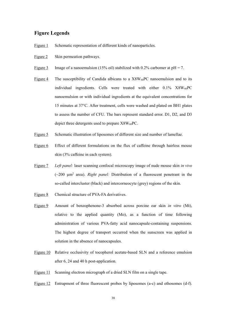
38
Figure Legends
Figure 1 Schematic representation of different kinds of nanoparticles.
Figure 2 Skin permeation pathways.
Figure 3 Image of a nanoemulsion (15% oil) stabilized with 0.2% carbomer at pH = 7.
Figure 4 The susceptibility of Candida albicans to a X8W60PC nanoemulsion and to its
individual ingredients. Cells were treated with either 0.1% X8W60PC
nanoemulsion or with individual ingredients at the equivalent concentrations for
15 minutes at 37°C. After treatment, cells were washed and plated on BH1 plates
to assess the number of CFU. The bars represent standard error. D1, D2, and D3
depict three detergents used to prepare X8W60PC.
Figure 5 Schematic illustration of liposomes of different size and number of lamellae.
Figure 6 Effect of different formulations on the flux of caffeine through hairless mouse
skin (3% caffeine in each system).
Figure 7 Left panel: laser scanning confocal microscopy image of nude mouse skin in vivo
(~200 µm2 area). Right panel: Distribution of a fluorescent penetrant in the
so-called intercluster (black) and intercorneocyte (grey) regions of the skin.
Figure 8 Chemical structure of PVA-FA derivatives.
Figure 9 Amount of benzophenone-3 absorbed across porcine ear skin in vitro (Mt),
relative to the applied quantity (Mo), as a function of time following
administration of various PVA-fatty acid nanocapsule-containing suspensions.
The highest degree of transport occurred when the sunscreen was applied in
solution in the absence of nanocapsules.
Figure 10 Relative occlusivity of tocopherol acetate-based SLN and a reference emulsion
after 6, 24 and 40 h post-application.
Figure 11 Scanning electron micrograph of a dried SLN film on a single tape.
Figure 12 Entrapment of three fluorescent probes by liposomes (a-c) and ethosomes (d-f).

39
The fluorescent probes were rhodamine red (a, d), D-289 (b, e) and calcein (e, f).
White represents the highest concentration of probe, with yellow and red
signifying progressively lower levels.
Figure 13 Proposed mechanisms for enhanced drug delivery across the skin from ethosomal
vehicles.
Figure 14 LSCM images of rhodamine red penetration into nude mouse skin following an 8
hour application of (a) ethosomes, (b) a hydroalcoholic solution of the dye, and (c)
liposomes.
Figure 15 LSCM images of D-289 penetration into nude mouse skin following an 8-hour
application of (a) liposomes, (b) a hydroalcoholic solution of the dye, and (c)
ethosomes.
Figure 16 Skin penetration profiles of (A) calcein, and (B) rhodamine red, following their
application to hairless mouse skin from either ethosomes, liposomes or a
hydroethanolic solution for 8 hours. Depth of skin penetration and fluorescence
intensity were determined by LSCM. Significantly different values (*p<0.001,
**p<0.05; ethosomes versus other systems) are highlighted.
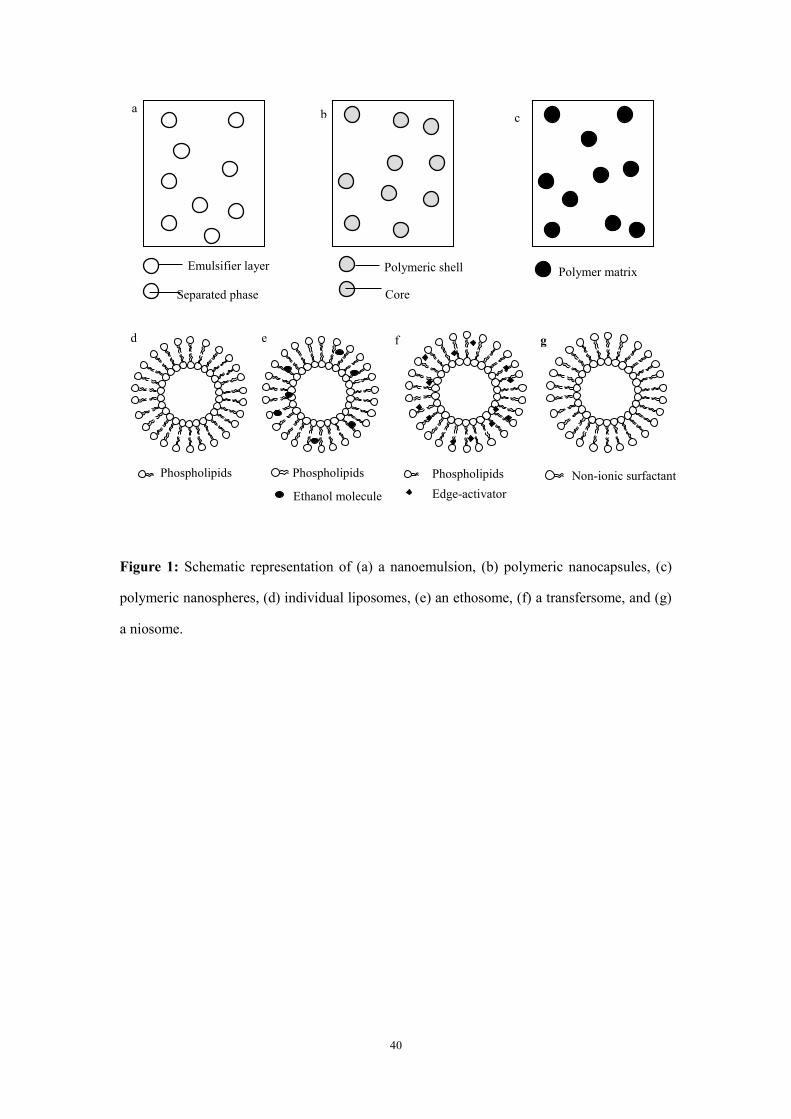
40
Figure 1: Schematic representation of (a) a nanoemulsion, (b) polymeric nanocapsules, (c)
polymeric nanospheres, (d) individual liposomes, (e) an ethosome, (f) a transfersome, and (g)
a niosome.
a
Emulsifier layer
Separated phase
b
Polymeric shell
Core
c
Polymer matrix
Phospholipids
d
Phospholipids
e
Ethanol molecule Edge-activator
Phospholipids
f
Non-ionic surfactant
g

41
Figure 2: Skin permeation pathways.
Stratum corneum
Intercellular route Transcellular route Appendageal route
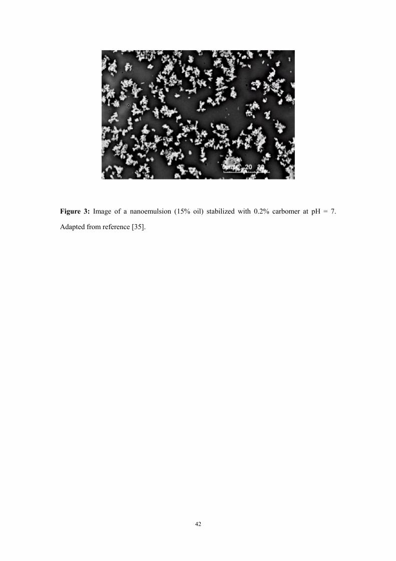
42
Figure 3: Image of a nanoemulsion (15% oil) stabilized with 0.2% carbomer at pH = 7.
Adapted from reference [35].
43
Control X8W60PC Solvent D1 D2 D3
log
(C
FU
/ml)
0
1
2
3
4
5
6
7
Figure 4: The susceptibility of Candida albicans to a X8W60PC nanoemulsion and to its
individual ingredients. Cells were treated with either 0.1% X8W60PC nanoemulsion or with
individual ingredients at the equivalent concentrations for 15 minutes at 37°C. After treatment,
cells were washed and plated on BH1 plates to assess the number of CFU. The bars represent
standard error. D1, D2, and D3 depict three detergents used to prepare X8W60PC. Redrawn
from reference [41].
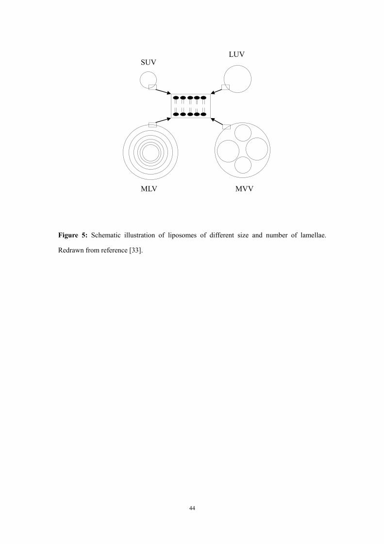
44
Figure 5: Schematic illustration of liposomes of different size and number of lamellae.
Redrawn from reference [33].
SUV LUV
MLV MVV
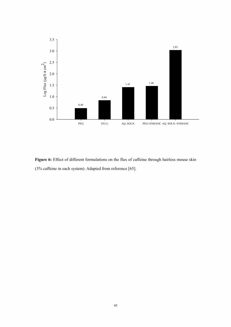
45
PEG SV(1) AQ. SOLN. PEG+ENHANC AQ. SOLN.+ENHANC
Lo
g F
lux
(μ
g/h
x c
m2
)
0.0
0.5
1.0
1.5
2.0
2.5
3.0
3.5
0.49
0.84
1.41 1.46
3.03
Figure 6: Effect of different formulations on the flux of caffeine through hairless mouse skin
(3% caffeine in each system). Adapted from reference [65].
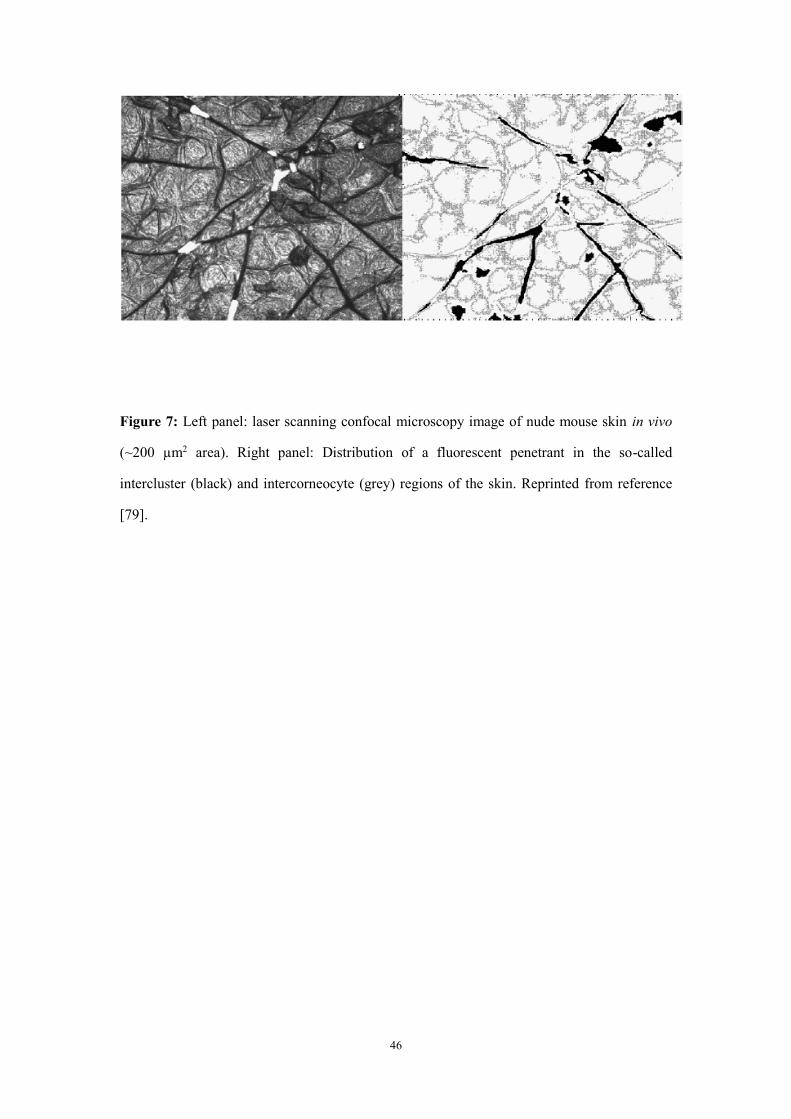
46
Figure 7: Left panel: laser scanning confocal microscopy image of nude mouse skin in vivo
(~200 µm2 area). Right panel: Distribution of a fluorescent penetrant in the so-called
intercluster (black) and intercorneocyte (grey) regions of the skin. Reprinted from reference
[79].
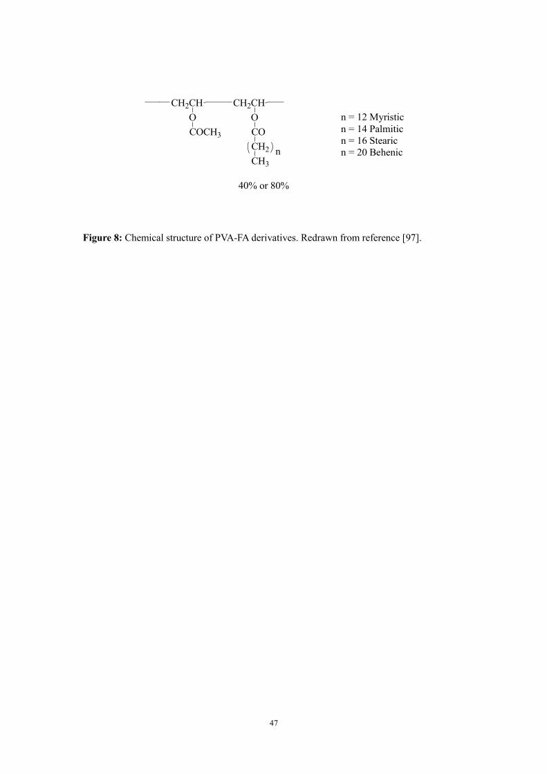
47
CH2CH
O
CH2CH
COCH3
O
CO
CH2
CH3
n
40% or 80%
n = 12 Myristic
n = 14 Palmitic
n = 16 Stearic
n = 20 Behenic
Figure 8: Chemical structure of PVA-FA derivatives. Redrawn from reference [97].

48
Time (h)
0 1 2 3 4 5 6
Mt/
Mo
0.000
0.005
0.010
0.015
0.020
0.025
PVA-BEH40
PVA-STEA40
PVA-PALM40
PVA-MIR40
PVA-BEH80
PVA-STEA80
PVA-PALM80
PVA-MIR80
BZP
Figure 9: Amount of benzophenone-3 absorbed across porcine ear skin in vitro (Mt), relative
to the applied quantity (Mo), as a function of time following administration of various
PVA-fatty acid nanocapsule-containing suspensions. The highest degree of transport occurred
when the sunscreen was applied in solution in the absence of nanocapsules. Redrawn from
reference [97].

49
6h 24h 48h
Rel
ativ
e o
cclu
siv
ity
0
10
20
30
40
50
60
SLN
Emulsion
Figure 10: Relative occlusivity of tocopherol acetate-based SLN and a reference emulsion
after 6, 24 and 40 h post-application. Redrawn from reference [123].
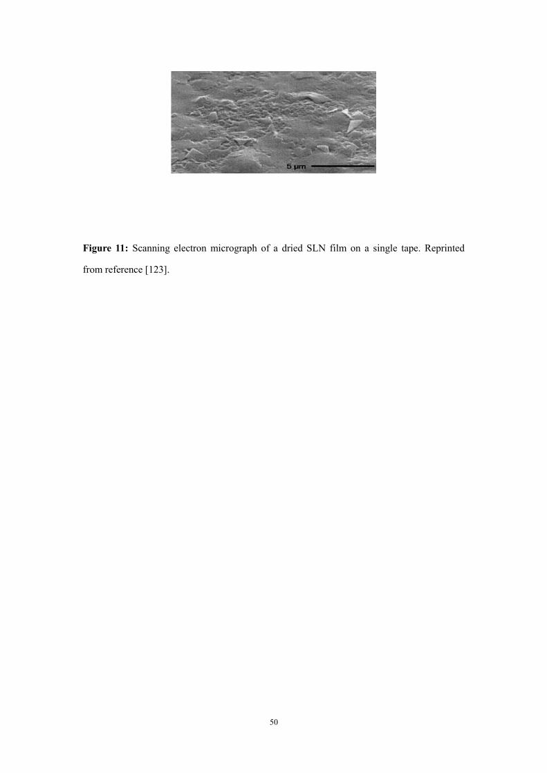
50
Figure 11: Scanning electron micrograph of a dried SLN film on a single tape. Reprinted
from reference [123].
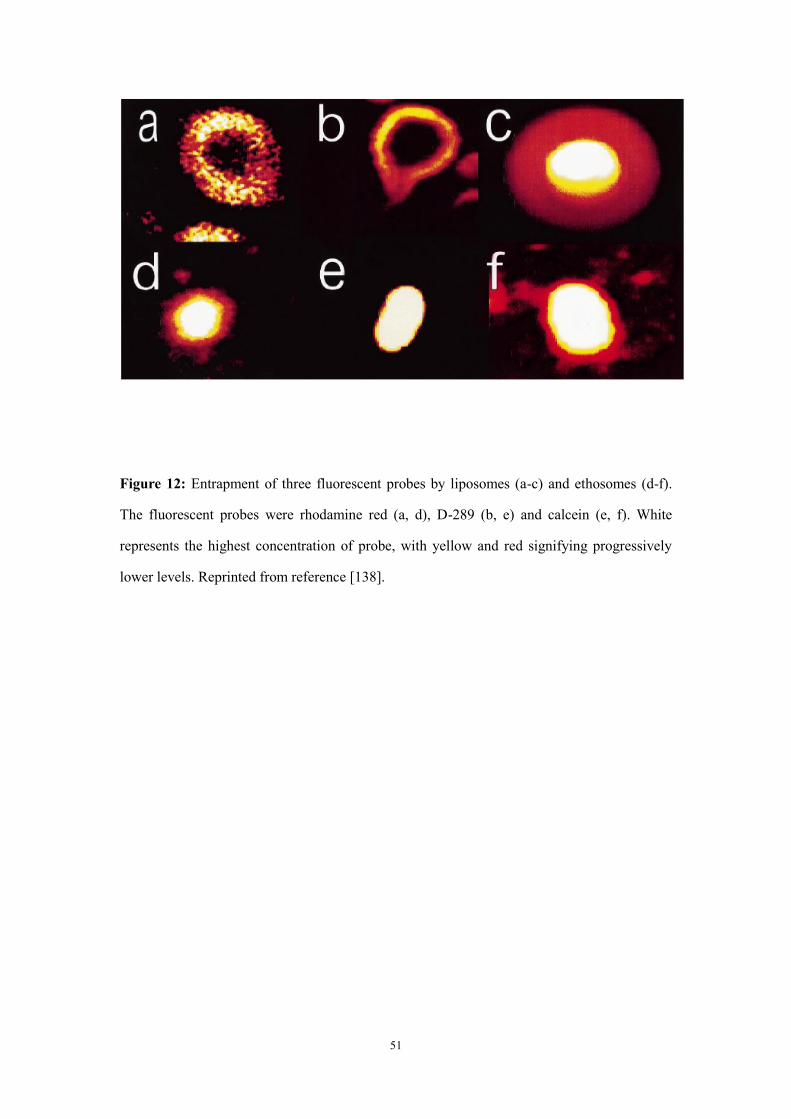
51
Figure 12: Entrapment of three fluorescent probes by liposomes (a-c) and ethosomes (d-f).
The fluorescent probes were rhodamine red (a, d), D-289 (b, e) and calcein (e, f). White
represents the highest concentration of probe, with yellow and red signifying progressively
lower levels. Reprinted from reference [138].
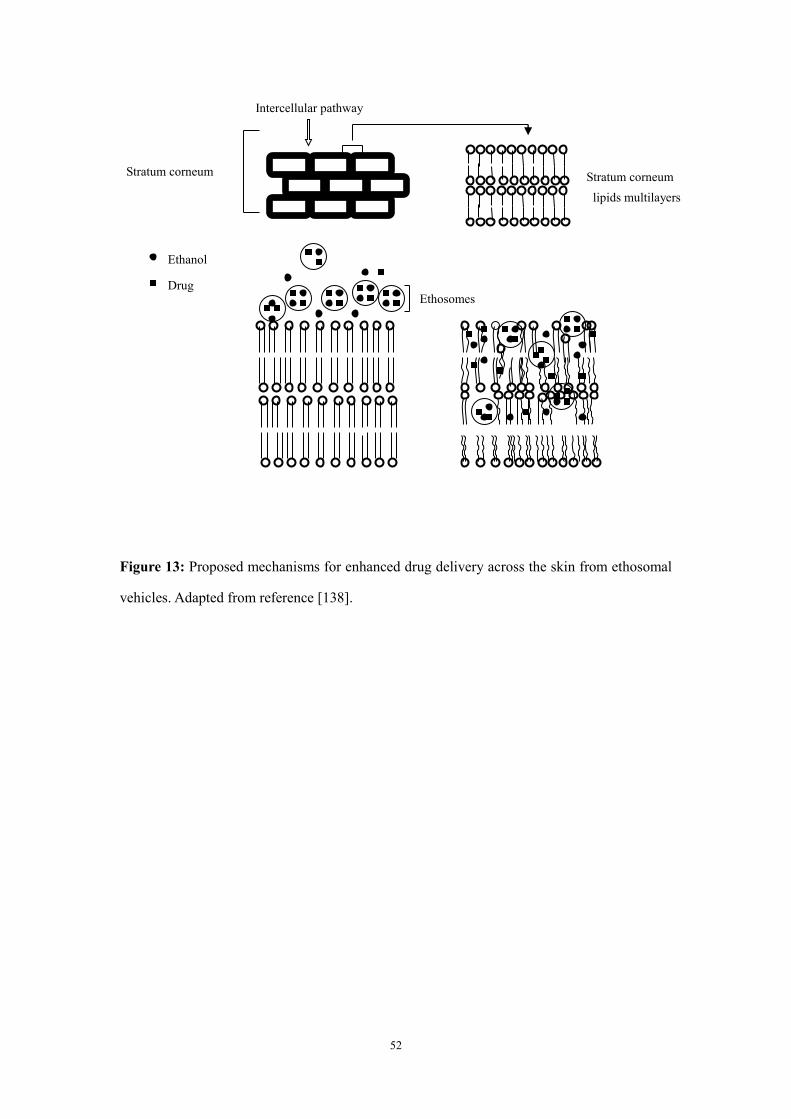
52
Figure 13: Proposed mechanisms for enhanced drug delivery across the skin from ethosomal
vehicles. Adapted from reference [138].
Intercellular pathway
Stratum corneum Stratum corneum
lipids multilayers
Ethanol
Drug Ethosomes
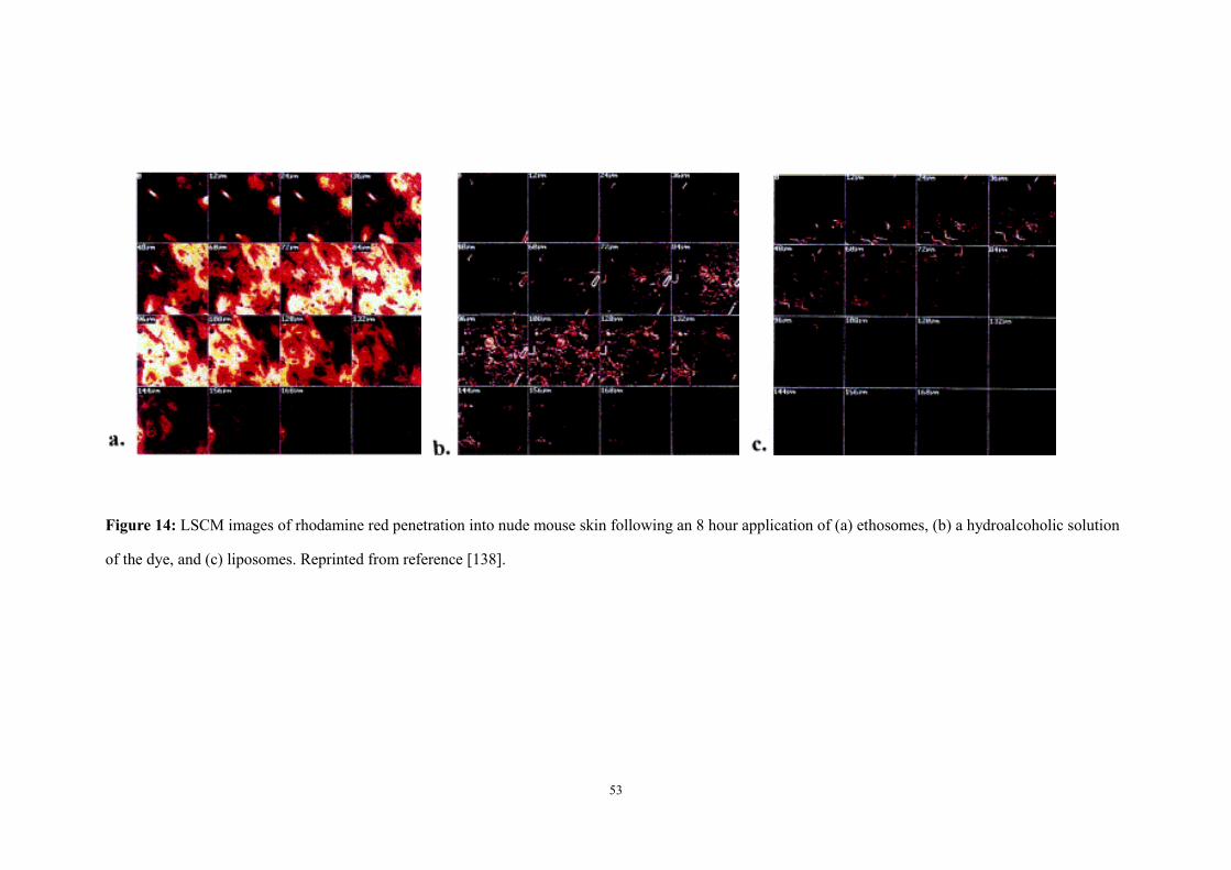
53
Figure 14: LSCM images of rhodamine red penetration into nude mouse skin following an 8 hour application of (a) ethosomes, (b) a hydroalcoholic solution
of the dye, and (c) liposomes. Reprinted from reference [138].
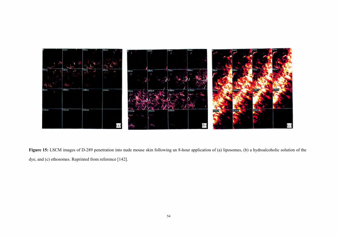
54
Figure 15: LSCM images of D-289 penetration into nude mouse skin following an 8-hour application of (a) liposomes, (b) a hydroalcoholic solution of the
dye, and (c) ethosomes. Reprinted from reference [142].

55
Skin depth (μm)
0 20 40 60 80 100 120 140 160
Flu
ore
scen
ce i
nte
nsi
ty (
arbit
rary
unit
s)
0
20
40
60
80
100
120
140
160
180 Ethosomes
Liposomes
Hydroethanolic solution
Blank
(A)
Skin depth (μm)
0 20 40 60 80 100 120 140 160 180 200 220 240 260
Flu
ore
scen
ce i
nte
nsi
ty (
arbit
rary
unit
s)
0
20
40
60
80
100
120
140
160Ethosomes
Liposomes
hydroethanolic solution
Blank
(B)
Figure 16: Skin penetration profiles of (A) calcein, and (B) rhodamine red, following their application
to hairless mouse skin from either ethosomes, liposomes or a hydroethanolic solution for 8 hours.
Depth of skin penetration and fluorescence intensity were determined by LSCM. Significantly
different values (*p<0.001, **p<0.05; ethosomes versus other systems) are highlighted. Redrawn from
reference [162].