Embed Size (px)
Citation preview

CHAPTER22Micellar Nanoparticles:Applications for Topicaland Passive TransdermalDrug DeliveryRobert W. Lee 1*, Dinesh B. Shenoy 2 and Rajiv Sheel 31 Particle Sciences Inc., 3894 Courtney Street, Bethlehem, PA 18017, USA; email:[email protected] Pharmaceuticals Ltd (CPL), India3Orgenus Pharma Inc., 116 Village Blvd Ste 200, Princeton, NJ 08540, USA
Contents
2.1 Introduction 372.1.1 MNP composition and structures 392.1.2 Physicochemical characterization 422.1.3 Antimicrobial properties 44
2.2 Transdermal drug delivery applications of MNP technology 452.2.1 Estrasorb� – commercial validation of MNP technology 452.2.2 Raloxifene MNP product 472.2.3 Nicotine MNP product 51
2.3 Topical drug delivery applications of MNP technology 532.4 Conclusion 54References 56Further reading 58
2.1 INTRODUCTION
Nanotechnology has evolved to be an integral part of the twenty-first cen-
tury. Nanotech-enabled products find applicability in almost everything we
touch on a day-to-day basis, such as medicine, pharmaceuticals, chemicals,
biologics, and information technology. In particular, the pharmaceutical
industry has been energized with breakthroughs in nano-engineering,
especially in the fields of drug delivery and formulation development. Over
the last few decades, there has been an explosion of research – at both aca-
demic and industrial levels – pertaining to nano-formulations: liposomes
*Author to whom correspondence should be addressed.
Handbook of Non-Invasive Drug Delivery Systems ISBN 9780815520252
� 2010 Elsevier Inc. All rights reserved.
37
(El Maghraby et al., 2006; Kazakov and Levon, 2006; Mainardes et al., 2006;
Minko et al., 2006; Sharma et al., 2006; Torchilin, 2006; Tyagi et al., 2006;
Weissig et al., 2006; Dutta, 2007; Elsayed et al., 2007; Karanth and Murthy,
2007; Koning and Krijger, 2007; Letchford and Burt, 2007; Malik et al.,
2007; Vyas and Khatri, 2007), nanoparticles (Asghar and Chandran, 2006;
Conti et al., 2006; Mainardes et al., 2006; Singhvi, 2006; Caruthers et al.,
2007; Emerich and Thanos, 2007; Garg and Saraf, 2007; Goldberg et al.,
2007; Illum, 2007; Kohane, 2007; Koning and Krijger, 2007; Navalakhe and
Nandedkar, 2007; Patel and Vavia, 2007; Silva, 2007; Torchilin, 2007; Wang
et al., 2007), nanoemulsions (Sarker, 2005; Pattani et al., 2006; Saupe et al.,
2006; Tiwari and Amiji, 2006; Date andNagarsenker, 2007; de Araujo et al.,
2007; Fatouros et al., 2007; Khandavilli and Panchagnula, 2007; Schaffazick
et al., 2007), dendrimers (Duncan and Izzo, 2005; Kitchens et al., 2005; Koo
et al., 2005; Bai et al., 2006; Gupta et al., 2006; Najlah and D’Emanuele,
2006; Qiu and Bae, 2006; Reddy et al., 2006; Yang and Kao, 2006), su-
pramolecular assemblies (Giraud-Guille et al., 2003; Lukin and Vogtle, 2005;
Hamacek et al., 2006; Perez-Garcia and Amabilino, 2007), and surface nano-
engineered products, to name a few.
Transdermal delivery involves application of a pharmacologically active
compound on to the skin to achieve therapeutic blood levels in order to
treat diseases remote from the site of application. Ever since the approval of
Transderm-Scop�, the first transdermal drug delivery system (TDDS) in
1981, there has been explosive research in the field of transdermal thera-
peutics for treatment of a variety of clinical conditions (Gordon and
Peterson, 2003). Unmatched clinical benefits (Gordon and Peterson, 2003),
profound industry interest, existence of strong and niche markets, and
regulatory precedence show why the TDDS has become a flourishing and
viable dosage form. The current transdermal therapeutics market is
segmented into traditional formulations (gels), advanced delivery systems
(patches), and novel physical technologies (microporation, iontophoresis,
and sonophoresis). Transdermal delivery is particularly advantageous for
those drugs having significant hepatic first-pass metabolism or degradation
in the gastrointestinal tract. Over the years, the US Food and Drug
Administration (FDA) has approved more than 40 transdermal products,
spanning about 15 molecules with sales of nearly $2.5 billion.
Micellar nanoparticle (MNP) technology was invented in the mid-1990s
(Wright, 1997; Simon, 2006; Singhvi, 2006). Scientists at Novavax de-
veloped and patented MNP technology and subsequently rolled out the first
nano-engineered transdermal lotion product (Estrasorb�) in 2003. Estra-
sorb is commercially manufactured on a kiloton scale and the manufacturing
38 Lee et al.
process is economical. The ingredients used in Estrasorb are all generally
recognized as safe (GRAS).
MNP is a nanotechnology-based formulation that has achieved a
breakthrough in transdermal therapeutics. The formulation represents a
robust and versatile delivery system that can accommodate a range of
therapeutic compounds having varying physicochemical properties. MNP-
based emulsions (lotions) are attractive alternatives for systemic drug de-
livery via topical application. The technology allows high concentrations of
drug to penetrate the skin and functionally create a drug depot in the
stratum corneum and epidermis. This route of delivery provides similar
advantages of patch technology in avoiding both contact with the gastro-
intestinal tract and hepatic first-pass effects, and is cosmetically more ac-
ceptable to many patients. MNP drug delivery offers a potentially fast and
inexpensive pharmaceutical development model by using drugs already
proven safe and effective to create new proprietary formulations.
2.1.1 MNP composition and structures
In broad terms, MNP is a multiphasic nanoemulsion. MNP technology
presents the active pharmaceutical ingredient (API) in a more readily bio-
available form. There are five basic components of an MNP system: (i) one
or more APIs; (ii) solvent; (iii) stabilizer; (iv) oil; and (v) aqueous medium.
When these components are mixed together and subjected to a milling
process (assisted by high-shear or high-pressure mixing), the API presents in
one or more composite fractions (Figure 2.1):
� Solid particulates (micro/nanoparticles)
� Micelle-associated
� Oil-associated
� Solubilized (in aqueous and/or solvent medium).
MNPs can accommodate both water-soluble and poorly water-soluble
APIs. While the technology can accommodate more traditional crystalline
compounds, surprisingly it can also be used with amorphous drugs.
Depending upon the physicochemical properties of the API and the dose
requirements, drug loading up to 20% (w/w) can be achieved. The range of
APIs that can be formulated in MNP technology is quite broad – from
physicochemical and therapeutic perspectives. This aspect will be elaborated
upon in the following sections.
A solvent is generally used to assist solubilization of the API during
processing – though it is not a prerequisite. The typical solvent used inMNP
Micellar Nanoparticles: Applications for Topical and Passive Transdermal Drug Delivery 39

preparation is ethanol. In addition, stable MNPs can be obtained using other
solvents such as propylene glycol, low-molecular-weight polyethylene
glycols, triacetin, and N-methylpyrrolidinone. The solvent plays an im-
portant role in controlling the solubilized fraction of the drug, which is
a key facilitator for rapid transmembrane permeation of the API.
The stabilizers used are generally non-ionic surfactants. Stable MNP
preparations have been prepared using both hydrophilic and lipophilic
stabilizers that encompass a wide hydrophilic–lipophilic balance (HLB)
range. The surfactants include such classes as sorbitan esters, glycerol esters,
block copolymers, polyethylene glycol esters, and ethoxylated fatty esters.
The surfactant helps to sterically stabilize the micro/nanoparticles and the
oil droplets, besides contributing to formation of the micellar phase.
The oil forms the internal phase of the emulsion. Depending upon the
properties of the drug, the oil phase can accommodate an API fraction in
soluble form. Some of the oils used are mineral oil, vegetable oils (soybean,
corn, etc.), medium-chain triglycerides, and squalane. The aqueous
medium used is generally purified water. A buffering agent may be included
to maintain the pH and maximize stability of the API. The product-specific
composition of the MNP formulation is dependent on the physicochemical
Figure 2.1 Schematic representation of the micro/nanostructures within an MNPformulation showing the different API components.
40 Lee et al.

properties of the API, the therapeutic need, intended site of action (local or
systemic), and target product profile.
For topical or transdermal administration, MNPs can be classified as
a type of microreservoir-dissolution-controlled system that can be tailored
to deliver drugs topically (skin being the site of action) or transdermally
(systemic availability). The physicochemical properties of MNP formula-
tions can be tailored for a given route of administration. This may en-
compass adjusting the viscosity appropriately for topical or transdermal
formulations, incorporating a mucoadhesive for vaginal or rectal adminis-
tration, changing the particle/droplet size, tuning the formulation com-
position and components, adjusting the zeta potential, or tailoring the
fraction of drug in solution versus in suspension. In a highly fragmented
transdermal drug delivery market, MNP is the only passive, nanotechnol-
ogy-enabled, cosmetically appealing, lotion-like topical dosage form of-
fering a tunable delivery profile for a wide range of APIs. Figure 2.2 depicts
a model for deposition of the MNP formulation within skin layers. The
composite structures within the MNP preparation are complementary to
the skin architecture and we hypothesize that this facilitates stratified po-
sitioning of the API within different skin layers.
Figure 2.2 Schematic representation (hypothesis) of deposition and disposition ofMNP structures within skin layers showing stratification of API.
Micellar Nanoparticles: Applications for Topical and Passive Transdermal Drug Delivery 41
2.1.2 Physicochemical characterization
The composite and multiphasic nature of the MNP formulation makes it
difficult to capture the complete picture with a single characterization tool.
Being the first of its kind, Estrasorb underwent tight scrutiny by the US
FDA during the approval process, which led to state-of-the-art quality
control test procedures (including particle sizing, crystal quantity, crystal
number, in vitro release test).
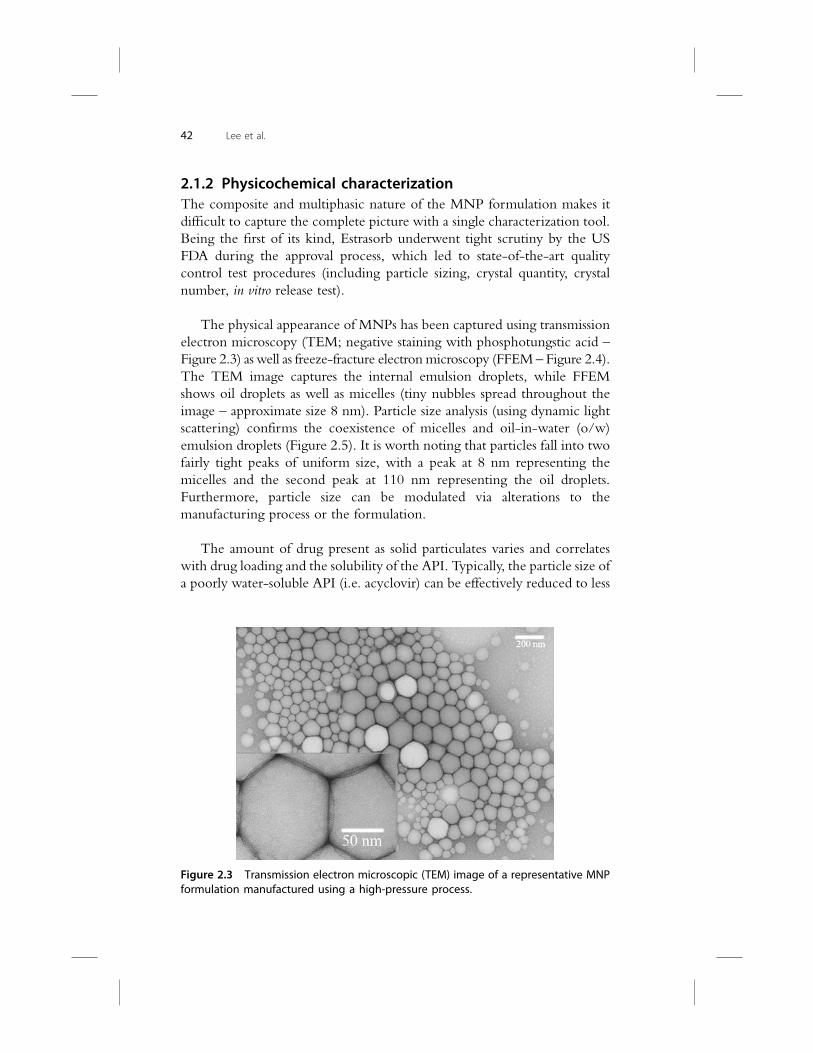
The physical appearance of MNPs has been captured using transmission
electron microscopy (TEM; negative staining with phosphotungstic acid –
Figure 2.3) as well as freeze-fracture electronmicroscopy (FFEM – Figure 2.4).
The TEM image captures the internal emulsion droplets, while FFEM
shows oil droplets as well as micelles (tiny nubbles spread throughout the
image – approximate size 8 nm). Particle size analysis (using dynamic light
scattering) confirms the coexistence of micelles and oil-in-water (o/w)
emulsion droplets (Figure 2.5). It is worth noting that particles fall into two
fairly tight peaks of uniform size, with a peak at 8 nm representing the
micelles and the second peak at 110 nm representing the oil droplets.
Furthermore, particle size can be modulated via alterations to the
manufacturing process or the formulation.
The amount of drug present as solid particulates varies and correlates
with drug loading and the solubility of the API. Typically, the particle size of
a poorly water-soluble API (i.e. acyclovir) can be effectively reduced to less
Figure 2.3 Transmission electron microscopic (TEM) image of a representative MNPformulation manufactured using a high-pressure process.
42 Lee et al.

than 3 mm in the MNP formulation. Figure 2.6a shows the particle size of
the API (raw material – before incorporation into MNP formulation) and
Figure 2.6b shows the size of the o/w emulsion (approximately 200 nm) and
the solid particulate drug upon formulating into the MNP product. The
particle size of the droplet and particulates can be tailored by choosing the
appropriate manufacturing technique (high-shear or high-pressure).
The viscosity of the MNP formulation can be tuned depending upon
the intended application. For a lotion-like appearance, a viscosity of about
80–350 mPa s can be achieved. A stiffer preparation (like a semisolid) can be
obtained by incorporating a suitable thickening agent (i.e. carbopol,
xanthan gum, stearyl alcohol). The nature and amount of oil and stabilizer
also play a role in modulating the viscosity of the final product.
Figure 2.4 Freeze-fracture electron microscopic (FFEM) image of a representativeMNP formulation manufactured using a high-pressure process.
0.10
5
10
15
20
10 100 1000 100001
size (d.nm)
In
ten
sity (%
)
o/w nanoemulsion droplets +nanoparticles (~ 125 nm)
Micelles (~ 8 nm)
Figure 2.5 Particle size data for a representative MNP formulation showing co-existence of o/w emulsion droplets and micelles.
Micellar Nanoparticles: Applications for Topical and Passive Transdermal Drug Delivery 43

Optimized MNPs are highly stable products. The validated shelf-life
claim for the commercial product based on MNP technology (Estrasorb�)
is 3 years at room temperature with excursions allowed to 40�C. It has beendemonstrated that there is no Oswald ripening phenomenon occurring in
the MNP product, and that both the number of crystals per unit volume and
crystal quantity of the API (estradiol) remain stable during room tempera-
ture and accelerated stability storage conditions. The MNP vehicle com-
position can be altered to offer enhanced thermal stability and some
preparations can withstand a standard terminal heat sterilization cycle (i.e.
120�C for 25 min at 15 psi).
2.1.3 Antimicrobial properties
The MNP composition is inherently antimicrobial. Figure 2.7 depicts the
results of the United States Pharmacopeia (USP) antimicrobial effectiveness
test (AET) for a representative placebo MNP formulation. The results in-
dicate that the MNPs are not only microbistatic, but are essentially
0.04 0.10
2
4
6
0.4 1 2 6 10 20 40 100 200 200010004004
Particle Diameter (μm)
Vo
lu
me (%
)
Differential Volume
0.04 0.1 0.4 1 2 4 6 10 20 40 100 200 400 1000 20000
5
10
15
20
Particle Diameter (µm)
Vo
lu
me (%
)
Differential Volume
093005.$03
093005.$02
a
b
Figure 2.6 Particle size data for acyclovir. (a) Raw material. (b) Upon formulating asMNP product.
44 Lee et al.

microbicidal. This can be attributed to the nano-size of the preparation and
the nature of the composition (i.e. the high concentration of non-ionic
surfactant). However, MNPs exhibit good safety profiles and they are rel-
atively non-irritating dermally. This property offers commercial benefits
such as the possible elimination of an antimicrobial preservative (especially
for product filled in a multi-dose container), or the possible synergy of the
microbicidal effect of an MNP preparation when formulated with an an-
tibacterial, antifungal, and/or antiviral API.
2.2 TRANSDERMAL DRUG DELIVERY APPLICATIONS OF MNP
TECHNOLOGY
2.2.1 Estrasorb� – commercial validation of MNP technology
MNP technology was originally developed for transdermal delivery of APIs.
MNP technology has been applied for estrogen replacement therapy with
17b-estradiol in Estrasorb (Estrasorb Package Insert, http://www.estra
sorb.com/EstrasorbBrief.pdf) – Novavax’s first internally developed FDA-
approved product and the only emulsion-based formulation in the topical
estrogen replacement market (primary indication being moderate-to-severe
vasomotor symptoms associated with menopause). Estrasorb is the world’s
first nano-engineered topical dosage form that is approved by the US FDA
for hormone replacement therapy, and represents commercial validation of
the MNP technology.
Figure 2.7 Antimicrobial effectiveness testing (USP) data for a representative pla-cebo MNP preparation.
Micellar Nanoparticles: Applications for Topical and Passive Transdermal Drug Delivery 45
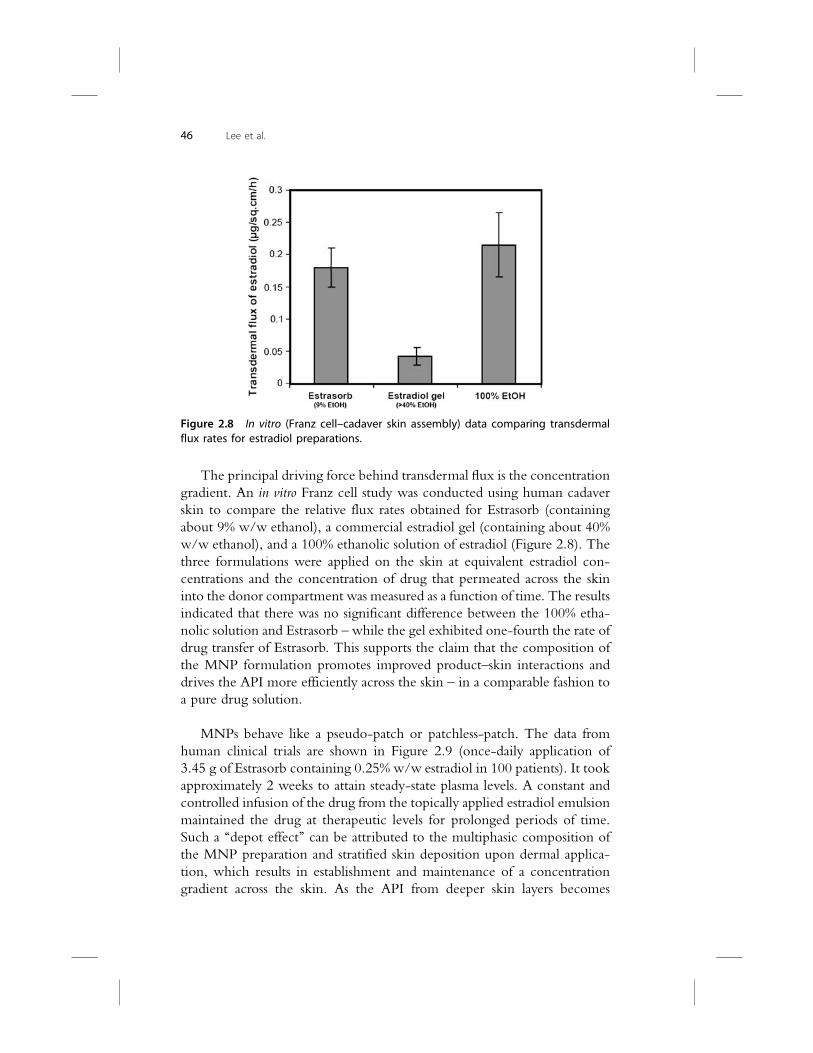
The principal driving force behind transdermal flux is the concentration
gradient. An in vitro Franz cell study was conducted using human cadaver
skin to compare the relative flux rates obtained for Estrasorb (containing
about 9% w/w ethanol), a commercial estradiol gel (containing about 40%
w/w ethanol), and a 100% ethanolic solution of estradiol (Figure 2.8). The
three formulations were applied on the skin at equivalent estradiol con-
centrations and the concentration of drug that permeated across the skin
into the donor compartment was measured as a function of time. The results
indicated that there was no significant difference between the 100% etha-
nolic solution and Estrasorb – while the gel exhibited one-fourth the rate of
drug transfer of Estrasorb. This supports the claim that the composition of
the MNP formulation promotes improved product–skin interactions and
drives the API more efficiently across the skin – in a comparable fashion to
a pure drug solution.
MNPs behave like a pseudo-patch or patchless-patch. The data from
human clinical trials are shown in Figure 2.9 (once-daily application of
3.45 g of Estrasorb containing 0.25% w/w estradiol in 100 patients). It took
approximately 2 weeks to attain steady-state plasma levels. A constant and
controlled infusion of the drug from the topically applied estradiol emulsion
maintained the drug at therapeutic levels for prolonged periods of time.
Such a ‘‘depot effect’’ can be attributed to the multiphasic composition of
the MNP preparation and stratified skin deposition upon dermal applica-
tion, which results in establishment and maintenance of a concentration
gradient across the skin. As the API from deeper skin layers becomes
Figure 2.8 In vitro (Franz cell–cadaver skin assembly) data comparing transdermalflux rates for estradiol preparations.
46 Lee et al.

depleted (through absorption into systemic circulation), more API dissolves
from the solid particulate drug reservoir (deposited in superficial skin layers),
maintaining a steady drug infusion. The effective plasma half-life for es-
tradiol in Estrasorb (57.6 h) is significantly higher as compared to the
commercial estradiol gel (36 h) or oral tablet (16.5 h). This provides strong
evidence of the patch-like delivery profile for the MNPs.
Several small-molecular-weight compounds have been evaluated to
prove the versatility and expandability of the MNP technology. A testos-
terone MNP formulation (Androsorb�) has completed phase I clinical
evaluation for two indications: hormone replacement therapy in hypo-
gonadal males, and to treat sexual dysfunction in females. A brief list of APIs
that have been successfully formulated asMNP products and have completed
key proof-of-concept (PoC) investigation has been compiled in Table 2.1.
Two case studies are presented, which will help define the MNP tech-
nology in terms of delivering a nontraditional transdermal API (raloxifene),
or tuning the delivery profile for a classical transdermal candidate (nicotine).
2.2.2 Raloxifene MNP product
Raloxifene is a selective estrogen receptor modulator that belongs to the
benzothiophene class of compounds. It is commercially available in tablet
form. Approximately 60% of the oral dose is absorbed, but extensive hepatic
conjugation to a number of inactive glucuronides results in an absolute
bioavailability of 2%. The rationale for developing a transdermal delivery
system for raloxifene was based on two considerations: (i) if therapeutic
concentrations of raloxifene could be delivered to the systemic circulation
transdermally, high hepatic concentrations would be avoided – thereby
Baseline week 2 week 4 week 8 Endpoint0
10
20
30
40
50
60
70
80
90
Tro
ug
h level, p
g/m
L
Estrasorb (n = 100)Placebo (n = 100)
Figure 2.9 Mean trough serum estradiol concentrations following daily topicalapplication of 3.45 g of Estrasorb� containing 2.5 mg/g estradiol for 12 weeks.
Micellar Nanoparticles: Applications for Topical and Passive Transdermal Drug Delivery 47

Table
2.1
Proof-of-conceptstudiesforvariousAPIsform
ulatedusingMNPtechnology
APIandindication
PoCinvestigationmodel
Keyoutcomes
Traditionaltransderm
alAPIs
Testosterone(horm
one
replacementtherapyin
males
orfemalesexual
dysfunction)
�PhaseIcompletedforboth
indications
�Pharmacokinetic
end-pointshavebeenmet
inphaseI
�Dose-dependentbloodlevelsseen
inmales
andfemales
�Sam
estrength
form
ulationcanbeusedforboth
indications–simply
byvaryingtheam
ountapplied
Nicotine
(smokingcessation)
�In
vitroFranzcell–cadaver
skin
studyforform
ulation
screening
�Preclinicalpharmacokinetic
evaluationin
rabbits
�Notareplacementproduct
forpatch
�Product
ideallysuited
forinterm
ediate
durationof
action(3–6h)to
addresswithdrawalsymptoms
�Deliveryprofilecanbetuned
tofitquickonsetofaction
(toaddresscraving)
Oxybutynin
(urinaryincontinence)
�Preclinicalpharmacokinetic
evaluationin
rabbits
�Datashowed
clinicallyexploitabletransdermaldelivery
profile
�Idealform
ulation
fortreating
urinary
incontinence
consideringthedrawbacksofthecommercialpatch
�Can
betuned
tocreate
once-a-day
applicationproduct
Fentanyl
(severepain)
�Preclinicalpharmacokinetic
evaluationin
rabbits
�Datadem
onstrateaproductwitharapid
onsetofaction
(andpainrelief)
�Opportunityto
create
anabuse-resistantproduct
throughform
ulationengineering
Clonidine
(hypertension)
�Preclinicalpharmacokinetic
evaluationin
rabbits
�Datashowed
clinicallyexploitabletransdermaldelivery
profile
�Can
betuned
tocreate
once-a-day
applicationproduct
48 Lee et al.
Nontraditionaltransderm
alAPIs
Raloxifene
(osteoporosisin
postmenopausalwomen)
�In
vitroFranzcell–cadaver
skin
studyforform
ulation
screening
�Datashowed
clinicallyexploitabletransdermaldelivery
profile–unlikethehydro-alcoholicgel(50%
ethanol),
whichshowed
zero
transdermaldelivery
Alprostadil
(erectiledysfunction)
�Preclinicalpharmacodynam
icevaluationin
rabbits(vasodila-
tationin
ears)
�Positive
datashowed
improvedefficacy
overpure
ethanolicsolution
�Significantdegree(visualscoring)andextent(vein
diameter)ofvasodilatationsuggestan
increased
probabilityofsuccessforuse
aslocalizedtreatm
entfor
erectiledysfunction
Cetirizine
(antihistaminic)
�Preclinicalpharmacokinetic
evaluationin
rabbits
�Datashowed
clinicallyexploitabletransdermaldelivery
profile
�In
addition,theproduct
islikelyto
offer
significant
benefitsatthesite
ofaction(i.e.localinflam
mation)
Naltrexone
(narcoticantagonist)
�Preclinicalpharmacokinetic
evaluationin
rabbits
�In
vitroFranzcell–cadaver
skin
studyforform
ulation
screening
�Datashowed
clinicallyexploitabletransdermal
deliveryprofile
Cyclobenzaprine
(antispasmodic)
�Preclinicalpharmacokinetic
evaluationin
rabbits
�Datashowed
clinicallyexploitablelocalandtransdermal
drugdeliveryprofile
Micellar Nanoparticles: Applications for Topical and Passive Transdermal Drug Delivery 49
reducing or avoiding adverse effects on coagulation factors and the conse-
quent risk of thromboembolism; and (ii) by avoiding extensive first-pass
metabolism to inactive metabolites, the total amount of raloxifene required
to achieve therapeutic concentrations is reduced – with an expected result of
a reduction in the adverse effects of metabolites. The physicochemical
properties of raloxifene (molecular weight 473, melting point 145�C, log P5.7, water solubility 0.25 mg/L) make it challenging to formulate using
conventional transdermal technologies, and difficult to create an elegant
topical formulation.
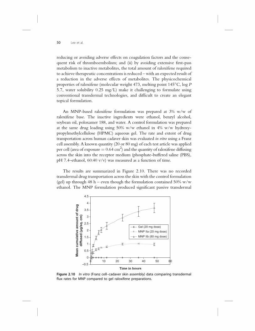
An MNP-based raloxifene formulation was prepared at 3% w/w of
raloxifene base. The inactive ingredients were ethanol, benzyl alcohol,
soybean oil, poloxamer 188, and water. A control formulation was prepared
at the same drug loading using 50% w/w ethanol in 4% w/w hydroxy-
propylmethylcellulose (HPMC) aqueous gel. The rate and extent of drug
transportation across human cadaver skin was evaluated in vitro using a Franz
cell assembly. A known quantity (20 or 80 mg) of each test article was applied
per cell (area of exposure¼ 0.64 cm2) and the quantity of raloxifene diffusing
across the skin into the receptor medium (phosphate-buffered saline (PBS),
pH 7.4–ethanol, 60:40 v/v) was measured as a function of time.
The results are summarized in Figure 2.10. There was no recorded
transdermal drug transportation across the skin with the control formulation
(gel) up through 48 h – even though the formulation contained 50% w/w
ethanol. The MNP formulation produced significant passive transdermal
–0.5
0
0.5
1
1.5
2
2.5
3
3.5
4
4.5
40 0 10 20 30 50 60
Time in hours
Mean
cu
mu
lative am
ou
nt o
f d
ru
g
diffu
sed
(μ
g/sq
. cm
)
Gel (20 mg dose) MNP IIa (20 mg dose) MNP IIb (80 mg dose)
Figure 2.10 In vitro (Franz cell–cadaver skin assembly) data comparing transdermalflux rates for MNP compared to gel raloxifene preparations.
50 Lee et al.

drug flux and showed a linear dose–response (MNP IIa and IIb) relationship
at two doses – a fourfold increase in the amount of raloxifene applied
resulted in a corresponding fourfold increase in transdermal drug flux. Based
on these data, 2 g of 3% w/w MNP product (60 mg raloxifene) applied on
two thighs (~375 cm2 each) could provide the targeted 1.2 mg/day dose
(equivalent to a single oral dose of 60 mg).
It is evident from this study that the MNP technology can facilitate
transdermal transportation of APIs that may not be considered as the ideal
candidates for transdermal delivery, or that cannot be formulated using
conventional dosage forms.
2.2.3 Nicotine MNP product
Nicotine is an alkaloid found in the nightshade family of plants, pre-
dominantly in tobacco. It functions as an antiherbivore chemical, being
a potent neurotoxin with particular specificity to insects. In low concen-
trations (an average cigarette yields about 1 mg of absorbed nicotine), the
substance acts as a stimulant in mammals and is one of the main factors
responsible for the dependence-forming properties of tobacco smoking.
The pharmacologic and behavioral characteristics that determine tobacco
addiction are similar to those that determine addiction to drugs such as
heroin and cocaine.
Current nicotine products include chewing gum, oral lozenge, nasal
spray, oral inhalant, and transdermal patch. The objective of the nicotine
MNP product is to provide controlled and continuous delivery of nicotine
through the skin. The MNP formulation can be customized with respect to
onset and duration of action. Based on in vitro data (given below), it is
expected that the MNP product will have an intermediate pharmacokinetic
profile falling between that of a gum and a patch.
The physicochemical properties of nicotine (molecular weight 162.26,
melting point�79�C, log P 1.305, oily liquid but miscible in water) make it
an ideal fit for transdermal therapeutics – this fact is evident through the
multitude of transdermal formulations commercially available. However, for
a nonpolymer-based topical lotion technology like MNP, the challenge is to
deliver the drug upon dermal application over longer periods of time. The
objective of the in vitro proof-of-concept study (Franz cell–cadaver skin
assembly) was to demonstrate the capability of the MNP technology to
modulate the rate and extent of nicotine delivery through variations in the
formulation.
Micellar Nanoparticles: Applications for Topical and Passive Transdermal Drug Delivery 51
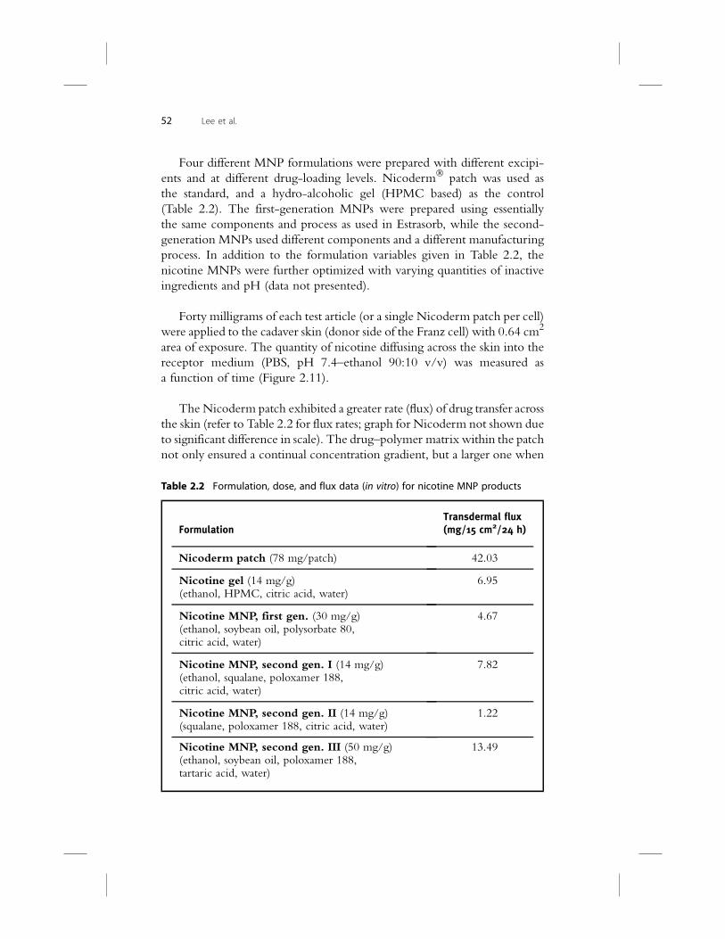
Four different MNP formulations were prepared with different excipi-
ents and at different drug-loading levels. Nicoderm� patch was used as
the standard, and a hydro-alcoholic gel (HPMC based) as the control
(Table 2.2). The first-generation MNPs were prepared using essentially
the same components and process as used in Estrasorb, while the second-
generation MNPs used different components and a different manufacturing
process. In addition to the formulation variables given in Table 2.2, the
nicotine MNPs were further optimized with varying quantities of inactive
ingredients and pH (data not presented).
Forty milligrams of each test article (or a single Nicoderm patch per cell)
were applied to the cadaver skin (donor side of the Franz cell) with 0.64 cm2
area of exposure. The quantity of nicotine diffusing across the skin into the
receptor medium (PBS, pH 7.4–ethanol 90:10 v/v) was measured as
a function of time (Figure 2.11).
TheNicoderm patch exhibited a greater rate (flux) of drug transfer across
the skin (refer to Table 2.2 for flux rates; graph for Nicoderm not shown due
to significant difference in scale). The drug–polymer matrix within the patch
not only ensured a continual concentration gradient, but a larger one when
Table 2.2 Formulation, dose, and flux data (in vitro) for nicotine MNP products
FormulationTransdermal flux(mg/15 cm2/24 h)
Nicoderm patch (78 mg/patch) 42.03
Nicotine gel (14 mg/g)(ethanol, HPMC, citric acid, water)
6.95
Nicotine MNP, first gen. (30 mg/g)(ethanol, soybean oil, polysorbate 80,citric acid, water)
4.67
Nicotine MNP, second gen. I (14 mg/g)(ethanol, squalane, poloxamer 188,citric acid, water)
7.82
Nicotine MNP, second gen. II (14 mg/g)(squalane, poloxamer 188, citric acid, water)
1.22
Nicotine MNP, second gen. III (50 mg/g)(ethanol, soybean oil, poloxamer 188,tartaric acid, water)
13.49
52 Lee et al.

compared to the MNPs or gel (0.64 cm2 of patch contained approximately
3.33 mg nicotine vs. 0.56, 1.2, and 2.0 mg of nicotine for 14, 30, and
50 mg/g strengths respectively). The nicotine gel had no effective control
over drug disposition as it followed a first-order release during the initial 8-h
release phase and reached a plateau within about 8 h. The two second-
generation MNP formulations (I and II) containing the same loading of
nicotine as the gel (14 mg/g) not only exhibited prominent control over
drug transportation rates (zero-order release), but also a significant difference
in the rate or drug transport (sixfold difference in flux). This was essentially
because of the difference in the composition of the two MNPs, which were
tailored to release the drug at faster or slower rates. In spite of having higher
drug loading (30 mg/g), the first-generation MNP showed a lower drug
transportation pattern than the second-generation MNP I, which can be
attributed to the difference in formulation components.
2.3 TOPICAL DRUG DELIVERY APPLICATIONS OF MNP
TECHNOLOGY
Although the transdermal drug delivery field has enjoyed a significant
amount of research effort and technological breakthrough, there has not
been much corresponding innovation taking place in the field of topical
drug delivery. The majority of the dosage forms are limited to traditional
–100
0
100
200
300
400
500
600
700
800
900
1000
1100
1200
0 5 10 15 20 25 30
Time in hours
Mean
cu
mu
lati
ve a
mo
un
t d
iffu
sed
(μ
g/s
q. cm
) Nicotine MNP 2nd Gen I, 1.4% w/w Nicotine MNP 2nd Gen II, 1.4% w/w Nicotine MNP 1st Gen, 3% w/w Nicotine Gel, 1.4% w/w Nicotine MNP 2nd Gen III, 5.0% w/w
Figure 2.11 In vitro (Franz cell–cadaver skin assembly) data comparing transdermalflux rates for MNP compared to gel nicotine preparations.
Micellar Nanoparticles: Applications for Topical and Passive Transdermal Drug Delivery 53

creams, ointments, and gels. Some of the new additions have been sprays,
foams, and patches. MNP technology can be exploited to design improved
topical dosage forms that deliver the API locally (at site of application) in an
efficient and effective manner. It is possible to tailor drug deposition, dis-
position, and permeation kinetics through formulation engineering (altered
composition, drug loading, droplet size, etc.). This concept has been
demonstrated using acyclovir as the model drug. Commercially, a topical
acyclovir product is available (Zovirax�) and is indicated for the treatment
of recurrent herpes labialis (cold sores), for genital herpes, and in limited
nonlife-threatening mucocutaneous herpes simplex viral infections in im-
munocompromised patients. The product needs to be applied topically five
to seven times a day for 4–7 days. A comparative investigation (in vitro using
Franz cell–cadaver skin assembly) was carried out with two MNP formu-
lations (designed to differentiate topical and transdermal delivery) and
Zovirax cream. All the formulations had a drug loading of 5% w/w. The
product was applied to the skin (donor compartment), and drug that per-
meated across the skin as well as that retained within skin layers was esti-
mated. The results are captured in Figure 2.12a and b. MNP I was designed
to retain the API preferentially in the skin layers, while MNP II was
engineered to facilitate transdermal permeation of the same drug. It is clear
from the data that the transdermal flux rate for MNP I was comparable to
that for Zovirax – but the amount of drug retained within skin was about
twofold higher for the former. If this effect can be translated into clinical use
(or in vivo systems), one can expect to witness:
� Increased rate of skin permeation, resulting in faster onset of action
� Greater degree of skin deposition, leading to higher local drug
concentrations
� Skin depot effects, leading to longer drug residence time
� Potential reductions of total dose, frequency of application, or both.
In addition, the inherent antimicrobial nature of the MNP vehicle would be
beneficial from a therapeutic and packaging perspective. Based on these
benefits, the MNP technology could offer a novel perspective to the field of
topical drug delivery – especially for nonsteroidal anti-inflammatory drugs
(NSAIDs), antifungals, antibacterials, antivirals, antispasmodics, and vaso-
dilatory drugs.
2.4 CONCLUSION
Transdermal drug delivery is not suited or clinically justified for all drugs,
yet it is viewed to be much more limited than is warranted. MNP tech-
nology helps to incorporate and deliver many therapeutic compounds
54 Lee et al.

that are otherwise viewed as unsuitable for transdermal delivery. MNP
technology allows fast, low-cost product development compared with the
typical development of new chemical entities. From proof of principle in
a validated preclinical model through beginning a phase I study in humans
requires approximately 12months to complete. The data from the preclinical
studies described here show a high probability of clinical success within
a shorter development time frame, and a lower cost than a typical NDA
(New Drug Application).
Research to date has shown the MNP drug delivery platform to be
a versatile technology for multiple routes of administration (data not
presented for ophthalmic, vaginal, and oral routes). Understanding the basic
0 0
0.1
0.2
0.3
0.4
0.5
0.6
0.7
0.8
5 10 15 20 25 30 Time in hours
[M
ean
cu
mu
lative d
ru
g released
]
(µg
/sq
. cm
)
Zovirax MNP I MNP II 0
0.05
0.1
0.15
0.2
0.25
0.3
0.35
Dru
g am
ou
nt in
skin
(µg
/g
)
Zovirax (0.006 µg/sq. cm/h)MNP I (0.009 µg/sq. cm/h)MNP II (0.024 µg/sq. cm/h)
a
b
Figure 2.12 In vitro (Franz cell–cadaver skin assembly) data comparing acyclovirpreparations. (a) Transdermal flux rates. (b) Amount retained within skin layersafter 23 h.
Micellar Nanoparticles: Applications for Topical and Passive Transdermal Drug Delivery 55

physicochemical properties of MNPs has enabled a degree of control over
pharmacokinetic parameters that may provide an attractive option for
pharmaceutical formulators. The technology is validated for transdermal
delivery and a commercial MNP product, Estrasorb, is manufactured on
a kiloton scale. The Estrasorb ingredients are GRAS and the manufacturing
process is attractive from a cost of goods perspective. The heterogeneous,
multiphasic nanoemulsion that comprises the MNPs is surprisingly stable
and, in some cases, amenable to terminal heat sterilization. Experiments
have shown the potential for MNPs to be used for intranasal, vaginal, rectal,
and parenteral routes of administration – for poorly water-soluble drugs in
particular. Better commercial exploitation of the MNP technology, to its
fullest potential, is expected in coming years.
REFERENCES
Asghar, L.F., Chandran, S., 2006. Multiparticulate formulation approach to colon specificdrug delivery: current perspectives. J. Pharm. Sci. 9 (3), 327–338.
Bai, S., Thomas, C., Rawat, A., Ahsan, F., 2006. Recent progress in dendrimer-basednanocarriers. Crit. Rev. Ther. Drug Carrier Syst. 23 (6), 437–495.
Caruthers, S.D., Wickline, S.A., Lanza, G.M., 2007. Nanotechnological applications inmedicine. Curr. Opin. Biotechnol. 18 (1), 26–30. Epub 2007, Jan 24.
Conti, M., Tazzari, V., Baccini, C., Pertici, G., Serino, L.P., De Giorgi, U., 2006. Anti-cancer drug delivery with nanoparticles. In Vivo 20 (6A), 697–701.
Date, A.A., Nagarsenker, M.S., 2007. Design and evaluation of self-nanoemulsifying drugdelivery systems (SNEDDS) for cefpodoxime proxetil. Int. J. Pharm. 329 (1–2), 166–172.Epub 2006, Sep 1.
deAraujo, S.C., deMattos, A.C.,Teixeira,H.F.,Coelho, P.M.,Nelson,D.L., deOliveira,M.C.,2007. Improvement of invitro efficacyof a novel schistosomicidal drug by incorporation intonanoemulsions. Int. J. Pharm. 337 (1–2), 307–315. Epub 2007, Jan 14.
Duncan, R., Izzo, L., 2005. Dendrimer biocompatibility and toxicity. Adv. Drug Deliv.Rev. 57 (15), 2215–2237. Epub 2005, Nov 16.
Dutta, R.C., 2007. Drug carriers in pharmaceutical design: promises and progress. Curr.Pharm. Des. 13 (7), 761–769.
El Maghraby, G.M., Williams, A.C., Barry, B.W., 2006. Can drug-bearing liposomespenetrate intact skin? J. Pharm. Pharmacol. 58 (4), 415–429.
Elsayed, M.M., Abdallah, O.Y., Naggar, V.F., Khalafallah, N.M., 2007. Lipid vesicles forskin delivery of drugs: reviewing three decades of research. Int. J. Pharm. 332 (1–2),1–16. Epub 2006, Dec 8.
Emerich, D.F., Thanos, C.G., 2007. Targeted nanoparticle-based drug delivery and di-agnosis. J. Drug Target. 15 (3), 163–183.
Fatouros, D.G., Deen, G.R., Arleth, L., Bergenstahl, B., Nielsen, F.S., Pedersen, J.S.,Mullertz, A., 2007. Structural development of self nano emulsifying drug deliverysystems (SNEDDS) during in vitro lipid digestion monitored by small-angle X-rayscattering. Pharm. Res. Epub ahead of print, Apr 26.
Garg, G., Saraf, S., 2007. Cubosomes: an overview. Biol. Pharm. Bull. 30 (2), 350–353.Giraud-Guille, M.M., Besseau, L., Martin, R., 2003. Liquid crystalline assemblies of col-
lagen in bone and in vitro systems. J. Biomech. 36 (10), 1571–1579.Goldberg, M., Langer, R., Jia, X., 2007. Nanostructured materials for applications in drug
delivery and tissue engineering. J. Biomater. Sci. Polym. Ed. 18 (3), 241–268.Gordon, R.D., Peterson, T.A., 2003. 4 myths about transdermal drug delivery. Drug Deliv.
Tech. 3 (4), 44–50.
56 Lee et al.

Gupta, U., Agashe, H.B., Asthana, A., Jain, N.K., 2006. A review of in vitro–in vivo in-vestigations on dendrimers: the novel nanoscopic drug carriers. Nanomedicine 2 (2),66–73.
Hamacek, J., Borkovec, M., Piguet, C., 2006. Simple thermodynamics for unraveling so-phisticated self-assembly processes. Dalton Trans. 12, 1473–1490. Epub 2006, Mar 1.
Illum, L., 2007. Nanoparticulate systems for nasal delivery of drugs: a real improvementover simple systems? J. Pharm. Sci. 96 (3), 473–483.
Karanth, H., Murthy, R.S., 2007. pH-sensitive liposomes – principle and application incancer therapy. J. Pharm. Pharmacol. 59 (4), 469–483.
Kazakov, S., Levon, K., 2006. Liposome–nanogel structures for future pharmaceutical ap-plications. Curr. Pharm. Des. 12 (36), 4713–4728.
Khandavilli, S., Panchagnula, R., 2007. Nanoemulsions as versatile formulations for pac-litaxel delivery: peroral and dermal delivery studies in rats. J. Invest. Dermatol. 127 (1),154–162. Epub 2006, Jul 20.
Kitchens, K.M., El-Sayed, M.E., Ghandehari, H., 2005. Transepithelial and endothelialtransport of poly (amidoamine) dendrimers. Adv. Drug Deliv. Rev. 57 (15), 2163–2176.Epub 2005, Nov 11.
Kohane, D.S., 2007. Microparticles and nanoparticles for drug delivery. Biotechnol. Bioeng.96 (2), 203–209.
Koning, G.A., Krijger, G.C., 2007. Targeted multifunctional lipid-based nanocarriers forimage-guided drug delivery. Anticancer Agents Med. Chem. 7 (4), 425–440.
Koo, O.M., Rubinstein, I., Onyuksel, H., 2005. Role of nanotechnology in targeted drugdelivery and imaging: a concise review. Nanomedicine 1 (3), 193–212.
Letchford, K., Burt, H., 2007. A review of the formation and classification of amphiphilicblock copolymer nanoparticulate structures: micelles, nanospheres, nanocapsules andpolymersomes. Eur. J. Pharm. Biopharm. 65 (3), 259–269. Epub 2006, Nov 23.
Lukin, O., Vogtle, F., 2005. Knotting and threading of molecules: chemistry and chirality ofmolecular knots and their assemblies. Angew. Chem. Int. Ed. Engl. 44 (10), 1456–1477.
Mainardes,R.M.,Urban,M.C.,Cinto, P.O.,Chaud,M.V., Evangelista,R.C.,Gremiao,M.P.,2006. Liposomes and micro/nanoparticles as colloidal carriers for nasal drug delivery.Curr. Drug Deliv. 3 (3), 275–285.
Malik, D.K., Baboota, S., Ahuja, A., Hasan, S., Ali, J., 2007. Recent advances in proteinand peptide drug delivery systems. Curr. Drug. Deliv. 4 (2), 141–151.
Minko, T., Pakunlu, R.I., Wang, Y., Khandare, J.J., Saad, M., 2006. New generation ofliposomal drugs for cancer. Anticancer Agents Med. Chem. 6 (6), 537–552.
Najlah, M., D’Emanuele, A., 2006. Crossing cellular barriers using dendrimer nanotech-nologies. Curr. Opin. Pharmacol. 6 (5), 522–527. Epub 2006, Aug 4.
Navalakhe, R.M., Nandedkar, T.D., 2007. Application of nanotechnology in biomedicine.Indian J. Exp. Biol. 45 (2), 160–165.
Patel, A.R., Vavia, P.R., 2007. Nanotechnology and pharmaceutical inhalation aerosols.Indian J. Exp. Biol. 45 (2), 166–174.
Pattani, A.S., Mandawgade, S.D., Patravale, V.B., 2006. Development and comparativeanti-microbial evaluation of lipid nanoparticles and nanoemulsion of Polymyxin B.J.Nanosci. Nanotechnol. 6 (9–10), 2986–2990.
Perez-Garcia, L., Amabilino, D.B., 2007. Spontaneous resolution, whence and whither: fromenantiomorphic solids to chiral liquid crystals, monolayers and macro- and supra-molecular polymers and assemblies. Chem. Soc. Rev. 36 (6), 941–967. Epub 2007, Feb 5.
Qiu, L.Y., Bae, Y.H., 2006. Polymer architecture and drug delivery. Pharm. Res. 23 (1),1–30. Epub 2006, Jan 11.
Reddy, S.T., Swartz, M.A., Hubbell, J.A., 2006. Targeting dendritic cells with biomaterials:developing the next generation of vaccines. Trends Immunol. 27 (12), 573–579. Epub2006, Oct 16.
Sarker, D.K., 2005. Engineering of nanoemulsions for drug delivery. Curr. Drug Deliv.2 (4), 297–310.
Micellar Nanoparticles: Applications for Topical and Passive Transdermal Drug Delivery 57

Saupe, A., Gordon, K.C., Rades, T., 2006. Structural investigations on nanoemulsions, solidlipid nanoparticles and nanostructured lipid carriers by cryo-field emission scanningelectron microscopy and Raman spectroscopy. Int. J. Pharm. 314 (1), 56–62. Epub 2006,Mar 30.
Schaffazick, S.R., Pohlmann, A.R., Guterres, S.S., 2007. Nanocapsules, nanoemulsion andnanodispersion containing melatonin: preparation, characterization and stability evalua-tion. Pharmazie 62 (5), 354–360.
Sharma, G., Anabousi, S., Ehrhardt, C., Ravi Kumar, M.N., 2006. Liposomes as targeteddrug delivery systems in the treatment of breast cancer. J. Drug Target. 14 (5), 301–310.
Silva, G.A., 2007. Nanotechnology approaches for drug and small molecule delivery acrossthe blood brain barrier. Surg. Neurol. 67 (2), 113–116.
Simon, J.A., 2006. Estrasorb Study Group. Estradiol in micellar nanoparticles: the efficacyand safety of a novel transdermal drug-delivery technology in the management ofmoderate to severe vasomotor symptoms. Menopause 13 (2), 222–231.
Singhvi, R., 2006. Micellar nanoparticles: a new drug delivery platform. Drug Deliv. Tech.6 (2), 72–75.
Tiwari, S.B., Amiji, M.M., 2006. Improved oral delivery of paclitaxel following adminis-tration in nanoemulsion formulations. J. Nanosci. Nanotechnol. 6 (9–10), 3215–3221.
Torchilin, V.P., 2006. Multifunctional nanocarriers. Adv. Drug Deliv. Rev. 58 (14),1532–1555. Epub 2006, Sep 28.
Torchilin, V.P., 2007. Micellar nanocarriers: pharmaceutical perspectives. Pharm. Res.24 (1), 1–16. Epub 2006, Nov 16.
Tyagi, P., Wu, P.C., Chancellor, M., Yoshimura, N., Huang, L., 2006. Recent advances inintravesical drug/gene delivery. Mol. Pharm. 3 (4), 369–379.
Vyas, S.P., Khatri, K., 2007. Liposome-based drug delivery to alveolar macrophages. ExpertOpin. Drug Deliv. 4 (2), 95–99.
Wang, M.D., Shin, D.M., Simons, J.W., Nie, S., 2007. Nanotechnology for targeted cancertherapy. Expert Rev. Anticancer Ther. 7 (6), 833–837.
Weissig, V., Boddapati, S.V., Cheng, S.M., D’Souza, G.G., 2006. Liposomes and liposome-like vesicles for drug and DNA delivery to mitochondria. J. Liposome Res. 16 (3),249–264.
Wright, C., 1997. Micellar nanoparticles. US patent, number 5,620.021.Yang, H., Kao, W.J., 2006. Dendrimers for pharmaceutical and biomedical applications.
J. Biomater. Sci. Polym. Ed. 17 (1–2), 3–19.
FURTHER READING
Biorise� technology from Eurand. <http://www.eurand.com/Technologies/Bioavailability-enhancement/Biorise/>.
Burgess, D. (Ed.), 2005. Injectable Dispersed Systems: Formulation, Processing and Per-formance. Taylor & Francis, Florida.
Insoluble Drug Delivery (IDD�) technology from SkyePharma.<http://www.skyepharma.com/solubilization_idd.html>.
Liu, R. (Ed.), 2000. Water-Insoluble Drug Formulations. Interpharm Press, Colorado.NanoCrystal� technology from elan Drug Delivery Inc. <http://www.elan.com/EDT/
nanocrystal_technology/>.NanoEdge� technology from Baxter BioPharma Solutions.<http://www.baxterbiopharma
solutions.com/content/formulation_tech/nanoedge.html>.
58 Lee et al.