Embed Size (px)
Citation preview
Hindawi Publishing CorporationCase Reports in OtolaryngologyVolume 2012, Article ID 931350, 4 pagesdoi:10.1155/2012/931350
Case Report
An Unusual Presentation of Ludwig’s AnginaComplicated by Cervical Necrotizing Fasciitis:A Case Report and Review of the Literature
Kristelle Chueng,1 David J. Clinkard,1 Danny Enepekides,1
Yousef Peerbaye,2 and Vincent Y. W. Lin1
1 Department of Otolaryngology-Head & Neck Surgery, Sunnybrook Health Sciences Centre,Toronto, ON, Canada M4N 3M5
2 Department of Emergency Medicine, Sunnybrook Health Sciences Centre, Toronto, ON, Canada M4N 3M5
Correspondence should be addressed to David J. Clinkard, [email protected]
Received 2 April 2012; Accepted 29 June 2012
Academic Editors: A. Rapoport and R. J. Stokroos
Copyright © 2012 Kristelle Chueng et al. This is an open access article distributed under the Creative Commons AttributionLicense, which permits unrestricted use, distribution, and reproduction in any medium, provided the original work is properlycited.
Ludwig’s angina can seldom be complicated by necrotizing fasciitis. Due to the rapidly progressing nature of this infection and thepotential for airway compromise and death, it is important to be aware of different ways in which this disease process can presentin order to recognize and treat it emergently. We report here an unusual presentation of a case of Ludwig’s angina complicated bynecrotizing fasciitis in an elderly patient. The clinical features, diagnosis, and treatment are discussed in detail as well as a briefliterature review on craniocervical necrotizing fasciitis.
1. Introduction
Ludwig’s angina is a rapidly progressing necrotizing cellulitisaffecting the posterior oropharynx, submaxillary, and sublin-gual spaces [1]. It usually arises following a dental extractionor infection and is potentially fatal due to airway obstruction[2]. Patients may present with dysphagia, trismus, drooling,and shortness of breath [2]. The management of Ludwig’sangina involves antibiotics, maintenance of a secure airwayto prevent asphyxia, and surgical drainage if necessary[3]. Infrequently, Ludwig’s angina has been documentedto extend deeper into the soft tissues and progress tocraniocervical necrotizing fasciitis (CCNF).
Necrotizing fasciitis is a rare soft tissue infection resultingin the death of subcutaneous and fascial tissue [4]. Theinfection spreads along the fascial planes and can extend intosurrounding vessels, nerves, and muscle tissue [5]. It usuallyoccurs in the extremities, abdomen, perineum, and rarelyin the head and neck [4]. In most cases of craniocervicalnecrotizing fasciitis, the original source of the infectionis odontogenic, usually an undiagnosed or treated dental
abscess [6]. Alternatively, CCNF has been documented toresult from periodontic disease, tooth impaction or failedtooth extraction [7, 8]. CCNF is thought to be caused bypolymicrobial infection, with both facultative aerobic andanaerobic bacteria being implicated (e.g., Peptostreptococ-cus, Staphylococcal species, and Streptococcal species) [7].Predisposing factors that have been identified in associationwith CCNF include extremes of age, immunosuppression,diabetes mellitus, alcohol, and tobacco smoking [4, 7].
Histopathology confirms the diagnosis of necrotizingfasciitis but early recognition of CCNF can usually beachieved based on a combination of clinical evaluationand laboratory investigations [4, 7]. The clinical diseaseprocess usually begins with painful edema and erythemafollowed by a purplish discolouration of the skin, gas-filledbullae, pus formation, and eventually frank necrosis of theaffected area. In the early stages of the disease, it may bedifficult to differentiate between CCNF and nonnecrotizingsoft tissue infections such as cellulitis or erysipelas. Althoughthere are no clinical features that are pathognomonic forCCNF, certain physical signs increase its likelihood. Pain,
2 Case Reports in Otolaryngology
hyperpyrexia, and tachycardia that are out of keeping withthe degree of soft tissue involvement are more suggestiveof CCNF. Furthermore, anesthesia of the affected area asa result of nerve involvement is an early sign of NF.Certain laboratory values (WBC > 14 × 109/L, serum Na <135 mmol/L, blood urea > 15 mg/dL) have also been foundto be associated with CCNF, although they are nonspecificfor this disease entity.
These clinical and laboratory findings are useful informulating a presumptive diagnosis of necrotizing fasciitis,but they are not always present. The following case is anexample of an atypical presentation of necrotizing fasciitisthat progressed from Ludwig’s angina.
2. Case Report
An 80 year-old healthy female presented to the emergencydepartment following a two-day history of dysphagia. Oninitial examination, the patient was leaning forward withher head in a sniffing position, her tongue was protruding,and soft but audible stridor could be heard. She was afebrileand had a heart rate of 84, blood pressure of 170/70,respiratory rate of 18, and an oxygen saturation of 96%on room air. Examination revealed anterior neck fullnesswith a deep purplish discoloration of her skin. There wassignificant swelling, and induration in the submandibularand submental regions extending down towards the base ofneck. No major cartilaginous landmarks could be palpated.Inferiorly, the sternal notch could be palpated. Intraoralexam demonstrated some mild trismus, swelling, and pur-plish discolouration of the base of the tongue, and a very firmand woody floor of mouth bilaterally. Bloodwork showedWBC (12.2), Na (141) and BUN (22.2), creatinine (226),and CK (787, reference is <170) values. She was administeredintravenous antibiotics and supplemental oxygen.
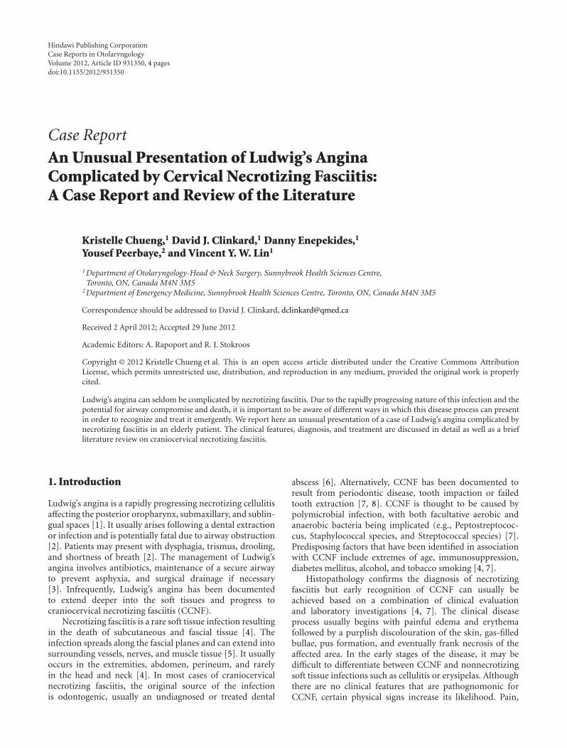
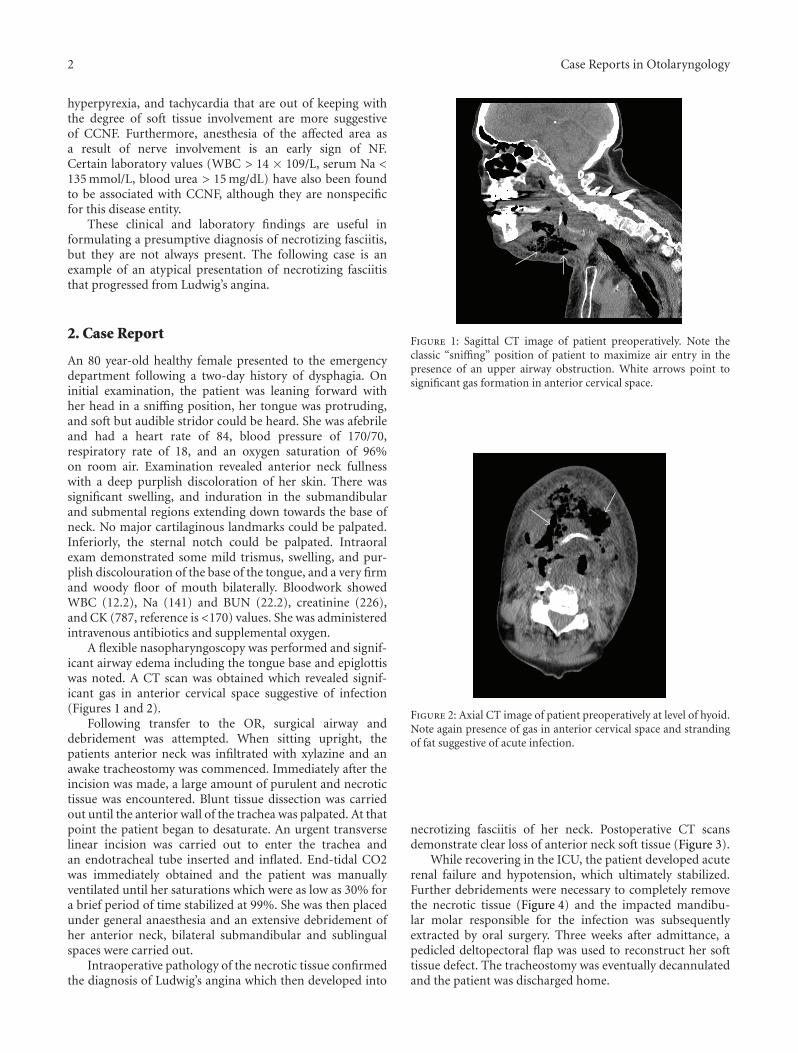
A flexible nasopharyngoscopy was performed and signif-icant airway edema including the tongue base and epiglottiswas noted. A CT scan was obtained which revealed signif-icant gas in anterior cervical space suggestive of infection(Figures 1 and 2).
Following transfer to the OR, surgical airway anddebridement was attempted. When sitting upright, thepatients anterior neck was infiltrated with xylazine and anawake tracheostomy was commenced. Immediately after theincision was made, a large amount of purulent and necrotictissue was encountered. Blunt tissue dissection was carriedout until the anterior wall of the trachea was palpated. At thatpoint the patient began to desaturate. An urgent transverselinear incision was carried out to enter the trachea andan endotracheal tube inserted and inflated. End-tidal CO2was immediately obtained and the patient was manuallyventilated until her saturations which were as low as 30% fora brief period of time stabilized at 99%. She was then placedunder general anaesthesia and an extensive debridement ofher anterior neck, bilateral submandibular and sublingualspaces were carried out.
Intraoperative pathology of the necrotic tissue confirmedthe diagnosis of Ludwig’s angina which then developed into
Figure 1: Sagittal CT image of patient preoperatively. Note theclassic “sniffing” position of patient to maximize air entry in thepresence of an upper airway obstruction. White arrows point tosignificant gas formation in anterior cervical space.
Figure 2: Axial CT image of patient preoperatively at level of hyoid.Note again presence of gas in anterior cervical space and strandingof fat suggestive of acute infection.
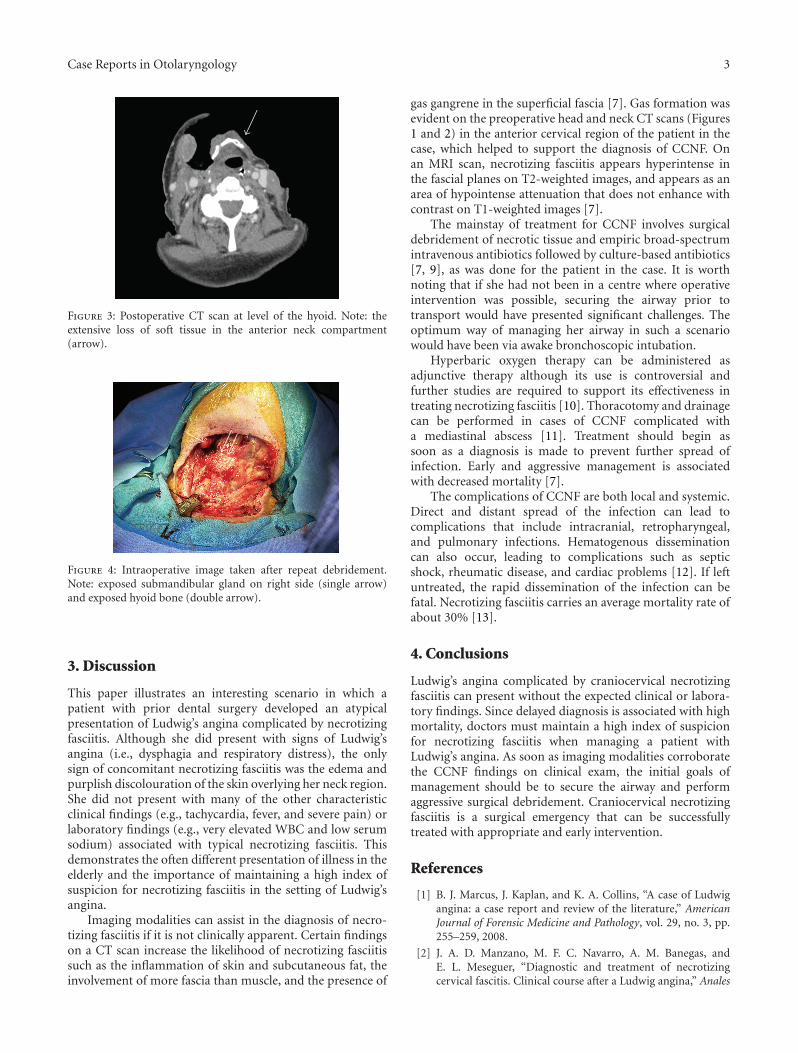
necrotizing fasciitis of her neck. Postoperative CT scansdemonstrate clear loss of anterior neck soft tissue (Figure 3).
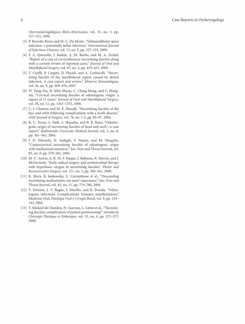
While recovering in the ICU, the patient developed acuterenal failure and hypotension, which ultimately stabilized.Further debridements were necessary to completely removethe necrotic tissue (Figure 4) and the impacted mandibu-lar molar responsible for the infection was subsequentlyextracted by oral surgery. Three weeks after admittance, apedicled deltopectoral flap was used to reconstruct her softtissue defect. The tracheostomy was eventually decannulatedand the patient was discharged home.
Case Reports in Otolaryngology 3
Figure 3: Postoperative CT scan at level of the hyoid. Note: theextensive loss of soft tissue in the anterior neck compartment(arrow).
Figure 4: Intraoperative image taken after repeat debridement.Note: exposed submandibular gland on right side (single arrow)and exposed hyoid bone (double arrow).
3. Discussion
This paper illustrates an interesting scenario in which apatient with prior dental surgery developed an atypicalpresentation of Ludwig’s angina complicated by necrotizingfasciitis. Although she did present with signs of Ludwig’sangina (i.e., dysphagia and respiratory distress), the onlysign of concomitant necrotizing fasciitis was the edema andpurplish discolouration of the skin overlying her neck region.She did not present with many of the other characteristicclinical findings (e.g., tachycardia, fever, and severe pain) orlaboratory findings (e.g., very elevated WBC and low serumsodium) associated with typical necrotizing fasciitis. Thisdemonstrates the often different presentation of illness in theelderly and the importance of maintaining a high index ofsuspicion for necrotizing fasciitis in the setting of Ludwig’sangina.
Imaging modalities can assist in the diagnosis of necro-tizing fasciitis if it is not clinically apparent. Certain findingson a CT scan increase the likelihood of necrotizing fasciitissuch as the inflammation of skin and subcutaneous fat, theinvolvement of more fascia than muscle, and the presence of
gas gangrene in the superficial fascia [7]. Gas formation wasevident on the preoperative head and neck CT scans (Figures1 and 2) in the anterior cervical region of the patient in thecase, which helped to support the diagnosis of CCNF. Onan MRI scan, necrotizing fasciitis appears hyperintense inthe fascial planes on T2-weighted images, and appears as anarea of hypointense attenuation that does not enhance withcontrast on T1-weighted images [7].
The mainstay of treatment for CCNF involves surgicaldebridement of necrotic tissue and empiric broad-spectrumintravenous antibiotics followed by culture-based antibiotics[7, 9], as was done for the patient in the case. It is worthnoting that if she had not been in a centre where operativeintervention was possible, securing the airway prior totransport would have presented significant challenges. Theoptimum way of managing her airway in such a scenariowould have been via awake bronchoscopic intubation.
Hyperbaric oxygen therapy can be administered asadjunctive therapy although its use is controversial andfurther studies are required to support its effectiveness intreating necrotizing fasciitis [10]. Thoracotomy and drainagecan be performed in cases of CCNF complicated witha mediastinal abscess [11]. Treatment should begin assoon as a diagnosis is made to prevent further spread ofinfection. Early and aggressive management is associatedwith decreased mortality [7].
The complications of CCNF are both local and systemic.Direct and distant spread of the infection can lead tocomplications that include intracranial, retropharyngeal,and pulmonary infections. Hematogenous disseminationcan also occur, leading to complications such as septicshock, rheumatic disease, and cardiac problems [12]. If leftuntreated, the rapid dissemination of the infection can befatal. Necrotizing fasciitis carries an average mortality rate ofabout 30% [13].
4. Conclusions
Ludwig’s angina complicated by craniocervical necrotizingfasciitis can present without the expected clinical or labora-tory findings. Since delayed diagnosis is associated with highmortality, doctors must maintain a high index of suspicionfor necrotizing fasciitis when managing a patient withLudwig’s angina. As soon as imaging modalities corroboratethe CCNF findings on clinical exam, the initial goals ofmanagement should be to secure the airway and performaggressive surgical debridement. Craniocervical necrotizingfasciitis is a surgical emergency that can be successfullytreated with appropriate and early intervention.
References
[1] B. J. Marcus, J. Kaplan, and K. A. Collins, “A case of Ludwigangina: a case report and review of the literature,” AmericanJournal of Forensic Medicine and Pathology, vol. 29, no. 3, pp.255–259, 2008.
[2] J. A. D. Manzano, M. F. C. Navarro, A. M. Banegas, andE. L. Meseguer, “Diagnostic and treatment of necrotizingcervical fascitis. Clinical course after a Ludwig angina,” Anales
4 Case Reports in Otolaryngology
Otorrinolaringologicos Ibero-Americanos, vol. 33, no. 3, pp.317–322, 2006.
[3] P. Boscolo-Rizzo and M. C. Da Mosto, “Submandibular spaceinfection: a potentially lethal infection,” International Journalof Infectious Diseases, vol. 13, no. 3, pp. 327–333, 2009.
[4] F. A. Quereshy, J. Baskin, A. M. Barbu, and M. A. Zechel,“Report of a case of cervicothoracic necrotizing fasciitis alongwith a current review of reported cases,” Journal of Oral andMaxillofacial Surgery, vol. 67, no. 2, pp. 419–423, 2009.
[5] T. Cutilli, P. Cargini, D. Placidi, and A. Corbacelli, “Necro-tizing fasciitis of the maxillofacial region caused by dentalinfection. A case report and review,” Minerva Stomatologica,vol. 56, no. 9, pp. 469–476, 2007.
[6] W. Tung-Yiu, H. Jehn-Shyun, C. Ching-Hung, and C. Hung-An, “Cervical necrotizing fasciitis of odontogenic origin: areport of 11 cases,” Journal of Oral and Maxillofacial Surgery,vol. 58, no. 12, pp. 1347–1352, 2000.
[7] C. I. Clement and M. E. Hassall, “Necrotizing fasciitis of theface and orbit following complications with a tooth abscess,”ANZ Journal of Surgery, vol. 74, no. 1-2, pp. 85–87, 2004.
[8] K. C. Toran, S. Nath, S. Shrestha, and B. B. Rana, “Odonto-genic origin of necrotizing fasciitis of head and neck—a casereport,” Kathmandu University Medical Journal, vol. 2, no. 8,pp. 361–363, 2004.
[9] J. D. Edwards, N. Sadeghi, F. Najam, and M. Margolis,“Craniocervical necrotizing fasciitis of odontogenic originwith mediastinal extension,” Ear, Nose and Throat Journal, vol.83, no. 8, pp. 579–582, 2004.
[10] M. U. Anwar, A. K. M. F. Haque, J. Rahman, R. Morris, and J.McDermott, “Early radical surgery and antimicrobial therapywith hyperbaric oxygen in necrotizing fasciitis,” Plastic andReconstructive Surgery, vol. 121, no. 1, pp. 360–361, 2008.
[11] R. Mora, B. Jankowska, U. Catrambone et al., “Descendingnecrotizing mediastinitis: ten years’ experience,” Ear, Nose andThroat Journal, vol. 83, no. 11, pp. 774–780, 2004.
[12] Y. Jimenez, J. V. Bagan, J. Murillo, and R. Poveda, “Odon-togenic infections. Complications. Systemic manifestations,”Medicina Oral, Patologia Oral y Cirugia Bucal, vol. 9, pp. 143–143, 2004.
[13] V. Medard de Chardon, N. Guevara, L. Lattes et al., “Necrotiz-ing fasciitis: complication of patient positionning?” Annales deChirurgie Plastique et Esthetique, vol. 53, no. 4, pp. 372–377,2008.

Submit your manuscripts athttp://www.hindawi.com
Stem CellsInternational
Hindawi Publishing Corporationhttp://www.hindawi.com Volume 2014
Hindawi Publishing Corporationhttp://www.hindawi.com Volume 2014
MEDIATORSINFLAMMATION
of
Hindawi Publishing Corporationhttp://www.hindawi.com Volume 2014
Behavioural Neurology
EndocrinologyInternational Journal of
Hindawi Publishing Corporationhttp://www.hindawi.com Volume 2014
Hindawi Publishing Corporationhttp://www.hindawi.com Volume 2014
Disease Markers
Hindawi Publishing Corporationhttp://www.hindawi.com Volume 2014
BioMed Research International
OncologyJournal of
Hindawi Publishing Corporationhttp://www.hindawi.com Volume 2014
Hindawi Publishing Corporationhttp://www.hindawi.com Volume 2014
Oxidative Medicine and Cellular Longevity
Hindawi Publishing Corporationhttp://www.hindawi.com Volume 2014
PPAR Research
The Scientific World JournalHindawi Publishing Corporation http://www.hindawi.com Volume 2014
Immunology ResearchHindawi Publishing Corporationhttp://www.hindawi.com Volume 2014
Journal of
ObesityJournal of
Hindawi Publishing Corporationhttp://www.hindawi.com Volume 2014
Hindawi Publishing Corporationhttp://www.hindawi.com Volume 2014
Computational and Mathematical Methods in Medicine
OphthalmologyJournal of
Hindawi Publishing Corporationhttp://www.hindawi.com Volume 2014
Diabetes ResearchJournal of
Hindawi Publishing Corporationhttp://www.hindawi.com Volume 2014
Hindawi Publishing Corporationhttp://www.hindawi.com Volume 2014
Research and TreatmentAIDS
Hindawi Publishing Corporationhttp://www.hindawi.com Volume 2014
Gastroenterology Research and Practice
Hindawi Publishing Corporationhttp://www.hindawi.com Volume 2014
Parkinson’s Disease
Evidence-Based Complementary and Alternative Medicine
Volume 2014Hindawi Publishing Corporationhttp://www.hindawi.com