Embed Size (px)
Citation preview
ORIGINAL ARTICLE
Cervical medullary syndrome secondary to craniocervicalinstability and ventral brainstem compression in hereditaryhypermobility connective tissue disorders: 5-year follow-upafter craniocervical reduction, fusion, and stabilization
Fraser C. Henderson Sr1,2 & C. A. Francomano1& M. Koby1 & K. Tuchman2
& J. Adcock3 & S. Patel4
Received: 10 October 2018 /Revised: 28 November 2018 /Accepted: 10 December 2018# The Author(s) 2019
AbstractA great deal of literature has drawn attention to the Bcomplex Chiari,^ wherein the presence of instability or ventral brainstemcompression prompts consideration for addressing both concerns at the time of surgery. This report addresses the clinical andradiological features and surgical outcomes in a consecutive series of subjects with hereditary connective tissue disorders(HCTD) and Chiari malformation. In 2011 and 2012, 22 consecutive patients with cervical medullary syndrome andgeneticist-confirmed hereditary connective tissue disorder (HCTD), with Chiari malformation (type 1 or 0) and kyphoticclivo-axial angle (CXA) enrolled in the IRB-approved study (IRB# 10-036-06: GBMC). Two subjects were excluded on thebasis of previous cranio-spinal fusion or unrelated medical issues. Symptoms, patient satisfaction, and work status were assessedby a third-party questionnaire, pain by visual analog scale (0–10/10), neurologic exams by neurosurgeon, function by Karnofskyperformance scale (KPS). Pre- and post-operative radiological measurements of clivo-axial angle (CXA), the Grabb-Mapstone-Oakes measurement, and Harris measurements were made independently by neuroradiologist, with pre- and post-operativeimaging (MRI and CT), 10/20 with weight-bearing, flexion, and extension MRI. All subjects underwent open reduction,stabilization occiput to C2, and fusion with rib autograft. There was 100% follow-up (20/20) at 2 and 5 years. Patients weresatisfied with the surgery and would do it again given the same circumstances (100%). Statistically significant improvement wasseen with headache (8.2/10 pre-op to 4.5/10 post-op, p < 0.001, vertigo (92%), imbalance (82%), dysarthria (80%), dizziness(70%), memory problems (69%), walking problems (69%), function (KPS) (p < 0.001). Neurological deficits improved in allsubjects. The CXA average improved from 127° to 148° (p < 0.001). The Grabb-Oakes and Harris measurements returned tonormal. Fusion occurred in 100%. There were no significant differences between the 2- and 5-year period. Two patients returnedto surgery for a superficial wound infections, and two required transfusion. All patients who had rib harvests had pain related thatprocedure (3/10), which abated by 5 years. The results support the literature, that open reduction of the kyphotic CXA to lessenventral brainstem deformity, and fusion/stabilization to restore stability in patients with HCTD is feasible, associated with a lowsurgical morbidity, and results in enduring improvement in pain and function. Rib harvest resulted in pain for several years inalmost all subjects.
Keywords Ehlers-Danlos syndrome . Craniocervical instability . Clivo-axial angle . Cervical medullary syndrome
Introduction
Many studies have drawn attention to the presence ofcraniocervical instability or basilar invagination in patientswith Chiari one and Chiari zero malformation [1–23]. Theneed for reduction and stabilization in basilar invaginationand craniocervical instability are recognized in connective tis-sue joint degenerative disorders, such as rheumatoid arthritisand lupus [10, 17, 24–36] and hereditary hypermobile and
* Fraser C. Henderson, [email protected]
1 Doctor’s Community Hospital, Lanham, MD, USA2 The Metropolitan Neurosurgery Group, LLC, Silver Spring, MD,
USA3 Harvey Institute of Human Genetics, Greater Baltimore Medical
Center, Baltimore, MD, USA4 Medical University of South Carolina, Charleston, SC, USA
Neurosurgical Reviewhttps://doi.org/10.1007/s10143-018-01070-4

developmental disorders, including osteogenesis imperfecta,achondroplasia, Down syndrome and Ehlers-Danlos syn-drome (EDS) [8, 18, 21, 26, 31, 37–50].
Emblematic of the approximately 50 heritable connectivetissue disorders characterized by joint hypermobility is Ehlers-Danlos syndrome (EDS). Though Ehlers-Danlos syndromewas described in 1905, its neurological and spinal manifesta-tions have only recently been appreciated [18, 41, 51–66].These heritable connective tissue disorders are characterizedby tissue fragility, skin extensibility, joint hypermobility, pre-mature disk degeneration and spinal problems, and numerouscomorbid conditions.
We report on an IRB-approved retrospective cohort studyof 20 consecutive patients with hereditary connective tissuedisorders and a kyphotic CXA, cerebellar ectopia (18/20), andcraniocervical instability or ventral brainstem compression,who underwent reduction and stabilization. This is the firstsuch study to critically assess 5-year outcomes aftercraniocervical reduction, stabilization, and fusion in a patientpopulation with hereditary connective tissue disorders.
In this study, the CXA (clivo-axial angle) was used to in-dicate potential brainstem deformity. The CXA has drawnincreasing attention as an important radiological metric to in-dicate the presence of neurological deficit and considerationfor craniocervical stabilization [4]. The line of reasoning that akyphotic CXA is associated with pathologic bending of thebrainstem (medullary kyphosis, or kink) began with Liszt,who first recognized that clivo-axial kyphosis may result inneurobehavioral effects. Van Gilder reported that CXA of lessthan 150° were often associated with neurological deficits[67]. Breig demonstrated the importance of mechanical ten-sion and deformation of the brainstem [68]. Menezes de-scribed the Bfulcrum effect in basilar invagination, by whichtraction is applied to the caudal brainstem and rostral cervicalspinal cord. Others have demonstrated the salutary conse-quences to the correction of the CXA [1, 10, 12, 15, 30, 49,69–75].
It is important to recognize that the CXA is simply a staticrepresentation of a dynamic phenomenon. It has been gener-ally considered that a CXA of less than 135° represents thethreshold below which chronic repetitive injury may occur asa result of mechanical deformation of the lower brainstem andupper spinal cord.
The authors’ hypothesis was that reduction of the Clivo-axial kyphosis and stabilization for craniocervical instabilitywere feasible and associated with clinical improvement in thehereditary connective tissue disorder (HCTD) population.
Materials and methods
Subject enrollment Over a 2-year period (2011–2012), a co-hort of 22 consecutive patients diagnosed with EDS, or in a
few cases, unspecified hereditary connective tissue disorders(HCTD), were enrolled in the study and underwent occipitalto C1/C2 fusion for craniovertebral instability and flexion de-formity. Of the original 22 consecutive subjects, two wereexcluded: one had previously undergone a cranio-spinal fu-sion, and the second declined to participate due to unrelatedmedical issues. The data analysis was, therefore, conducted onthe remaining 20 subjects, all of whom were enrolled in theIRB-approved study (IRB# 10-036-06: Greater BaltimoreMedical Center). In 18 patients, cerebellar ectopia was alsopresent.
Evaluation Symptoms were assessed by a standardized ques-tionnaire administered by third party at 2 and 5 years. Painwas assessed by the visual analog scale for pain (0–10/10).The neurologic exams were performed by the neurosurgeon.Function and the ability to return to work were assessed withthe Karnofsky Performance Scale (Fig. 1). Radiological mea-surements were performed by a neuroradiologist (MK) after2 years.
Pre- and post-operative radiological measurements weremade or reviewed by the neuroradiologist (MK). Subjectsunderwent pre-operative and post-operative imaging withMRI and CT of the cervical spine. Upright, weight-bearingflexion and extension MRI of the cervical spine was obtainedin 10/20 of the subjects.
Radiometrics were performed at the 2-year follow-up andincluded the clivo-axial angle (CXA), Grabb-Mapstone-Oaksmeasurement (the pBC2), and the horizontal HarrisMeasurement (Basion axis interval or BAI). CXA is the mea-surement in degrees between the line drawn along the lowerthird of the clivus, and a line drawn along the posterior aspect
Fig. 1 The Karnofsky Performance Status Scale
Neurosurg Rev
of the axis [1, 76] (Fig. 2a). The CXA measurements weretaken from the flexion image, when it was available (Fig. 2b,c).
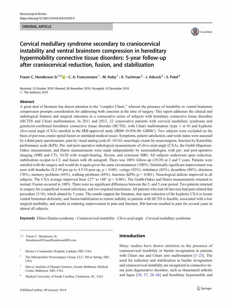
The pBC2, or Grabb, Oakes measurement (Fig. 3) is theperpendicular measurement from the dura to a line drawnfrom the basion to the posterior inferior aspect of C2 [7, 76,77].
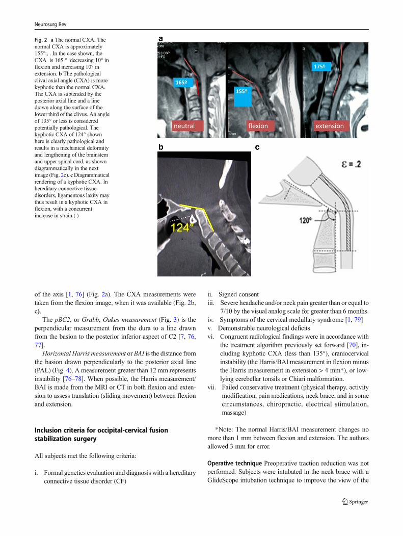
Horizontal Harris measurement or BAI is the distance fromthe basion drawn perpendicularly to the posterior axial line(PAL) (Fig. 4). A measurement greater than 12 mm representsinstability [76–78]. When possible, the Harris measurement/BAI is made from the MRI or CT in both flexion and exten-sion to assess translation (sliding movement) between flexionand extension.
Inclusion criteria for occipital-cervical fusionstabilization surgery
All subjects met the following criteria:
i. Formal genetics evaluation and diagnosis with a hereditaryconnective tissue disorder (CF)
ii. Signed consentiii. Severe headache and/or neck pain greater than or equal to
7/10 by the visual analog scale for greater than 6 months.iv. Symptoms of the cervical medullary syndrome [1, 79]v. Demonstrable neurological deficitsvi. Congruent radiological findings were in accordance with
the treatment algorithm previously set forward [70], in-cluding kyphotic CXA (less than 135°), craniocervicalinstability (the Harris/BAI measurement in flexion minusthe Harris measurement in extension > 4 mm*), or low-lying cerebellar tonsils or Chiari malformation.
vii. Failed conservative treatment (physical therapy, activitymodification, pain medications, neck brace, and in somecircumstances, chiropractic, electrical stimulation,massage)
*Note: The normal Harris/BAI measurement changes nomore than 1 mm between flexion and extension. The authorsallowed 3 mm for error.
Operative technique Preoperative traction reduction was notperformed. Subjects were intubated in the neck brace with aGlideScope intubation technique to improve the view of the
Fig. 2 a The normal CXA. Thenormal CXA is approximately155°;, . In the case shown, theCXA is 165 ° decreasing 10° inflexion and increasing 10° inextension. b The pathologicalclival axial angle (CXA) is morekyphotic than the normal CXA.The CXA is subtended by theposterior axial line and a linedrawn along the surface of thelower third of the clivus. An angleof 135° or less is consideredpotentially pathological. Thekyphotic CXA of 124° shownhere is clearly pathological andresults in a mechanical deformityand lengthening of the brainstemand upper spinal cord, as showndiagrammatically in the nextimage (Fig. 2c). cDiagrammaticalrendering of a kyphotic CXA. Inhereditary connective tissuedisorders, ligamentous laxity maythus result in a kyphotic CXA inflexion, with a concurrentincrease in strain ( )
Neurosurg Rev
glottis and to avoid hyperextension of the neck. Sensoryevoked potentials were performed throughout the surgery. Athree-pronged Mayfield head holder was placed, and the sub-ject positioned prone on chest rolls. The cervical spine wascarefully aligned to eliminate tilt and rotation, and then placedin a neutral position, as confirmed by cross table fluoroscopy.After sterile prep and drape, the incision was made from inionto C4, but the subperiosteal exposure was limited to the occi-put, C1and C2. Care was taken to preserve the ligaments at-tached to the dorsal aspect of the spinous process of C2 and tothe caudal aspect of the C2 lamina.
A limited sub occipital decompression was performed withhigh speed burr and Kerrison rongeur from the foramen
magnum upward 14 mm, but carried laterally to the full me-ridian of the dura. The dura was not opened, and thus, noexpansion duroplasty was performed.
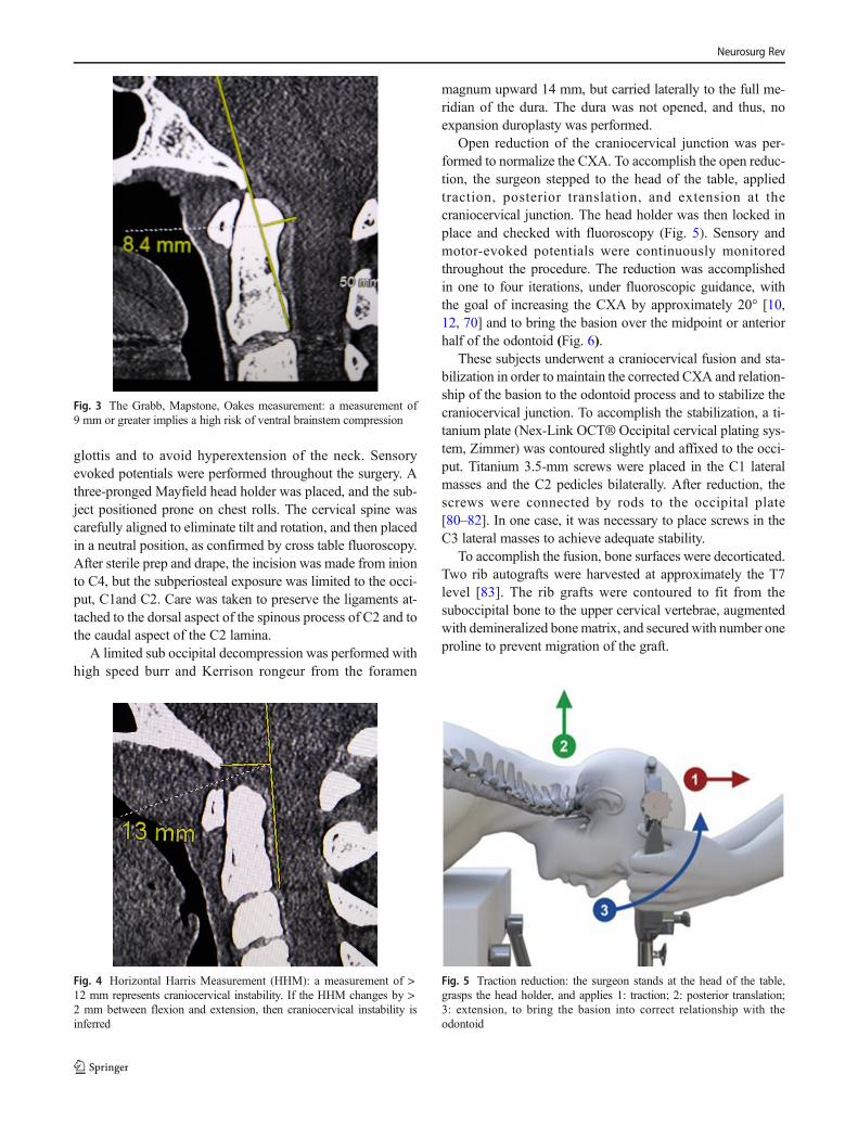
Open reduction of the craniocervical junction was per-formed to normalize the CXA. To accomplish the open reduc-tion, the surgeon stepped to the head of the table, appliedtraction, posterior translation, and extension at thecraniocervical junction. The head holder was then locked inplace and checked with fluoroscopy (Fig. 5). Sensory andmotor-evoked potentials were continuously monitoredthroughout the procedure. The reduction was accomplishedin one to four iterations, under fluoroscopic guidance, withthe goal of increasing the CXA by approximately 20° [10,12, 70] and to bring the basion over the midpoint or anteriorhalf of the odontoid (Fig. 6).
These subjects underwent a craniocervical fusion and sta-bilization in order to maintain the corrected CXA and relation-ship of the basion to the odontoid process and to stabilize thecraniocervical junction. To accomplish the stabilization, a ti-tanium plate (Nex-Link OCT® Occipital cervical plating sys-tem, Zimmer) was contoured slightly and affixed to the occi-put. Titanium 3.5-mm screws were placed in the C1 lateralmasses and the C2 pedicles bilaterally. After reduction, thescrews were connected by rods to the occipital plate[80–82]. In one case, it was necessary to place screws in theC3 lateral masses to achieve adequate stability.
To accomplish the fusion, bone surfaces were decorticated.Two rib autografts were harvested at approximately the T7level [83]. The rib grafts were contoured to fit from thesuboccipital bone to the upper cervical vertebrae, augmentedwith demineralized bonematrix, and secured with number oneproline to prevent migration of the graft.
Fig. 5 Traction reduction: the surgeon stands at the head of the table,grasps the head holder, and applies 1: traction; 2: posterior translation;3: extension, to bring the basion into correct relationship with theodontoid
Fig. 4 Horizontal Harris Measurement (HHM): a measurement of >12 mm represents craniocervical instability. If the HHM changes by >2 mm between flexion and extension, then craniocervical instability isinferred
Fig. 3 The Grabb, Mapstone, Oakes measurement: a measurement of9 mm or greater implies a high risk of ventral brainstem compression
Neurosurg Rev
Both the neck and graft harvest wounds were then closedover drains. The patients were usually mobilized 1 day aftersurgery and kept in a neck brace (Miami J™, or equivalent)for 4 weeks. Physical therapy was then started.
Statement of human and animal rights All procedures per-formed in studies involving human participants were carriedout in accordance with the ethical standards of the institutionaland/or national research committee in the United States, andwith the 1964Helsinki declaration and its later amendments orcomparable ethical standards. Informed consent was obtainedfrom all individual patients and participants included in thestudy.
Results
Nineteen subjects were female and one male, with an averageage of 24 years (range of 12–53 years). All patients werediagnosed with a hereditary connective tissue disorder(HCTD): ten had hypermobile EDS (h-EDS), two classicalEDS, four unspecified EDS, and four hypermobility spectrumdisorder. All subjects (20/20) had a kyphotic CXA (less thanor equal to 135°) and craniocervical instability (HarrisMeasurement/BAI of 4 mm or greater). Eighteen subjectshad cerebellar ectopia.
Pre-operative findings
The most prominent symptoms prior to surgery includedheadache (100%), fatigue (100%), dizziness (100%), musclepain, vertigo, arm weakness, neck pain, balance problems,memory problems, night awakenings, numbness and weak-ness of the arms and legs, and gait problems (Table 1).
Patient satisfaction
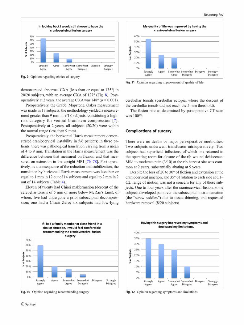
There was 100% follow-up at 2 years and 5 years (Figs. 7 and8). All patients were satisfied with the surgery and wouldrepeat the surgery given similar circumstances, and reportedimproved quality of life (Figs. 9, 10, and 11). All but onepatient would recommend the surgery to a family member(Fig. 10). Eighteen of the twenty patients reported that thecraniocervical fusion surgery had decreased their limitations;the remaining two patients, who responded that the limitationshad not decreased with surgery, explained that there remainedlimitations from other medical problems and spinal instabilityelsewhere (Fig. 12).
Postoperative findings
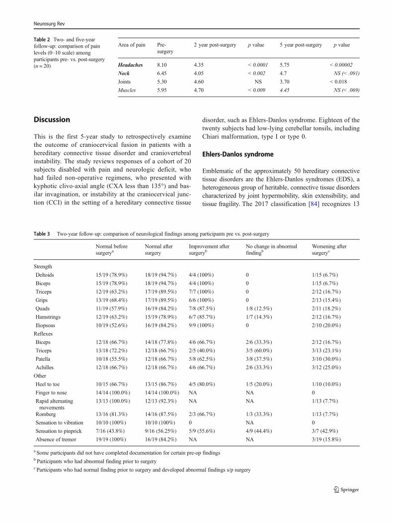
Postoperatively at 2 years, statistically significant improve-ments were seen in vertigo (92%), headaches (85%), imbal-ance (82%), dysarthria (80%), dizziness (70%), memory(69%), walking (69%), and frequent daytime urination(42%) (Table 1). The average headache decreased from 8.1/10 pre-op to 4.35/10 post-op (p < 0.0001). Neck pain meandecreased in 71% of patients, from 6.45/10 to 4.05/10 post-op(p < 0.002), and muscle pain decreased from 6/10 to 4.7/10post-op (p < 0.009) (Table 2).
Improvement, though not statistically significant, includedtremors (87%), syncope (86%), numbness of the arms andhands (73%), upper extremity numbness (73%), lower ex-tremity weakness (69%), back numbness (67%), swallowingdifficulty (63%), upper extremity weakness (61%), hearingproblems (61%), lower extremity numbness (55%), andGERDS (55%) (Table 1).
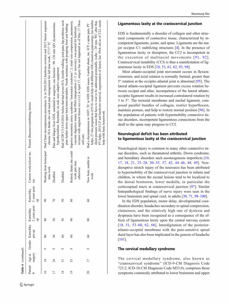
Similarly, at 5 years, there remained statistically significantimprovement in dizziness (75%), walking problems (69%),speech problems (67%), frequent daytime urination (67%),headaches (65%), and imbalance (59%). Improvement in up-per extremity numbness, syncope, lower extremity weakness,back numbness, swallowing difficulty, upper extremity weak-ness, hearing problems, and lower extremity numbness wereimproved but not with statistical significance (Tables 2, 3, 4,and 5).
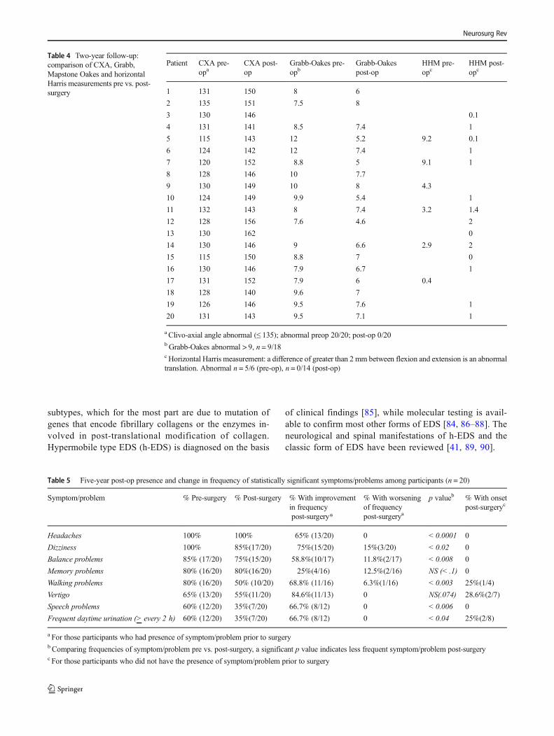
On neurological examination, those who were weak beforesurgery improved, though not completely. The ability to walkheel-to-toe, Romberg, and sensation were all improved. Therewas no significant improvement in reflexes (Table 3).
Functional outcome
Function and the ability to return to work, as assessed with theKarnofsky Performance Scale, demonstrated a highly statical-ly significant improvement (p < 0.001). Preoperatively, 12/20subjects were completely disabled, and 4/20 were able to carefor themselves only, but unable to go to work or school.
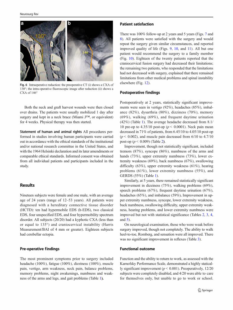
Fig. 6 Intraoperative reduction: the preoperative CT (i) shows a CXA of130°; the intra-operative fluoroscopic image after reduction (ii) shows aCXA of 146°
Neurosurg Rev
Table1
Two-year
follo
w-up:
presence
andchange
infrequencyof
symptom
s/problemsam
ongparticipants(n=20)
Symptom
/problem
%Pre-surgery
%Post-surgery
%With
improvem
ent
infrequencypost-surgery
a%
With
worsening
offrequencypost-surgery
apvalueb
%With
onset
post-surgery
c
Headaches
100%
95%
(19/20)
85%
(17/20)
0<0.001
0
Fatig
ue100%
100%
30%
(6/20)
15%
(3/20)
NS
0
Dizziness
100%
95%
(19/20)
70%
(14/20)
10%
(2/20)
<0.0007
0
Musclepain
95%
(19/20)
95%
(19/20)
36.8%
(7/19)
10.5%
(2/19)
NS
0
Upper
extrem
ityweakness
90%
(18/20)
85%
(17/20)
61.1%
(11/18)
22.2%
(4/18)
NS
0
Jointp
ain
85%
(17/20)
85%
(17/20)
29.4%
(5/17)
11.8%
(2/17)
NS
0
Neckpain
85%
(17/20)
90%
(18/20)
70.6%
(12/17)
5.9%
(1/17)
NS
33.3%
(1/3)
Balance
problems
85%
(17/20)
85%
(17/20)
82.4%
(14/17)
5.9%
(1/17)
<0.0001
0
Night
awakenings
85%
(17/20)
85%
(17/20)
23.5%
(4/17)
11.8%
(2/17)
NS
0
Mem
oryproblems
80%
(16/20)
80%
(16/20)
68.9%
(11/16)
0<0.002
0
Walking
problems
80%
(16/20)
70%
(14/20)
68.9%
(11/16)
6.3%
(1/16)
<0.002
0
Upper
extrem
itynumbness
75%
(15/20)
85%
(17/20)
73.3%
(11/15)
6.7%
(1/15)
NS
40%
(2/5)
Hands
andfeetturningcold
75%
(15/20)
70%
(14/20)
26.75%
(4/15)
6.7%
(1/15)
NS
0
Low
erextrem
itynumbness
75%
(15/20)
70%
(14/20)
60%
(9/15)
13.3%
(2/15)
NS
0
Visualp
roblem
s75%
(15/20)
80%
(16/20)
53.3%
(8/15)
13.3%
(2/15)
NS
20%
(1/5)
Low
erextrem
ityweakness
65%
(13/20)
70%
(14/20)
69.2%
(9/13)
15.4%
(2/13)
NS
14.3%
(1/7)
Vertigo
65%
(13/20)
50%
(10/20)
92.3%
(12/13)
0<0.0006
0
Hearing
problems
65%
(13/20)
65%
(13/20)
61.5%
(8/13)
15.4%
(2/13)
NS(0.053)
14.3%
(1/7)
Speech
problems
60%
(12/20)
55%
(11/20)
80%
(8/12)
8.3%
(1/12)
<0.03
0
Frequentd
aytim
eurination(>
every2h)
60%
(12/20)
45%
(9/20)
41.7%
(5/12)
0<0.02
0
GERD
55%
(11/20)
55%
(11/20)
36.4%
(4/11)
0NS
11.1%
(1/9)
Swallowing/chokingproblems
55%
(11/20)
55%
(11/20)
63.4%
(7/11)
18.2%
(2/11)
NS
22.2%
(2/9)
Nocturia(>
twiceanight)
55%
(11/20)
55%
(11/20)
27.3%
(3/11)
9.1%
(1/11)
NS
11.1%
(1/9)
IBS
50%
(10/20)
50%
(10/20)
30%
(3/10)
0NS
0
Tremors
40%
(8/20)
40%
(8/20)
87.5%
(7/8)
0NS
0
Faintin
g35%
(7/20)
25%
(5/20)
85.7%
(6/7)
0NS
14.3%
(1/7)
Num
bnessin
back
30%
(6/20)
40%
(8/20)
66.7%
(4/6)
0NS
14.3%
(2/14)
Sleepapnea
25%
(5/20)
25%
(5/20)
20%
(1/5)
0NS
0
aFo
rthoseparticipantswho
hadpresence
ofsymptom
/problem
priorto
surgery
bCom
paring
frequenciesof
symptom
/problem
prevs.post-surgery,asignificantp
valueindicatesless
frequent
symptom
/problem
post-surgery
cFo
rthoseparticipantswho
didnoth
avethepresence
ofsymptom
/problem
priorto
surgery
Neurosurg Rev
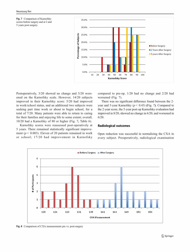
Postoperatively, 3/20 showed no change and 3/20 wors-ened on the Karnofsky scale. However, 14/20 subjectsimproved in their Karnofsky score: 5/20 had improvedin work/school status, and an additional two subjects wereseeking part time work or about to begin school, for atotal of 7/20. Many patients were able to return to caringfor their families and enjoying life to some extent; overall,10/20 had a Karnofsky of 80 or higher (Fig. 7, Table 6).
Karnofsky scores were reassessed post-operatively at5 years. There remained statistically significant improve-ment (p < 0.003). Eleven of 20 patients remained in workor school; 17/20 had improvement in Karnofsky
compared to pre-op, 1/20 had no change and 2/20 hadworsened (Fig. 7).
There was no significant difference found between the 2-year and 5-year Karnofsky (p < 0.43) (Fig. 7). Compared tothe 2-year score, the 5-year post-op Karnofsky evaluation hadimproved in 8/20, showed no change in 6/20, and worsened in6/20.
Radiological outcomes
Open reduction was successful in normalizing the CXA inevery subject. Preoperatively, radiological examination
0.0%
5.0%
10.0%
15.0%
20.0%
25.0%
30.0%
35.0%
10 20 30 40 50 60 70 80 90 100
Perc
enta
ge o
f Sub
ject
sKarnofsky Score
Before Surgery
2 Years After Surgery
5 years After Surgery
Fig. 7 Comparison of Karnofskyscores before surgery and at 2 and5 years post-surgery
Fig. 8 Comparison of CXA measurements pre vs. post-surgery
Neurosurg Rev
demonstrated abnormal CXA (less than or equal to 135°) in20/20 subjects, with an average CXA of 127° (Fig. 8). Post-operatively at 2 years, the average CXAwas 148° (p < 0.001).
Preoperatively, the Grabb, Mapstone, Oakes measurementwas made in 18 subjects; the methodology yielded a measure-ment greater than 9 mm in 9/18 subjects, constituting a high-risk category for ventral brainstem compression [7].Postoperatively at 2 years, all subjects (20/20) were withinthe normal range (less than 9 mm).
Preoperatively, the horizontal Harris measurement demon-strated craniocervical instability in 5/6 patients; in these pa-tients, there was pathological translation varying from a meanof 4 to 9 mm. Translation in the Harris measurement was thedifference between that measured on flexion and that mea-sured on extension in the upright MRI [76–78]. Post-opera-tively, as a consequence of the reduction and stabilization, thetranslation by horizontal Harris measurement was less than orequal to 1 mm in 12 out of 14 subjects and equal to 2 mm in 2out of 14 subjects (Table 4).
Eleven of twenty had Chiari malformation (descent of thecerebellar tonsils of 5 mm or more below McRae’s Line), ofwhom, five had undergone a prior suboccipital decompres-sion; one had a Chiari Zero; six subjects had low-lying
cerebellar tonsils (cerebellar ectopia, where the descent ofthe cerebellar tonsils did not reach the 5 mm threshold).
The fusion rate as determined by postoperative CT scanwas 100%.
Complications of surgery
There were no deaths or major peri-operative morbidities.Two subjects underwent transfusion intraoperatively. Twosubjects had superficial infections, of which one returned tothe operating room for closure of the rib wound dehiscence.Mild to moderate pain (3/10) at the rib harvest site was com-mon at 2 years, substantially abating at 5 years.
Despite the loss of 20 to 30° of flexion and extension at thecraniocervical junction, and 35° of rotation to each side at C1–C2, range of motion was not a concern for any of these sub-jects. One to four years after the craniocervical fusion, somesubjects developed pain over the suboccipital instrumentation(the Bscrew saddles^) due to tissue thinning, and requestedhardware removal (8/20 subjects).
0%
10%
20%
30%
40%
50%
60%
70%
Strongly
Agree
Agree Somewhat
Agree
Somewhat
Disagree
Disagree Strongly
Disagree
% o
f Sub
ject
s
If I had a family member or close friend in asimilar situa�on, I would feel comfortablerecommending the craniovertebral fusion
surgery
Fig. 10 Opinion regarding recommending surgery
0%10%20%30%40%50%60%70%
StronglyAgree
Agree SomewhatAgree
SomewhatDisagree
Disagree StronglyDisagree
% o
f Sub
ject
sIn looking back I would s�ll choose to have the
craniovertebral fusion surgery
Fig. 9 Opinion regarding choice of surgery
0%
5%
10%
15%
20%
25%
30%
35%
40%
Strongly
Agree
Agree Somewhat
Agree
Somewhat
Disagree
Disagree Strongly
Disagree
% o
f Sub
ject
s
Having this surgery improved my symptoms and decreased my limita�ons.
Fig. 12 Opinion regarding symptoms and limitations
0%
10%
20%
30%
40%
50%
60%
Strongly
Agree
Agree Somewhat
Agree
Somewhat
Disagree
Disagree Strongly
Disagree
% o
f Sub
ject
s
My quality of life was improved by having the craniovertebral fusion surgery
Fig. 11 Opinion regarding improvement of quality of life
Neurosurg Rev
Discussion
This is the first 5-year study to retrospectively examinethe outcome of craniocervical fusion in patients with ahereditary connective tissue disorder and craniovertebralinstability. The study reviews responses of a cohort of 20subjects disabled with pain and neurologic deficit, whohad failed non-operative regimens, who presented withkyphotic clivo-axial angle (CXA less than 135°) and bas-ilar invagination, or instability at the craniocervical junc-tion (CCI) in the setting of a hereditary connective tissue
disorder, such as Ehlers-Danlos syndrome. Eighteen of thetwenty subjects had low-lying cerebellar tonsils, includingChiari malformation, type I or type 0.
Ehlers-Danlos syndrome
Emblematic of the approximately 50 hereditary connectivetissue disorders are the Ehlers-Danlos syndromes (EDS), aheterogeneous group of heritable, connective tissue disorderscharacterized by joint hypermobility, skin extensibility, andtissue fragility. The 2017 classification [84] recognizes 13
Table 2 Two- and five-yearfollow-up: comparison of painlevels (0–10 scale) amongparticipants pre- vs. post-surgery(n = 20)
Area of pain Pre-surgery
2 year post-surgery p value 5 year post-surgery p value
Headaches 8.10 4.35 < 0.0001 5.75 < 0.00002
Neck 6.45 4.05 < 0.002 4.7 NS (< .091)
Joints 5.30 4.60 NS 3.70 < 0.018
Muscles 5.95 4.70 < 0.009 4.45 NS (< .069)
Table 3 Two-year follow-up: comparison of neurological findings among participants pre vs. post-surgery
Normal beforesurgerya
Normal aftersurgery
Improvement aftersurgeryb
No change in abnormalfindingb
Worsening aftersurgeryc
Strength
Deltoids 15/19 (78.9%) 18/19 (94.7%) 4/4 (100%) 0 1/15 (6.7%)
Biceps 15/19 (78.9%) 18/19 (94.7%) 4/4 (100%) 0 1/15 (6.7%)
Triceps 12/19 (63.2%) 17/19 (89.5%) 7/7 (100%) 0 2/12 (16.7%)
Grips 13/19 (68.4%) 17/19 (89.5%) 6/6 (100%) 0 2/13 (15.4%)
Quads 11/19 (57.9%) 16/19 (84.2%) 7/8 (87.5%) 1/8 (12.5%) 2/11 (18.2%)
Hamstrings 12/19 (63.2%) 15/19 (78.9%) 6/7 (85.7%) 1/7 (14.3%) 2/12 (16.7%)
Iliopsoas 10/19 (52.6%) 16/19 (84.2%) 9/9 (100%) 0 2/10 (20.0%)
Reflexes
Biceps 12/18 (66.7%) 14/18 (77.8%) 4/6 (66.7%) 2/6 (33.3%) 2/12 (16.7%)
Triceps 13/18 (72.2%) 12/18 (66.7%) 2/5 (40.0%) 3/5 (60.0%) 3/13 (23.1%)
Patella 10/18 (55.5%) 12/18 (66.7%) 5/8 (62.5%) 3/8 (37.5%) 3/10 (30.0%)
Achilles 12/18 (66.7%) 12/18 (66.7%) 4/6 (66.7%) 2/6 (33.3%) 3/12 (25.0%)
Other
Heel to toe 10/15 (66.7%) 13/15 (86.7%) 4/5 (80.0%) 1/5 (20.0%) 1/10 (10.0%)
Finger to nose 14/14 (100.0%) 14/14 (100.0%) NA NA 0
Rapid alternatingmovements
13/13 (100.0%) 12/13 (92.3%) NA NA 1/13 (7.7%)
Romberg 13/16 (81.3%) 14/16 (87.5%) 2/3 (66.7%) 1/3 (33.3%) 1/13 (7.7%)
Sensation to vibration 10/10 (100%) 10/10 (100%) 0 NA 0
Sensation to pinprick 7/16 (43.8%) 9/16 (56.25%) 5/9 (55.6%) 4/9 (44.4%) 3/7 (42.9%)
Absence of tremor 19/19 (100%) 16/19 (84.2%) NA NA 3/19 (15.8%)
a Some participants did not have completed documentation for certain pre-op findingsb Participants who had abnormal finding prior to surgeryc Participants who had normal finding prior to surgery and developed abnormal findings s/p surgery
Neurosurg Rev
subtypes, which for the most part are due to mutation ofgenes that encode fibrillary collagens or the enzymes in-volved in post-translational modification of collagen.Hypermobile type EDS (h-EDS) is diagnosed on the basis
of clinical findings [85], while molecular testing is avail-able to confirm most other forms of EDS [84, 86–88]. Theneurological and spinal manifestations of h-EDS and theclassic form of EDS have been reviewed [41, 89, 90].
Table 4 Two-year follow-up:comparison of CXA, Grabb,Mapstone Oakes and horizontalHarris measurements pre vs. post-surgery
Patient CXA pre-opa
CXA post-op
Grabb-Oakes pre-opb
Grabb-Oakespost-op
HHM pre-opc
HHM post-opc
1 131 150 8 6
2 135 151 7.5 8
3 130 146 0.1
4 131 141 8.5 7.4 1
5 115 143 12 5.2 9.2 0.1
6 124 142 12 7.4 1
7 120 152 8.8 5 9.1 1
8 128 146 10 7.7
9 130 149 10 8 4.3
10 124 149 9.9 5.4 1
11 132 143 8 7.4 3.2 1.4
12 128 156 7.6 4.6 2
13 130 162 0
14 130 146 9 6.6 2.9 2
15 115 150 8.8 7 0
16 130 146 7.9 6.7 1
17 131 152 7.9 6 0.4
18 128 140 9.6 7
19 126 146 9.5 7.6 1
20 131 143 9.5 7.1 1
a Clivo-axial angle abnormal (≤ 135); abnormal preop 20/20; post-op 0/20bGrabb-Oakes abnormal > 9, n = 9/18cHorizontal Harris measurement: a difference of greater than 2 mm between flexion and extension is an abnormaltranslation. Abnormal n = 5/6 (pre-op), n = 0/14 (post-op)
Table 5 Five-year post-op presence and change in frequency of statistically significant symptoms/problems among participants (n = 20)
Symptom/problem % Pre-surgery % Post-surgery % With improvementin frequencypost-surgery*
% With worseningof frequencypost-surgerya
p valueb % With onsetpost-surgeryc
Headaches 100% 100% 65% (13/20) 0 < 0.0001 0
Dizziness 100% 85%(17/20) 75%(15/20) 15%(3/20) < 0.02 0
Balance problems 85% (17/20) 75%(15/20) 58.8%(10/17) 11.8%(2/17) < 0.008 0
Memory problems 80% (16/20) 80%(16/20) 25%(4/16) 12.5%(2/16) NS (< .1) 0
Walking problems 80% (16/20) 50% (10/20) 68.8% (11/16) 6.3%(1/16) < 0.003 25%(1/4)
Vertigo 65% (13/20) 55%(11/20) 84.6%(11/13) 0 NS(.074) 28.6%(2/7)
Speech problems 60% (12/20) 35%(7/20) 66.7% (8/12) 0 < 0.006 0
Frequent daytime urination (> every 2 h) 60% (12/20) 35%(7/20) 66.7% (8/12) 0 < 0.04 25%(2/8)
a For those participants who had presence of symptom/problem prior to surgeryb Comparing frequencies of symptom/problem pre vs. post-surgery, a significant p value indicates less frequent symptom/problem post-surgeryc For those participants who did not have the presence of symptom/problem prior to surgery
Neurosurg Rev
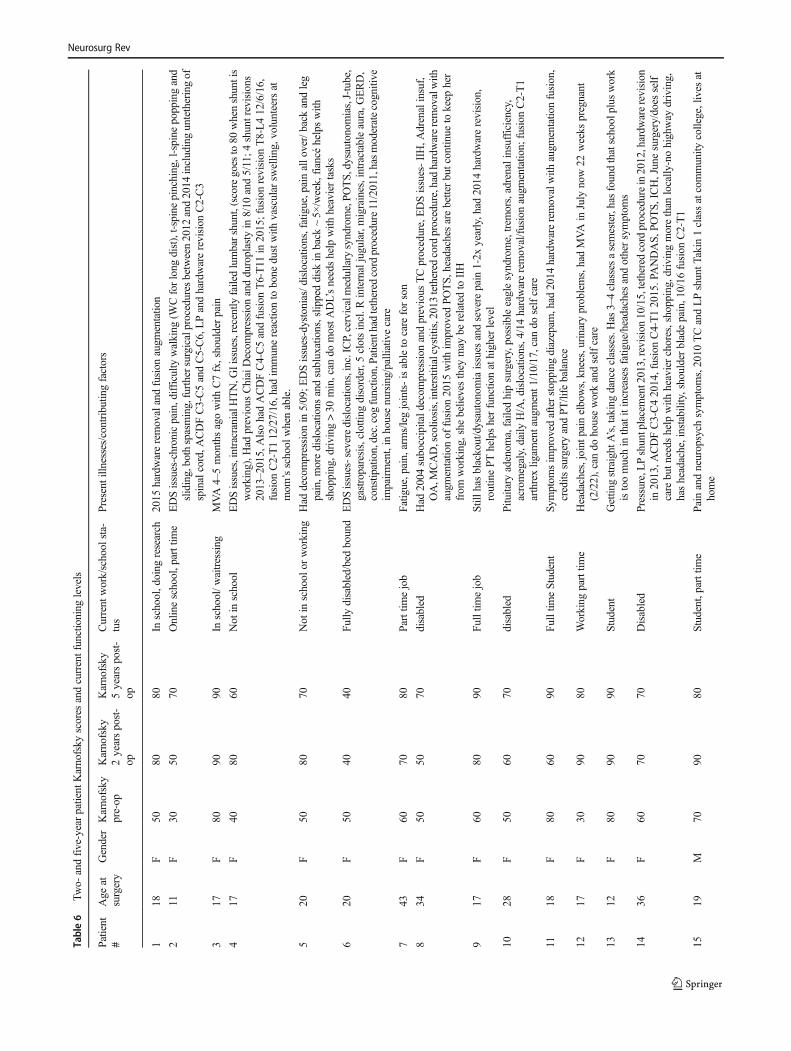
Table6
Two-
andfive-yearpatient
Karnofsky
scores
andcurrentfunctioning
levels
Patient
#Age
atsurgery
Gender
Karnofsky
pre-op
Karnofsky
2yearspost-
op
Karnofsky
5yearspost-
op
Current
work/school
sta-
tus
Presentilln
esses/contributin
gfactors
118
F50
8080
Inschool,doing
research
2015
hardwareremovalandfusion
augm
entatio
n
211
F30
5070
Onlineschool,parttim
eEDSissues-chronicpain,difficulty
walking
(WCforlong
dist),t-spinepinching,l-spine
poppingand
sliding,bothspasming,furthersurgicalproceduresbetween2012
and2014
includinguntetheringof
spinalcord,A
CDFC3-C5andC5-C6,LPandhardwarerevision
C2-C3
317
F80
9090
Inschool/w
aitressing
MVA4–5monthsagowith
C7fx,shoulderpain
417
F40
8060
Not
inschool
EDSissues,intracranialH
TN,G
Iissues,recently
failedlumbarshunt,(scoregoes
to80
whenshuntis
working),Had
previous
ChiaiDecom
pression
andduroplasty
in8/10
and5/11;4
shuntrevisions
2013–2015,AlsohadACDFC4-C5andfusion
T6-T11
in2015;fusionrevision
T8-L412/6/16,
fusion
C2-T112/27/16,had
immunereactio
nto
bone
dustwith
vascular
swellin
g,volunteersat
mom
’sschool
whenable.
520
F50
8070
Not
inschool
orworking
Had
decompression
in5/09;E
DSissues-dystonias/d
islocatio
ns,fatigue,painallo
ver/back
andleg
pain,m
oredislocations
andsubluxations,slip
peddisk
inback
~5×
/week,fiancé
helpswith
shopping,driving
>30
min,can
domostA
DL’sneedshelp
with
heaviertasks
620
F50
4040
Fully
disabled/bed
bound
EDSissues-severedislocations,inc.ICP,cervicalmedullary
syndrome,POTS,dysautonom
ias,J-tube,
gastroparesis,clottin
gdisorder,5
clotsincl.R
internaljugular,migraines,intractableaura,G
ERD,
constip
ation,dec.cogfunctio
n,Patienthadtethered
cord
procedure11/2011,hasmoderatecognitive
impairment,in
housenursing/palliativecare
743
F60
7080
Parttim
ejob
Fatig
ue,pain,arms/legjoints-isableto
care
forson
834
F50
5070
disabled
Had
2004
suboccipitald
ecom
pression
andprevious
TCprocedure,EDSissues-IIH,A
drenalinsuf,
OA,M
CAD,scolio
sis,interstitialcystitis,2013tethered
cord
procedure,hadhardwareremovalwith
augm
entatio
nof
fusion
2015
with
improved
POTS,
headachesarebetterbutcontin
ueto
keep
her
from
working,she
believesthey
may
berelatedto
IIH
917
F60
8090
Fulltim
ejob
Still
hasblackout/dysautonomiaissues
andsevere
pain
1-2x
yearly,had
2014
hardwarerevision,
routinePT
helpsherfunctio
nathigher
level
1028
F50
6070
disabled
Pitu
itary
adenom
a,failedhipsurgery,possibleeaglesyndrome,trem
ors,adrenalinsufficiency,
acromegaly,daily
H/A,dislocatio
ns,4/14hardwareremoval/fusionaugm
entatio
n;fusion
C2-T1
arthrexlig
amentaugment1
/10/17,can
doselfcare
1118
F80
6090
Fulltim
eStudent
Symptom
sim
proved
afterstopping
diazepam
,had
2014
hardwareremovalwith
augm
entatio
nfusion,
credits
surgeryandPT/life
balance
1217
F30
9080
Working
parttim
eHeadaches,joint
pain
elbows,knees,urinaryproblems,hadMVAin
July
now22
weeks
pregnant
(2/22),can
dohouseworkandselfcare
1312
F80
9090
Student
Gettin
gstraightA’s,takingdanceclasses.Has
3–4classesasemester,hasfoundthatschoolplus
work
istoomuchin
thatitincreasesfatig
ue/headaches
andothersymptom
s
1436
F60
7070
Disabled
Pressure,LPshuntplacement2013,revision
10/15,tethered
cordprocedurein2012,hardw
arerevision
in2013,A
CDFC3-C42014,fusionC4-T12015.PANDAS,
POTS,
ICH,Junesurgery/does
self
care
butn
eeds
help
with
heavierchores,shopping,drivingmorethan
locally
-nohighway
driving,
hasheadache,instability,shoulder
bladepain,10/16
fusion
C2-T1
1519
M70
9080
Student,parttim
ePainandneuropsych
symptom
s,2010
TCandLPshuntT
akin
1classatcommunity
college,lives
athome
Neurosurg Rev
Ligamentous laxity at the craniocervical junction
EDS is fundamentally a disorder of collagen and other struc-tural components of connective tissue, characterized by in-competent ligaments, joints, and spine. Ligaments are the ma-jor occiput–C1 stabilizing structures [4]. In the presence ofligamentous laxity or disruption, the CCJ is incompetent inthe execution of multiaxial movements [91, 92].Craniocervical instability (CCI) is thus a manifestation of lig-amentous laxity in EDS [18, 53, 61, 62, 93, 94].
Most atlanto-occipital joint movement occurs in flexion-extension, and axial rotation is normally limited; greater than5° rotation at the occipito-atlantal joint is abnormal [95]. Thelateral atlanto-occipital ligament prevents excess rotation be-tween occiput and atlas; incompetence of the lateral atlanto-occipito ligament results in increased contralateral rotation by3 to 5°. The tectorial membrane and nuchal ligament, com-posed parallel bundles of collagen, restrict hyperflexion,maintain posture, and help to restore normal position [96]. Inthe population of patients with hypermobility connective tis-sue disorders, incompetent ligamentous connections from theskull to the spine may progress to CCI.
Neurological deficit has been attributedto ligamentous laxity at the craniocervical junction
Neurological injury is common in many other connective tis-sue disorders, such as rheumatoid arthritis, Down syndrome,and hereditary disorders such asosteogenesis imperfecta [10,17, 18, 21, 25–28, 30–35, 37, 42, 44–46, 48, 49]. Non-disruptive stretch injury of the neuroaxis has been attributedto hypermobility of the craniocervical junction in infants andchildren, in whom the axonal lesions tend to be localized tothe dorsal brainstem, lower medulla, in particular thecorticospinal tracts at craniocervical junction [97]. Similarhistopathological findings of nerve injury were seen in thelower brainstem and spinal cord, in adults [30, 71, 98–100].
In the EDS population, motor delay, developmental coor-dination disorder, headaches secondary to spinal compression,clumsiness, and the relatively high rate of dyslexia anddyspraxia have been recognized as a consequence of the ef-fects of ligamentous laxity upon the central nervous system[18, 51, 53–60, 62, 66]. Interdigitation of the posterior-atlanto-occipital membrane with the pain-sensitive spinaldural layer has also been implicated in the genesis of headache[101].
The cervical medullary syndrome
The cervical medullary syndrome, also known asBcraniocervical syndrome^ (ICD-9-CM Diagnosis Code723.2; ICD-10-CM Diagnosis Code M53.0), comprises thosesymptoms commonly attributed to lower brainstem and upperT
able6
(contin
ued)
Patient
#Age
atsurgery
Gender
Karnofsky
pre-op
Karnofsky
2yearspost-
op
Karnofsky
5yearspost-
op
Current
work/school
sta-
tus
Presentilln
esses/contributin
gfactors
1618
F80
8090
Working
from
home/part
timework
Had
Chiarid
ecom
pression
procedures
3xin
2010,2012hardwarerevision
and2015
fusion
augm
ent/
chronicH/A
thinks
shewill
need
pain
managem
entfor
fulltim
ework,3/7/17
TC
1726
F50
5070
Disabled
Painandfatigue
from
EDS,unrelated
tosurgeries,canfunctio
n~90–120
min
QD,dysautonomias,
hypothyroidism
,Raynauds,on
ssdi,usesadaptiv
eequipm
ent
1831
F60
5050
Disabled
Had
ACDFC5-C66/2016,beforethathadsevereH/A,troublewalking,jointpain,hipproblems,neck
pain
furtherdownupperback/arm
s/shoulders.Needs
assistance
with
prepping
food
andbathing
1953
F60
9080
Retired,thinksshecould
workparttim
eotherw
ise
Superior
mesentericartery
syndromeBnutcrackersyndrome^,L
renalv
eincompression,pneum
onia,
vascular
digestiveissues,had
SMAtransposition
(1–2
yr.recovery),w
asposted
forhardware
revision
with
augm
entfusionocc-c1/c2in
April17,surgery
hasnoth
appenedas
ofMay
17.D
oes
self-care
2017
F60
8060
New
born,unableto
work
Had
adecompression
in10/07,TCin
8/09;E
DSissues-Lum
barshunt,,
inc.ICP,c-spinepain,P
Thelps,27
wks
pregnantas
of9/16-n
eeds
helpwith
differentA
DLsbasedon
pain/energy,haslumbar
shuntp
ressureissues
thathave
towaittobe
addressedpostnatally,w
alks
~20
min,W
Cafterthat,
PThelps,2014
LPandhardwareremoval,augmentatio
nof
fusion,has
10wkoldas
of2/22,needs
help
with
basichousew
ork
Neurosurg Rev
cervical spinal cord pathology, usually in the presence of aBcomplex Chiari^ (Chiari malformation with basilar invagina-tion or craniocervical instability) [1, 3–5, 77, 79].
In the present study, all subjects presented with headache,fatigue and dizziness, and most reported, in descending orderof frequency: weakness, neck pain, imbalance, night awaken-ings, memory difficulties, walking problems, sensory chang-es, visual problems, vertigo, altered hearing, speech impedi-ments, micturition issues and dysphagia, and syncopal epi-sodes. In aggregate, these symptoms are reasonably describedas the BCervical Medullary Syndrome^ [1, 77].
While there is an overlap of clinical findings, the clinicalpresentation of the pure Chiari malformation differs from thecomplex Chiari malformation. Chiari I malformations arecharacterized primarily by the suboccipital Bcough headache^exacerbated by Valsalva, cough or straining-dizziness, ele-ments of cerebellar dysfunction, lower cranial nerve deficits,and gait problems [102]. On the other hand, the BComplexChiari^ with ventral brainstem compression or craniocervicalinstability present with other genetic conditions—such asHOX D3 homeotic transformation, Klippel Feil malforma-tion, hereditary connective tissue disorders [102–104]—andis characterized by pyramidal changes, with weakness,hyperreflexia, pathological reflexes, paresthesias, and sphinc-ter problems, in addition to headache, neck pain, dizziness,vertigo, dyspnea, dysphonia, altered vision and hearing, syn-cope, gait changes, and altered sleep architecture [5, 7, 10, 30,70, 71, 105–107]. Dysautonomia has also been attributed tobasilar impression [108].
Radiological metrics in the diagnosis of basilarinvagination and CCI
Three radiologic metrics used in this study, the Clivo-axialangle (CXA), the horizontal Harris Measurement [78], andthe Grabb, Mapstone, Oakes measurement [7, 78] have beenadopted as common data elements (CDEs) by the NIH/NINDS, and characterized useful in identifying possible CCIand basilar invagination [1, 76, 77]. The CXA of less than135° is considered potentially pathological [10, 12, 18, 30,70–75, 79, 109]. Salutary consequences have been attributedto the correction of the CXA [10, 12, 69, 70, 107].
The Grabb, Mapstone, Oakes measurement of 9 mmormore suggests high risk of ventral brainstem compression,requiring consideration for craniospinal reduction or transoraldecompression, and fusion stabilization [7, 77, 79].
The horizontal Harris measurement (or BAI) was useful indemonstrating craniocervical instability. Normally, the basionpivots on a point above the odontoid, and there is no measur-able translatory movement between flexion and extension. Achange in the horizontal Harris measurement of 2 mm ormore, as measured in flexion and extension images, represents
pathological translation between the basion and odontoid [1,10, 76, 77, 79, 110–114].
Non-operative management of patientswith craniocervical instability due to hereditaryconnective tissue disorder
Patients should be given a specific diagnosis to validate theirconcerns, and allay their fears. Rigorous instruction shouldfollow to avoid aggravating activities—impact sports andprolonged sitting or driving, the importance of frequent restperiods, physical therapy—for strengthening, sagittal balance,posture and cardiorespiratory fitness, and judicious use of ap-propriate bracing, to be accompanied by isometric exercises.When possible, treatment of co-morbid conditions should beundertaken.
Craniocervical fusion should be considered the last option,to be engaged when non-operative treatment has failed.
Indications for surgery
Posterior occipito-cervical fusion is indicated in patients whopresent with basilar invagination, instability or abnormal bio-mechanics, and cervical medullary syndrome [13, 21, 25, 112,115].
Therefore, at the time of decompression of a Chiari malfor-mation, the finding of basilar invagination or craniocervicalinstability should prompt consideration of fusion and stabili-zation [2, 3, 11, 18, 19, 21–23, 116, 117].
In this study, indications for surgery included disablingheadache or neck pain, symptoms constituting the cervicalmedullary syndromewith demonstrable neurological findings,congruent radiological findings, a determination on the part ofthe patient that they were unable to continue in the normalactivities of daily living, and failed non-operative treatment.
Headache should not be attributed a priori to craniocervicalinstability. In the hereditary connective tissue disorders, head-ache may have many origins: cervicogenic, vessel dissection,or venous occlusive disease or thrombosis, intracranial hyper-tension or hypotension, temporomandibular joint syndrome,inflammatory and infectious disorders, neuralgia and migrain-ous conditions, postural orthostatic tachycardia syndrome(POTS), or mast cell activation syndrome (MCAS) [41, 118,119].
Radiological metrics are useful guidelines, but not indica-tions, per se, for surgery. The radiological indications werecongruent with the treatment algorithm previously established[70]. Abnormal radiological metrics may exist in patients withno neurological symptoms.
A number of subjects with CCI were also found to haveatlantoaxial instability, a radiological and clinical finding thatdid add weight to the decision to proceed to surgery.Occipitocervical fusion is indicated in some circumstances
Neurosurg Rev
for atlantoaxial instability alone, or for complex cervical de-formities [21, 27].
A patient with hereditary disorder is at risk for multilevelinstability issues; any injury or period of disability may resultin exacerbation of instability [120]. The complexity of thesepatients warrants a rigorous selection process. Selection ofcandidates for surgery should follow standard guidelines andindications for instability, the diagnosis of which often re-quires dynamic imaging [13, 14, 70]. Occipitocervical fusionshould be considered the last treatment option in this patientpopulation [41].
Surgical open reduction
The reduction should be executed in a thoughtful and deliber-ate manner to avoid incorrect or painful malalignment, Bstargazing^ from excessive extension or conversely a downwardgaze. If the cranium is inadequately extended, the oropharyn-geal space may be decreased, and the patient may exhibitsevere dysphagia or potentially life-threatening dyspnea[121]. To maintain appropriate oropharyngeal space, the sur-geons extended the cervical spine to maintain 2 cm betweenthe anterior spinal line and the posterior edge of the mandible,as seen on lateral fluoroscopy. In most cases, the basion wastranslated posteriorly to lie above the midpoint of theodontoid. The kyphotic angulation of the brainstem over theodontoid process, as measured by the CXA, was normalizedby extension of the cranium at the craniocervical junction,thereby decreasing the fulcrum effect of the odontoid [49],and the mechanical stress on the brainstem [10, 12, 30, 41,109, 122]. We attempted to achieve a mild cervical lordosis.
Reduction, fusion/stabilization appears to improvepain and neurological deficit
There was 100% follow-up at 2-year and 5-year follow-up.Except for the neurological exam, the clinical data was col-lected by a third party, and de-identified. All patients weresatisfied with the surgery, would repeat the surgery given thesame circumstances, and reported improved quality of life. Allbut one patient would recommend the surgery to a familymember. Eighteen of the twenty patients reported that thecraniocervical fusion surgery decreased their limitations; tworeported continued limitations from other medical problemsand spinal instability elsewhere.
Postoperatively, at the 2-year follow-up, patients demon-strated a statistically significant improvement in in frequencyand severity of headache, speech, memory, vertigo, dizziness,gait, balance, and urinary frequency. There were also improve-ments in most patients with tremors, syncope, imbalance,hearing problems, dysarthria, swallowing difficulty, numb-ness of the upper and lower extremities and back, neck painand upper extremity weakness.
At 5 years, there remained statistically significant improve-ment in headaches, dizziness and imbalance, gait, speechproblems, and frequent daytime urination. Though not statis-tically significant, there was also continued improvement inupper extremity, back and lower numbness, syncope, upperand lower extremity weakness, swallowing difficulty, andhearing problems.
At the 2-year period, the improvement of the Karnofskyperformance score was statistically significant and remainedsignificantly improved over the 5-year follow-up period, withthe majority of subjects returning to employment, school, orwork in the home. This improvement was supported by theobserved improvement in neurological deficits; weakness,heel-to-toe walking, Romberg and sensation.
Co-morbid conditions in this population thatconfounded the outcome
At 5 years, 8/20 patients reported disability from co-morbidconditions. In keeping with the literature, most patients pre-sented with postural orthostatic tachycardia syndrome andother manifestations of dysautonomia; many patients receiveddiagnoses of abnormalities of CSF hydrodynamics with intra-cranial hypertension or hypotension, abnormalities of intracra-nial venous drainage due to sinus stenosis or jugular veinstenosis. Migraine headaches and temporomandibular jointdysfunction were very common. A majority of patients hadvitamin and trace element deficiencies. Many patients demon-strated cervical instability with cervicogenic headaches.Gastroparesis, superior mesenteric artery syndrome, mast cellactivation syndrome occurred and endocrine disorders.Several patients were diagnosed with movement disorders,Tarlov cysts, kypho-scoliosis, tethered cord syndrome, neuro-muscular disorders, anxiety, and depression [18, 41,123–129].
A multi-disciplinary team, familiar with the many co-morbidities and the generalized ligament laxity throughoutthe spinal column, is necessary to address the many issues inorder to improve the well-being of the patient with a heredi-tary connective tissue disorder.
Complications of surgery
There were no deaths or major peri-operative morbidities.There were two patients who underwent transfusion intraop-eratively, two with superficial infections of whom onereturned to the operating room for closure of the rib wounddehiscence. Mild to moderate pain at the rib harvest site wascommon at 2 years, substantially abating at 5 years. Spinalinstability is a potential complication of rib harvest, but wasnot reported in this group.
The absence of screw malposition and vertebral artery in-jury [29, 130] is attributed in part to improvement in
Neurosurg Rev
instrumentation, preoperative CT to examine the anatomy, andintra-operative fluoro-CT to assess the construct real-time.
No patient complained of decreased neck range of motionafter surgery. Despite the loss of approximately 20° to 30° offlexion and extension at the craniocervical junction, and 35°of rotation to each side at C1–C2, range of motion was not aconcern for any of these patients.
One to four years after the craniocervical fusion, some pa-tients developed pain over the suboccipital instrumentation(the Bscrew saddles^) due to tissue thinning, and requestedhardware removal (8/20 subjects). The authors have, there-fore, adopted lower profile craniocervical instrumentation. Asmaller profile generally requires a smaller size and smootherouter contour of the instrumentation. The instrumentationshould be configured to allow placement as low as possibleover the cranium, to increase the thickness of the tissue over-lying the instrumentation.
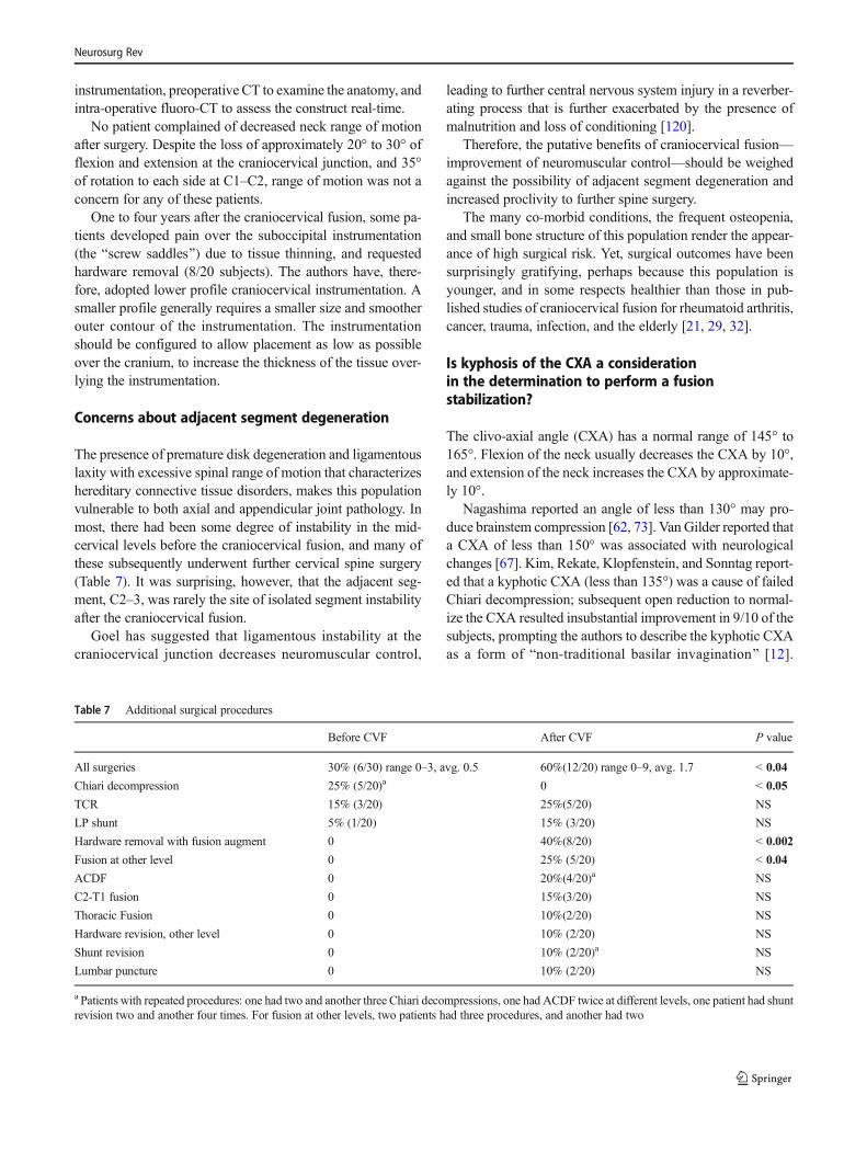
Concerns about adjacent segment degeneration
The presence of premature disk degeneration and ligamentouslaxity with excessive spinal range of motion that characterizeshereditary connective tissue disorders, makes this populationvulnerable to both axial and appendicular joint pathology. Inmost, there had been some degree of instability in the mid-cervical levels before the craniocervical fusion, and many ofthese subsequently underwent further cervical spine surgery(Table 7). It was surprising, however, that the adjacent seg-ment, C2–3, was rarely the site of isolated segment instabilityafter the craniocervical fusion.
Goel has suggested that ligamentous instability at thecraniocervical junction decreases neuromuscular control,
leading to further central nervous system injury in a reverber-ating process that is further exacerbated by the presence ofmalnutrition and loss of conditioning [120].
Therefore, the putative benefits of craniocervical fusion—improvement of neuromuscular control—should be weighedagainst the possibility of adjacent segment degeneration andincreased proclivity to further spine surgery.
The many co-morbid conditions, the frequent osteopenia,and small bone structure of this population render the appear-ance of high surgical risk. Yet, surgical outcomes have beensurprisingly gratifying, perhaps because this population isyounger, and in some respects healthier than those in pub-lished studies of craniocervical fusion for rheumatoid arthritis,cancer, trauma, infection, and the elderly [21, 29, 32].
Is kyphosis of the CXA a considerationin the determination to perform a fusionstabilization?
The clivo-axial angle (CXA) has a normal range of 145° to165°. Flexion of the neck usually decreases the CXA by 10°,and extension of the neck increases the CXA by approximate-ly 10°.
Nagashima reported an angle of less than 130° may pro-duce brainstem compression [62, 73]. Van Gilder reported thata CXA of less than 150° was associated with neurologicalchanges [67]. Kim, Rekate, Klopfenstein, and Sonntag report-ed that a kyphotic CXA (less than 135°) was a cause of failedChiari decompression; subsequent open reduction to normal-ize the CXA resulted insubstantial improvement in 9/10 of thesubjects, prompting the authors to describe the kyphotic CXAas a form of Bnon-traditional basilar invagination^ [12].
Table 7 Additional surgical procedures
Before CVF After CVF P value
All surgeries 30% (6/30) range 0–3, avg. 0.5 60%(12/20) range 0–9, avg. 1.7 < 0.04
Chiari decompression 25% (5/20)a 0 < 0.05
TCR 15% (3/20) 25%(5/20) NS
LP shunt 5% (1/20) 15% (3/20) NS
Hardware removal with fusion augment 0 40%(8/20) < 0.002
Fusion at other level 0 25% (5/20) < 0.04
ACDF 0 20%(4/20)a NS
C2-T1 fusion 0 15%(3/20) NS
Thoracic Fusion 0 10%(2/20) NS
Hardware revision, other level 0 10% (2/20) NS
Shunt revision 0 10% (2/20)a NS
Lumbar puncture 0 10% (2/20) NS
a Patients with repeated procedures: one had two and another three Chiari decompressions, one had ACDF twice at different levels, one patient had shuntrevision two and another four times. For fusion at other levels, two patients had three procedures, and another had two
Neurosurg Rev
Morishita suggested that a clivo-axial angle of less than 135°is a risk factor for spinal cord compression [131]. Kubota, in aretrospective series of foramen magnum decompression forChiari and syringomyelia, reported that the syrinxes failed toabate in those patients in whom the CXAwas less than 130°[15].
Brockmeyer, in a retrospective pediatric series of Chiaridecompressions, reported that 20% of patients were returnedto surgery for reduction and stabilization for kyphotic CXA,craniocervical instability, or the presence of a Chiari 1.5 [4], afinding echoed by Klekamp and others in the adult population[6, 7, 9, 13, 19, 21, 132, 133]. The medulla becomes kinked asthe CXA becomes more kyphotic; increasing kyphosis ofclivo-axial angle creates a fulcrum by which the odontoiddeforms the brainstem [49, 132, 134]. A more complete trea-tise on the importance of the CXA has been presented else-where [70]. In this study, the mean preoperative CXA of 129°was increased to 148° by open reduction of the kyphosis,which correlated with patient improvement. However, the ob-served improvement may have been the result ofcraniocervical stabilization.
Pathophysiology
A kyphotic CXA is associated with bending and strain ofthe lower brainstem and upper spinal cord, and a preludeto neurological deficit [9, 22, 32, 68, 135]. Stretching aneuron nerve decreases neural firing rate and amplitude[136]. The predominant substrate for deformity-inducedinjury is the axon: electron micrographs show clumping,loss of microtubules and neurofilaments, loss of axontransport and accumulations of axoplasmic material iden-tified as the retraction ball, or retraction bulb, analogousto diffuse axonal injury (DAI) in the brain [137–142].Axon retraction bulbs result from stretch/deformation in-jury and injury in animal models [98, 100, 143, 144], inthe cortico-spinal tracts of the brainstem in infants withshaken baby syndrome, adults with spinal cord injuries,and in basilar invagination [30, 109, 143, 145, 146]. Atthe molecular level, stretching of nervous tissue deformsNa+ channels, causing increased membrane depolariza-tion and a consequent deleterious influx of Ca++ [147].The epigenetic effects of mechanical strain are manifest inthe observation of increased expression of N-methyl-d-aspartate in the stretched neuron, altered mitochondrialfunction, and apoptosis [148–150].
The clinical improvement observed in this cohort is thepresumed consequence of reduction of mechanical deformityof the nervous system and elimination or mitigation ofmicrotrauma from craniocervical instability [10, 16, 49,107], consistent with the experimental models of axons sub-jected to strain [149, 151–154].
Controversy
Treatment of other forms of degenerative and hereditary con-nective tissue disorders is firmly established in the literature.However, treatment of the EDS patient has been problematicfor several reasons. First, though EDS was first described in1901, the recognition of spinal and neurological manifesta-tions has been only recent [56, 62, 18, 41, 51, 53–55, 57–61,66]. Because this information is new, there is a dearth of ev-idence upon which to base the management of these geneticdisorders.
Second, EDS is considered an Binvisible disorder.^ EDSpatients are characterized by youthful skin, and the appear-ance of good health, belying their severe pain and disability.
Third, the legion of disparate symptoms due to ligamentousweakness of the joints and spine, and the many co-morbidconditions that accompany EDS, result, understandably, indismissal by healthcare providers because of the large numberof seemingly disparate symptoms.
The authors advocate that the indications for craniocervicalfusion should be no different than for the non-EDS popula-tion, with the caveat that conditions of ligamentous laxityoften require dynamic imaging to demonstrate the pathology[1, 13, 14, 27, 77, 112, 155]. Occiput to C2 bone fusion, asopposed to atlantoaxial fusion in conjunction with fixation,has been discussed [3, 38, 69, 82, 115, 155]. A comparisonof various methodologies for bone fusion has also beendiscussed [83].
The economic significance of hypermobilityconnective tissue disorders
Treatment of the EDS population is problematic because ofthe diverse spectrum of disease severity and presentation forwhom, in the majority of cases, there is no genetic testingavailable. In the experience and belief of most EDS care pro-viders, EDS patients suffer through scores of visits to special-ists over a mean of 10 years before the diagnosis of EDS ismade, during which time they consume vast medical re-sources through emergency room visits, and unscheduled, of-ten prolonged, admissions to hospital.
The epidemiology of EDS is not known; however, there islittle phenotypic difference between patients with h-EDS andthe very large population previously diagnosed with joint hy-permobility disorder (now referred to as hypermobility spec-trum disorder) sharing the same early degeneration of thespine and joints, and the same co-morbid conditions[156–158]. Therefore, the authors believe that earlier recogni-tion of these hereditary disorders would substantially reducecostly specialty visits, improve care of this patient population.Early recognition, prudent management, and non-operativetherapy may be adequate to stabilize the patient and obviateneed of surgery in many cases.
Neurosurg Rev
In this cohort, 55% of subjects have returned to work andare paying taxes, or attending school full or part-time with theprospect of future employment, or serving society throughcaring for their families.
Limitations of the study
This IRB study is a single-center, non-controlled analysisof a small cohort of subjects, referred by medical pro-viders from a broad geographical area (USA andCanada). The study was conceived prior to any surgery,but the subjects were not enrolled until after surgery.Therefore, this should be considered a retrospective study.The outcomes data is to some extent obfuscated by thepresence of previous Chiari surgery (five), multiple co-morbid conditions common to EDS, and multiple surger-ies within the 5-year follow-up period (12/20). The com-plexity of the co-morbid conditions and other surgeriesare prohibitive to more complex statistical methods.These patients appeared to be the most seriously affectedpatients within the spectrum of hereditary connective tis-sue disorders.
Not every patient had cerebellar ectopia (18/20). Subjectsmay have inaccurately reported the severity of their preoper-ative symptoms upon questioning at the 2-year follow-up andmay have exaggerated the degree of improvement. However,accuracy of reporting was improved through the employmentof two independent researchers, who performed the subjects’interviews at 2 and 5 years. Some subjects may have seensurgery as means validation of their suffering. There was nocontrol for a placebo effect [159].
Conclusion
This study supports the hypothesis that craniocervical reduc-tion, stabilization, and fusion are feasible and associated withclinical improvement in patients in the HCTD population withChiari malformation or cerebellar ectopia, kyphotic clivo-axial angle, ventral brainstem compression, and/orcraniocervical instability. The neurological and functional im-provements associated with craniocervical fusion/stabilizationappear to be clinically significant and durable. That said,craniocervical fusion should be considered as a last resort aftera reasonable course of non-operative treatments.
Acknowledgements To Betsy G. Henderson for help with layout andediting, and to the patients who have informed this study.
Compliance with ethical standards
Funding This study was solely funded by the MetropolitanNeurosurgery Group, LLC.
Conflict of interest One of the senior authors (FCH Sr.) was first authorof a patent for a craniocervical stabilization device, which is currentlybeing developed by LifeSpine, Inc. (Huntley, IL) and related patentsdiscussing finite element analysis (spinal cord stress injury analysis) ofthe central nervous system, mathematical prediction of neurobehavioralchange, and related devices pertinent to disorders of the craniocervicaljunction. The same author (FCH Sr) has donated his royalties for thecraniocervical device to the Chiari Syringomyelia Foundation (CSF).
Ethical approval The study was conducted under the auspices of theGreater Baltimore Medical Center (IRB# 10-036-06: GBMC). This studyfollowed all requirements of the 1964 Helsinki declaration and its lateramendments or comparable ethical standards.
Informed consent Every patient signed an informed consent form toparticipate in the study.
Open Access This article is distributed under the terms of the CreativeCommons At t r ibut ion 4 .0 In te rna t ional License (h t tp : / /creativecommons.org/licenses/by/4.0/), which permits unrestricted use,distribution, and reproduction in any medium, provided you give appro-priate credit to the original author(s) and the source, provide a link to theCreative Commons license, and indicate if changes were made.
Publisher’s Note Springer Nature remains neutral with regard to jurisdic-tional claims in published maps and institutional affiliations.
References
1. Batzdorf U HF, Rigamonti D. et al (2016) Consensus statement inproceedings of CSF colloquium 2014. In: Batzdorf U (ed) Co-morbidities that complicate the treatment and outcomes of Chiarimalformation. Chiari Syringomyelia Foundation, Inc., Lulu, p 3
2. Bekelis K, Duhaime AC,Missios S, Belden C, Simmons N (2010)Placement of occipital condyle screws for occipitocervical fixationin a pediatric patient with occipitocervical instability after decom-pression for Chiari malformation. J Neurosurg Pediatr 6:171–176.https://doi.org/10.3171/2010.4.peds09551
3. Bollo RJ, Riva-Cambrin J, Brockmeyer MM, Brockmeyer DL(2012) Complex Chiari malformations in children: an analysis ofpreoperative risk factors for occipitocervical fusion. J NeurosurgPediatr 10:134–141. https://doi.org/10.3171/2012.3.peds11340
4. Brockmeyer DL (2011) The complex Chiari: issues and manage-ment strategies. Neurol Sci 32(Suppl 3):S345–S347. https://doi.org/10.1007/s10072-011-0690-5
5. Caetano de Barros M, Farias W, Ataide L, Lins S (1968) Basilarimpression and Arnold-Chiari malformation. A study of 66 cases.J Neurol Neurosurg Psychiatry 31:596–605
6. Felbaum D, Spitz S, Sandhu FA (2015) Correction of clivoaxialangle deformity in the setting of suboccipital craniectomy: techni-cal note. J Neurosurg Spine 23:8–15. https://doi.org/10.3171/2014.11.spine14484
7. Grabb PA, Mapstone TB, Oakes WJ (1999) Ventral brain stemcompression in pediatric and young adult patients with Chiari Imalformations. Neurosurgery 44:520–527 discussion 527-528
8. Henderson FC (2016) Cranio-cervical Instability in Patients withHypermobility Connective Disorders. J Spine
9. Henderson FC, Wilson WA, Benzel EC (2010) Pathophysiologyof cervical myelopathy: biomechanics and deformative stress. In:Benzel EC (ed), Spine surgery: Techniques, complication avoid-ance, and management, vol 1, 1st edn. Elsevier: ChurchillLivingstone, p 188–195
Neurosurg Rev
10. Henderson FC, Wilson WA, Mott S, Mark A, Schmidt K, BerryJK, Vaccaro A, Benzel E (2010) Deformative stress associatedwith an abnormal clivo-axial angle: a finite element analysis.Surg Neurol Int 1:30. https://doi.org/10.4103/2152-7806.66461
11. Joseph V, Rajshekhar V (2003) Resolution of syringomyelia andbasilar invagination after traction. Case illustration. J Neurosurg98:298
12. Kim LJ, Rekate HL, Klopfenstein JD, Sonntag VK (2004)Treatment of basilar invagination associated with Chiari Imalformations in the pediatric population: cervical reduction andposterior occipitocervical fusion. J Neurosurg 101:189–195.https://doi.org/10.3171/ped.2004.101.2.0189
13. Klekamp J (2012) Neurological deterioration after foramen mag-num decompression for Chiari malformation type I: old or newpathology? J Neurosurg Pediatr 10:538–547. https://doi.org/10.3171/2012.9.peds12110
14. Klekamp J (2015) Chiari I malformation with and without basilarinvagination: a comparative study. Neurosurg Focus 38:E12.https://doi.org/10.3171/2015.1.focus14783
15. Kubota M, Yamauchi T, Saeki N Surgical Results of ForamenMagnum Decompression for Chiari Type 1 Malformation associ-ated with Syringomyelia: A Retrospective Study onNeuroradiological Characters influencing Shrinkage of SyringesY1–2004. - Spinal Surg M1 - Journal Article:- 81
16. Menezes AH (2012) Craniovertebral junction abnormalities withhindbrain herniation and syringomyelia: regression of syringomy-elia after removal of ventral craniovertebral junction compression.J Neurosurg 116:301–309. https://doi.org/10.3171/2011.9.jns11386
17. Menezes AH, VanGilder JC, Clark CR, el-Khoury G (1985)Odontoid upward migration in rheumatoid arthritis. An analysisof 45 patients with "cranial settling". J Neurosurg 63:500–509.https://doi.org/10.3171/jns.1985.63.4.0500
18. Milhorat TH, Bolognese PA, Nishikawa M, McDonnell NB,Francomano CA (2007) Syndrome of occipitoatlantoaxial hyper-mobility, cranial settling, and chiari malformation type I in pa-tients with hereditary disorders of connective tissue. J NeurosurgSpine 7:601–609. https://doi.org/10.3171/spi-07/12/601
19. Nishikawa M, Ohata K, Baba M, Terakawa Y, Hara M (2004)Chiari I malformation associated with ventral compression andinstability: one-stage posterior decompression and fusion with anew instrumentation technique. Neurosurgery 54:1430–1434 dis-cussion 1434-1435
20. Nishikawa M, Sakamoto H, Hakuba A, Nakanishi N, Inoue Y(1997) Pathogenesis of Chiari malformation: a morphometricstudy of the posterior cranial fossa. J Neurosurg 86:40–47.https://doi.org/10.3171/jns.1997.86.1.0040
21. Singh SK, Rickards L, Apfelbaum RI, Hurlbert RJ, Maiman D,Fehlings MG (2003) Occipitocervical reconstruction with theOhio medical instruments loop: results of a multicenter evaluationin 30 cases. J Neurosurg 98:239–246
22. Smith JS, Shaffrey CI, Abel MF, Menezes AH (2010) Basilarinvagination. Neurosurgery 66:39–47. https://doi.org/10.1227/01.neu.0000365770.10690.6f
23. Tubbs RS, Beckman J, Naftel RP, Chern JJ, Wellons JC 3rd,Rozzelle CJ, Blount JP, Oakes WJ (2011) Institutional experiencewith 500 cases of surgically treated pediatric Chiari malformationtype I. J Neurosurg Pediatr 7:248–256. https://doi.org/10.3171/2010.12.peds10379
24. Braca J, Hornyak M, Murali R (2005) Hemifacial spasm in apatient with Marfan syndrome and Chiari I malformation. Casereport. J Neurosurg 103:552–554. https://doi.org/10.3171/jns.2005.103.3.0552
25. Crockard HA, Stevens JM (1995) Craniovertebral junction anom-alies in inherited disorders: part of the syndrome or caused by thedisorder? Eur J Pediatr 154:504–512
26. Gabriel KR, Mason DE, Carango P (1990) Occipito-atlantal trans-lation in Down's syndrome. Spine 15:997–1002
27. Fehlings MG, Cooper P, Errico TJ Rheumatoid arthritis of thecervical spine, Neurosurgical topics: Degenerative disease of thecervical spine Y1–1992. - AANS M1 - Journal Article:- 125–139
28. Grob D, Schutz U, Plotz G (1999) Occipitocervical fusion in pa-tients with rheumatoid arthritis. Clin Orthop Relat Res 366:46–53
29. Grob D, Dvorak J, Panjabi MM, Antinnes JA (1994) The role ofplate and screw fixation in occipitocervical fusion in rheumatoidarthritis. Spine 19:2545–2551
30. Henderson FC, Geddes JF, Crockard HA (1993) Neuropathologyof the brainstem and spinal cord in end stage rheumatoid arthritis:implications for treatment. Ann Rheum Dis 52:629–637
31. Ibrahim AG, Crockard HA (2007) Basilar impression and osteo-genesis imperfecta: a 21-year retrospective review of outcomes in20 patients. J Neurosurg Spine 7:594–600. https://doi.org/10.3171/spi-07/12/594
32. Menezes AH, VanGilder JC (1988) Transoral-transpharyngeal ap-proach to the anterior craniocervical junction. Ten-year experiencewith 72 patients. J Neurosurg 69:895–903. https://doi.org/10.3171/jns.1988.69.6.0895
33. Nockels RP, Shaffrey CI, Kanter AS, Azeem S, York JE (2007)Occipitocervical fusion with rigid internal fixation: long-term fol-low-up data in 69 patients. J Neurosurg Spine 7:117–123. https://doi.org/10.3171/spi-07/08/117
34. Sandhu FA, Pait TG, Benzel E, Henderson FC (2003)Occipitocervical fusion for rheumatoid arthritis using the inside-outside stabilization technique. Spine 28:414–419. https://doi.org/10.1097/01.brs.0000048460.58471.db
35. Zygmunt SC, Christensson D, Saveland H, Rydholm U, Alund M(1995) Occipito-cervical fixation in rheumatoid arthritis–an anal-ysis of surgical risk factors in 163 patients. Acta Neurochir 135:25–31
36. Yoshizumi TMH, Ikenishi Y et al (2014) Occipitocervical fusionwith relief of odontoid invagination: atlantoaxial distraction meth-od using cylindrical titanium cage for basilar invagination—casereport. Neurosurg Rev 37:519–525
37. Bick S, Dunn R (2010) Occipito-cervical fusion: review of surgi-cal indications, techniques and clinical outcomes. SA Orthop J 3:26–32
38. Brockmeyer D (1999) Down syndrome and craniovertebral insta-bility. Topic review and treatment recommendations. PediatrNeurosurg 31:71–77. https://doi.org/10.1159/000028837
39. Gordon N (2000) The neurological complications of achondropla-sia. Brain Dev 22:3–7
40. Harkey HL, Crockard HA, Stevens JM, Smith R, Ransford AO(1990) The operative management of basilar impression in osteo-genesis imperfecta. Neurosurgery 27:782–786 discussion 786
41. Henderson FC Sr, Austin C, Benzel E, Bolognese P, EllenbogenR, Francomano CA, Ireton C, Klinge P, KobyM, Long D, Patel S,Singman EL, Voermans NC (2017) Neurological and spinal man-ifestations of the Ehlers-Danlos syndromes. Am J Med Genet C:Semin Med Genet 175:195–211. https://doi.org/10.1002/ajmg.c.31549
42. Jain VK, Mittal P, Banerji D, Behari S, Acharya R, Chhabra DK(1996) Posterior occipitoaxial fusion for atlantoaxial dislocationassociated with occipitalized atlas. J Neurosurg 84:559–564.https://doi.org/10.3171/jns.1996.84.4.0559
43. Keiper GL Jr, Koch B, Crone KR (1999) Achondroplasia andcervicomedullary compression: prospective evaluation and surgi-cal treatment. Pediatr Neurosurg 31:78–83. https://doi.org/10.1159/000028838
44. Kosnik-Infinger L, Glazier SS, Frankel BM (2014) Occipital con-dyle to cervical spine fixation in the pediatric population. JNeurosurg Pediatr 13:45–53. https://doi.org/10.3171/2013.9.peds131
Neurosurg Rev
45. Menezes AH (2008) Specific entities affecting the craniocervicalr e g i o n : o s t e o g e n e s i s i m p e r f e c t a a n d r e l a t e dosteochondrodysplasias: medical and surgical management ofbasilar impression. Childs Nerv. Syst. 24:1169–1172. https://doi.org/10.1007/s00381-008-0602-z
46. Menezes AH, Ryken TC (1992) Craniovertebral abnormalities inDown's syndrome. Pediatr Neurosurg 18:24–33
47. National Down Syndrome C the position statement. In:BContinuing the Revolution^, 1991
48. Noske DP, van Royen BJ, Bron JL, Vandertop WP (2006) Basilarimpression in osteogenesis imperfecta: can it be treated with halotraction and posterior fusion? Acta Neurochir 148:1301–1305;discussion 1305. https://doi.org/10.1007/s00701-006-0870-x
49. Sawin PD, Menezes AH (1997) Basilar invagination in osteogen-esis imperfecta and related osteochondrodysplasias: medical andsurgical management. J Neurosurg 86:950–960. https://doi.org/10.3171/jns.1997.86.6.0950
50. Tredwell SJ, Newman DE, Lockitch G (1990) Instability of theupper cervical spine in down syndrome. J Pediatr Orthop 10:602–606
51. Adib N, Davies K, Grahame R, Woo P, Murray KJ (2005) Jointhypermobility syndrome in childhood. A not so benign multisys-tem disorder? Rheumatology (Oxford, England) 44:744–750.https://doi.org/10.1093/rheumatology/keh557
52. Castori M, Camerota F, Celletti C, Danese C, Santilli V, SaraceniVM, Grammatico P (2010) Natural history and manifestations ofthe hypermobility type Ehlers-Danlos syndrome: a pilot study on21 patients. Am J Med Genet A 152a:556–564. https://doi.org/10.1002/ajmg.a.33231
53. De Paepe A, Malfait F (2012) The Ehlers-Danlos syndrome, adisorder with many faces. Clin Genet 82:1–11. https://doi.org/10.1111/j.1399-0004.2012.01858.x
54. Di Palma F, Cronin AH (2005) Ehlers-Danlos syndrome: correla-tion with headache disorders in a young woman. J Headache Pain6:474–475. https://doi.org/10.1007/s10194-005-0256-0
55. Easton V, Bale P, Bacon H, Jerman E, Armon K, Macgregor AJ(2014) The relationship between benign joint hypermobility syn-drome and developmental coordination disorders in children.Arthritis Rheumatol 124
56. el-Shaker M, Watts HG (1991) Acute brachial plexus neuropathysecondary to halo-gravity traction in a patient with Ehlers-Danlossyndrome. Spine 16:385–386
57. Galan E, Kousseff BG (1995) Peripheral neuropathy in Ehlers-Danlos syndrome. Pediatr Neurol 12:242–245
58. Halko GJ, Cobb R, AbelesM (1995) Patients with type IV Ehlers-Danlos syndrome may be predisposed to atlantoaxial subluxation.J Rheumatol 22:2152–2155
59. Jelsma LD, Geuze RH, Klerks MH, Niemeijer AS, Smits-Engelsman BC (2013) The relationship between joint mobilityand motor performance in children with and without the diagnosisof developmental coordination disorder. BMC Pediatr 13:35.https://doi.org/10.1186/1471-2431-13-35
60. Kirby A, Davies R (2007) Developmental coordination disorderand joint hypermobility syndrome–overlapping disorders?Implications for research and clinical practice. Child Care HealthDev 33:513–519. https://doi.org/10.1111/j.1365-2214.2006.00694.x
61. Milhorat TH, Nishikawa M, Kula RW, Dlugacz YD (2010)Mechanisms of cerebellar tonsil herniation in patients withChiari malformations as guide to clinical management. ActaNeurochir 152:1117–1127. https://doi.org/10.1007/s00701-010-0636-3
62. Nagashima C, Tsuji R, Kubota S, Tajima K (1981) [Atlanto-axial,Atlanto-occipital dislocations, developmental cervical canal steno-sis in the Ehlers-Danlos syndrome (author's transl)]. No shinkeigeka. Neurol Surg 9:601–608
63. Palmeri S, Mari F, Meloni I, Malandrini A, Ariani F, VillanovaM,Pompilio A, Schwarze U, Byers PH, Renieri A (2003)Neurological presentation of Ehlers-Danlos syndrome type IV ina family with parental mosaicism. Clin Genet 63:510–515
64. Rombaut L, De Paepe A, Malfait F, Cools A, Calders P (2010)Joint position sense and vibratory perception sense in patients withEhlers-Danlos syndrome type III (hypermobility type). ClinRheumatol 29:289–295. https://doi.org/10.1007/s10067-009-1320-y
65. Voermans NC, Drost G, van Kampen A, Gabreels-Festen AA,Lammens M, Hamel BC, Schalkwijk J, van Engelen BG (2006)Recurrent neuropathy associated with Ehlers-Danlos syndrome. JNeurol 253:670–671. https://doi.org/10.1007/s00415-005-0056-0
66. Voermans NC, van Alfen N, Pillen S, Lammens M, Schalkwijk J,Zwarts MJ, van Rooij IA, Hamel BC, van Engelen BG (2009)Neuromuscular involvement in various types of Ehlers-Danlossyndrome. Ann Neurol 65:687–697. https://doi.org/10.1002/ana.21643
67. VanGilder JC, Menezes AH, Dolan KD (1987) Thecraniovertebral junction and its abnormalities. Futura PublishingCompany
68. Breig A (1978) Effects of pincer and clamping actions on thespinal cord Adverse Mechanical Tension in the Central NervousSystem:61
69. Goel A (2004) Treatment of basilar invagination by atlantoaxialjoint distraction and direct lateral mass fixation. J Neurosurg Spine1:281–286. https://doi.org/10.3171/spi.2004.1.3.0281
70. Henderson FC Sr, Henderson FC Jr, WAt W, Mark AS, Koby M(2018) Utility of the clivo-axial angle in assessing brainstem de-formity: pilot study and literature review. Neurosurg Rev 41:149–163. https://doi.org/10.1007/s10143-017-0830-3
71. Howard RS, Henderson F, Hirsch NP, Stevens JM, Kendall BE,Crockard HA (1994) Respiratory abnormalities due tocraniovertebral junction compression in rheumatoid disease. AnnRheum Dis 53:134–136
72. Menezes A, Ryken T, Brockmeyer D Abnormalities of thecraniocervical junction Y1–2001. - Pediatric Neurosurgery:Surgery of the Developing Nervous System:- 400–422
73. Nagashima C, Kubota S (1983) Craniocervical abnormalities.Modern diagnosis and a comprehensive surgical approach.Neurosurg Rev 6:187–197
74. Scoville WB, Sherman IJ (1951) Platybasia, report of 10 caseswith comments on familial tendency, a special diagnostic sign,and the end results of operation. Ann Surg 133:496–502
75. Smoker WR (1994) Craniovertebral junction: normal anatomy,craniometry, and congenital anomalies. Radiographics 14:255–277. https://doi.org/10.1148/radiographics.14.2.8190952
76. Elements NCD (2016) Clinical research common data Elements(CDEs): radiological metrics standardization for Craniocervicalinstability. National Institute of Neurological Disorders andStroke common data element project - approach and methods.Clin Trials 9(33):322–329
77. Batzdorf U B E,Henderson F. et al (2013) Consensus Statement InProceedings of CSF Colloquium 2013. In: U B (ed) BasilarImpression & Hypermobility at the Craniocervical Junction.Chiari Syringomyelia Foundation, Lulu,
78. Harris JH Jr, Carson GC,Wagner LK (1994) Radiologic diagnosisof traumatic occipitovertebral dissociation: 1. Normaloccipitovertebral relationships on lateral radiographs of supinesubjects. AJR Am J Roentgenol 162:881–886. https://doi.org/10.2214/ajr.162.4.8141012
79. Elements NCD Clinical Research Common Data Elements(CDEs): Radiological Metrics Standardization for CraniocervicalInstability Y1–2016
80. Aryan HE, Newman CB, Nottmeier EW, Acosta FL Jr, Wang VY,Ames CP (2008) Stabilization of the atlantoaxial complex via C-1
Neurosurg Rev
lateral mass and C-2 pedicle screw fixation in a multicenter clin-ical experience in 102 patients: modification of the harms andGoel techniques. J Neurosurg Spine 8:222–229. https://doi.org/10.3171/spi/2008/8/3/222
81. Dickman CA, Sonntag VK (1998) Posterior C1-C2 transarticularscrew fixation for atlantoaxial arthrodesis. Neurosurgery 43:275–280 discussion 280-271
82. Goel A, Bhatjiwale M, Desai K (1998) Basilar invagination: astudy based on 190 surgically treated patients. J Neurosurg 88:962–968. https://doi.org/10.3171/jns.1998.88.6.0962
83. Sawin PD, Traynelis VC, Menezes AH (1998) A comparativeanalysis of fusion rates and donor-site morbidity for autogeneicrib and iliac crest bone grafts in posterior cervical fusions. JNeurosurg 88:255–265. https://doi.org/10.3171/jns.1998.88.2.0255
84. Malfait F, Francomano C, Byers P, Belmont J, Berglund B, BlackJ, Bloom L, Bown JM et al (2017) The 2017 international classi-fication of the Ehlers–Danlos syndromes. Am J Med Genet 175:8–26. https://doi.org/10.1002/ajmg.c.31552
85. Tinkle B, Castori M, Berglund B, Cohen H, Grahame R, KazkazH, Levy H (2017) Hypermobile Ehlers–Danlos syndrome (a.k.a.Ehlers–Danlos syndrome type III and Ehlers–Danlos syndromehypermobility type): clinical description and natural history. AmJ Med Genet 175C:48–69. https://doi.org/10.1002/ajmg.c.31538
86. Byers PH (2001) Folding defects in fibrillar collagens. PhilosTrans R Soc.B 356:151–158. https://doi.org/10.1098/rstb.2000.0760
87. Malfait F, Coucke P, Symoens S, Loeys B, Nuytinck L, De PaepeA (2005) The molecular basis of classic Ehlers-Danlos syndrome:a comprehensive study of biochemical and molecular findings in48 unrelated patients. Hum Mutat 25:28–37
88. Steinmann B, Royce PM, Superti-Furga A The Ehlers-DanlosSyndrome Y1–2003. - Connective Tissue and Its HeritableDisorders: Molecular, Genetic, and Medical Aspects:- 431
89. Castori M, Voermans NC (2014) Neurological manifestations ofEhlers-Danlos syndrome(s): a review. Iran J Neurol 13:190–208
90. Savasta S, Merli P, Ruggieri M, Bianchi L, Sparta MV (2011)Ehlers-Danlos syndrome and neurological features: a review.Childs Nerv Syst 27:365–371. https://doi.org/10.1007/s00381-010-1256-1
91. Martin MD, Bruner HJ, Maiman DJ (2010) Anatomic and biome-chanical considerations of the craniovertebral junction.Neurosurgery 66:2–6. https://doi.org/10.1227/01.neu.0000365830.10052.87
92. Tubbs RS, Hallock JD, Radcliff V, Naftel RP, Mortazavi M, ShojaMM, Loukas M, Cohen-Gadol AA (2011) Ligaments of thecraniocervical junction. J Neurosurg Spine 14:697–709. https://doi.org/10.3171/2011.1.spine10612
93. Koby M (2016) The discordant report - pathological radiologicalfindings: A peripatetic review of salient features of neuropatholo-gy in the setting of an erstwhile standard 'normal' radiologicalassessment. In: Batzdorf U (ed) Co-Morbidities that Complicatethe Treatment and Outcomes of Chiari Malformation. ChiariSyringomyelia Foundation Inc., Lulu, p 50
94. Steinmetz MP, Mroz TE, Benzel EC (2010) Craniovertebral junc-tion: biomechanical considerations. Neurosurgery 66:7–12.https://doi.org/10.1227/01.neu.0000366109.85796.42
95. Dvorak J, Hayek J, Zehnder R (1987) CT-functional diagnostics ofthe rotatory instability of the upper cervical spine. Part 2. Anevaluation on healthy adults and patients with suspected instabil-ity. Spine 12:726–731
96. Tubbs RS, StetlerW, ShojaMM, LoukasM,Hansasuta A, LiechtyP, Acakpo-Satchivi L, Wellons JC, Blount JP, Salter EG, OakesWJ (2007) The lateral atlantooccipital ligament. Surg Radiol Anat29:219–223. https://doi.org/10.1007/s00276-007-0196-2
97. Geddes JF, Hackshaw AK, Vowles GH, Nickols CD, WhitwellHL (2001) Neuropathology of inflicted head injury in children.I. Patterns of brain damage. Brain 124:1290–1298
98. Hardman JM (1979) The pathology of traumatic brain injuries.Adv Neurol 22:15–50
99. Lindenberg R, Freytag E (1970) Brainstem lesions characteristicof traumatic hyperextension of the head. Arch Pathol 90:509–515
100. Riggs JE, Schochet SS Jr (1995) Spastic quadriparesis, dysarthria,and dysphagia following cervical hyperextension: a traumaticpontomedullary syndrome. Mil Med 160:94–95
101. Nash L, Nicholson H, Lee AS, Johnson GM, Zhang M (2005)Configuration of the connective tissue in the posterior atlanto-occipital interspace: a sheet plastination and confocal microscopystudy. Spine 30:1359–1366
102. Milhorat TH, Chou MW, Trinidad EM, Kula RW, Mandell M,Wolpert C, Speer MC (1999) Chiari I malformation redefined:clinical and radiographic findings for 364 symptomatic patients.Neurosurgery 44:1005–1017
103. Pang D, Thompson DN (2011) Embryology and bonymalformations of the craniovertebral junction. Childs Nerv Syst27:523–564. https://doi.org/10.1007/s00381-010-1358-9
104. Thakar S, Sivaraju L, Jacob KS, ArunAA, Aryan S,Mohan D, SaiKiran NA, Hegde AS (2018) A points-based algorithm for prog-nosticating clinical outcome of Chiari malformation type I withsyringomyelia: results from a predictive model analysis of 82 sur-gically managed adult patients. J Neurosurg Spine 28:23–32.https://doi.org/10.3171/2017.5.spine17264
105. Celletti C, Galli M, Cimolin V, Castori M, Albertini G, CamerotaF (2012) Relationship between fatigue and gait abnormality injoint hypermobility syndrome/Ehlers-Danlos syndrome hypermo-bility type. Res Dev Disabil 33:1914–1918. https://doi.org/10.1016/j.ridd.2012.06.018
106. Dyste GN, Menezes AH, VanGilder JC (1989) SymptomaticChiari malformations. An analysis of presentation, management,and long-term outcome. J Neurosurg 71:159–168. https://doi.org/10.3171/jns.1989.71.2.0159
107. Goel A, Shah A (2009) Reversal of longstanding musculoskeletalchanges in basilar invagination after surgical decompression andstabilization. J Neurosurg Spine 10:220–227. https://doi.org/10.3171/2008.12.spine08499
108. da Silva JA, Brito JC, da Nobrega PV (1992) Autonomic nervoussystem disorders in 230 cases of basilar impression and Arnold-Chiari deformity. Neurochirurgia 35:183–188
109. Henderson FC, Geddes JF, Vaccaro AR, Woodard E, Berry KJ,Benzel EC (2005) Stretch-associated injury in cervical spondyloticmyelopathy: new concept and review. Neurosurgery 56:1101–1113 discussion 1101-1113
110. Fielding JW (1957) Cineroentgenography of the normal cervicalspine. J Bone Joint Surg Am 39:1280–1288
111. Werne S (1957) Studies in spontaneous atlas dislocation. ActaOrthop Scand Suppl 23:1–150
112. White AA, Panjabi MM Clinical biomechanics of the spine 2ndedition Y1–1990
113. Wiesel SW, Rothman RH (1979) Occipitoatlantal hypermobility.Spine 4:187–191
114. Wolfla CE (2006) Anatomical, biomechanical, and practical con-siderations in posterior occipitocervical instrumentation. The spinejournal : official journal of the North American Spine Society 6:225s–232s. https://doi.org/10.1016/j.spinee.2006.09.001
115. Dickman CA, Douglas RA, Sonntag VH (1990) Occipitocervicalfusion: posterior stabilization of the craniovertebral junction andupper cervical spine. BNI Quarterly 6:2–14
116. Batzdorf U, Henderson FC, Rigamonti D (2016) Co-Morbiditiesthat Complicate the Treatment and Outcomes of ChiariMalformation. Proceedings of the CSF Colloquium 2014. ChiariSyringomyelia Foundation, Inc., Lulu
Neurosurg Rev
117. Menezes A (1995) Primary Craniovertebral anomalies and thehindbrain herniation syndrome (Chiari I): Data Base analysis.Pediatr Neursurg 23:260–269. https://doi.org/10.1159/000120969
118. Jacome DE (1999) Headache in Ehlers-Danlos syndrome.Cephalalgia 19:791–796. https://doi.org/10.1046/j.1468-2982.1999.1909791.x
119. Satti SR, Leishangthem L, Chaudry MI (2015) Meta-analysis ofCSF diversion procedures and Dural venous sinus stenting in thesetting of medically refractory idiopathic intracranial hyperten-sion. AJNR Am J Neuroradiol 36:1899–1904. https://doi.org/10.3174/ajnr.A4377
120. Goel A (2012) Instability and basilar invagination. Journal ofcraniovertebral junction & spine 3:1–2. https://doi.org/10.4103/0974-8237.110115
121. Izeki M, Neo M, Takemoto M, Fujibayashi S, Ito H, Nagai K,Matsuda S (2014) The O-C2 angle established at occipito-cervical fusion dictates the patient's destiny in terms of postoper-ative dyspnea and/or dysphagia. Eur Spine J 23:328–336. https://doi.org/10.1007/s00586-013-2963-6
122. Breig A (1970) Overstretching of and circumscribed pathologicaltension in the spinal cord—a basic cause of symptoms in corddisorders. J Biomech Eng 3:7–9
123. Bendik EM, Tinkle BT, Al-shuik E, Levin L, Martin A, Thaler R,Atzinger CL, Rueger J, Martin VT (2011) Joint hypermobilitysyndrome: a common clinical disorder associated with migrainein women. Cephalalgia 31:603–613. https://doi.org/10.1177/0333102410392606
124. Bulbena A, Baeza-Velasco C, Bulbena-Cabré A, Pailhez G,Critchley H, Chopra P, Mallorquí-Bagué N, Frank C, Porges S(2017) Psychiatric and psychological aspects in the Ehlers–Danlos syndromes. Am J Med Genet 175:237–245
125. Bulbena A, Pailhez G, Bulbena-Cabré A, Mallorquí-Bagué N,Baeza-Velasco C (2015) Joint hypermobility, anxiety and psycho-somatics: two and a half decades of progress toward a new phe-notype. Clinical Challenges in the Biopsychosocial Interface(Karger Publishers) 34:143–157
126. Castori M, Morlino S, Ghibellini G, Celletti C, Camerota F,Grammatico P (2015) Connective tissue, Ehlers-Danlos syn-drome(s), and head and cervical pain. American journal of medi-cal genetics part C, seminars in medical. genetics 169c:84–96.https://doi.org/10.1002/ajmg.c.31426
127. Farb RI, Vanek I, Scott JN, Mikulis DJ, Willinsky RA, TomlinsonG, terBrugge KG (2003) Idiopathic intracranial hypertension: theprevalence and morphology of sinovenous stenosis. Neurology60:1418–1424
128. Hamonet C, Ducret L, Marié-Tanay C, Brock I (2016) Dystonia inthe joint hypermobility syndrome (aka Ehlers-Danlos syndrome,hypermobility type). SOJ Neurol 3:1–3
129. Tinkle BT (2014) Joint hypermobility and headache. Headache54:1412–1413. https://doi.org/10.1111/head.12416
130. Sasso RC, Jeanneret B, Fischer K, Magerl F (1994)Occipitocervical fusion with posterior plate and screw instrumen-tation. A long-term follow-up study. Spine 19:2364–2368
131. Morishita YFJ, Naito M, Hymanson HJ, Taghavi C, Wang JC(2009) The kinematic relationships of the upper cervical spine.Spine 34:2642–2645
132. Klimo P Jr, Kan P, Rao G, Apfelbaum R, Brockmeyer D (2008)Os odontoideum: presentation, diagnosis, and treatment in a seriesof 78 patients. Journal of neurosurgery Spine 9:332–342. https://doi.org/10.3171/spi.2008.9.10.332
133. Menezes A (2014) Clival and Craniovertebral junctionChordomas. World Neurosurg 81:690–692. https://doi.org/10.1016/j.wneu.2013.03.050
134. Tubbs RS, McGirt MJ, Oakes WJ (2003) Surgical experience in130 pediatric patients with Chiari I malformations. J Neurosurg99:291–296. https://doi.org/10.3171/jns.2003.99.2.0291
135. Breig A (1989) Skull traction and cervical cord injury: a newapproach to improved rehabilitation. Springer-Verlag, New York
136. Shi R, Whitebone J (2006) Conduction deficits and membranedisruption of spinal cord axons as a function of magnitude andrate of strain. J Neurophysiol 95:3384–3390
137. Gennarelli TA (1997) The pathobiology of traumatic brain injury.Neu ro sc i en t i s t 3 : 73–81 . h t t p s : / / do i . o rg / 10 .1177 /107385849700300117
138. Jafari SS, Maxwell WL, Neilson M, Graham DI (1997) Axonalcytoskeletal changes after non-disruptive axonal injury. JNeurocytol 26:207–221
139. MaxwellWL, Domleo A,McColl G, Jafari SS, GrahamDI (2003)Post-acute alterations in the axonal cytoskeleton after traumaticaxonal injury. J Neurotrauma 20:151–168. https://doi.org/10.1089/08977150360547071
140. Maxwell WL, Islam MN, Graham DI, Gennarelli TA (1994) Aqualitative and quantitative analysis of the response of the retinalganglion cell soma after stretch injury to the adult Guinea-pigoptic nerve. J Neurocytol 23:379–392
141. Maxwell WL, Kosanlavit R, McCreath BJ, Reid O, Graham DI(1999) Freeze-fracture and cytochemical evidence for structuraland functional alteration in the axolemma and myelin sheath ofadult Guinea pig optic nerve fibers after stretch injury. JNeurotrauma 16:273–284. https://doi.org/10.1089/neu.1999.16.273
142. Povlishock JT (1992) Traumatically induced axonal injury: path-ogenesis and pathobiological implications. Brain Pathol (Zurich,Switzerland) 2:1–12
143. Chung RS, Staal JA, McCormack GH, Dickson TC, Cozens MA,Chuckowree JA, Quilty MC, Vickers JC (2005) Mild axonalstretch injury in vitro induces a progressive series of neurofilamentalterations ultimately leading to delayed axotomy. J Neurotrauma22:1081–1091. https://doi.org/10.1089/neu.2005.22.1081
144. Saatman KE, Abai B, Grosvenor A, Vorwerk CK, Smith DH,Meaney DF (2003) Traumatic axonal injury results in biphasiccalpain activation and retrograde transport impairment in mice. JCereb Blood Flow Metab 23:34–42. https://doi.org/10.1097/01.wcb.0000035040.10031.b0
145. Bunge RP, Puckett WR, Becerra JL, Marcillo A, Quencer RM(1993)Observations on the pathology of human spinal cord injury.A review and classification of 22 new cases with details from acase of chronic cord compression with extensive focal demyelin-ation. Adv Neurol 59:75–89
146. Geddes JF, Whitwell HL, Graham DI (2000) Traumatic axonalinjury: practical issues for diagnosis in medicolegal cases.Neuropathol Appl Neurobiol 26:105–116
147. Wolf JA, Stys PK, Lusardi T, Meaney D, Smith DH (2001)Traumatic axonal injury induces calcium influx modulated bytetrodotoxin-sensitive sodium channels. J Neurosci 21:1923–1930
148. Arundine M, Aarts M, Lau A, Tymianski M (2004) Vulnerabilityof central neurons to secondary insults after in vitro mechanicalstretch. J Neurosci 24:8106–8123. https://doi.org/10.1523/jneurosci.1362-04.2004
149. Li GL, Brodin G, Farooque M, Funa K, Holtz A, Wang WL,Olsson Y (1996) Apoptosis and expression of Bcl-2 after com-pression trauma to rat spinal cord. J Neuropathol Exp Neurol 55:280–289
150. Liu XZ, Xu XM, Hu R, Du C, Zhang SX, McDonald JW, DongHX, Wu YJ, Fan GS, Jacquin MF, Hsu CY, Choi DW (1997)Neuronal and glial apoptosis after traumatic spinal cord injury. JNeurosci 17:5395–5406
151. Galbraith JA, Thibault LE, Matteson DR (1993) Mechanical andelectrical responses of the squid giant axon to simple elongation. JBiomech Eng 115:13–22
152. Povlishock JT, Jenkins LW (1995) Are the pathobiological chang-es evoked by traumatic brain injury immediate and irreversible?Brain Pathol (Zurich, Switzerland) 5:415–426
Neurosurg Rev
153. Shi R, Pryor JD (2002) Pathological changes of isolated spinalcord axons in response to mechanical stretch. Neuroscience 110:765–777
154. Torg JS, Thibault L, Sennett B, Pavlov H (1995) The NicolasAndry award. The pathomechanics and pathophysiology of cervi-cal spinal cord injury. Clin Orthop Relat Res:259–269
155. Botelho RVNE, Patriota GC, Daniel JW, Dumont PA, Rotta JM(2007) Basilar invagination: craniocervical instability treated withcervical traction and occipitocervical fixation. J Neurosurg Spine7:444–449
156. Grahame R, Bird HA, Child A (2000) The revised (Brighton1998) criteria for the diagnosis of benign joint hypermobility syn-drome (BJHS). J Rheumatol 27:1777–1779
157. Sacheti A, Szemere J, Bernstein B, Tafas T, Schechter N,Tsipouras P (1997) Chronic pain is a manifestation of theEhlers-Danlos syndrome. J Pain Symptom Manag 14:88–93
158. Tinkle BT, Bird HA, Grahame R, Lavallee M, Levy HP, SillenceD (2009) The lack of clinical distinction between the hypermobil-ity type of Ehlers-Danlos syndrome and the joint hypermobilitysyndrome (a.k.a. hypermobility syndrome). Am J Med Genet A149a:2368–2370. https://doi.org/10.1002/ajmg.a.33070
159. Wartolowska K, Judge A, Hopewell S, Collins GS, Dean BJ,Rombach I, Brindley D, Savulescu J, Beard DJ, Carr AJ (2014)Use of placebo controls in the evaluation of surgery: systematicreview. BMJ (Clinical research ed) 348:g3253. https://doi.org/10.1136/bmj.g3253
Neurosurg Rev