Embed Size (px)
Citation preview
Antihyperlipidemic Studies
140
CHAPTER-VIII- ANTI HYPERLIPIDEMIC STUDIES
8.1 INTRODUCTION
Hyperlipidemia is a broad term which is also called hyperlipoproteinemia, is a
common disorder in developed countries and is the major cause of coronary heart
diseases. It results from abnormalities in lipid metabolism or plasma lipid
transport or a disorder in the synthesis and degradation of plasma lipoproteins.
The term “dyslipidemia” now a days is increasingly being used to describe
abnormal changes in lipid profile, replacing the old term hyperlipidemia.
Hyperlipidemia means abnormal increase in fat levels of blood. These fats include
cholesterol and triglycerides. These are important for our body to function, but
when their levels are high they, can cause heart disorders.
Hyperlipidemia is manifested as hypercholesterolemia and
hypertriglycerlomia. Hypercholesterolemia is the most common hyperlipidemia.
The lipids that are involved in hypercholesterolemia are cholesterol, an essential
component of cell membrane and a precursor of steroid hormone synthesis and
triglycerides are important energy source, they are transported in blood as
lipoproteins. The consequence of hyperlipidemia is to cause atherosclerosis, thus
the risk of coronary heart diseases and strokes. The risk of heart diseases in future
also depends on many other factors that influence the health of a person’s level of
cholesterol, blood vessels and blood circulation1.
LDL is strongly associated with a higher risk, and HDL is associated with
a lower risk, of coronary heart diseases (CHD). Lowering lipids through dietary or
pharmacological therapy has been shown to decrease the incidence of
Antihyperlipidemic Studies
141
atherosclerotic events. Since lipid levels have been observed to track into adult
hood, adolescents with hyperlipidemia are also at greater CHD risk. The extent of
abnormal lipids and other cardiovascular risk factors during childhood and
adolescence is related to the severity of atherosclerosis seen in autopsies of young
adults.2
Patients with Diabetes mellitus DM are at significantly increased risk of
CHD compared with non diabetic patients of similar age. DM patients without
known CHD appear to have a risk for first myocardial infarction (MI) similar to
the risk for recurrent MI of non-DM patients with CHD and a prior coronary
event. Patients with type 2 diabetes commonly have other risk factors
(hypertension, high LDL-C, low HDL-C, obesity) that increase risk for cardiac
events 3.
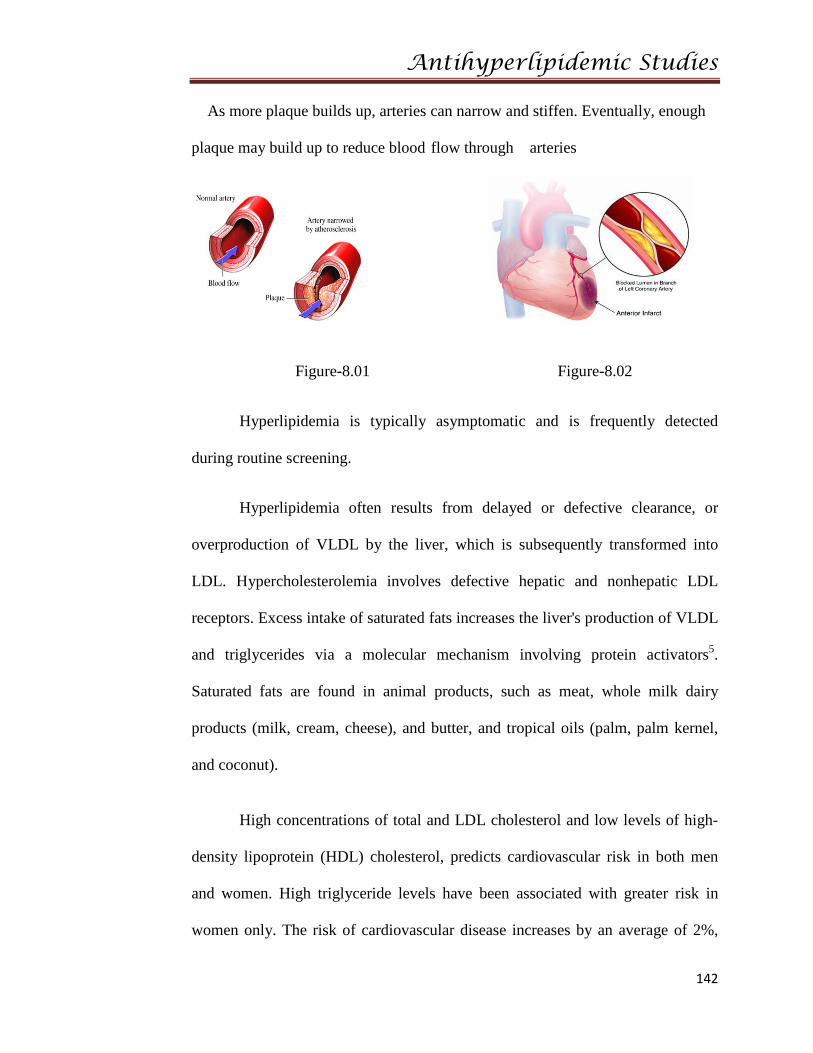
High lipid levels can speed up a process called atherosclerosis, or
hardening of the arteries. From inside, arteries are normally smooth and
unobstructed , but as increase in age, a sticky substance called plaque forms in
the walls of arteries, which is made of lipids and other materials circulating in
blood.
Antihyperlipidemic Studies
142
As more plaque builds up, arteries can narrow and stiffen. Eventually, enough
plaque may build up to reduce blood flow through arteries
Figure-8.01 Figure-8.02
Hyperlipidemia is typically asymptomatic and is frequently detected
during routine screening.
Hyperlipidemia often results from delayed or defective clearance, or
overproduction of VLDL by the liver, which is subsequently transformed into
LDL. Hypercholesterolemia involves defective hepatic and nonhepatic LDL
receptors. Excess intake of saturated fats increases the liver's production of VLDL
and triglycerides via a molecular mechanism involving protein activators5.
Saturated fats are found in animal products, such as meat, whole milk dairy
products (milk, cream, cheese), and butter, and tropical oils (palm, palm kernel,
and coconut).
High concentrations of total and LDL cholesterol and low levels of high-
density lipoprotein (HDL) cholesterol, predicts cardiovascular risk in both men
and women. High triglyceride levels have been associated with greater risk in
women only. The risk of cardiovascular disease increases by an average of 2%,
Antihyperlipidemic Studies
143
for each corresponding 1% rise in total cholesterol. Adolescents with high TC or
LDL may have a genetic disorder of lipid metabolism such as familial
hypercholesterolemia or familial combined hypercholesterolemia. Those with
homozygous forms of these disorders can experience myocardial infarction or
other events during childhood or early adolescence. Familial
hypercholesterolemia is often diagnosed in adolescence and is characterized by
high LDL levels that can be refractory to dietary treatment. These patients can
present clinically with xanthomas or xanthelasma– cholesterol deposits under the
skin on the hands, elbows, knees, heel or eyelids 6.
8.1.1 Types of Hyperlipidemia:
Depending on the complexity of the disease, Hyperlipidemia classified
into two types.
1) Primary Hyperlipidemia.
2) Secondary / Acquired Hyperlipidemia.
I) Primary Hyperlipidemia7,8:
Several genetic conditions are known to responsible for primary
Hyperlipidemia, such as lipoprotein lipase deficiency, apolipoprotein C-II
deficiency etc. The primary hyperlipidemia may be treated by anti-lipidemic
drugs. Primary Hyperlipidemia are again classified into 5 types
1. Type-I Hyperlipidemia: Severe elevation of chylomicrons (CMs) with
resultant elevation of TGs.
2. Type-II (A) Hyperlipidemia: Elevations of LDL –C only.
Antihyperlipidemic Studies
144
3. Type-II (B) Hyperlipidemia: Elevations of both LDL-C and triglycerides
(TG’s).
4. Type-III Hyperlipidemia: It develops due to defect in VLDL remnant
Clearance.
5. Type-IV Hyperlipidemia: It is characterized by hyper TG’s
6. Type-V Hyperlipidemia: Characterized by elevated levels of CMs and
VLDL.
II) Secondary Hyperlipidemia:
In this many factors can influence the level of TGs in circulation like
diabetes, obesity etc. Secondary Hyperlipidemia demands treatment of original
diseases rather than Hyperlipidemia.
Causes of secondary Hyperlipidemia:
A. Metabolic influences: Diabetes, obesity, hyperuricemia, glycogen storage
diseases.
B. Harmonal influences: Insulin, estrogen, thyroxine
C. Nutritional influences:-Alcohol, high carbohydrate intake
D. Disease states:-Renal diseases, renal failure, nephrotic syndrome
E. Drugs: - Diuretics
Beta-blockers
Glucocorticoids
Estrogen replacement therapy9
Antihyperlipidemic Studies
145
There are several secondary causes of abnormal lipids that may occur in
adolescence. Children, infants and geriatritics have been shown to have higher
levels of cholesterol, especially those who exhibit poor catch-up growth4. The
starved state that occurs in anorexia and the use of anabolic steroids are both
associated with abnormal lipids. Certain medications for acne, seizure disorders,
immunosuppression, and contraception can adversely affect lipids as can a high
carbohydrate diet or a ketogenic diet sometimes prescribed for refractory
epilepsy. Adolescents with a history of a transplant also tend to have an abnormal
lipoprotein panel despite a TC in the normal range.
8.1.2 Risk factors 3:
a. Positive risk factors:
1. Age (males > 45 years, females > 55 years or menopause < age 40)
2. Family history of premature coronary artery disease; definite myocardial
infarction (MI) or sudden death before age 55 in father or other male first-
degree relative, or before age 65 in mother or other female first-degree
relative
3. Current cigarette smoker
4. Hypertension (systolic blood pressure > 140 mmHg or diastolic blood
pressure > 90 mmHg confirmed on more than one occasion, or current
therapy with antihypertensive medications)
5. Diabetes mellitus (DM)
6. High-density lipoprotein (HDL)-cholesterol < 40 mg/dl
Antihyperlipidemic Studies
146
b. Negative risk factor:
1. Elevated HDL cholesterol, > 60 mg/dl
8.1.3 Etiology 10:
The etiology can be classified into primary and secondary causes.
Primary causes are due to single or multiple gene mutations resulting in a
disturbance of LDL and triglyceride production or clearance. They vary in
location of genetic defect, inheritance pattern, prevalence, clinical features, and
treatment. At least 18 separate entities have been described. The suspicion for a
primary lipid disorder should be especially high in patients with premature
atherosclerotic disease, a family history of early atherosclerotic disease, a
significantly elevated serum cholesterol level (>240 mg/dl), are physical signs of
hyperlipidemia. Primary dyslipidemia are most commonly seen in children, young
adults and a small percentage of adults were prone to this disorder.
Most adult cases of dyslipidemia are secondary in nature. In Western
civilizations, sedentary lifestyle and excessive consumption of saturated fats,
trans-fatty acids, and cholesterol are the most important secondary causes. Certain
medical conditions are commonly associated with dyslipidemia, including chronic
renal insufficiency, renal failure, diabetes mellitus, hypothyroidism, cholestatic
liver disease, and alcohol dependency. Certain drugs, including high-dose thiazide
diuretics, oral estrogens, glucocorticoids, anabolic steroids, and atypical
antipsychotics such as olanzapine and clozapine have also been implicated in
causing mild-to-moderate degrees of dyslipidemia. Use of atypical
Antihyperlipidemic Studies
147
antipsychotics, such as olanzapine and clozapine, and of beta-blockers without
intrinsic sympathomimetic or alpha-blocking activities is associated with reduced
HDL-cholesterol levels.
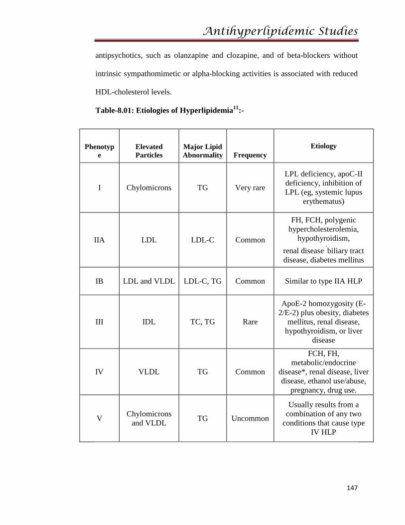
Table-8.01: Etiologies of Hyperlipidemia11:-
Phenotype
Elevated Particles
Major Lipid Abnormality Frequency
Etiology
I Chylomicrons TG Very rare
LPL deficiency, apoC-II deficiency, inhibition of LPL (eg, systemic lupus
erythematus)
IIA LDL LDL-C Common
FH, FCH, polygenic hypercholesterolemia,
hypothyroidism, renal disease
,biliary tract
disease, diabetes mellitus
IB LDL and VLDL LDL-C, TG Common Similar to type IIA HLP
III IDL TC, TG Rare
ApoE-2 homozygosity (E-2/E-2) plus obesity, diabetes
mellitus, renal disease, hypothyroidism, or liver
disease
IV VLDL TG Common
FCH, FH, metabolic/endocrine
disease*, renal disease, liver disease, ethanol use/abuse,
pregnancy, drug use.
V Chylomicrons and VLDL TG Uncommon
Usually results from a combination of any two
conditions that cause type IV HLP
Antihyperlipidemic Studies
148
8.1.4 Pathophysiology 11:-
Hypercholesterolemia develops as a consequence of abnormal lipoprotein
metabolism, mainly reduction of LDL receptor expression or activity, and
consequently diminishing hepatic LDL clearance from the plasma. It is a major
predisposing risk factor for the development of atherosclerosis. This mechanism
is classically seen in familial hypercholesterolemia and when excess saturated fat
or cholesterol is ingested. In addition, excessive production of VLDL by the liver,
as seen in familial combined hyperlipidemia and insulin resistance states such as
abdominal obesity and Type -II diabetes, can also induce hypercholesterolemia or
mixed dyslipidemia.
A current theory for the initiating event in atherogenesis is that apoprotein
B-100 containing lipoproteins are retained in the sub endothelial space, by means
of a charge-mediated interaction with extracellular matrix and
proteoglycans. This allows reactive oxygen species to modify the surface
phospholipids and unesterified cholesterol of the small LDL particles. Circulating
LDL can also be taken up into macrophages through unregulated scavenger
receptors. As a result of LDL oxidation, isoprostanes are formed. Isoprostanes are
chemically stable, free radical-catalyzed products of arachidonic acid, and are
structural isomers of conventional prostaglandins. Isoprostane levels are increased
in atherosclerotic lesions, but they may also be found as F2 isoprostanes in the
urine of asymptomatic patients with hypercholesterolemia.
A strong association exists between elevated plasma concentrations of
oxidized LDL and CHD. The mechanisms through which oxidized LDL
Antihyperlipidemic Studies
149
promotes atherosclerosis are multiple and include damage to the endothelium,
induction of growth factors, and recruitment of macrophages and monocytes.
Vasoconstriction in the setting of high levels of oxidized LDL seem to be
related to a reduced release of the vasodilator nitric oxide from the damaged
endothelial wall as well as increased platelet aggregation and thromboxane
release. Smooth muscle proliferation has been linked to the release of cytokines
from activated platelets.
The state of hypercholesterolemia leads invariably to an excess
accumulation of oxidized LDL within the macrophages, thereby transforming
them into "foam" cells. The rupture of these cells can lead to further damage of
the vessel wall due to the release of oxygen free radicals, oxidized LDL, and
intracellular enzymes.
This is a metabolically complex disease of lipid -lipoprotein metabolism
and the exact etiology is not fully appreciated. The familial type in schnauzers
may involve defects lipoprotein lipase and/or Apoprotein C-II, a required cofactor
for lipoprotein lipase activity. This defect causes a failure to
breakdown chylomicrons and VLDL, and results in excessive levels of circulating
triglycerides. It is the elevated concentration of triglycerides that is responsible for
the clinical signs.
Antihyperlipidemic Studies
150
8.1.5 Lipids12: Lipids are a group of naturally occurring fatty substances, which
are present in the blood and tissues of the body. They include cholesterol,
cholesterol esters, triglycerides, and phospholipids. Lipids are essential dietary
constituents because of their important functions.
8.1.6 Classification of lipids:-
• Fatty acids (palmetic, linoleic, etc)
• Glycerol esters (triglycerides)
• Sterols (cholesterol, hormones, vitamin D)
• Terpenes (vitamin A, E, K)
• Sphingosine derivatives (sphingomyelin)
8.1.7 Lipid functions 13:-
• Provide energy required by the body
• Serve as the major structural components of cell membranes
• Aid in the efficient absorption of fat-soluble vitamins
• Serve as insulating material beneath the skin and around certain organs
(e.g. kidneys)
• Serve as biosynthetic precursors (e.g., Cholesterol is a precursor for
adrenal and gonadal steroid hormones and hepatic bile acids.)
Antihyperlipidemic Studies
151
Lipids are insoluble in blood (plasma), they must be transported to the
cells by special carriers called lipoproteins. Lipoproteins are spherical particles of
high molecular weight. Each lipoprotein particle contains a non-polar core and a
hydrophilic surface. The hydrophilic surface makes the lipoprotein soluble in
plasma and acts as an interface between the plasma and lipid core. The core
consists of hydrophobic lipids, triglycerides and cholesterol esters, surrounded by
a hydrophilic surface coat of phospholipids, unesterified cholesterol, and specific
proteins termed apolipoproteins or apoproteins. The apolipoproteins provide
structural integrity to the lipoproteins and determine the lipoproteins’ metabolic
fate by serving as binding sites for receptors and activating enzymes involved in
lipid metabolism.
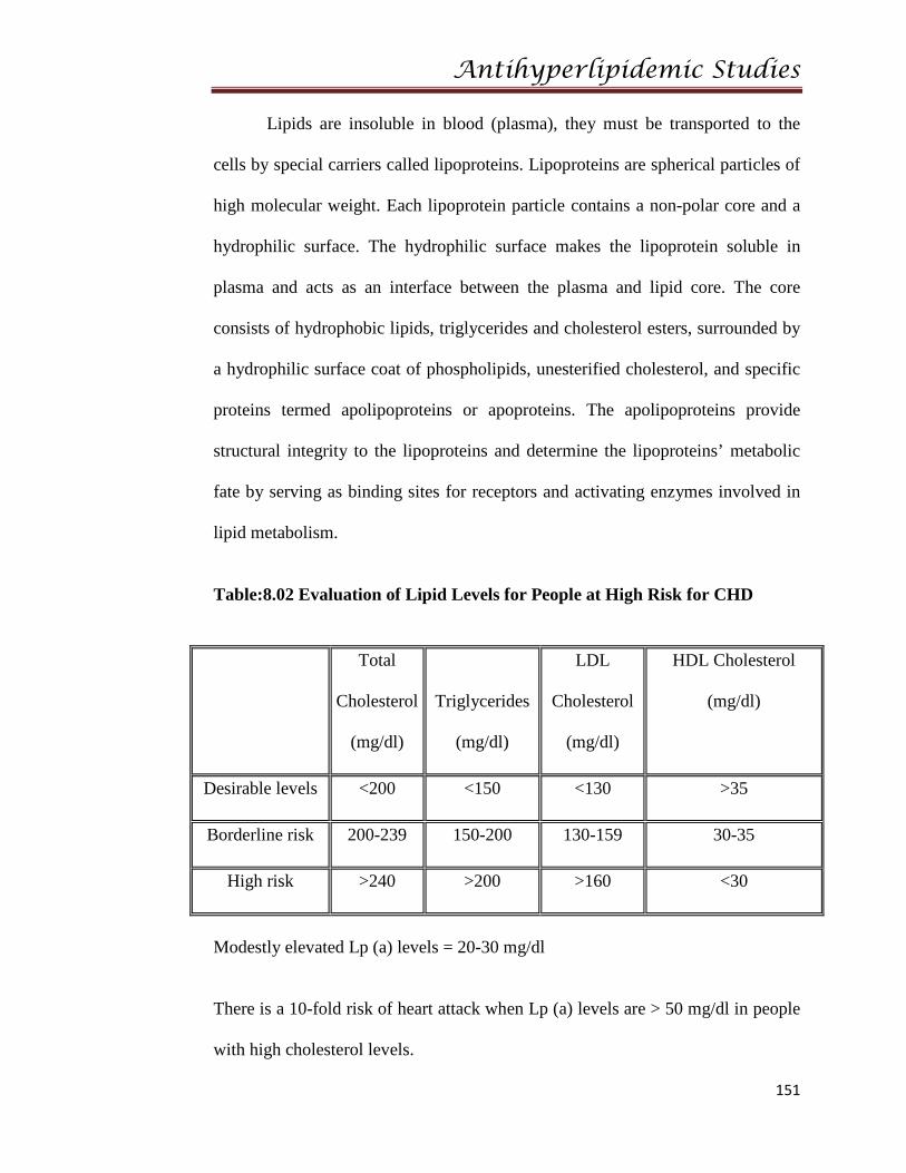
Table:8.02 Evaluation of Lipid Levels for People at High Risk for CHD
Total
Cholesterol
(mg/dl)
Triglycerides
(mg/dl)
LDL
Cholesterol
(mg/dl)
HDL Cholesterol
(mg/dl)
Desirable levels <200 <150 <130 >35
Borderline risk 200-239 150-200 130-159 30-35
High risk >240 >200 >160 <30
Modestly elevated Lp (a) levels = 20-30 mg/dl
There is a 10-fold risk of heart attack when Lp (a) levels are > 50 mg/dl in people
with high cholesterol levels.
Antihyperlipidemic Studies
152
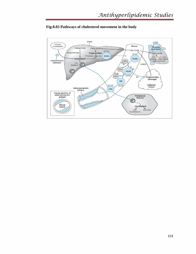
8.1.8 Pathways of Lipid Transport 14:-
Cholesterol is absorbed from the intestine and transported to the liver by
chylomicron reminants, which are taken up by the low density lipoprotein (LDL)-
receptor related protein (LRP). Hepatic cholesterol enters the circulation as very-
low-density lipoprotein (VLDL) and is metabolized to remnant lipoproteins after
lipoprotein lipase removes triglyceride. The remnant lipoproteins are removed by
LDL receptors (LDL-R) or further metabolized to LDL and then removed by
these receptors. Cholesterol is transported from peripheral cells to the liver by
high-density lipoprotein (HDL). Cholesterol is recycled to LDL and VLDL by
cholesterol-ester transport protein (CETP) or is taken up in the liver by hepatic
lipase. Cholesterol is excreted in bile. The points in the process that are affected
by the five primary lipoprotein disorders and familial hypertriglyceridemia
(FHTG), Familial combined hyperlipidemia (FCHL), remnant removal disease
(RRD, also known as familial dys-beta-lipoproteinemia), Familial
hypercholesterolemia (FH), and hypo-alpha- lipoproteinemia shown. The effects
of drug therapy can also be understood from these pathways. Statins decrease the
synthesis of cholesterol and the secretion of VLDL and increase the activity of
LDL receptors. Bile-acid binding resins increase the secretion of bile acids.
Nicotinic acid decreases the secretion of VLDL and the formation of LDL and
increases the formation of HDL. Fibrates decrease the secretion of VLDL and
increase the activity of lipoprotein lipase, thereby increasing the removal of
triglycerides.
Antihyperlipidemic Studies
153
Fig:8.03 Pathways of cholesterol movement in the body
Antihyperlipidemic Studies
154
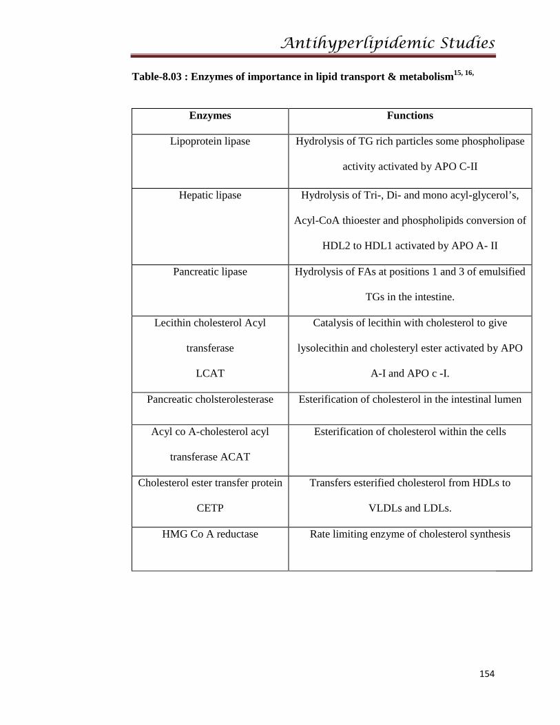
Table-8.03 : Enzymes of importance in lipid transport & metabolism15, 16,
Enzymes Functions
Lipoprotein lipase Hydrolysis of TG rich particles some phospholipase
activity activated by APO C-II
Hepatic lipase Hydrolysis of Tri-, Di- and mono acyl-glycerol’s,
Acyl-CoA thioester and phospholipids conversion of
HDL2 to HDL1 activated by APO A- II
Pancreatic lipase Hydrolysis of FAs at positions 1 and 3 of emulsified
TGs in the intestine.
Lecithin cholesterol Acyl
transferase
LCAT
Catalysis of lecithin with cholesterol to give
lysolecithin and cholesteryl ester activated by APO
A-I and APO c -I.
Pancreatic cholsterolesterase Esterification of cholesterol in the intestinal lumen
Acyl co A-cholesterol acyl
transferase ACAT
Esterification of cholesterol within the cells
Cholesterol ester transfer protein
CETP
Transfers esterified cholesterol from HDLs to
VLDLs and LDLs.
HMG Co A reductase Rate limiting enzyme of cholesterol synthesis
Antihyperlipidemic Studies
155
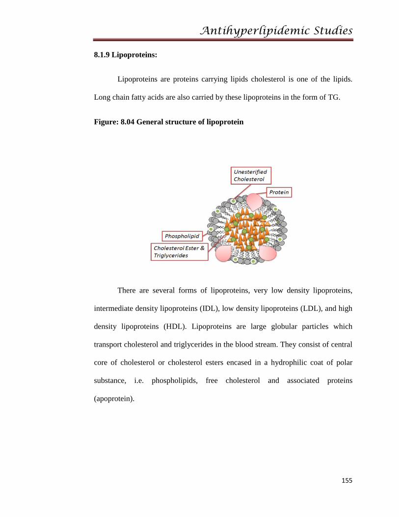
8.1.9 Lipoproteins:
Lipoproteins are proteins carrying lipids cholesterol is one of the lipids.
Long chain fatty acids are also carried by these lipoproteins in the form of TG.
Figure: 8.04 General structure of lipoprotein
There are several forms of lipoproteins, very low density lipoproteins,
intermediate density lipoproteins (IDL), low density lipoproteins (LDL), and high
density lipoproteins (HDL). Lipoproteins are large globular particles which
transport cholesterol and triglycerides in the blood stream. They consist of central
core of cholesterol or cholesterol esters encased in a hydrophilic coat of polar
substance, i.e. phospholipids, free cholesterol and associated proteins
(apoprotein).
Antihyperlipidemic Studies
156
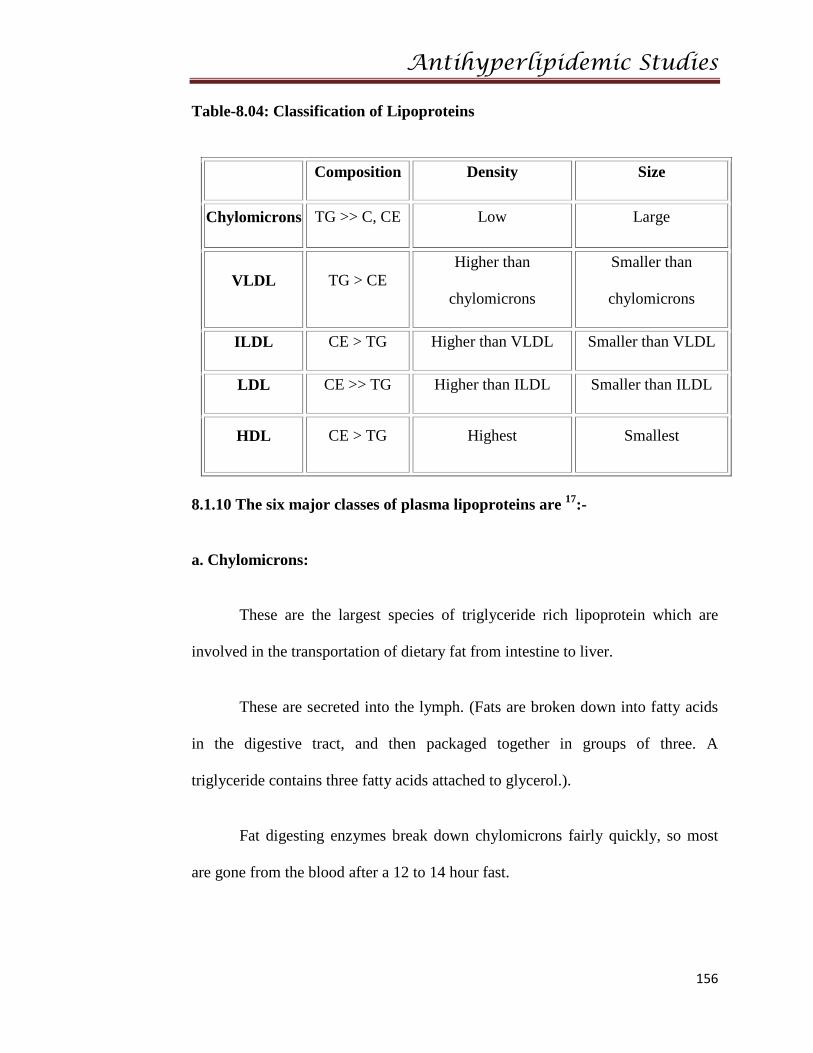
Table-8.04: Classification of Lipoproteins
Composition Density Size
Chylomicrons TG >> C, CE Low Large
VLDL TG > CE Higher than
chylomicrons
Smaller than
chylomicrons
ILDL CE > TG Higher than VLDL Smaller than VLDL
LDL CE >> TG Higher than ILDL Smaller than ILDL
HDL CE > TG Highest Smallest
8.1.10 The six major classes of plasma lipoproteins are 17:-
a. Chylomicrons:
These are the largest species of triglyceride rich lipoprotein which are
involved in the transportation of dietary fat from intestine to liver.
These are secreted into the lymph. (Fats are broken down into fatty acids
in the digestive tract, and then packaged together in groups of three. A
triglyceride contains three fatty acids attached to glycerol.).
Fat digesting enzymes break down chylomicrons fairly quickly, so most
are gone from the blood after a 12 to 14 hour fast.
Antihyperlipidemic Studies
157
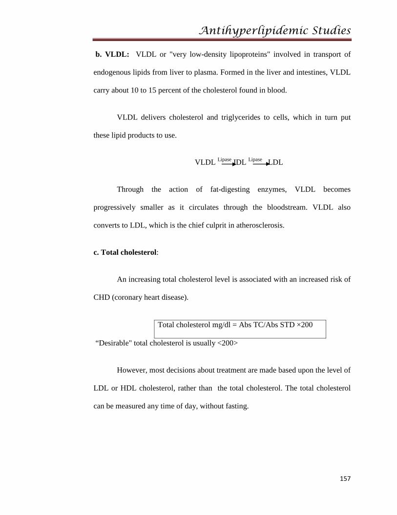
b. VLDL: VLDL or "very low-density lipoproteins" involved in transport of
endogenous lipids from liver to plasma. Formed in the liver and intestines, VLDL
carry about 10 to 15 percent of the cholesterol found in blood.
VLDL delivers cholesterol and triglycerides to cells, which in turn put
these lipid products to use.
VLDL Lipase IDL Lipase LDL
Through the action of fat-digesting enzymes, VLDL becomes
progressively smaller as it circulates through the bloodstream. VLDL also
converts to LDL, which is the chief culprit in atherosclerosis.
c. Total cholesterol:
An increasing total cholesterol level is associated with an increased risk of
CHD (coronary heart disease).
“Desirable" total cholesterol is usually <200>
However, most decisions about treatment are made based upon the level of
LDL or HDL cholesterol, rather than the total cholesterol. The total cholesterol
can be measured any time of day, without fasting.
Total cholesterol mg/dl = Abs TC/Abs STD ×200
Antihyperlipidemic Studies
158
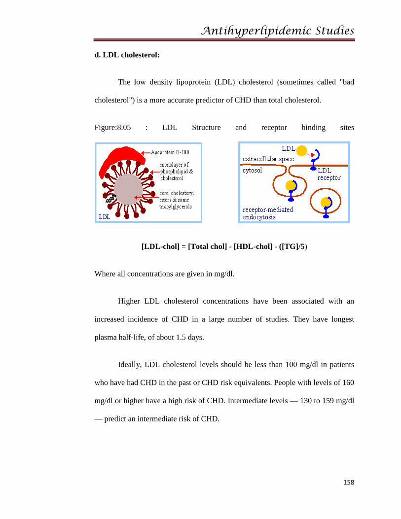
d. LDL cholesterol:
The low density lipoprotein (LDL) cholesterol (sometimes called "bad
cholesterol") is a more accurate predictor of CHD than total cholesterol.
Figure:8.05 : LDL Structure and receptor binding sites
[LDL-chol] = [Total chol] - [HDL-chol] - ([TG]/5)
Where all concentrations are given in mg/dl.
Higher LDL cholesterol concentrations have been associated with an
increased incidence of CHD in a large number of studies. They have longest
plasma half-life, of about 1.5 days.
Ideally, LDL cholesterol levels should be less than 100 mg/dl in patients
who have had CHD in the past or CHD risk equivalents. People with levels of 160
mg/dl or higher have a high risk of CHD. Intermediate levels — 130 to 159 mg/dl
— predict an intermediate risk of CHD.
Antihyperlipidemic Studies
159
LDL particles are finally delivered to hepatic and certain extra hepatic
tissues for further liposomal degradation to release the cholesterol which can be
utilized in cell membrane formation.
The LDL cholesterol can only be determined accurately on a blood test
after fasting for 12 to 14 hours.
e. IDL cholesterol:-
These are the lipoproteins obtained when the triglyceride content of VLDL
are partially digested in capillaries by the action of extra hepatic lipoprotein lipase
and having the diameter of 20-35 nm.
f. Triglycerides:
Elevated levels of triglycerides are also associated with an increased risk of CHD.
• Normal - less than 150 mg/dl (1.69 mmol/l)
• Borderline high - 150 to 199 mg/dl (1.69 to 2.25 mmol/l)
• High - 200 to 499 mg/dl (2.25 to 5.63 mmol/l)
• Very high - greater than 500 mg/dl (5.65 mmol/l)
Triglycerides in mg/dl = Abs T/Abs STD×200
Like LDL cholesterol, triglycerides should only be measured in a blood
specimen obtained after fasting for 12 to 14 hours.
Antihyperlipidemic Studies
160
g. HDL cholesterol:
This is a group of heterogeneous lipoprotein having low lipid content and
is also called as good cholesterol. HDL enhances the removal of cholesterol from
the arterial wall. Hence, chances of development of atherosclerotic lesions are
more when HDL value falls below normal.
Similar to total cholesterol, the HDL-cholesterol can be measured in
blood specimen without fasting.
HDL cholesterol mg/dl = Abs TH/Abs STD×50
Conversion Factors:
Cholesterol: mmol/L x 38.7 = mg/dl mg/dl x 0.026 = mmol/L
Triglycerides: mmol/L x 885.5 = mg/dl mg/dl x 0.0113 = mmol/L
Phospholipids: g/L x 0.01 = mg/dl mg/dl x 10 = g/L
Simple blood tests can determine levels of Lipoproteins. Including Total
cholesterol, LDL and HDL cholesterol, and triglycerides.
Antihyperlipidemic Studies
161
8.1.11 Some of the risk factors for developing hyperlipoproteinemia are:
• Obesity
• Diabetes
• Hyperthyroidism
• Nephrotic disorder
• Liver disease
• Hypertension
• Family history of high cholesterol or heart disease
• Diet high in fat and cholesterol
• Cigarette smoking
8.1.12 Causes:
Hyperlipidemia is caused by lifestyle habits or treatable medical
conditions. Lifestyle habits include obesity, sedentary life without exercise,
smoking. Medical diseases that may result in Hyperlipidemia are diabetes, kidney
disorders, pregnancy, and an under active thyroid gland. Common secondary
causes of hypercholesterolemia are hypothyroidism, pregnancy, and Kidney
failure. Common secondary causes of hypertriglyceridemia are diabetes, excess
alcohol intake, obesity, and certain prescription medications18.
Antihyperlipidemic Studies
162
8.1.13 Symptoms and diagnoses of Hyperlipidemia19:
Generally hyperlipidemia condition does not show apparent symptoms
and it is discovered and diagnosed during routine examination or evaluation for
atherosclerotic cardiovascular disease. However, deposits of cholesterol may be
formed under the skin in individuals with familial forms of the disorder or in
persons with very high levels of cholesterol in the blood. In individuals with
hypertriglyceridemia, several pimple-like lesions may be developed across their
bodies. Pancreatitis, a severe inflammation of the pancreas that may be life-
threatening can also be developed due to extremely high levels of triglycerides.
For diagnosis of hyperlipidemia, levels of total cholesterol, low density
lipoprotein cholesterol, high density lipoprotein cholesterol, and triglycerides are
measured in blood sample. It is important to note that the lipid profile should be
measured in all adults 20 years and older, and the measurement should be
repeated after every 5 years. Food or beverages may increase triglyceride levels
temporarily, so people must fast at least 12 hours before giving their blood
samples. Blood tests are carried out to identify the specific disorders, when lipid
levels in the blood are very high. Specific disorders may include several
hereditary disorders, which produce different lipid abnormalities and have
different risks.

Antihyperlipidemic Studies
163
8.1.14 Laboratory Testing: Patients are subjected to fasting for at least 12
hours before collecting the blood sampling. Because, chylomicron clearance can
take up to 10 hours. However, a fasted sample is not required for simple
cholesterol screening.
Laboratory testing of the lipid profile measures total plasma cholesterol,
HDL, and triglycerides levels directly. VLDL cholesterol levels are calculated by
dividing the triglyceride value by 5. LDL cholesterol is calculated by subtracting
HDL cholesterol and VLDL cholesterol from total cholesterol. When triglycerides
are above 400 mg/dl, LDL calculation is inaccurate, and specialized laboratory
tests are required.
8.1.15 Treatment 20,21:
The goals of treatment are to lower total and LDL cholesterol in order to reduce
the risk of first or recurrent events such as
• Myocardial infarction,
• Angina,
• Heart failure,
• Ischemic stroke, or other forms of peripheral arterial disease such
as carotid stenosis or abdominal aortic aneurysm
Antihyperlipidemic Studies
164
Most patients should receive 3 month TLC trial before initiating
pharmacologic therapy unless very high risk
If patient unable to reach goals with TLC alone choose lipid-lowering
drugs based on lipoprotein disorder
Combination therapy may be necessary to monitor closely: increased risk
of drug interactions, adverse effects
8.1.16 General approach:
The National Cholesterol Education Program Adult Treatment Panel III
(NCEP ATP III) recommends that a fasting lipoprotein profile and risk
factor assessment be used in the initial classification of adults.
There are four categories of risk that modify the goals and modalities of
LDL-lowering therapy.
The highest risk category is having known CHD or CHD risk equivalents;
the risk for major coronary events is equal to or greater than that for
established CHD (i.e., >20% per 10 years, or 2% per year).
The next category is moderately high risk, consisting of patients with two
or more risk factors in which 10-year risk for CHD is 10% to 20%.
The lowest risk category is persons with zero to one risk factor, which is
usually associated with a 10-year CHD risk of <10%.
ATP III recognizes the metabolic syndrome as a secondary target of risk
reduction after LDL-C has been addressed.
Antihyperlipidemic Studies
165
This syndrome is characterized by abnormal obesity, atherogenic
dyslipidemia (elevated triglycerides, small LDL particles, low HDL
cholesterol), increased blood pressure, insulin resistance (with or without
glucose intolerance), and pro thrombotic and pro inflammatory states.
If the metabolic syndrome is present, the patient is considered to have a
CHD risk equivalent.
Other targets include non-HDL goals for patients with triglycerides
>200mg/dl.
Non-HDL cholesterol is calculated by subtracting HDL from total
cholesterol, and the targets are 30 mg/dl greater than for LDL at each risk
stratum.
8.1.17 Pharmacologic Therapy:22
Treat all secondary problems resulting from acute or chronic disease (e.g.
Diabetes, seizures)
After proper diagnosis of hyper lipoproteinemia and before medicinal
intervention, lifestyle changes and risk factor reduction is warranted. This
includes diet modification, weight loss, exercise, smoking cessation, and control
of underlying disorders such as diabetes and hypertension. Such changes can lead
to significant reductions in plasma lipoproteins. If dietary and lifestyle changes
fail, hypolipoproteinemic (cholesterol-reducing) drugs are advised. The types of
drugs used to lower blood lipids and block atherogenesis are:
Antihyperlipidemic Studies
166
1. Cholesterol biosynthesis inhibitors (e.g. Lovastatin)
2. Fibrates (e.g. Clofibrate)
3. Bile acid sequesterants (e.g. Cholestyramine)
4. Cholesterol absorption inhibitors
5. LDL oxidation inhibitors (e.g. Probucol)
8.1.18 Experimental Induced Methods of Hyperlipidemia:
I. Cholesterol Induced Hyperlipidemia:
Hyperlipidemia has been ranked as one of the greatest risk factors
contributing to the prevalence and severity of coronary heart diseases. Coronary
heart disease, stroke, atherosclerosis and hyperlipidemia are the primary cause of
death. Hyperlipidemia is characterized by elevated serum total cholesterol, low
density lipoprotein, very low density lipoprotein and decreased high density
lipoprotein levels. Hyperlipidemia associated lipid disorders are considered to
cause atherosclerotic cardiovascular disease. Among these hypercholesterolemia
and hypertriglyceridemia are closely related to ischemic heart disease. The main
aim of treatment in patients with hyperlipidemia is to reduce the risk of
developing ischemic heart disease or the occurrence of further cardiovascular
disease or cerebrovascular disease. It is actively involved in the screening of
herbal formulations and synthetic drugs for its anti-hyperlipidemic activity 23.
Antihyperlipidemic Studies
167
II. Atherogenic Diet Induced Hyperlipidemia in Rats:
In rats, hyperlipidemia can be induced by daily oral administration of 1%
cholesterol, 0.5% Cholic acid suspended in 25% coconut oil over a period of 26
days. The test compounds were administered simultaneously along with
cholesterol diet.
High intake of saturated fat and cholesterol increases serum LDL-C,
probably by decreasing the amount of and/or activity of LDL receptors in the
liver. Elevated and modified LDL is one of the principal factors in the
development of atherosclerosis. Feeding the high fat diets causes fatty liver with
accumulation of TG and TC 24
.
III. Fructose Induced Hyperlipidemia in Rats 25:
Carbohydrate, fructose plays an important role in the pathogenesis of
experimental and clinical hypertriglyceridemia and hyperinsulinemia. High
fructose fed (HFF) diet induces significant hyperinsulinemia and
hypertriglyceridemia in rats. An adverse effect of fructose on insulin sensitivity of
rat is well established. This phenomenon is believed to be related
hypertriglyceridemic effect of fructose. In rats hyperlipidemia can be produced by
administration of fructose (66% fructose), once daily for 30 days.
Fructose feeding stimulates the hepatic production TGs, both by
promoting the reesterification of circulating non-esterified FAs and by stimulating
fatty acid synthesis. Increased delivery of TGs to the muscle interferes with the
utilization of glucose, through the principles of Randle cycle, impairing the
insulin action.
Antihyperlipidemic Studies
168
IV. Triton WR 1339(TR) Induced Hyperlipidemia in Rats:
The systemic administration of the nonionic surfactant TR (iso octyl
polyoxyethylene phenol /Tyloxipal) to rats results in a biphasic elevation of
plasma cholesterol and TGs. TR induced hyperlipidemia occurs in 2 phases.
Phase –I (synthesis phase):
It is thought to be due to increased hepatic synthesis of cholesterol, which
reaches the elevated lipid level at the end of 24th hr through the ability to interfere
with the uptake of plasma lipid levels, by the tissues. Drugs interfering with
cholesterol biosynthesis were shown to be active in this phase.
Phase –II (excretory phase):
In this phase, the elevated lipid levels almost reach normal by the end of
48th
hr .while drugs interfering with cholesterol excretion and metabolism were
shown to be active in this phase.
The biphasic nature of TR induced hyperlipidemia is helpful in
understanding the mode of action of hypolipidemic agents27.
Antihyperlipidemic Studies
169
8.2 MATERIALS AND METHODS:
Wistar albino rats of either sex (150-200gm)
Petroleum ether extract of V.mung
Chloroform extract of V.mung
Ethanolic extract of V.mung
Petroleum ether extract of V.radiata
Chloroform extract of V.radiata
Ethanolic extract of V.radiata
Petroleum ether extract of V.ungiculata
Chloroform extract of V.ungiculata
Ethanolic extract of V.ungiculata
High Cholesterol diet pellets
5% Aqueous gum acacia
Animal models: Wistar albino rats
Standard drug: Atorvastatin- ATOCORTM 80 supplied by Dr.Reddy Labs
Antihyperlipidemic Studies
170
8.3 EXPERIMENTAL PROCEDURE
In the present study we aimed to screen various extracts of selected plants on
Cholesterol induced hyperlipidemic rat model27,28.
Healthy Wistar albino rats weighing between 150-200gm were
acclimatized to the laboratory at temperature (25±1)0c, relative humidity(50±15)
%, 12hrs light-dark cycles, kept in standard polypropylene cages and given
standard diet and water ad-libitum. The animals were divided into control, toxic,
standard and test groups of V.M.P.E 100mg/kg, V.M.C.E 100mg/kg, V.M.E.E
100mg/kg b.w p.o, suspended in 5% gum acacia solution, daily once . Each
comprising of 6 animals in all sets of experiments. Animals in the normal control
group, received normal saline orally. Except control group rest other groups were
fed with rich cholesterol diet pellets supplied by M/s Rayans biotechnologies
Pvt.Ltd., Hyderabad. Standard group received Atorvastatin 10mg/kg b.w p.o
suspended in 5% gum acacia solution. The treatment was given for 20 days. In
between mean body weight of the animals was checked time to time. Feeding
the animals with cholesterol supplied diet induces hyperlipidemia, especially
hypercholesterolemia and hypertriglyceridemia. Cholesterol feeding has been
often used to elevate serum or tissue cholesterol levels to assess
hypercholesterolemia-related metabolic disturbances in experimental animal .On
21st day the blood samples were withdrawn from the arterial damage. All the lipid
profile parameters were determined. Total cholesterol (TC), triglycerides (TG),
high density lipoproteins (HDL), very low density lipoproteins (VLDL), low
density lipoproteins (LDL) were analysed from serum.
Antihyperlipidemic Studies
171
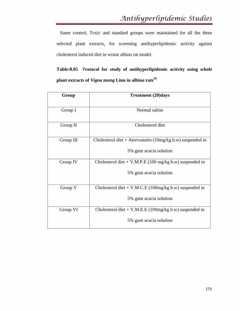
Same control, Toxic and standard groups were maintained for all the three
selected plant extracts, for screening antihyperlipidemic activity against
cholesterol induced diet in wistar albino rat model.
Table:8.05 Protocol for study of antihyperlipidemic activity using whole
plant extracts of Vigna mung Linn in albino rats29
Group Treatment (20)days
Group I Normal saline
Group II Cholesterol diet
Group III Cholesterol diet + Atorvastatin (10mg/kg b.w) suspended in
5% gum acacia solution
Group IV Cholesterol diet + V.M.P.E (100 mg/kg b.w) suspended in
5% gum acacia solution
Group V Cholesterol diet + V.M.C.E (100mg/kg b.w) suspended in
5% gum acacia solution
Group VI Cholesterol diet + V.M.E.E (100mg/kg b.w) suspended in
5% gum acacia solution
Antihyperlipidemic Studies
172
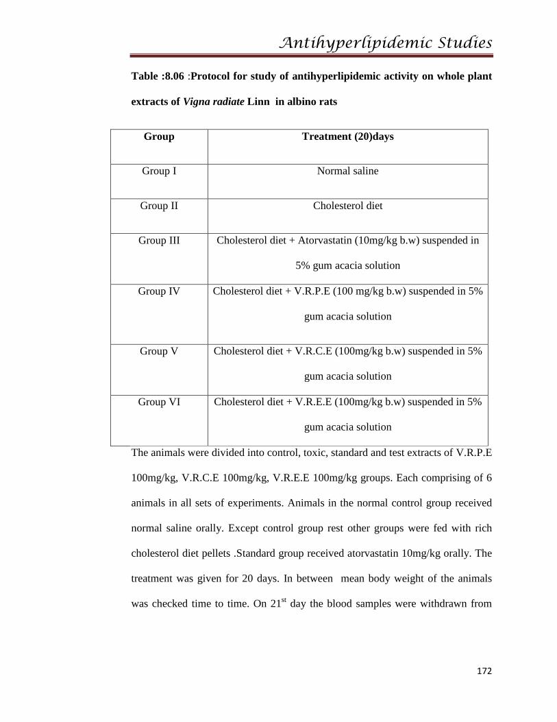
Table :8.06 :Protocol for study of antihyperlipidemic activity on whole plant
extracts of Vigna radiate Linn in albino rats
Group Treatment (20)days
Group I Normal saline
Group II Cholesterol diet
Group III Cholesterol diet + Atorvastatin (10mg/kg b.w) suspended in
5% gum acacia solution
Group IV Cholesterol diet + V.R.P.E (100 mg/kg b.w) suspended in 5%
gum acacia solution
Group V Cholesterol diet + V.R.C.E (100mg/kg b.w) suspended in 5%
gum acacia solution
Group VI Cholesterol diet + V.R.E.E (100mg/kg b.w) suspended in 5%
gum acacia solution
The animals were divided into control, toxic, standard and test extracts of V.R.P.E
100mg/kg, V.R.C.E 100mg/kg, V.R.E.E 100mg/kg groups. Each comprising of 6
animals in all sets of experiments. Animals in the normal control group received
normal saline orally. Except control group rest other groups were fed with rich
cholesterol diet pellets .Standard group received atorvastatin 10mg/kg orally. The
treatment was given for 20 days. In between mean body weight of the animals
was checked time to time. On 21st day the blood samples were withdrawn from
Antihyperlipidemic Studies
173
the arterial damage. Total cholesterol (TC), triglycerides (TG), high density
lipoproteins (HDL), low density lipoproteins (LDL) were analysed from serum.
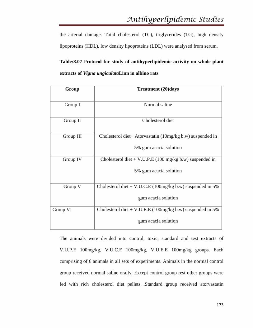
Table:8.07 Protocol for study of antihyperlipidemic activity on whole plant
extracts of Vigna ungiculataLinn in albino rats
Group Treatment (20)days
Group I Normal saline
Group II Cholesterol diet
Group III Cholesterol diet+ Atorvastatin (10mg/kg b.w) suspended in
5% gum acacia solution
Group IV Cholesterol diet + V.U.P.E (100 mg/kg b.w) suspended in
5% gum acacia solution
Group V Cholesterol diet + V.U.C.E (100mg/kg b.w) suspended in 5%
gum acacia solution
Group VI Cholesterol diet + V.U.E.E (100mg/kg b.w) suspended in 5%
gum acacia solution
The animals were divided into control, toxic, standard and test extracts of
V.U.P.E 100mg/kg, V.U.C.E 100mg/kg, V.U.E.E 100mg/kg groups. Each
comprising of 6 animals in all sets of experiments. Animals in the normal control
group received normal saline orally. Except control group rest other groups were
fed with rich cholesterol diet pellets .Standard group received atorvastatin
Antihyperlipidemic Studies
174
10mg/kg orally. The treatment was given for 20 days. In between mean body
weight of the animals was checked time to time. On 21st day the blood samples
were withdrawn from the arterial damage. Total cholesterol (TC), triglycerides
(TG), high density lipoproteins (HDL), low density lipoproteins (LDL) were
analysed from serum.
8.3.1 Biochemical Estimations:-
At the end of experimental period, rats were anesthetized with ether.
Blood samples were collected by cardiac puncture method. Serum total
cholesterol, triglycerides, high density lipoproteins- cholesterol using beacon
diagnostic Pvt ltd kits. Serum LDL, VLDL was determined by calculation.
8.3.2 Procedures for testing parameters30:
1. Estimation of serum of triglycerides:
Diagnostic kit was used for estimation of triglycerides, which followed
end point colorimetry enzymatic test using glycerol-3-phosphate oxidase.
Principle:
The enzyme, lipoprotein lipase catalyzes hydrolysis of TGs to glycerol
and Free acids. Glycerol then is phosphorylated in an ATP - requiring reaction
catalyzed by glycerophosphate. The formed glycerophosphate is oxidized to
dihydroxyacetone and H2O2 in a glycerophosphate oxidase catalyzed reaction.
H2O2 then reacts with 4 -AAP and 4 -chlorophenol under the catalytic influence of
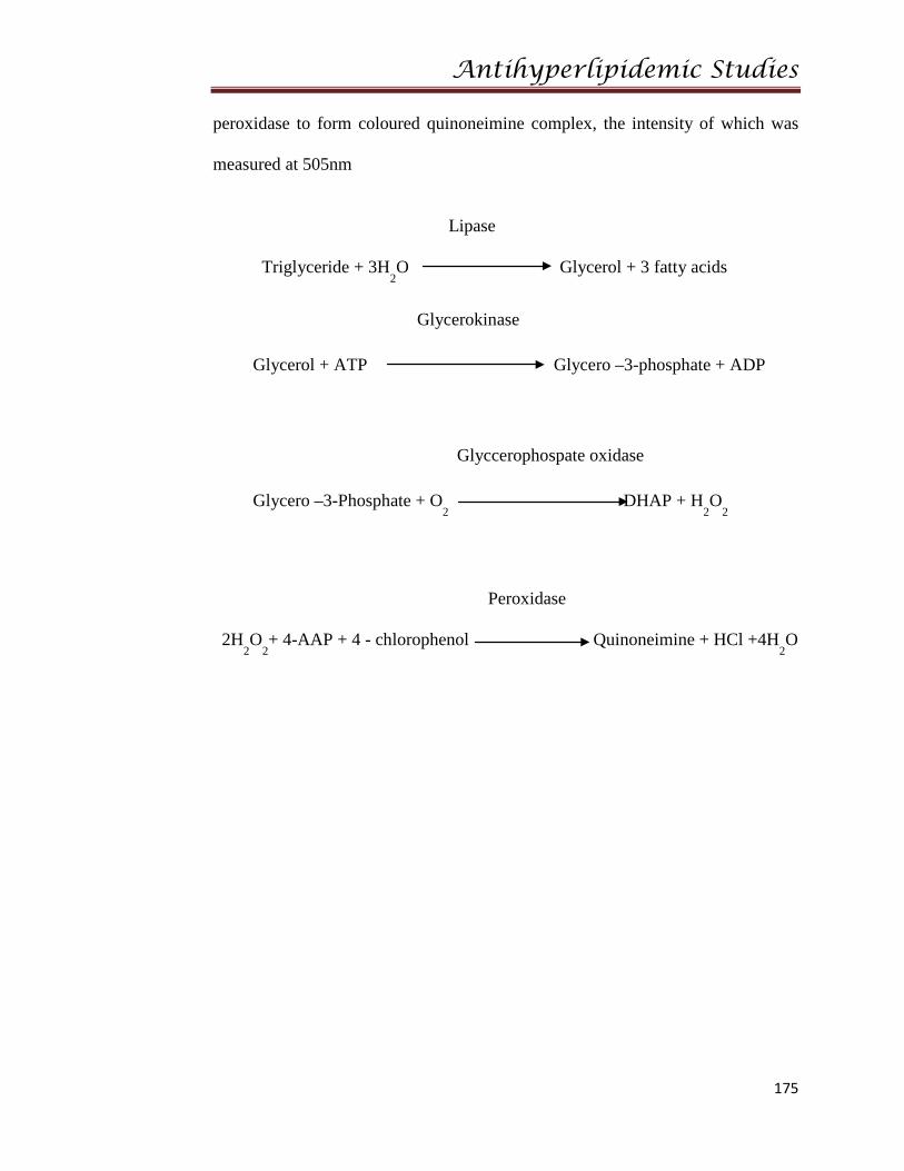
Antihyperlipidemic Studies
175
peroxidase to form coloured quinoneimine complex, the intensity of which was
measured at 505nm
Lipase
Triglyceride + 3H2O Glycerol + 3 fatty acids
Glycerokinase
Glycerol + ATP Glycero –3-phosphate + ADP
Glyccerophospate oxidase
Glycero –3-Phosphate + O2
DHAP + H2O
2
Peroxidase
2H2O
2+ 4-AAP + 4 - chlorophenol Quinoneimine + HCl +4H
2O
Antihyperlipidemic Studies
176
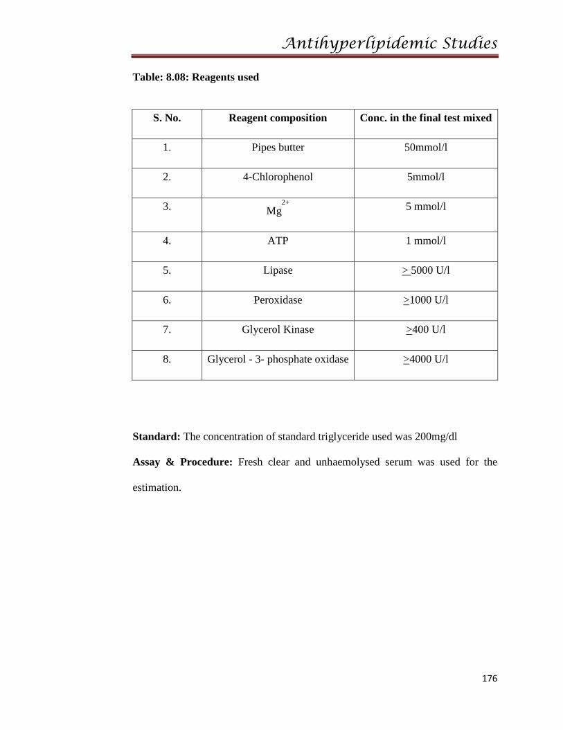
Table: 8.08: Reagents used
S. No. Reagent composition Conc. in the final test mixed
1. Pipes butter 50mmol/l
2. 4-Chlorophenol 5mmol/l
3. Mg2+
5 mmol/l
4. ATP 1 mmol/l
5. Lipase > 5000 U/l
6. Peroxidase >1000 U/l
7. Glycerol Kinase >400 U/l
8. Glycerol - 3- phosphate oxidase >4000 U/l
Standard: The concentration of standard triglyceride used was 200mg/dl
Assay & Procedure: Fresh clear and unhaemolysed serum was used for the
estimation.
Antihyperlipidemic Studies
177
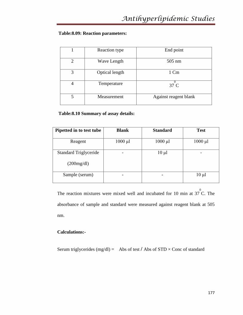
Table:8.09: Reaction parameters:
1 Reaction type End point
2 Wave Length 505 nm
3 Optical length 1 Cm
4 Temperature 370C
5 Measurement Against reagent blank
Table:8.10 Summary of assay details:
Pipetted in to test tube Blank Standard Test
Reagent 1000 μl 1000 μl 1000 μl
Standard Triglyceride
(200mg/dl)
- 10 μl -
Sample (serum) - - 10 μl
The reaction mixtures were mixed well and incubated for 10 min at 370C. The
absorbance of sample and standard were measured against reagent blank at 505
nm.
Calculations:-
Serum triglycerides (mg/dl) = Abs of test / Abs of STD × Conc of standard
Antihyperlipidemic Studies
178
2. Estimation of serum Total Cholesterol:-
The reagents kits intended for the In-vitro quantitative determination of
cholesterol in serum/plasma.
Principle: -
The cholesterol esters are hydrolysed by enzyme cholesterol esterase to
give free cholesterol and fatty acid molecules. This free cholesterol gets oxidised
in presence of cholesterol oxidase to liberate cholset4en-3one and H2O2.
Liberated H2O2 by this reaction combines with phenol and 4 amino antipyrine in
presence of peroxidase to form red colour quinonimine complex, the intensity of
which is measured at 505 nm.
General system parameters:-
Wave length: 505 nm (490-530nm)
Incubation: 5 min
Sample volume: 10µl
Reagent volume: 1.0ml
Standard concentration: 200mg/dl
Antihyperlipidemic Studies
179
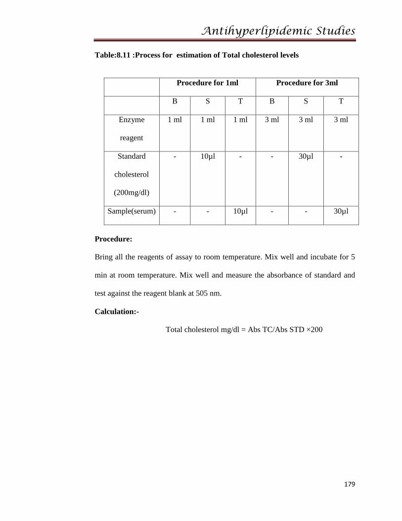
Table:8.11 :Process for estimation of Total cholesterol levels
Procedure for 1ml Procedure for 3ml
B S T B S T
Enzyme
reagent
1 ml 1 ml 1 ml 3 ml 3 ml 3 ml
Standard
cholesterol
(200mg/dl)
- 10µl - - 30µl -
Sample(serum) - - 10µl - - 30µl
Procedure:
Bring all the reagents of assay to room temperature. Mix well and incubate for 5
min at room temperature. Mix well and measure the absorbance of standard and
test against the reagent blank at 505 nm.
Calculation:-
Total cholesterol mg/dl = Abs TC/Abs STD ×200
Antihyperlipidemic Studies
180
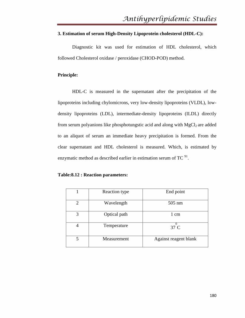
3. Estimation of serum High-Density Lipoprotein cholesterol (HDL-C):
Diagnostic kit was used for estimation of HDL cholesterol, which
followed Cholesterol oxidase / peroxidase (CHOD-POD) method.
Principle:
HDL-C is measured in the supernatant after the precipitation of the
lipoproteins including chylomicrons, very low-density lipoproteins (VLDL), low-
density lipoproteins (LDL), intermediate-density lipoproteins (ILDL) directly
from serum polyanions like phosphotungstic acid and along with MgCl2 are added
to an aliquot of serum an immediate heavy precipitation is formed. From the
clear supernatant and HDL cholesterol is measured. Which, is estimated by
enzymatic method as described earlier in estimation serum of TC 91.
Table:8.12 : Reaction parameters:
1 Reaction type End point
2 Wavelength 505 nm
3 Optical path 1 cm
4 Temperature 370C
5 Measurement Against reagent blank
Antihyperlipidemic Studies
181
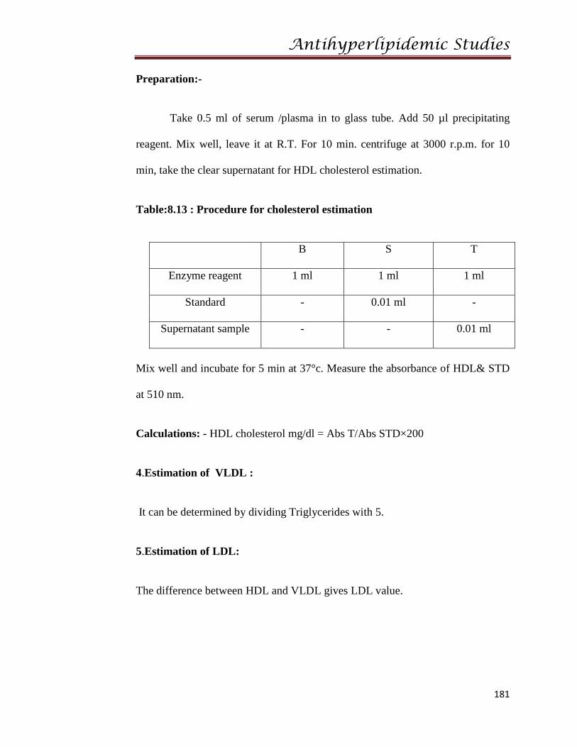
Preparation:-
Take 0.5 ml of serum /plasma in to glass tube. Add 50 µl precipitating
reagent. Mix well, leave it at R.T. For 10 min. centrifuge at 3000 r.p.m. for 10
min, take the clear supernatant for HDL cholesterol estimation.
Table:8.13 : Procedure for cholesterol estimation
B S T
Enzyme reagent 1 ml 1 ml 1 ml
Standard - 0.01 ml -
Supernatant sample - - 0.01 ml
Mix well and incubate for 5 min at 37°c. Measure the absorbance of HDL& STD
at 510 nm.
Calculations: - HDL cholesterol mg/dl = Abs T/Abs STD×200
4.Estimation of VLDL :
It can be determined by dividing Triglycerides with 5.
5.Estimation of LDL:
The difference between HDL and VLDL gives LDL value.
Antihyperlipidemic Studies
182
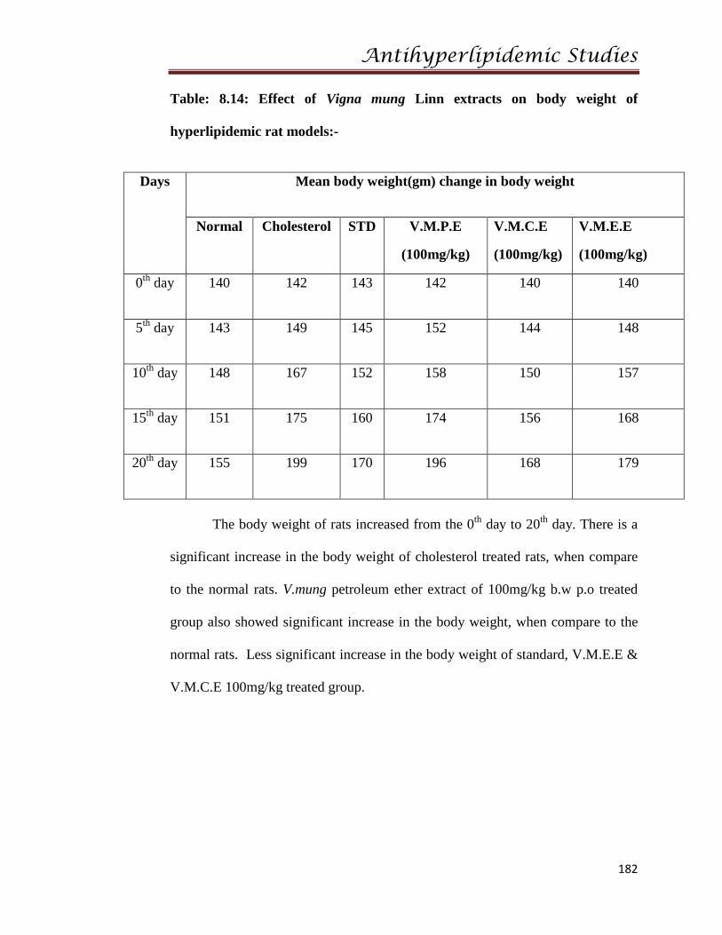
Table: 8.14: Effect of Vigna mung Linn extracts on body weight of
hyperlipidemic rat models:-
Days Mean body weight(gm) change in body weight
Normal Cholesterol STD V.M.P.E
(100mg/kg)
V.M.C.E
(100mg/kg)
V.M.E.E
(100mg/kg)
0th day 140 142 143 142 140 140
5th day 143 149 145 152 144 148
10th day 148 167 152 158 150 157
15th day 151 175 160 174 156 168
20th day 155 199 170 196 168 179
The body weight of rats increased from the 0th day to 20th day. There is a
significant increase in the body weight of cholesterol treated rats, when compare
to the normal rats. V.mung petroleum ether extract of 100mg/kg b.w p.o treated
group also showed significant increase in the body weight, when compare to the
normal rats. Less significant increase in the body weight of standard, V.M.E.E &
V.M.C.E 100mg/kg treated group.
Anti Hyperlipidemic Studies
183
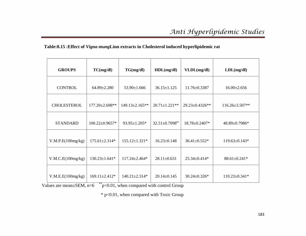
Table:8.15 :Effect of Vigna mungLinn extracts in Cholesterol induced hyperlipidemic rat
GROUPS
TC(mg/dl)
TG(mg/dl)
HDL(mg/dl)
VLDL(mg/dl)
LDL(mg/dl)
CONTROL
64.89±2.280
53.90±1.666
36.15±1.125
11.76±0.3387
16.00±2.656
CHOLESTEROL
177.20±2.698**
149.13±2.165**
20.71±1.221**
29.23±0.4326**
116.26±3.507**
STANDARD
100.22±0.9657*
93.95±1.205*
32.51±0.7098ns
18.78±0.2407*
48.89±0.7986*
V.M.P.E(100mg/kg)
175.61±2.314*
155.12±1.321*
16.23±0.148
36.41±0.552*
119.63±0.143*
V.M.C.E(100mg/kg)
130.23±1.641*
117.24±2.464*
28.11±0.631
25.34±0.414*
88.61±0.241*
V.M.E.E(100mg/kg)
169.11±2.412*
140.21±2.314*
20.14±0.145
30.24±0.326*
110.23±0.341*
Values are mean±SEM, n=6 **p<0.01, when compared with control Group
* p<0.01, when compared with Toxic Group
Anti Hyperlipidemic Studies
184
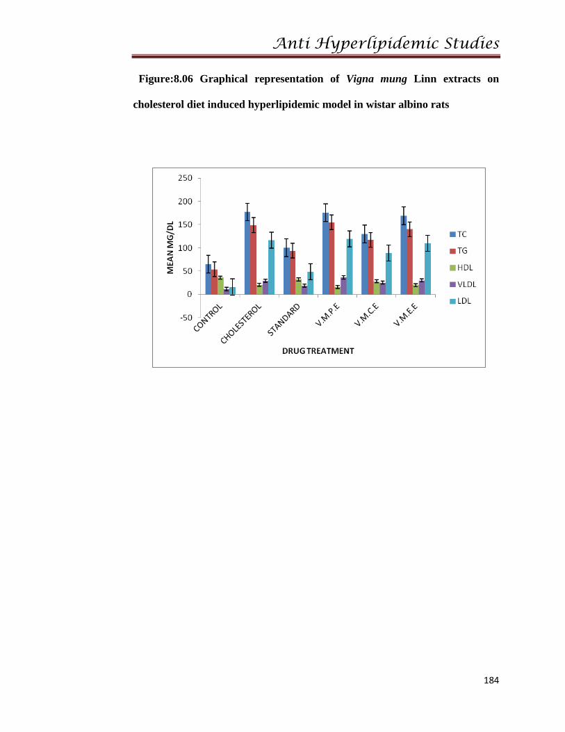
Figure:8.06 Graphical representation of Vigna mung Linn extracts on
cholesterol diet induced hyperlipidemic model in wistar albino rats
Anti Hyperlipidemic Studies
185
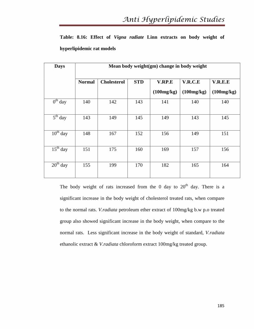
Table: 8.16: Effect of Vigna radiate Linn extracts on body weight of
hyperlipidemic rat models
Days Mean body weight(gm) change in body weight
Normal Cholesterol STD V.RP.E
(100mg/kg)
V.R.C.E
(100mg/kg)
V.R.E.E
(100mg/kg)
0th day 140 142 143 141 140 140
5th day 143 149 145 149 143 145
10th day 148 167 152 156 149 151
15th day 151 175 160 169 157 156
20th day 155 199 170 182 165 164
The body weight of rats increased from the 0 day to 20th day. There is a
significant increase in the body weight of cholesterol treated rats, when compare
to the normal rats. V.radiata petroleum ether extract of 100mg/kg b.w p.o treated
group also showed significant increase in the body weight, when compare to the
normal rats. Less significant increase in the body weight of standard, V.radiata
ethanolic extract & V.radiata chloroform extract 100mg/kg treated group.
Anti Hyperlipidemic Studies
186
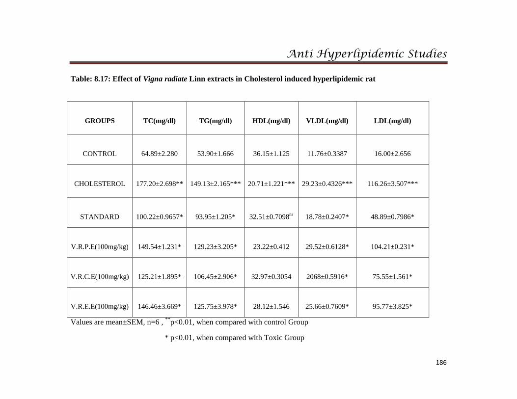
Table: 8.17: Effect of Vigna radiate Linn extracts in Cholesterol induced hyperlipidemic rat
GROUPS
TC(mg/dl)
TG(mg/dl)
HDL(mg/dl)
VLDL(mg/dl)
LDL(mg/dl)
CONTROL
64.89±2.280
53.90±1.666
36.15±1.125
11.76±0.3387
16.00±2.656
CHOLESTEROL
177.20±2.698**
149.13±2.165***
20.71±1.221***
29.23±0.4326***
116.26±3.507***
STANDARD
100.22±0.9657*
93.95±1.205*
32.51±0.7098ns
18.78±0.2407*
48.89±0.7986*
V.R.P.E(100mg/kg)
149.54±1.231*
129.23±3.205*
23.22±0.412
29.52±0.6128*
104.21±0.231*
V.R.C.E(100mg/kg)
125.21±1.895*
106.45±2.906*
32.97±0.3054
2068±0.5916*
75.55±1.561*
V.R.E.E(100mg/kg)
146.46±3.669*
125.75±3.978*
28.12±1.546
25.66±0.7609*
95.77±3.825*
Values are mean±SEM, n=6 , **p<0.01, when compared with control Group
* p<0.01, when compared with Toxic Group
Anti Hyperlipidemic Studies
187
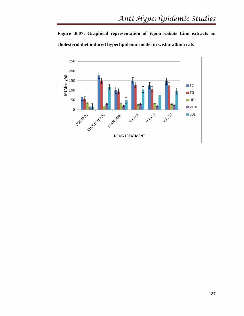
Figure :8.07: Graphical representation of Vigna radiate Linn extracts on
cholesterol diet induced hyperlipidemic model in wistar albino rats
Anti Hyperlipidemic Studies
188
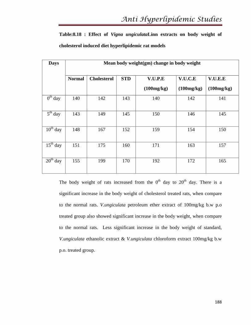
Table:8.18 : Effect of Vigna ungiculataLinn extracts on body weight of
cholesterol induced diet hyperlipidemic rat models
Days Mean body weight(gm) change in body weight
Normal Cholesterol STD V.U.P.E
(100mg/kg)
V.U.C.E
(100mg/kg)
V.U.E.E
(100mg/kg)
0th day 140 142 143 140 142 141
5th day 143 149 145 150 146 145
10th day 148 167 152 159 154 150
15th day 151 175 160 171 163 157
20th day 155 199 170 192 172 165
The body weight of rats increased from the 0th day to 20th day. There is a
significant increase in the body weight of cholesterol treated rats, when compare
to the normal rats. V.ungiculata petroleum ether extract of 100mg/kg b.w p.o
treated group also showed significant increase in the body weight, when compare
to the normal rats. Less significant increase in the body weight of standard,
V.ungiculata ethanolic extract & V.ungiculata chloroform extract 100mg/kg b.w
p.o. treated group.
Anti Hyperlipidemic Studies
189
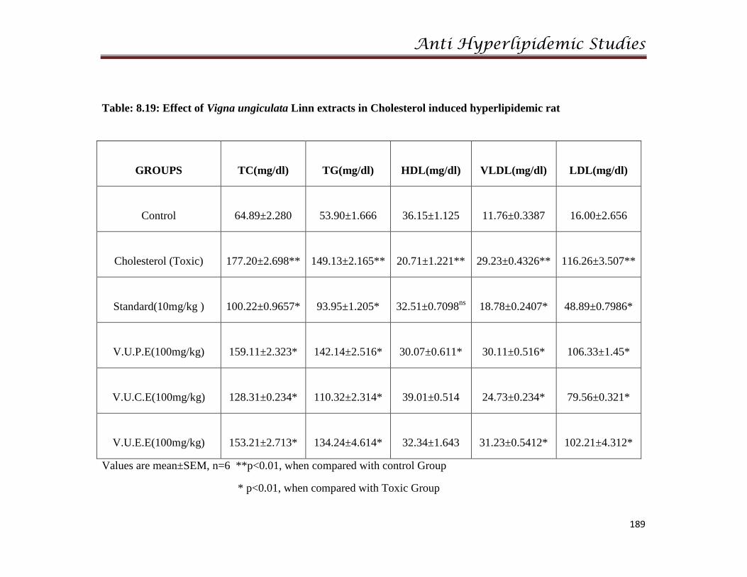
Table: 8.19: Effect of Vigna ungiculata Linn extracts in Cholesterol induced hyperlipidemic rat
GROUPS
TC(mg/dl)
TG(mg/dl)
HDL(mg/dl)
VLDL(mg/dl)
LDL(mg/dl)
Control
64.89±2.280
53.90±1.666
36.15±1.125
11.76±0.3387
16.00±2.656
Cholesterol (Toxic)
177.20±2.698**
149.13±2.165**
20.71±1.221**
29.23±0.4326**
116.26±3.507**
Standard(10mg/kg )
100.22±0.9657*
93.95±1.205*
32.51±0.7098ns
18.78±0.2407*
48.89±0.7986*
V.U.P.E(100mg/kg)
159.11±2.323*
142.14±2.516*
30.07±0.611*
30.11±0.516*
106.33±1.45*
V.U.C.E(100mg/kg)
128.31±0.234*
110.32±2.314*
39.01±0.514
24.73±0.234*
79.56±0.321*
V.U.E.E(100mg/kg)
153.21±2.713*
134.24±4.614*
32.34±1.643
31.23±0.5412*
102.21±4.312*
Values are mean±SEM, n=6 **p<0.01, when compared with control Group
* p<0.01, when compared with Toxic Group
Anti Hyperlipidemic Studies
190
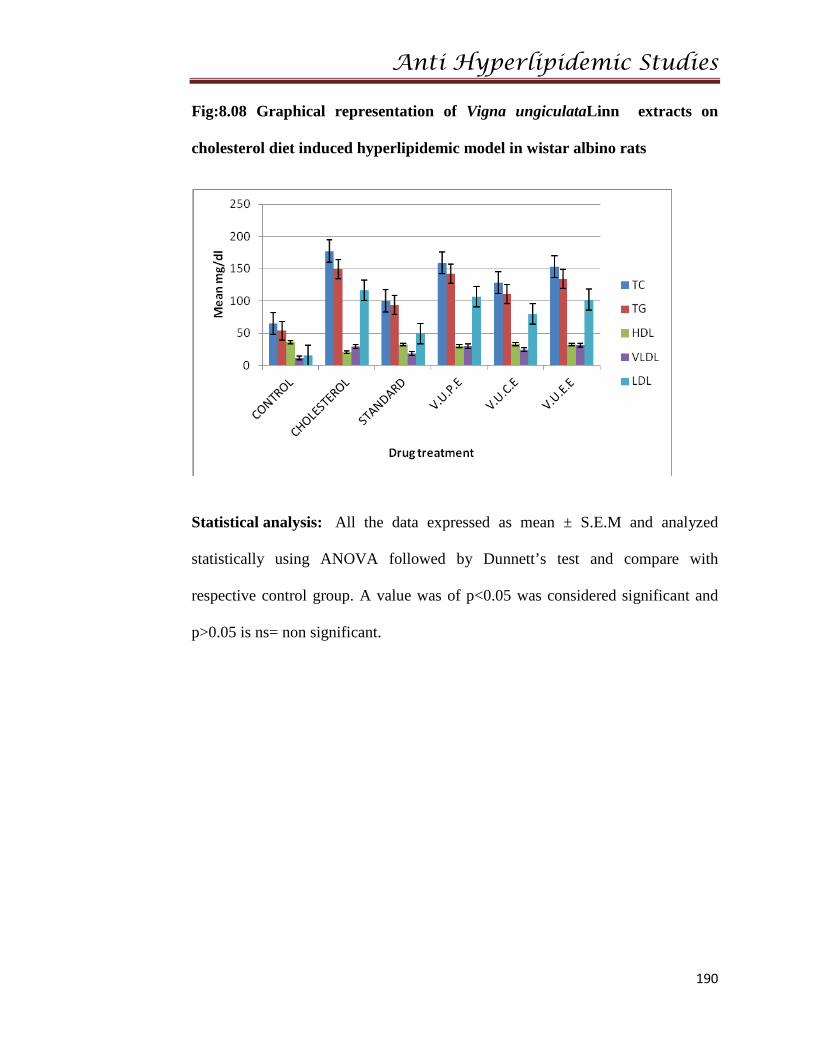
Fig:8.08 Graphical representation of Vigna ungiculataLinn extracts on
cholesterol diet induced hyperlipidemic model in wistar albino rats
Statistical analysis: All the data expressed as mean ± S.E.M and analyzed
statistically using ANOVA followed by Dunnett’s test and compare with
respective control group. A value was of p<0.05 was considered significant and
p>0.05 is ns= non significant.
Anti Hyperlipidemic Studies
191
8.4 RESULTS AND DISCUSSION:
Cholesterol induced hyperlipidemia:
Effect of administration of selected plant extracts (100 mg/kg, p.o., once daily)
/Atorvastatin (10mg/kg, p.o, once daily) on serum lipid Parameter levels in rats
fed with Cholesterol Diet for 20days.
Effect on serum total cholesterol (serum TC) level:-
• Rats fed with Cholesterol for 20 days had serum TC level of (177.20±2.698
mg/dl) when measured on day 21. This was significantly higher (p<0.001)
when compared to serum TC levels in normal control rats (64.89±2.280
mg/dl).
• Cholesterol induced hyperlipidemic rats treated with Atorvastatin (10mg/kg,
p.o., once daily) had serum level of 100.22±0.9657 mg/dl when measured
on day 21. This was significantly lower (p<0.001) when compared to the
serum TC levels in Cholesterol treated toxic control groups (177.20±2.698
mg/dl).
• Cholesterol induced hyperlipidemic rats treated with V.M.P.E 100mg/kg,
V.M.C.E 100mg/kg , V.M.E.E 100mg/kg b.w p.o, once daily, had serum
TC level of 175.61±2.314, 130.23±1.641 and 169.11±2.412 mg/dl
respectively when measured on day 21. These values were significantly
lower (P<0.001) when compared to the serum TC level in Cholesterol
control rats (177.20±2.698 mg/dl).
Anti Hyperlipidemic Studies
192
• Cholesterol induced hyperlipidemic rats treated with V.R.P.E 100mg/kg,
V.R.C.E 100mg/kg , V.R.E.E 100mg/kg b.w p.o, once daily, had serum TC
level of 149.54±1.231, 125.21±1.895 and 146.46±3.669 mg/dl respectively
when measured on day 21. These values were significantly lower (p<0.001)
when compared to the serum TC level in Cholesterol control rats
(177.20±2.698 mg/dl).
• Cholesterol induced hyperlipidemic rats treated with V.U.P.E 100mg/kg,
V.U.C.E 100mg/kg , V.U.E.E 100mg/kg b.w p.o, once daily, had serum
TC level of 159.11±2.323, 128.31±0.234 and 153.21±2.713 mg/dl
respectively when measured on day 21. These values were significantly
lower (p<0.001) when compared to the serum TC level in Cholesterol
control rats (177.20±2.698 mg/dl).
Anti Hyperlipidemic Studies
193
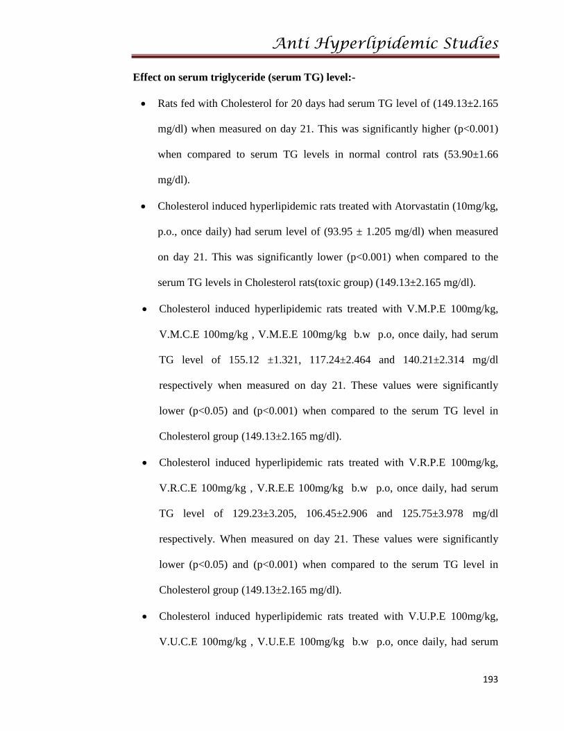
Effect on serum triglyceride (serum TG) level:-
• Rats fed with Cholesterol for 20 days had serum TG level of (149.13±2.165
mg/dl) when measured on day 21. This was significantly higher (p<0.001)
when compared to serum TG levels in normal control rats (53.90±1.66
mg/dl).
• Cholesterol induced hyperlipidemic rats treated with Atorvastatin (10mg/kg,
p.o., once daily) had serum level of (93.95 ± 1.205 mg/dl) when measured
on day 21. This was significantly lower (p<0.001) when compared to the
serum TG levels in Cholesterol rats(toxic group) (149.13±2.165 mg/dl).
• Cholesterol induced hyperlipidemic rats treated with V.M.P.E 100mg/kg,
V.M.C.E 100mg/kg , V.M.E.E 100mg/kg b.w p.o, once daily, had serum
TG level of 155.12 ±1.321, 117.24±2.464 and 140.21±2.314 mg/dl
respectively when measured on day 21. These values were significantly
lower (p<0.05) and (p<0.001) when compared to the serum TG level in
Cholesterol group (149.13±2.165 mg/dl).
• Cholesterol induced hyperlipidemic rats treated with V.R.P.E 100mg/kg,
V.R.C.E 100mg/kg , V.R.E.E 100mg/kg b.w p.o, once daily, had serum
TG level of 129.23±3.205, 106.45±2.906 and 125.75±3.978 mg/dl
respectively. When measured on day 21. These values were significantly
lower (p<0.05) and (p<0.001) when compared to the serum TG level in
Cholesterol group (149.13±2.165 mg/dl).
• Cholesterol induced hyperlipidemic rats treated with V.U.P.E 100mg/kg,
V.U.C.E 100mg/kg , V.U.E.E 100mg/kg b.w p.o, once daily, had serum
Anti Hyperlipidemic Studies
194
TG level of 142.14±2.516, 110.32±2.314 and 134.24±4.614 mg/dl
respectively when measured on day 21. These values were significantly
lower (p<0.05) and (p<0.001) when compared to the serum TG level in
Cholesterol group (149.13±2.165 mg/dl).
Effect on serum HDL cholesterol (serum HDL-C) level:-
• Rats fed with Cholesterol for 20 days had serum HDL-C level of
(20.71±1.221 mg/dl) when measured on day 21.This was significantly lower
(p<0.001) when compared to serum HDL-C levels in normal control rats
(36.15±1.125 mg/dl).
• Cholesterol induced hyperlipidemic rats treated with atorvastatin (10mg/kg,
p.o. once daily) had serum HDL-C level of 32.51±0.7098 mg/dl when
measured on day 21.This was significantly higher (p<0.001) when
compared to the serum HDL-C levels in Cholesterol control rats
(20.71±1.221 mg/dl).
• Cholesterol induced hyperlipidemic rats treated with V.M.P.E 100mg/kg,
V.M.C.E 100mg/kg , V.M.E.E 100mg/kg b.w p.o, once daily, had serum
HDL-C level of 16.23 ± 0.148, 28.11 ± 0.631 and 20.14 ±0.145 mg/dl
respectively, when measured on day 21.These values were significantly
higher (p<0.05) and (p<0.001) when compared to the serum HDL-C level in
Cholesterol control rats(20.71±1.221 mg/dl).
• Cholesterol induced hyperlipidemic rats treated with V.R.P.E 100mg/kg,
V.R.C.E 100mg/kg , V.R.E.E 100mg/kg b.w p.o, once daily, had serum
Anti Hyperlipidemic Studies
195
HDL-C level of 23.22±0.412, 32.97±0.3054 and 28.12±1.546 mg/dl
respectively when measured on day 21.These values were significantly
higher (p<0.05) and (p<0.001) when compared to the serum HDL-C level in
Cholesterol control rats (20.71±1.221 mg/dl).
• Cholesterol induced hyperlipidemic rats treated with V.U.P.E 100mg/kg,
V.U.C.E 100mg/kg, V.U.E.E 100mg/kg b.w p.o, once daily, had serum
HDL-C level of 30.07±0.611, 33.01±0.514 and 32.34±1.643mg/dl
respectively when measured on day 21.These values were significantly
higher (p<0.05) and (p<0.001) when compared to the serum HDL-C level in
Cholesterol control rats(20.71±1.221 mg/dl).
Anti Hyperlipidemic Studies
196
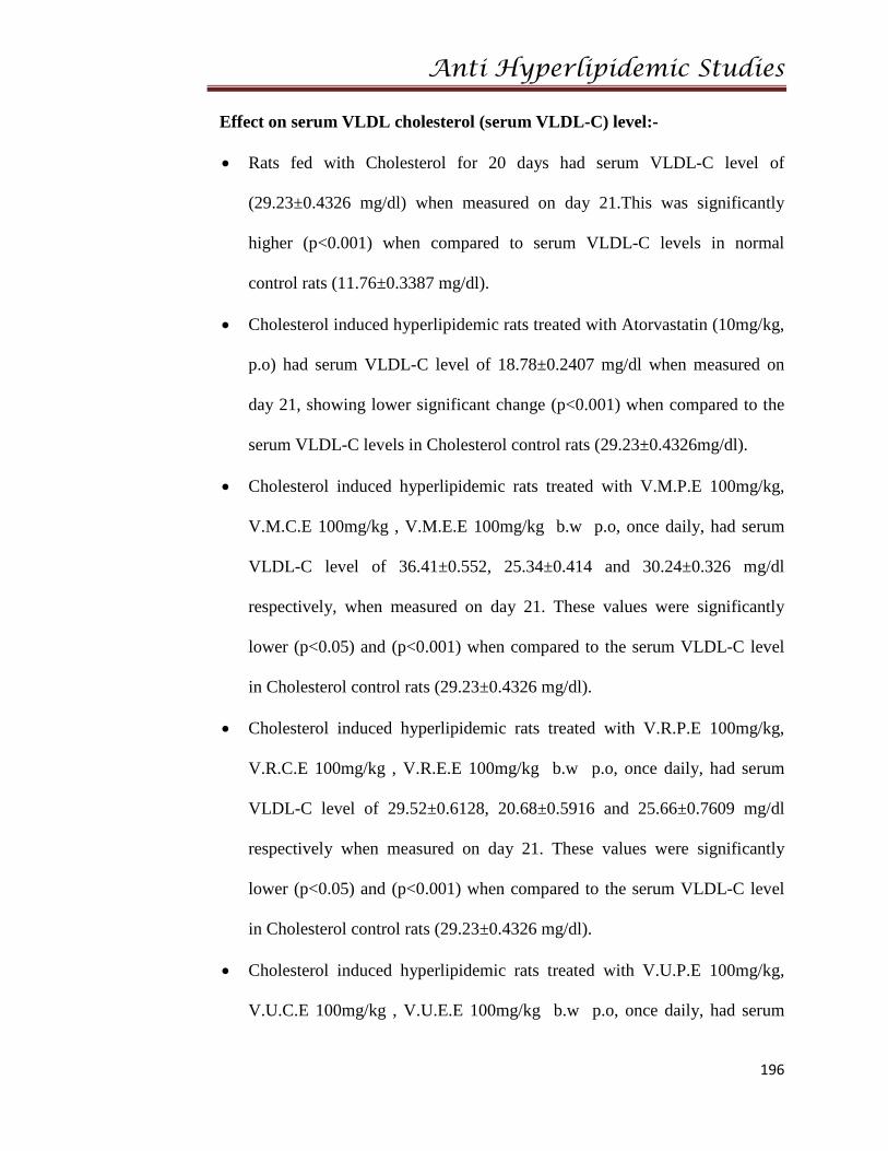
Effect on serum VLDL cholesterol (serum VLDL-C) level:-
• Rats fed with Cholesterol for 20 days had serum VLDL-C level of
(29.23±0.4326 mg/dl) when measured on day 21.This was significantly
higher (p<0.001) when compared to serum VLDL-C levels in normal
control rats (11.76±0.3387 mg/dl).
• Cholesterol induced hyperlipidemic rats treated with Atorvastatin (10mg/kg,
p.o) had serum VLDL-C level of 18.78±0.2407 mg/dl when measured on
day 21, showing lower significant change (p<0.001) when compared to the
serum VLDL-C levels in Cholesterol control rats (29.23±0.4326mg/dl).
• Cholesterol induced hyperlipidemic rats treated with V.M.P.E 100mg/kg,
V.M.C.E 100mg/kg , V.M.E.E 100mg/kg b.w p.o, once daily, had serum
VLDL-C level of 36.41±0.552, 25.34±0.414 and 30.24±0.326 mg/dl
respectively, when measured on day 21. These values were significantly
lower (p<0.05) and (p<0.001) when compared to the serum VLDL-C level
in Cholesterol control rats (29.23±0.4326 mg/dl).
• Cholesterol induced hyperlipidemic rats treated with V.R.P.E 100mg/kg,
V.R.C.E 100mg/kg , V.R.E.E 100mg/kg b.w p.o, once daily, had serum
VLDL-C level of 29.52±0.6128, 20.68±0.5916 and 25.66±0.7609 mg/dl
respectively when measured on day 21. These values were significantly
lower (p<0.05) and (p<0.001) when compared to the serum VLDL-C level
in Cholesterol control rats (29.23±0.4326 mg/dl).
• Cholesterol induced hyperlipidemic rats treated with V.U.P.E 100mg/kg,
V.U.C.E 100mg/kg , V.U.E.E 100mg/kg b.w p.o, once daily, had serum
Anti Hyperlipidemic Studies
197
VLDL-C level of 30.11±0.516, 24.73±0.234 and 31.23±0.5412 mg/dl
respectively, when measured on day 21. These values were significantly
lower (p<0.05) and (p<0.001) when compared to the serum VLDL-C level
in Cholesterol control rats (29.23±0.4326 mg/dl).
Effect on serum LDL cholesterol (serum LDL-C) level:-
• Rats fed with Cholesterol for 20 days had serum LDL-C level of
(116.26±3.507 mg/dl) when measured on day 21.This was significantly
higher (p<0.001) when compared to serum LDL-C levels in normal control
rats (16.00±2.656 mg/dl).
• Cholesterol induced hyperlipidemic rats treated with Atrovastatin (10mg/kg,
p.o., once daily) had serum LDL-C level of 48.89±0.7986 mg/dl when
measured on day 21.This was significantly lower (p<0.001) when compared
to the serum LDL-C levels in Cholesterol control rats 116.26±3.507 mg/dl).
• Cholesterol induced hyperlipidemic rats treated with V.M.P.E 100mg/kg,
V.M.C.E 100mg/kg , V.M.E.E 100mg/kg b.w p.o, once daily, had serum
LDL-C level of 119.63±0.143, 88.61±0.241 and 110.23±0.341 mg/dl
respectively, when measured on day21.These vales were significantly lower
(p<0.001) when compared to the serum LDL-C level in Cholesterol control
rats (116.26±3.507 mg/dl).
• Cholesterol induced hyperlipidemic rats treated with V.R.P.E 100mg/kg,
V.R.C.E 100mg/kg , V.R.E.E 100mg/kg b.w p.o, once daily, had serum
LDL-C level of 104.21±0.231, 75.55±1.561 and 95.77±3.825 mg/dl
respectively, when measured on day21.These values were significantly
Anti Hyperlipidemic Studies
198
lower (p<0.001) when compared to the serum LDL-C level in Cholesterol
control rats (116.26±3.507 mg/dl).
• Cholesterol induced hyperlipidemic rats treated with V.U.P.E 100mg/kg,
V.U.C.E 100mg/kg , V.U.E.E 100mg/kg b.w p.o, once daily, had serum
LDL-C level of 106.33±1.45, 79.56±0.321 and 102.21±4.312 mg/dl
respectively, when measured on day21.These values were significantly
lower (p<0.001) when compared to the serum LDL-C level in Cholesterol
control rats (116.26±3.507 mg/dl).
Discussion:
It has been well established that nutrition plays an important role in the
etiology of hyperlipidimia and atherosclerosis. In our study we choose cholesterol
diet which contains the common ingredients in our daily food. Cholesterol feeding
has been often used to elevate serum or tissue cholesterol levels to assess the
hypercholesterolemia- related metabolic disturbances in animals. Cholesterol
feeding alone however does not affect the serum TG level. It is assumed that a
high level of saturated fat in addition to cholesterol is required to significantly
elevate serum TG level in rat model.
Hyperlipidemia has been documented as one of the causative factor for
atherosclerosis, resulting in coronary heart disease. Elevated cholesterol
particularly LDL are the major reasons attributed to CVD. Accordingly to WHO
by 2020, 60% of the CVD causes will be of Indian origin 31.
Anti Hyperlipidemic Studies
199
Development of atherosclerotic disease is a complicated process involving
accumulation of lipid-containing particles in the walls of coronary arteries other
major arteries in the body. Similarly the present study there was a significant
weight gain in cholesterol control (toxic), as compared to normal control groups.
Treatment with Vigna genus plant extracts significantly reduced the weight gain
Lowering high cholesterol levels significantly reduce the risk of heart
attacks, strokes, and death. A rise in the LDL may cause deposition of cholesterol
in arteries and aorta and it is a direct risk factor for CHD32.
In the present study there was a elevation in serum and tissue cholesterol,
LDL-C, and VLDL-C level in response to cholesterol induced(toxic) compare
to normal control group. Treatment with Vigna genus selected plant extracts
significantly reduced serum and tissue cholesterol, LDL-C, and VLDL-C
levels 33.
The decrease in triglyceride level is an important finding of experiment.
Recent days studies shows that triglycerides are independently related with
coronary artery disease. Treatment with Vigna genus selected plant extracts
showed significant decreased in triglyceride.
HDL is synthesized mainly in intestine and liver. HDL is considered to be
a beneficial lipoprotein as it has an inhibitory effect in the pathogenesis of
atherosclerosis. Low level of HDL is associated with high risk of coronary artery
Anti Hyperlipidemic Studies
200
disease. In the present study HDL-C level in serum were significantly increased
by chloroform extract and ethanolic extract .
The activity of Cholesteryl Ester Transfer Protein (CETP), a key enzyme
in reverse cholesterol transport and HDL metabolism increase in high fat diet and
mediates the transfer of cholesterol esters from HDL-C to triglyceride-rich
particles in exchange for triglycerides. This leads to increased plasma
concentrations of TG’s & decreased concentrations of HDL-C.
Lipid profile of Cholesterol induced Hyperlipidemia rats in our study
revealed higher levels of serum triglycerides, cholesterol, LDL and VLDL
accompanied by decrease of serum HDL-C as compared to controls.
Treatment of Cholesterol induced Hyperlipidemia rats with selected plant
extracts and reference standard atorvastatin (10mg/kg b.w), an HMG CoA
inhibitor showed a significant decrease of serum triglycerides, cholesterol, LDL
and VLDL and significant increase of serum HDL-C levels.
The effect of Vigna genus plant extract cholesterol induced hyperlipidemic rat
models observes the changes in lipid levels compare to the control groups.
According the results observed upon comparing the results of control group all
the lipid levels like TC, TG, LDL, VLDL levels are increased and only HDL
levels are decrease when compare to normal rats. The test groups shows
significant to the control groups. The standard group shows non- significant when
compare to the control group. Decrease of lipid levels in the test group of is the
Anti Hyperlipidemic Studies
201
following order V.R.C.E > V.U.C.E > V.M.C.E > V.R.E.E > V.U.E.E > V.M.E.E
> V.R.P.E > V.U.P.E > V.M.P.E and shows better activity towards reducing
hyperlipidemic condition.
8.5 CONCLUSION
From this work we conclude that all the Vigna genus selected plants posses
ability to decrease cholesterol levels in the body. Vigna radiate Linn chloroform
extract possess highly significant action towards reducing the body cholesterol.
Hence the folklore usage has been validated. Vigna genus selected plants can be
treated as Nutraceuticals.
Anti Hyperlipidemic Studies
202
8.6 REFERENCES
1. Kaplan A, Lavernel L S. Disorder of carbohydrate. In: Clinical Chemistry:
Interpretation and Techniques. 2nd Edn. Philadelphia.1983:752.
2. Shamir R, Fisher E A. Dietary therapy for children with
hypercholesterolemia. Am fam Physician 2000; 61(3):675-682, 685-676.
3. http://www.infovet.ca/medias/media/mediacenterportal/64123/hyperlipede
mia.pdf
4. Brown M S, Goldstein J L. Lipoprotein receptors in the liver. Control
signals for plasma cholesterol traffic. J Clin Invest. 1983;72:743-747.
5. Angeles Zulet M, Ana Barber, Henri Garcin, Paul Higueret and José
Alfredo Martinez. Alterations in Carbohydrate and Lipid Metabolism
Induced by a Diet Rich in Coconut Oil and Cholesterol in a Rat Model.
Journal of the American College of Nutrition 1999; 18(1): 36-42.
6. HyperlipidemiaIntroduction:http://www.prr.hec.gov.pk/Chapters/48S-pdf
7. Sharpless KB, Snyder TE, Spencer TT, Maheswari KK, Guhn G, and
Clayton RB. J Am Chem Soc. 1969; 91(13): 3394.
8. Vinay Kumar, Abul K Abbas and Nelson F. Patholog. basis of disease.7th
Edn. New Delhi: Elsevier; 2002:46-49.
9. Brendan J Coughlan and Matthew J Sorrentino .Does hypertriglyceridemia
increases risk for CAD. Post graduate medicine, the practical peer-
reviewed; J for Prim Care Phys.2000; 108(7):56.
Anti Hyperlipidemic Studies
203
10. https://online.epocrates.com/u/2924170/Hypercholesterolemia/Basics/Etio
logy
11. William Winter and Desmond Schatz. Pediatric lipid disorders in clinical
practice. Medicine Specialties>Pediatrics>Cardiology. Available from
URL:file:///C:/Documents%20and%20Settings/Administrator/Desktop/eti
ology.html.
12. Kinosian B, Glick H, Garland G. Cholesterol and coronary heart disease:
predicting risk by levels and ratio. Ann Inter Med.1994;121:641- 647.
13. Fungwe TV, Cagen LM, Cook GA, et al. Dietary cholesterol stimulates
hepatic biosynthesis of triglyceride and reduces oxidation of fatty acids in
the rat. J Lipid Res. 1993; 34: 933 – 941.
14. Inadera H, Shirai K and Saito Y. The enzymes related to lipoprotein
metabolism (Hmg-coa reductase, 7-alpha hydroxylase). Japanese J of
Clinical Medicine. 1990; 48(11): 2483-91.
15. American heart association. Lipoproteins. Available from
URL: http://www.americanheart.org/presenter.jhtml?identifier=4600.
16. Ching Kuang Chow. Fatty acids in food and their health complications.
2nd
Edn, USA: Marcel Dekker publishers; 1999: 459.
17. Inadera H, Shirai K and Saito Y. The enzymes related to lipoprotein
metabolism (Hmg-coA reductase, 7-alpha hydroxylase). Japanese J of
Clinl Med 1990; 48(11): 2483-91.
Anti Hyperlipidemic Studies
204
18. Vinay Kumar, Abul K Abbas and Nelson F. Pathological basis of
disease.7th
Edn. New Delhi: Elsevier; 2002:576.
19. Nawrocki J W, Weiss S R, Davidson M H, Sprecher D L, Schwartz S L,
Lupien P J, Jones P H, Haber H E, Black D M. J.Pharmacol. 1995;
3(4):58.
20. Assy N, Kaita K, Mymin D, et al. Fatty infiltration of liver in
hyperlipidemic patients. Dig Dis Sci. 2000; 45:1929 -1934.
21. Ridker PM, Danielson E, Fonseca FA, et al. Rosuvastatin to prevent
vascular events in men and women with elevated C-reactive protein. N
Engl J Med. 2008; 359:2195-2207.
22. Glynn RJ, Danielson E, Fonseca FA, et al. A randomized trial of
rosuvastatin in the prevention of venous thromboembolism. N Engl J Med.
2009; 360:1851-61.
23. Riordan M .The side effects of statins: Heart healthy and head harmful?
The Wall Street Journal. 2010:10-24.
24. Demacker PN, Reijnen IG, Katan MB, et al. Increased removal of
remnants of triglyceride rich lipoproteins on a diet rich in polyunsaturated
fatty acids. Eur J Clin Invest. 1991; 21:197.
25. Prasad K. Hypocholesterolemic and antiatherosclerotic effect of flax
lignin complex isolated from flaxseed. Atherosclerosis. 2005; 179: 269-
275.
26. Jain GC, Agarwal S. Favourable effect of Cleome viscosa L. on serum and
hepatic lipid in hyperlipidemic rats. Asian J Exp Sci. 2006; 20: 331-336.
Anti Hyperlipidemic Studies
205
27. Fungwe TV, Cagen LM, Cook GA, et al. Dietary cholesterol stimulates
hepatic biosynthesis of triglyceride and reduces oxidation of fatty acids in
the rat. J Lipid Res. 1993; 34: 933-941.
28. Yokozawa T, Cho E J, Sasaki S, et al. The protective role of Chinese
prescription Kangen-karyu extract on diet-induced hypercholesterolemia
in rats. Biol Pharm Bull. 2006; 29: 760-765.
29. Wang YM, Zhang B, Xue Y, et al. The mechanism of dietary cholesterol
effects on lipids metabolism in rats. Lipids Health Dis. 2010; 92 - 96.
30. Herbert K. Lipids. In: Kaplan LA, Pesce AJ. Clin Chem: Theory,
Analysis and Correlation. St Louis, MO: Mosby; 1984:1182-1230.
31. Zhang HW, Zhang YH, Lu MJ, et al. Comparison of hypertension
dyslipidemia and hyperglycaemia between buckwheat seed consuming
and non-consuming Mongolian-Chinese populations in Inner Mongolia,
China. Clin Exp Pharmacol Physiol. 2007; 34: 838-844.
32. Brown MS, Goldstein JL. Lipoprotein receptors in the liver. Control
signals for plasma cholesterol traffic. J Clin Invest.1983; 72: 743-747.
33. Prasad K. Hypocholesterolemic and antiatherosclerotic effect of flax
lignan complex isolated from flaxseed. J Atherosclerosis. 2005; 179: 269-
275.