Embed Size (px)
Citation preview
IOSR Journal of Pharmacy and Biological Sciences (IOSR-JPBS)
e-ISSN:2278-3008, p-ISSN:2319-7676. Volume 12, Issue 4 Ver. II (Jul – Aug 2017), PP 39-50
www.iosrjournals.org
DOI: 10.9790/3008-1204023950 www.iosrjournals.org 39 | Page
Antihyperlipidemic And Glucose Lowering Effect Of Extract Of
Bioregulator Treated Okra (Abelmoschus Esculentus L.) Fruits in
Triton-Induced Hyperlipidemia Rats
A.M. Esan, 1 *
C.O. Olaiya
1, V. Sameer,
2 K. Elango
2, S.P. Dhanabal
2
1 Department of Biochemistry, Faculty of Basic Medical Sciences, University of Ibadan, Oyo state, Nigeria
2 JSS College of pharmacy, Tamil Nadu, Ooty, India
Corresponding Author: A.M. Esan
Abstract: Extracts from okra have been used to alleviate not only hyperglycemia but also hyperlipidemia in
diabetic mice induced by alloxan and streptozocin. However, its hypolipidemic activity has not yet been clearly
studied. This study was aimed to study the antihyperlipidemic potential of bioregulator treated okra fruit in
triton-X induced hyperlipidemic rats. Hyperlipidemia was induced in rats by administration of triton-X
(100mg/kg i.p.). After 14 days of induction, hyperlipidemic rats received AWB (okra extract treated with
bioregulator, AWOB (okra extract without bioregulator) and atorvastatin up to 28 days. The extracts activities
were assessed on serum blood glucose, lipid profiles, carbohydrate enzymes, antioxidant and non-enzymes
antioxidant enzymes, malondialdehyde level, and HMG-CoA reductase activity of control and drug-treated rats.
The extract exhibited a strong dose-dependent antihyperlipidemic activity and at dose level 400mg/kg (AWB)
p.o. the extract showed a significant (P≤0.05) decrease in blood glucose, TC, TG, LDL, VLDL, MDA,
gluconeogenic enzymes and significant (P≤0.05) increase in HDL-C, glycogenic enzymes, antioxidant enzymes
and non-enzymes antioxidant in a dose- dependent manner comparing with standard atorvastatin- treated
group. The present study demonstrated that the extract of bioregulator treated okra exhibits a potent lipid
lowering activity and well compared with the atorvastatin than extract without bioregulator.
Keywords: Bioregulator, Carbohydrate enzymes, lipid profiles, Okra, Triton-X.
----------------------------------------------------------------------------------------------------------------------------- ----------
Date of Submission: 23-05-2017 Date of acceptance: 17-07-2017
----------------------------------------------------------------------------------------------------------------------------- ----------
I. Introduction
Hyperlipidemia is a complicated disorder with a hallmark of elevated serum total cholesterol, low-
density and very low-density lipoprotein cholesterol, triglycerides, and decreased high-density lipoprotein levels
[1]. Hyperlipidemia has been implicated in atherosclerosis, which is the primary cause of heart disease and
stroke [2]. About two-third of the cholesterol made in the body is synthesized by the liver. Lipid metabolism is
controlled majorly in different ways. Enzymes play a pivot role in lipid metabolism; in cholesterol synthesis, the
rate limiting enzyme is 3-hydroxy-3-methylglutaryl (HMG)-CoA reductase that regulates cholesterol
concentrations in the cells by feedback inhibition. Insulin resistance and obesity are closely linked with the
infiltration of adipose tissue by inflammation cells [3]. Insulin resistance, a common accompaniment of obesity,
is a major risk factor for diabetes mellitus [4]. Increased extracellular and intracellular glucose concentrations
resulted in oxidative stress is due to increased production of ROS and a sharp decrease in antioxidant body
defenses [5]. The onset of diabetes complications, oxidative stress plays a key role notably diabetic nephropathy
[6]. Treatment of hyperlipidemia involves diet control, exercise, and the use of lipid-lowering diets and drugs
[7]. For the treatment of hyperlipidemia, the most common drugs used include 3-hydroxy-3-methylglutaryl
(HMG)-CoA reductase inhibitors, also known as statins. Bile acid sequestrants (anion-exchange resins) such as
cholestyramine and colestipol; fibrates such as clofibrate, gemfibrozil, fenofibrate, ciprofibrate, and bezafibrate;
niacin; cholesterol absorption inhibitors such as ezetimibe; and omega-3-fatty acids are others drugs employed
for the treatment of hyperlipidemia [8]. Many hypolipidemic drugs have already been proved to be useful in
lowering serum lipid levels in patients. However, its side effects in long-term treatment have been frequently
reported and its therapy is still deprived of the efficiency, safety, For example, severe muscle damage is
reported with statins, which are particularly well suited for lowering Low- density lipoprotein [9]. Niacin, a drug
for reducing triglycerides levels may cause hyperlipidemia and which in turn can lead to liver damage [10].
Adverse reactions of Achilles tendon xanthomas have been reported after the addition of niacin and bile acid
sequestrants to ongoing statin therapy in patients with hypercholesterolemia [11]. Prices are still expensive.
Thus, efforts to develop effective and better hypolipidemic drugs had led to the discovery of natural agents.
Antihyperlipidemic and glucose lowering effect of extract of bioregulator treated okra (Abelmoschus
DOI: 10.9790/3008-1204023950 www.iosrjournals.org 40 | Page
Abelmoschus esculentus is used for ages as an edible vegetable in many countries and commonly eaten in
Vietnam because of its nourishing components. Traditionally, it is believed that the plant is useful in the
treatment of inflammatory disorders, constipation, retention of urine, and etc. On the other hand, a number of
previous studies have reported that Abelmoschus sp. possessed hypoglycemic effect. However, there is a little
study regarding its hypolipidemic effect. The aim of this present study is to investigate and evaluate the
hypolipidemic effect of bioregulator treated A. esculentus extracts on triton-induced hyperlipidemia in rats to
provide scientific evidence for the development of A. esculentus as a potential natural oral hypolipidemic agent
or functional food.
II. Materials And Methods 2.1 Plant material
2.2 Preparation of plant extract
450kg of dried fruit powdered of both treated (AWB) and untreated (AWOB) okra samples of the same
genotype were used for this experiment; fruit powdered of Abelmoschus esculentus L. seeds treated with 0.4
mM of indole acetic acid and fruit powdered of Abelmoschus esculentus L. seeds without any treatment were
extracted with 70% hydro-alcohol by maceration in a round bottom flask for 72hrs. The solvent was filtered, and
the combined filtrate was concentrated in-vacuo using a water bath and kept in a desiccator to obtain paste
extract (Abelmoschus esculentus).
2.3 Chemicals and Reagents
Triton-X- 100 (100mg/kg), CMC and atorvastatin 30mg/L were purchased from Sigma-Aldrich. All
other biochemicals used in this experiment were purchased from Sigma-Aldrich, USA. All the chemicals were
of analytical grade.
2.4 Experimental animals
Sprague Dawley male rats, weighing 150-200g were obtained from the institutional animal house for
the present investigations. The animals were housed at a room temperature of 25±20C, relative humidity of
75±5% and 12hrs dark-light cycle, animals were provided basal standard rat diet and water ad libitum. The
animals were acclimatized to laboratory condition for five days before commencement of the experiment. The
experiments were conducted according to the ethical norms and Institutional Animal Ethics Committee
Guidelines.
2.5 Experimental design
The rats were divided into seven groups comprising of five animals each (n=5) (35 animals). The
standard drug atorvastatin and extracts were suspended in 0.2% w/v carboxymethyl cellulose (CMC) for oral
administration.
Group I: Normal control fed with standard diet, and rats received 0.2% CMC (10ml) orally 2 h before feeding
the animals with a normal diet.
Group II: served as obese control received a single dose of triton administered at a dose of 100mg/kg, i.p. After
72 hours of triton injection, this group received a daily dose of 0.2% CMC (p.o) for 7days.
Group I11: served as Abelmoschus esculentus without bioregulator (AWOB) were administered with a daily
dose of extract 200mg/kg (Sabitha et al., 2011) dissolved in 0.2% CMC (p.o) treated obese rats, for 14 days
after induction of hyperlipidemia.
Group 1V: served as Abelmoschus esculentus without bioregulator (AWOB) were administered with a daily
dose of extract 400mg/kg dissolved in 0.2% CMC (p.o) treated obese rats, for 14 days after induction of
hyperlipidemia.
Group V: served as Abelmoschus esculentus with bioregulator (AWB) were administered with a daily dose of
extract 200mg/kg dissolved in 0.2% CMC (p.o) treated obese rats, for 14 days after induction of hyperlipidemia.
Group VI: served as Abelmoschus esculentus with bioregulator (AWB) were administered with a daily dose of
extract 400mg/kg dissolved in 0.2% CMC (p.o) treated obese rats, for 14 days after induction of hyperlipidemia.
Group V11: served as a standard drug, were administered with a daily dose of atorvastatin 30mg/kg dissolved
in 0.2% CMC (p.o) treated obese rats for 14 days after induction of hyperlipidemia.
Blood samples were taken from a tail vein on 0, 7th
, 14th
, 21st and 28
th day for blood glucose level determination
using one touch glucometer purchased from Lifescan Europe Division of Cilag GmbH International 6300 Zug
Switzerland.
2.6 Collection of blood for serum glucose and lipid profiles
Blood samples after 24 hours of the last dose were collected from retro-orbital plexus and allowed to
coagulate at room temperature which was then centrifuged at 3000 rpm for 10 minutes. The serum was
Antihyperlipidemic and glucose lowering effect of extract of bioregulator treated okra (Abelmoschus
DOI: 10.9790/3008-1204023950 www.iosrjournals.org 41 | Page
separated and used for the biochemical estimations of TC, TG and HDL-C using commercially available kits
[12]. The fractions of LDL-C and VLDL-C in the serum were calculated by using Friedewald's equation [13] as
follows:
LDL-C = Total cholesterol − (HDL-C + VLDL-C)
VLDL-C = Triglyceride/5
Finally, at the 28th
day, all rats were sacrificed, liver isolated and washed with 1.15% KCl, dried and weighed
for biochemical estimations.
2.7 Measurement of Body Weight, Relative Liver, and Heart
Body weights of animals of all groups were measured every week for consecutive four weeks. But
relative liver and heart weight of the animals were measured on the 28th day after sacrificing the animals.
Relative liver and heart weight were calculated by applying the following formula [14]:
Relative weight =
I
2.8 Biochemical Estimations in Liver Homogenate
2.8.1 Estimation of carbohydrate metabolizing enzymes activities
2.8.1.1 Assay for hexokinase activity
Hexokinase -D was assayed by the method of [15] with some modifications. Briefly, the reaction was
initiated by the addition of 1.3 mL of tissue homogenate. 1.0 mL aliquot of the reaction mixture was taken
immediately (zero time) to tubes containing 1.0 mL of 10% TCA. A second aliquot was removed after 30 min of
incubation at 37°C and added to tubes containing 1.0 mL of 10% TCA. The precipitated protein was removed
by centrifugation and the residual glucose in the supernatant was estimated. A reagent blank was run with each
test. The difference between the two values gave the amount of glucose phosphorylated. The enzyme activity
was expressed as μ / mol of glucose phosphorylated /h1mg protein.
2.8.1.2 Assay for glucose - 6 - phosphate dehydrogenase activity
Glucose- 6-phosphate dehydrogenase in the erythrocytes and liver was assayed by the method of [16]
with some modifications. Briefly, the incubation in a total volume of 5.5 mL contained 1.0 mL of tris buffer, 0.1
mL of magnesium chloride, 0.1 mL of NADP+, 0.5 mL of phenazine methosulphate, 0.4 mL of the dye solution
and the requisite amount of the enzyme extract. The mixture was allowed to stand at room temperature for 10
min to permit the oxidation of endogenous materials. The reaction was initiated by the addition of 0.5 mL of
glucose 6-phosphate. The absorbance was read at 640 nm against water blank: at one- minute intervals for 3.5
min in a UV spectrophotometer. The activity of the enzyme was calculated in units by multiplying the change in
OD/min by the factor 6/17.6, which is the molar extinction coefficient of the reduced enzyme activity.
2.8.1.3 Assay for glucose-6-phosphatase activity
Glucose 6-phosphatase was assayed by the method of [17] with slight modifications. Briefly, the
incubation mixture contained 0.3 mL buffer, 0.5 mL glucose 6-phosphate and 0.2 mL tissue homogenate. This
was incubated at 37°C for 1 hr. 1 mL 10% TCA was added to the tubes to terminate the enzyme activity, then
centrifuged and the phosphate content of the supernatant was estimated by [18] method. To 1 mL of the aliquot
of the supernatant, 1 mL of ammonium molybdate and 0.4 mL ANSA were added. The blue color developed
was read after 20 min at 620 nm. A tube devoid of the enzyme served as control. A series of standards
containing 8-40 μg of phosphorus was treated similarly along with a blank containing only the reagent. The
enzyme activity was expressed as μ mol of inorganic phosphorus liberated/min/mg of protein.
2.8.1.4 Assay for fructose 1, 6-bisphosphatase activity
Fructose 1, 6-bisphosphatase was assayed by the method of [19] with some modifications. Briefly, The
assay medium in a final volume of 2.0 mL contained 1.0 mL buffer, 0.4 mL of the substrate, 0.1 mL each of
magnesium chloride, 0.2 mL potassium chloride, 0.1 mL of EDTA and 0.2 mL of enzyme source. The
incubation was carried out at 37 C for 15 min. The reaction was terminated by the addition of 1.0 mL of 10%
TCA. The suspension was centrifuged and the phosphorus content of the supernatant was estimated according to
the method described by [18]. To 1 mL of an aliquot of the supernatant, 0.3 mL of distilled water and 0.5 mL of
ammonium molybdate were added. After 10 min, 0.2 mL of ANSA was added. The tubes were shaken well,
kept aside for 20 min and the blue color developed was read at 620 nm. The values were expressed as μ /mol of
inorganic phosphorus liberated/h1mg protein.
Antihyperlipidemic and glucose lowering effect of extract of bioregulator treated okra (Abelmoschus
DOI: 10.9790/3008-1204023950 www.iosrjournals.org 42 | Page
2.8.2 Estimation of Malondialdehyde Levels
Malondialdehyde (MDA), an index of free radical generation/lipid peroxidation, was measured in liver
homogenate by using the procedure as reported earlier [20] with slight modification. Briefly, the tissue sample
(100 mg of the liver) was homogenized in 9.0mL of 1.15% KCL. Briefly, the reaction mixture consisted of
0.2mL of 8.1% sodium lauryl sulphate, 1.5mL of 20% acetic acid (pH 3.5), and 1.5mL of 0.8% aqueous solution
of thiobarbituric acid added to 0.2mL of liver homogenate. The mixture was made up to 4.0mL with distilled
water and heated at 95°C for 60min. After cooling the contents under running tap water, 5.0mL of n-butanol and
pyridine (15: 1 v/v) and 1.0mL of distilled water were added. The contents were centrifuged at about 4000 rpm
for 10min. The organic layer was separated out and its absorbance was measured at 532nm using double beam
UV-Visible spectrophotometer against a reagent blank. MDA values were calculated using the extinction
coefficient of MDA-thiobarbituric acid complex 1.56 × 10⁵ l/mol × cm and expressed as nmol/mg tissue.
2.8.3 Estimation of HMG-CoA Reductase Activity (HMGCoA/Mevalonate Ratio)
HMG-CoA reductase activity was measured in liver homogenate using the procedure of [21] with
slight modification. Briefly, the ratio of HMG-CoA to mevalonate was taken as an index of enzyme activity
which catalyzes the conversion of HMG to mevalonate, the lower the ratio the higher the enzyme activity. The
liver sample (100mg) was homogenized in 1.0mL of arsenate (1 gm/L) solution. Equal volumes (0.5mL each) of
fresh liver 10% tissue homogenate and diluted perchloric acid (50 mL/L) were mixed together. This was
allowed to stand for 5 minutes and centrifuged at about 2000rpm for 10min. This was filtered and 1mL of the
filtrate was mixed with 0.5mL of freshly prepared hydroxyl amine (2mol/L) reagent of pH 5.5 for HMG-CoA
and with 0.5mL of freshly prepared hydroxyl amine (2mol/L) reagent of pH 2.1 for mevalonate. After 5
minutes, 1.5mL of ferric chloride reagent (prepared by dissolving 5.2 gm of trichloroacetic acid and 10 gm of
ferric chloride in 50mL 0.65 mol/L HCl) was added to each of the test tubes for HMG-CoA and mevalonate.
The tubes were shaken well. Absorbance was read after 10 min at 540nm versus a similarly treated arsenate
blank using a double beam UV-Visible spectrophotometer.
2.8.4 Estimation of antioxidants activities levels
2.8.4.1 Assay for catalase activity
Catalase activity was determined by the method of [22] with some modification. Briefly, the reaction
solution of CAT activities contained 2.5 ml of 50 mM phosphate buffer (pH 5.0), 0.4 ml of 5.9 mM H2O2 and
0.1 ml enzyme extract. Changes in absorbance of the reaction solution at 240 nm were determined after one
minute. One unit of CAT activity was defined as an absorbance change of 0.01as units/min.
2.8.4.2 Assay for superoxide dismutase activity
Superoxide dismutase was assayed for by the method of [23] with slight modifications. Briefly, the
assay is based on the inhibition of the formation of NADH phenazine methosulphate, nitroblue tetrazolium
formazon. The reaction was initiated by the addition of NADH. After incubation for 90 sec, the reaction was
stopped by adding glacial acetic acid. The color developed at the end of the reaction was extracted into the n-
butanol layer and measured in a Spectronic 20 at 520 nm.
2.8.4.3 Assay for glutathione peroxidase activity
The activity of GPx was measured by the method of [24] with some modifications. Briefly, a known amount of
enzyme preparation was allowed to react with H2O2 in the presence of GSH for a specified time period. Then the
remaining GSH content was measured.
2.8.5 Estimation of non -enzymatic antioxidants levels
2.8.5.1 Assay for ascorbic acid (vitamin C)
Ascorbic acid was estimated by the method of [25] with some modifications. Briefly, the ascorbic acid was
converted to dehydroascorbic acid by mixing with norit and then was coupled with 2, 4 dinitrophenylhydrazines
(DNPH) in the presence of thiourea, a mild reducing agent. The coupled dinitrophenylhydrazine was converted
into a red colored compound when treated with sulphuric acid and read in a Spectronic 20 at 540 nm.
2.8.5.2 Assay for α--tocopherol (vitamin E)
α-Tocopherol was estimated by the method of [26] with slight modifications. Briefly, the method involves the
reduction of ferric ions to ferrous ions by α--tocopherol and the formation of a red colored complex with 2, 2'
dipyridyl. The absorbance of the chromophore was measured at 520 nm.
Antihyperlipidemic and glucose lowering effect of extract of bioregulator treated okra (Abelmoschus
DOI: 10.9790/3008-1204023950 www.iosrjournals.org 43 | Page
III. Statistical Analysis All values were expressed as mean ± SEM. The data were statistically analyzed using one-way (ANOVA)
followed by LSD test to detect differences between the groups. The differences were considered to be
statistically significant at < 0.05.
IV. Results
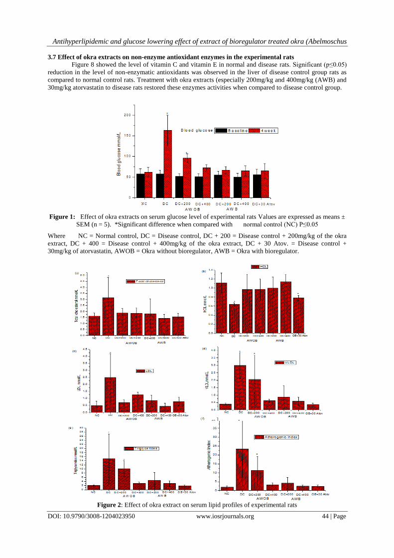
3.1 Effect of okra extracts on serum glucose level
As shown in figure 1, on the fourth week of the experiment, serum glucose levels of disease control and
DC+200mg/kg (AWOB) groups were significantly (P≤0.05) increased as compared to control levels. But in
other treated experimental groups there was no significant increased in serum glucose levels on the fourth week
of the experiment as compared to the control group. The administration of the extract was observed to be dose-
dependent in the reduction of serum glucose levels.
3.2 Effect of okra extracts on serum lipid profiles
Triton-induced significant increased in total cholesterol, LDL, VLDL, and atherogenic index (AI). Rats
in the disease control group and those treated with DC+ 200mg/kg (AWOB) showed significantly increased in
the levels of TC, LDL, VLDL and AI in comparison with normal control group. However, serum HDL level of
rats in disease control group was reduced when compared with other groups as well as a control group. Treated
Abelmoschus esculentus extracts (400mg/kg p.o) and atorvastatin (30mg/kg) decreased serum total cholesterol,
triglycerides, VLDL, LDL, and atherogenic index with increased HDL levels when compared with disease
control group, although the reduction did not return to baseline of normal control group. Higher doses
(400mg/kg) of both treated (AWB) and untreated (AWOB) extracts significantly decreased LDL and total
cholesterol but decreased in the atherogenic index were only seen in treated (AWB) extract at both 200mg/kg
and 400mg/kg respectively when compared with disease control group. However, there were no significant
decreased serum VLDL and TG and increased HDL levels (Figure 2).
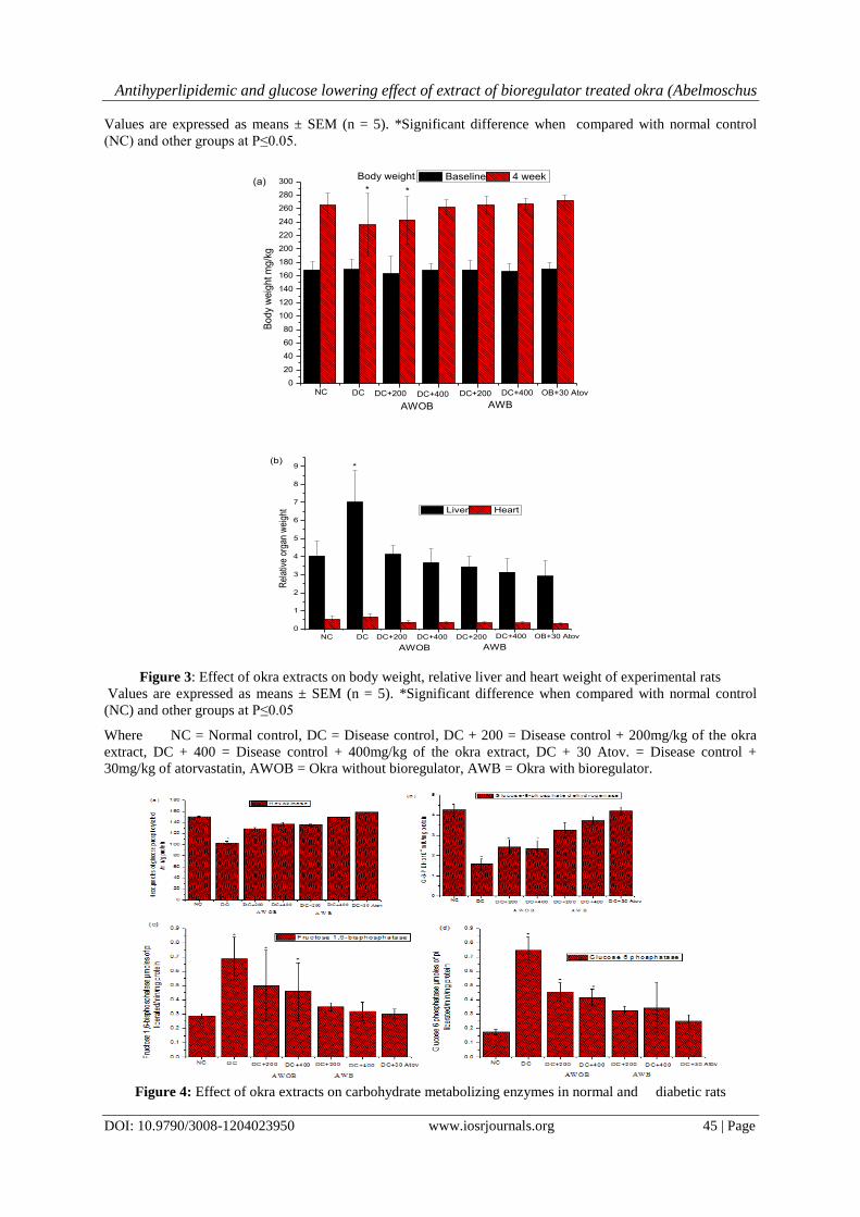
3.3 Effect of okra extracts on Body weight and relative liver and heart weight
After four weeks of the experiment, the body weights were measured. Significant (P≤0.05) decreased
in body weight were seen in disease control and DC+200mg/kg (AWOB) at about 25% when compared with
normal control and other experimental groups. However, significant increased liver relative weight was recorded
in disease control group when compared with other experimental groups. The liver relative weight was almost
times ten of heart relative weight in all the experimental groups (Figure 3).
3.4 Effect of okra extracts on carbohydrate metabolizing enzymes
Figure 4 showed the change in the activities of hexokinase, glucose-6-phosphate dehydrogenase,
fructose-1, 6- bisphosphatase, glucose-6-phosphatase in the liver of normal and experimental rats. The activities
of hepatic gluconeogenic enzymes (fructose-1, 6 - bisphosphatase and glucose-6-phosphatase) were
significantly ((P≤0.05) increased in disease control group as compared to normal control group, whereas
glycolytic enzyme (hexokinase), and pentose phosphate pathway enzyme (glucose-6-phosphate dehydrogenase)
were significantly ((P≤0.05) decreased in disease control group when compared to normal control group.
However, administration of ethanol extract of okra and glibenclamide to disease rats reversed the changes in the
activities of these hepatic enzymes in groups treated with 200mg/kg and 400mg/kg (AWB) and 30mg/kg
atorvastatin respectively as compared to disease control group.
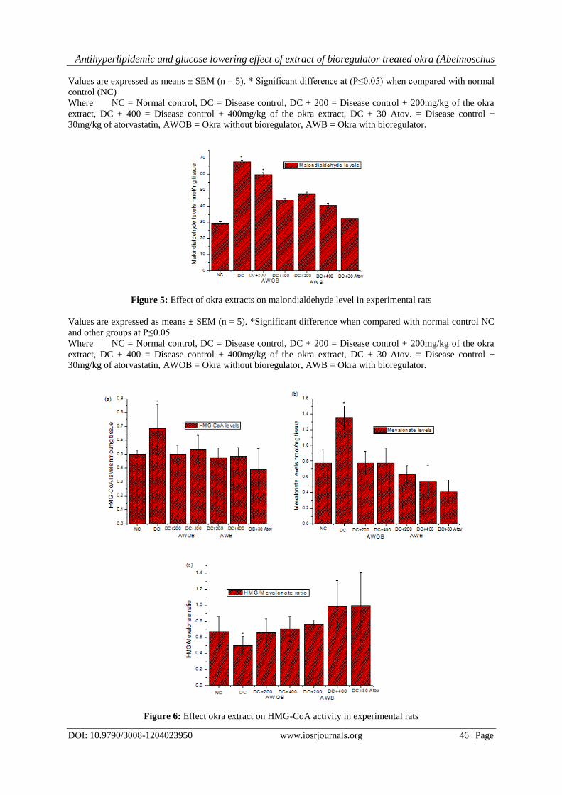
3.4 Effect of okra extracts on Malondialdehyde level
Figure 5 showed the level of lipid peroxidation in the experimaental rats. Malondialdehyde level was
significantly (P≤0.05) increased in disease control group as compared to normal control group. But the
administration of ethanol extract okra significantly ((P≤0.05) decreased the level of malondialdehyde as
compared to disease control group with the exception of the group treated with 200mg/kg (AWOB).
3.5 Effect of okra extracts on liver HMG- CoA reductase activity
Figure 6 showed the effect of the okra extracts on Liver HMG/Mevalonate Ratio. Significant (P≤0.05)
increased in HMG/Mevalonate ratio were observed in groups treated with 400mg/kg (AWB) and 30mg/kg
atorvastatin, which in turn significantly (P≤0.05) decreased the HMG-CoA reductase activity when compared
with the disease control group.
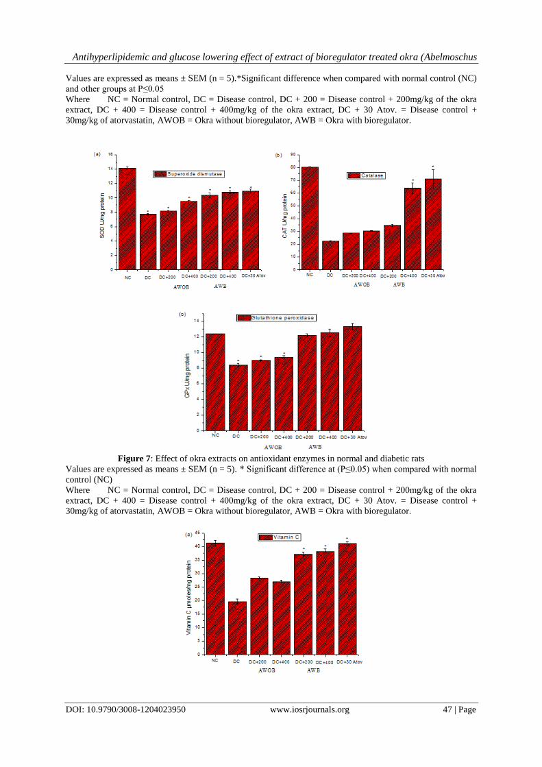
3.6 Effect of okra extracts on antioxidant enzymes in the experimental rats
Figure 7 showed the activities of CAT, SOD, and GPx in normal and disease rats. There was a
significant (p≤0.05) reduction in the activities of CAT, SOD, and GPx in disease control rats. After
administration with okra extracts the antioxidant activities in the groups treated with 400mg/kg (AWB) and
30mg/kg atorvastatin were well compared with normal control group.
Antihyperlipidemic and glucose lowering effect of extract of bioregulator treated okra (Abelmoschus
DOI: 10.9790/3008-1204023950 www.iosrjournals.org 44 | Page
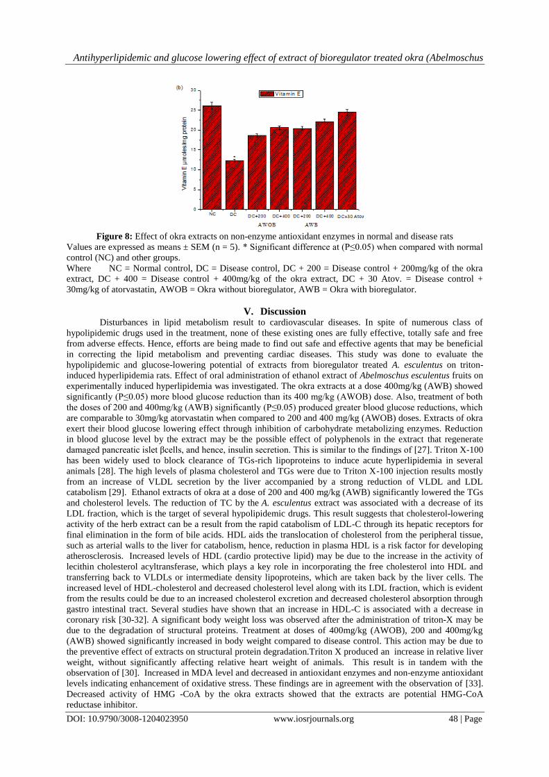
3.7 Effect of okra extracts on non-enzyme antioxidant enzymes in the experimental rats
Figure 8 showed the level of vitamin C and vitamin E in normal and disease rats. Significant (p≤0.05)
reduction in the level of non-enzymatic antioxidants was observed in the liver of disease control group rats as
compared to normal control rats. Treatment with okra extracts (especially 200mg/kg and 400mg/kg (AWB) and
30mg/kg atorvastatin to disease rats restored these enzymes activities when compared to disease control group.
Figure 1: Effect of okra extracts on serum glucose level of experimental rats Values are expressed as means ±
SEM (n = 5). *Significant difference when compared with normal control (NC) P≤0.05
Where NC = Normal control, DC = Disease control, DC + 200 = Disease control + 200mg/kg of the okra
extract, DC + 400 = Disease control + 400mg/kg of the okra extract, DC + 30 Atov. = Disease control +
30mg/kg of atorvastatin, AWOB = Okra without bioregulator, AWB = Okra with bioregulator.
Figure 2: Effect of okra extract on serum lipid profiles of experimental rats
Antihyperlipidemic and glucose lowering effect of extract of bioregulator treated okra (Abelmoschus
DOI: 10.9790/3008-1204023950 www.iosrjournals.org 45 | Page
Values are expressed as means ± SEM (n = 5). *Significant difference when compared with normal control
(NC) and other groups at P≤0.05.
0
20
40
60
80
100
120
140
160
180
200
220
240
260
280
300
Bo
dy
we
igh
t m
g/k
g
Baseline 4 week
NC DC DC+200 DC+400 DC+200 DC+400 OB+30 Atov
AWOB AWB
Body weight
* *(a)
0
1
2
3
4
5
6
7
8
9
Rel
ativ
e or
gan
wei
ght Liver Heart
NC DC DC+200 DC+400 DC+200 DC+400 OB+30 Atov
AWOB AWB
*(b)
Figure 3: Effect of okra extracts on body weight, relative liver and heart weight of experimental rats
Values are expressed as means ± SEM (n = 5). *Significant difference when compared with normal control
(NC) and other groups at P≤0.05
Where NC = Normal control, DC = Disease control, DC + 200 = Disease control + 200mg/kg of the okra
extract, DC + 400 = Disease control + 400mg/kg of the okra extract, DC + 30 Atov. = Disease control +
30mg/kg of atorvastatin, AWOB = Okra without bioregulator, AWB = Okra with bioregulator.
Figure 4: Effect of okra extracts on carbohydrate metabolizing enzymes in normal and diabetic rats
Antihyperlipidemic and glucose lowering effect of extract of bioregulator treated okra (Abelmoschus
DOI: 10.9790/3008-1204023950 www.iosrjournals.org 46 | Page
Values are expressed as means ± SEM (n = 5). * Significant difference at (P≤0.05) when compared with normal
control (NC)
Where NC = Normal control, DC = Disease control, DC + 200 = Disease control + 200mg/kg of the okra
extract, DC + 400 = Disease control + 400mg/kg of the okra extract, DC + 30 Atov. = Disease control +
30mg/kg of atorvastatin, AWOB = Okra without bioregulator, AWB = Okra with bioregulator.
Figure 5: Effect of okra extracts on malondialdehyde level in experimental rats
Values are expressed as means ± SEM (n = 5). *Significant difference when compared with normal control NC
and other groups at P≤0.05
Where NC = Normal control, DC = Disease control, DC + 200 = Disease control + 200mg/kg of the okra
extract, DC + 400 = Disease control + 400mg/kg of the okra extract, DC + 30 Atov. = Disease control +
30mg/kg of atorvastatin, AWOB = Okra without bioregulator, AWB = Okra with bioregulator.
Figure 6: Effect okra extract on HMG-CoA activity in experimental rats
Antihyperlipidemic and glucose lowering effect of extract of bioregulator treated okra (Abelmoschus
DOI: 10.9790/3008-1204023950 www.iosrjournals.org 47 | Page
Values are expressed as means ± SEM (n = 5).*Significant difference when compared with normal control (NC)
and other groups at P≤0.05
Where NC = Normal control, DC = Disease control, DC + 200 = Disease control + 200mg/kg of the okra
extract, DC + 400 = Disease control + 400mg/kg of the okra extract, DC + 30 Atov. = Disease control +
30mg/kg of atorvastatin, AWOB = Okra without bioregulator, AWB = Okra with bioregulator.
Figure 7: Effect of okra extracts on antioxidant enzymes in normal and diabetic rats
Values are expressed as means ± SEM (n = 5). * Significant difference at (P≤0.05) when compared with normal
control (NC)
Where NC = Normal control, DC = Disease control, DC + 200 = Disease control + 200mg/kg of the okra
extract, DC + 400 = Disease control + 400mg/kg of the okra extract, DC + 30 Atov. = Disease control +
30mg/kg of atorvastatin, AWOB = Okra without bioregulator, AWB = Okra with bioregulator.
Antihyperlipidemic and glucose lowering effect of extract of bioregulator treated okra (Abelmoschus
DOI: 10.9790/3008-1204023950 www.iosrjournals.org 48 | Page
Figure 8: Effect of okra extracts on non-enzyme antioxidant enzymes in normal and disease rats
Values are expressed as means ± SEM (n = 5). * Significant difference at (P≤0.05) when compared with normal
control (NC) and other groups.
Where NC = Normal control, DC = Disease control, DC + 200 = Disease control + 200mg/kg of the okra
extract, DC + 400 = Disease control + 400mg/kg of the okra extract, DC + 30 Atov. = Disease control +
30mg/kg of atorvastatin, AWOB = Okra without bioregulator, AWB = Okra with bioregulator.
V. Discussion
Disturbances in lipid metabolism result to cardiovascular diseases. In spite of numerous class of
hypolipidemic drugs used in the treatment, none of these existing ones are fully effective, totally safe and free
from adverse effects. Hence, efforts are being made to find out safe and effective agents that may be beneficial
in correcting the lipid metabolism and preventing cardiac diseases. This study was done to evaluate the
hypolipidemic and glucose-lowering potential of extracts from bioregulator treated A. esculentus on triton-
induced hyperlipidemia rats. Effect of oral administration of ethanol extract of Abelmoschus esculentus fruits on
experimentally induced hyperlipidemia was investigated. The okra extracts at a dose 400mg/kg (AWB) showed
significantly (P≤0.05) more blood glucose reduction than its 400 mg/kg (AWOB) dose. Also, treatment of both
the doses of 200 and 400mg/kg (AWB) significantly (P≤0.05) produced greater blood glucose reductions, which
are comparable to 30mg/kg atorvastatin when compared to 200 and 400 mg/kg (AWOB) doses. Extracts of okra
exert their blood glucose lowering effect through inhibition of carbohydrate metabolizing enzymes. Reduction
in blood glucose level by the extract may be the possible effect of polyphenols in the extract that regenerate
damaged pancreatic islet βcells, and hence, insulin secretion. This is similar to the findings of [27]. Triton X-100
has been widely used to block clearance of TGs-rich lipoproteins to induce acute hyperlipidemia in several
animals [28]. The high levels of plasma cholesterol and TGs were due to Triton X-100 injection results mostly
from an increase of VLDL secretion by the liver accompanied by a strong reduction of VLDL and LDL
catabolism [29]. Ethanol extracts of okra at a dose of 200 and 400 mg/kg (AWB) significantly lowered the TGs
and cholesterol levels. The reduction of TC by the A. esculentus extract was associated with a decrease of its
LDL fraction, which is the target of several hypolipidemic drugs. This result suggests that cholesterol-lowering
activity of the herb extract can be a result from the rapid catabolism of LDL-C through its hepatic receptors for
final elimination in the form of bile acids. HDL aids the translocation of cholesterol from the peripheral tissue,
such as arterial walls to the liver for catabolism, hence, reduction in plasma HDL is a risk factor for developing
atherosclerosis. Increased levels of HDL (cardio protective lipid) may be due to the increase in the activity of
lecithin cholesterol acyltransferase, which plays a key role in incorporating the free cholesterol into HDL and
transferring back to VLDLs or intermediate density lipoproteins, which are taken back by the liver cells. The
increased level of HDL-cholesterol and decreased cholesterol level along with its LDL fraction, which is evident
from the results could be due to an increased cholesterol excretion and decreased cholesterol absorption through
gastro intestinal tract. Several studies have shown that an increase in HDL-C is associated with a decrease in
coronary risk [30-32]. A significant body weight loss was observed after the administration of triton-X may be
due to the degradation of structural proteins. Treatment at doses of 400mg/kg (AWOB), 200 and 400mg/kg
(AWB) showed significantly increased in body weight compared to disease control. This action may be due to
the preventive effect of extracts on structural protein degradation.Triton X produced an increase in relative liver
weight, without significantly affecting relative heart weight of animals. This result is in tandem with the
observation of [30]. Increased in MDA level and decreased in antioxidant enzymes and non-enzyme antioxidant
levels indicating enhancement of oxidative stress. These findings are in agreement with the observation of [33].
Decreased activity of HMG -CoA by the okra extracts showed that the extracts are potential HMG-CoA
reductase inhibitor.
Antihyperlipidemic and glucose lowering effect of extract of bioregulator treated okra (Abelmoschus
DOI: 10.9790/3008-1204023950 www.iosrjournals.org 49 | Page
VI. Conclusion
In conclusion, the antihyperlipidemic and anti- glycemic activities of okra extracts at 200 and 400
mg/kg (AWB) against Triton X-100 shows a significant (P≤0.05) decrease in blood glucose, TC, TG, LDL-C,
VLDL, AI, MDA, HMG-CoA reductase activity, gluconeogenic enzymes and significant (P≤0.05) increase in
HDL-C, glycogenic enzymes, antioxidant enzymes and non-enzymes antioxidant in a dose- dependent manner
comparing with standard atorvastatin- treated group. However, further studies are still going on to evaluate the
actual active constituents responsible for the activity and mechanisms of these effects.
Acknowledgements
Authors are grateful to the director of INSA JRD TATA Dr. Amudeswari of CICS and JSS College of
Pharmacy, Ooty, Tamil Nadu, India for their constant support throughout this research work.
Funding
We are grateful for INSA-JRD TATA FELLOWSHIP of India for financing this project.
Abbreviations
AWB = Abelmoschus esculentus with bioregulator , AWOB = Abelmoschus esculentus without bioregulator,
ATOV= Atorvastatin, CAD = Coronary artery diseases, CMC= Carboxymethyl cellulose, HDL-C= High
density lipoproteins cholesterol, HL= Hyperlipidemic control group, LDL-C= low density lipoproteins
cholesterol, , P.o.= Per oral, S.D= Standard deviation, MDA = Malondialdehyde, TC= Serum total cholesterol,
TG= Triglycerides, VLDL-C= Very low density lipoproteins cholesterol, CAT = Catalase, SOD = Superoxide
dismutase, GPx = Glutathione peroxidase, HMG- CoA = 3-hydroxy-3-methylglutaryl, AI = Atherogenic index.
References [1] G.D. Kolovou, K. K. Anagnostopoulou,D. V. Cokkinos,“Pathophysiology of dyslipidaemia in themetabolic
syndrome,”Postgraduate Medical Journal 81 (956), 2005, 358–366.
[2] R.M. Krauss, Y.A. Kesaniemi, “Cardiovascular disease and hyperlipidaemia,” Current Opinion in Lipidology, 5 (4), 1994, 249–251.
[3] G.S. Hotamisligil, E. Erbay, Nutrient sensing and inflammation in metabolic diseases. Nat Rev Immunol. 8, 2009, 923-34 [4] J.H. Goedecke, J.A. Dave, M.V. Faulenbach, Insulin response in relation to insulin sensitivity: an appropriatebeta-cell response in
black South African women. Diabetes Care. 32(5), 2009, 860-65
[5] D. Hayoz, T. Ziegler, Diabetes mellitus and vascular lesions. Metabolism. Curr. Ther. Res., 47,1998, 16-19 [6] D. Bonnefont, J.P. Bastard M.C. Jaudon, J. Dellatre, Consequences of diabetes status on the oxidant/antioxidant balance. Diabetes
Metabol. 26, 2000, 163-76. [7] N. J. Stone, “Lipidmanagement: current diet and drug treatment options,” The American Journal of Medicine, 101(4), 1996,
supplement 1, 40S–49S,
[8] Y. S. Lin, S. Mousa, N. Elshourbagy, S. A. Mousa, “Current status and future directions in lipid management: emphasizing low-density lipoproteins, high-density lipoproteins, and triglycerides as targets for therapy,” Vascular Health and Risk Management, 6
(1),2010,73–85.
[9] M. Kobayashi, T. Kagawa, K. Narumi, S. Itagaki, T. Hirano, K. Iseki, “Bicarbonate supplementation as a preventive way in statins-induced muscle damage,” Journal of Pharmacy and Pharmaceutical Sciences, 11(1), 2008,1–8.
[10] J. R Guyton, H. E. Bays, “Safety considerations with niacin therapy,”The American Journal of Cardiology, 99 (6), 2007, S22–S31.
[11] W. C. Lakey, N. Greyshock, J.R.Guyton, “Adverse reactions of Achilles tendon xanthomas in three hypercholesterolemic patients after treatment intensification with niacin and bile acid sequestrants,” Journal of Clinical Lipidology, 7 (2), 2013,178– 181.
[12] C.C. Allain, L.S. Poon, C.S.G. Chan, W. Richmondn, P.C. Fu, Clin. Chem. 20, 1974, 470
[13] W.T. Friedewald, R.I. Levy, D.S. Fredrickson, Estimation of the concentration of low-density lipoprotein cholesterol in plasma, without the use of the preparative ultracentrifuge ClinChem. 18, 1972, 499–502. [PubMed]
[14] N. Taleb-Dida, D. Krouf, M Bouchenak, “Globularia alypum aqueous extract decreases hypertriglyceridemia and ameliorates
oxidative status of the muscle, kidney, and heart in rats fed a high-fructose diet,” Nutrition Research. 31(6), 2011, 488–495 [15] N. Brandstrup, J.E. Kirk, C. Bruni, Determination of hexokinase in tissues. J Gerontol 12, 1957,166-71.
[16] H.A. Ellis, H.N. Kirkman, A colorimetric method for assay of erythrocyte glucose-6-phosphate dehydrogenase. Proc Soc Exp Biol
Med. 106, 1961, 607-609.
[17] H. Koide, T. Oda, Pathological occurrence of glucose-6-phosphatase in liver disease. Clin Chim Acta. 4, 1959, 554-561
[18] C.H. Fiske, Y. Subbarow The colorimetric determination of phosphorus J BiolChem, 66, 1925, 375–400
[19] J.M. Gancedo, C. Gancedo, Fructose-1, 6-Bisphosphatase, phospho fructokinase and glucose-6-phosphate dehydrogenase from fermenting and non-fermenting yeasts. Arch Microbiol 76, 1971, 132-138..
[20] H. Ohkawa, N. Ohishi, K. Yagi, “Assay for lipid peroxides in animal tissues by the thiobarbituric acid reaction,” Analytical
Biochemistry. 1979; vol. 95, no. 2, pp. 351–358 [21] Venugopala Rao A and Ramakrishnan S. “Indirect assessment of hydroxymethyl glutarylCoA reductase (NADPH) activity in the
liver tissue,” Clinical Chemistry 21(10) 1975, 1523– 1525.
[22] K.A. Sinha, Colorimetric assay of catalase. Anal. Biochem., 47, 1972, 389-394. [23] D. Kakkar, B. Das, P.N. Viswanathan, A modified spectro-photometric assay of superoxide dismutase.Ind.J.Biochem. Biophys., 21,
1984, 130-132.
[24] ] J.T. Rotruck, A.L. Pope, H.E. Ganther, A.B. Swanson, D.G. Hafeman, W.G. Hoekstra, Selenium:Biochemicalroles as a component of glutathione peroxidase. Science., 179, 1984, 588-590.
[25] J.H. Roe, C.A. Kuether, The determination of ascorbic acid in whole blood and urine through the 2,4-dinitrophenylhydrazine
derivative of dehydroascorbic acid. J. Biol. Chem. 147, 1943, 399 [26] R. T. M. Baker, S. J. Davies, Changes in tissue α- tocopherol status and degree of lipid peroxidation with varying α-tocopheryl
acetate inclusion in diets for the African catfish. Aquacult. Nutr. 2, 1996, 71-79.
Antihyperlipidemic and glucose lowering effect of extract of bioregulator treated okra (Abelmoschus
DOI: 10.9790/3008-1204023950 www.iosrjournals.org 50 | Page
[27] N. Møller, K.S. Nair, Diabetes and protein metabolism. Diabetes. 57, 2008,3–4. [PubMed]
[28] A. Kellner, J.W. correll, AT. Ladd, Sustained hyperlipemia induced in rabbits by means of intravenously injected surface-active
agents. J Exp Med. 93,1951,373–384. [PMC free article] [PubMed] [29] S. Otway, D.S. Robinson, The effect of a non-ionic detergent (Triton WR 1339) on the removal of triglyceride fatty acids from the
blood of the rat J Physiol. 190, 1967, 309–319. [PMC free article] [PubMed]
[30] N. Taleb-Dida, D. Krouf, M. Bouchenak, “Globularia alypum aqueous extract decreases hypertriglyceridemia and ameliorates oxidative status of the muscle, kidney, and heart in rats fed a high-fructose diet,” Nutrition Research, 31(6), 2011, 488–495.
[31] A. R Borate, A. A. Suralkar, S. S. Birje, P. V. Malusare, and P. A. Bangale, “Antihyperlipidemic effect of protocatechuic acid in
fructose induced hyperlipidemia in rats,” International Journal of Pharma and Bio Sciences, 2 (4), 2011, 456–460. [32] G. L. Kelley, G. Allan, S. Azhar, “High dietary fructose induces a hepatic stress response resulting in cholesterol and lipid
dysregulation,” Endocrinology, 145(2), 2004, 548–555.
[33] S. Reddy, S. P. Ramatholisamma, R. Karuna, D. Saralakumari, “Preventive effect of Tinospora cordifolia against high fructose diet-induced insulin resistance and oxidative stress in male Wistar rats,” Food and Chemical Toxicology, 47(9), 2009, 2224–2229.
IOSR Journal of Pharmacy and Biological Sciences (IOSR-JPBS) is UGC approved Journal
with Sl. No. 5012, Journal no. 49063.
A.M. Esan. "Antihyperlipidemic And Glucose Lowering Effect Of Extract Of Bioregulator
Treated Okra (Abelmoschus Esculentus L.) Fruits in Triton-Induced Hyperlipidemia Rats."
IOSR Journal of Pharmacy and Biological Sciences (IOSR-JPBS) 12.4 (2017): 39-50.