Embed Size (px)
Citation preview
www.scholarsresearchlibrary.comt Available online a
Scholars Research Library
Der Pharmacia Lettre, 2016, 8 (14):109-118
(http://scholarsresearchlibrary.com/archive.html)
ISSN 0975-5071
USA CODEN: DPLEB4
109 Scholar Research Library
Evaluation of Antihyperlipidemic Activity of HMG-Co A Reductase Inhibitor and Curcumin in Combination on Diet Induced Hyperlipidemic Rats
Aijaz A. Sheikh1*, Tanwir Z. Khan1 and Sajid R. Sheikh2
1Anuradha College of Pharmacy, Chikhli, Maharashtra 2Dr. Vedprakash Patil Pharmacy College, Aurangabad, Maharashtra
_____________________________________________________________________________________________
ABSTRACT Hyperlipidemia a well known risk factor for cardiovascular disease, especially atherosclerotic coronary artery disease is one of the major cause of premature death globally. Cholesterol feeding has been often used to elevate serum or tissue cholesterol levels to assess hypercholesterolemia-related metabolic disturbances in different animal models. Hyperlipidemia was induced in rats by giving high cholesterol diet for seven days in standard rat chow diet. High cholesterol fed diet rats exhibited significant increase in total serum cholesterol, triglycerides, low density lipoprotein, very low density lipoprotein, atherogenic index and lipid per-oxidation and significant decrease in high density lipoprotein. The CCl4-induced hepatotoxicity model is extensively used to evaluate the hepatoprotective effects of drugs and extracts. In this study we diagnosed the different liver function parameter like, total cholesterol (TC), triglycerides (TG), high density lipoprotein cholesterol (HDLC) in serum by using kits. Also other diagnosed parameters such as low density lipoprotein (LDLC), atherogenic index, very low density lipoprotein (VLDL-C), HDL-C ratio and lipid per-oxidation (LPO). From obtained result it was observed that keeping the animal on atherogenic diet model (AD) significantly increased the TC, TG, LDL-C level in serum as compared to rats on normal diet. When AD was co-administered with curcumin and rosuvastatin in combination, the elevated levels of TC, TG and LDL-C and LPO condition has shown considerable decline. It was noted that TC, TG and LDL-C lowering activity of curcumin (300mg/kg) and rosuvastatin in combination was more significant as compared to curcumin and simvastatin. Key words: Curcumin, HMG-Co A reductase inhibitor, Antihyperlipidemic, CCL4 induced hyperlipidemia, Histopathological study _____________________________________________________________________________________________
INTRODUCTION
Cholesterol levels have become the source of health concerns, even though cholesterol is one of the most valuable substances in the human body. Over the past few years, the amount of cholesterol information and dietary advice bombarding the public has grown exponentially. Cholesterol plays a major role in the assembly of membranes and performs other important biological functions in human heart health [1]. However, when plasma cholesterol exceeds the level required for these functions, it results in the development of atherosclerotic cardiovascular disease such as coronary heart disease and stroke.
Hyperlipidemia may also induce other abnormalities like oxidation of free fatty acids, leading to the formation of ketone- bodies as well as masking liver and muscles resistance to insulin which initiates the progress of diabetes in patients [2].
Aijaz A. Sheikh et al Der Pharmacia Lettre, 2016, 8 (14):109-118 ______________________________________________________________________________
110 Scholar Research Library
Hyperlipidemia is a major cause of atherosclerosis and atherosclerosis related conditions like coronary heart disease, ischemic cerebrovascular disease, peripheral vascular disease and pancreatitis. The increase in lipids like low density lipoproteins (LDL), cholesterol (esters derivatives) and triglycerides are mainly responsible for this condition [3]. Simvastatin and rosuvastatin was shown to be a highly effective member of statins for lowering blood cholesterol, acts by stabilizing plaque and preventing strokes through anti-inflammatory and other mechanisms [4]. Among the plant foods that possess hypocholesterolemic property in clinical, as well as experimental studies are curcumin, garlic, gum guggul and capsicum. The aim of present study was the evaluation of antihyperlipidemic activity of HMG-CoA reductase inhibitor and curcumin in combination on diet induced hyperlipidemic rats.
MATERIALS AND METHODS
Male albino wistar rat 6-8 weeks of age, weighing between 180–240 gm of either sex were procured from animal house of Anuradha College of pharmacy, Chikhli, district Buldana (MS) India. Animals were housed in polypropylene cages under the standard conditions of temperature, pressure and humidity. (12 hrs light and dark cycles, at 25+3˚C and 35-60% humidity) [5].The animals were maintained under standard condition as per CPCSEA guideline. Animals were fed with standard normal pellet diet and water ad libitum during the course of the experiment. The animals were given standard diet supplied by local market of Chikhli. The composition of the diet are 3615 (Kcal/kg), crude protein 22.05%, crude oil 4.5%, crude fibre 4.10%, ash 11.10%, sand silica 0.75% [6,7]. The experimental design and research plan along with animal handling and disposal procedure were placed before the animal ethics committee. The committee granted approval after careful evaluation of research project. Proposal No. 751/03/abc/CPCSEA) and Ref. No. IAEC/ 2011-2012. Extract was gifted by Aldrich Pharma, simvastatin and rosuvastatin gifted by Dr. Reddy Hyderabad. Kits (total cholesterol, triglycerides and HDL-C) were purchased from Ashrita Dia Tech Hyderabad. Induction of Hyperlipidemia Atherogenic Diet Induced Method Hyperlipidemia was induced in rats by giving high cholesterol diet for seven days in standard rat chow diet. The drug solution was administered orally. High cholesterol diet was prepared by mixing cholesterol 2%, sodium cholate 1% and coconut oil 2% with standard powdered standard animal food. The diet was placed in the cage carefully and was administered for seven days [8]. CCL4 Induced Method Hyperlipidemia was induced in rats by giving CCl4 for seven days orally [9]. Animal were administered with daily dosage of CCl4 as 1:1 dilution with liquid paraffin for every 72 hours. Atherogenic Diet Model Healthy wistar albino rats divided into seven groups. Each group contains six rats. Group I was taken as a normal treated with normal diet, group II treated with atherogenic diet was kept as toxicant, group III treated with atherogenic diet and simvastatin(10mg/kg), group IV treated with atherogenic diet and rosuvastatin (10mg/kg) , group V treated with atherogenic diet and curcumin (300mg/kg), group VI was treated with atherogenic diet, simvastatin(2.5mg/kg) and curcumin(300mg/kg) and group VII was treated with atherogenic diet, rosuvastatin (2.5 mg/kg) and curcumin (300mg/kg) administered orally for seven days [10]. Blood was collected on 8th day by retro orbital puncture technique under mild anesthesia after 8 hour fasting and allowed to clot for 30 min at room temperature. Blood samples were centrifuged at 3000 rpm for 20 min. Serum was separated and stored at -20oC until biochemical estimation carried out. Serum sample were analyzed spectrophotometrically for total serum cholesterol (TC), triglycerides (TG) and high density lipoprotein cholesterol (HDL-C) was estimated by using diagnostic kit [11]. Very low density lipo-protein (VLDL), High density lipoprotein ratio (HDL-ratio), Atherogenic-index (AI) and low density lipoprotein cholesterol (LDL-C) were calculated by using the formula of Modi and colleague.
Aijaz A. Sheikh et al Der Pharmacia Lettre, 2016, 8 (14):109-118 ______________________________________________________________________________
111 Scholar Research Library
Table 1, Experimental design for atherogenic diet model
Group No. Group Name Treatment Observations I Normal control Normal diet
Estimation of parameters
II Toxicant control Atherogenic diet III Standard I Atherogenic diet+ simvastatin(10mg/kg) IV Standard II Atherogenic diet+ rosuvastatin(10mg/kg) V Curcumin Atherogenic diet+ curcumin(300mg/kg) VI Curcumin+ Standard I Atherogenic diet+ simvastatin(2.5mg/kg)+ curcumin(300mg/kg) VII Curcumin + Standard II Atherogenic diet+ rosuvastatin(2.5mg/kg)+ curcumin(300mg/kg)
CCL4 Induced Hyperlipidemia Healthy wistar albino rats were divided into seven groups. Each group contains six rats. Group I was taken as a normal treated with tween 80, group II treated with CCL4 (1ml/kg) was kept as toxicant, group III was treated with CCL4 (1ml/kg) and simvastatin (10mg/kg), group IV was treated CCL4 (1ml/kg) and rosuvastatin (10mg/kg), group V treated with CCL4 (1ml/kg) and curcumin (300mg/kg), group VI was treated with CCL4 (1ml/kg), simvastatin (2.5mg/kg) and curcumin (300mg/kg) and group VII treated with CCL4 (1ml/kg), rosuvastatin (2.5mg/kg) and curcumin (300mg/kg) administered orally once daily for 7 days and simultaneously the animal were treated with CCL4 [12,13].
Table 2, Experimental design for CCL4 model
Group No. Group Name Treatment Observations I Normal control Normal diet
Estimation of parameters
II Toxicant control CCL4 (1ml/kg) III Standard I CCL4 (1ml/kg)+ simvastatin 10mg/kg) IV Standard II CCL4 (1ml/kg) + rosuvastatin (10mg/kg) V Curcumin CCL4 (1ml/kg)+ curcumin (300mg/kg) VI Curcumin+ Standard I CCL4 (1ml/kg)+ simvastatin (2.5mg/kg)+ curcumin (300mg/kg) VII Curcumin+ Standard II CCL4 (1ml/kg)+ rosuvastatin (2.5mg/kg)+ curcumin (300mg/kg)
Dosage of CCL4 was administered as 1:1 dilution with liquid paraffin (Christina et al, 2006) for every 72 hours. The rats were sacrificed after 48 hour of last dose by cervical decapitation. The blood samples were collected separately by cardiac puncture and allow to clots for 30 min at room temp. The clear serum was separated by centrifugation at 2500 rpm for 10 min [14]. The TC, TG and HDL-C were determined using standard kits. 1. Total Cholesterol It was estimated by CHOD-PAP method using kit. The procedure was given in following Table No.3.
Table3, Procedure for total cholesterol determination
Pipette into tubes marked Blank Standard Test Serum/plasma - - 10µl Reagent 2 - 10µl - Reagent 1 1000µl 1000µl 1000µl
Mix well and incubate at 37 o C for 10 minutes or at room temperature (15-30 oC) for 30 minutes. Total cholesterol (mg/dl) = Absorbance of test _____________________× 200 Absorbance of standard 2. HDL-Cholesterol Determination: CHOD-PAP method was used to estimate the serum HDL- cholesterol. Step A – HDL Cholesterol separation
Table 4, Procedure for HDLC separation
Pipette in centrifuge tube Quantity Sample 200µl Precipitating reagent 200µl
Aijaz A. Sheikh et al Der Pharmacia Lettre, 2016, 8 (14):109-118 ______________________________________________________________________________
112 Scholar Research Library
Mix well and keep at room temperature for 10 minutes and then centrifuge at 2000 rpm to obtain a clear supernatant. Proceed to step- B of procedure. Step B – Colour development
Table 5, Procedure for HDLC determination
Pipette into tubes marked Blank solution Standard sample Test sample Supernatant from step A - - 10µl HDL-cholesterol Standard - 10µl - Cholesterol reagent 1000µl 1000µl 1000µl
Mix well and incubates at 37oC for 10 minutes. Read absorbance against reagent blank at 505 nm within 60 minutes. HDL cholesterol (mg/dl) = Absorbance of test ____________________× 50 Absorbance of standard 3. Triglycerides: It was estimated by GPO-PAP method by using kit.
Table 6, Procedure for triglycerides determination
Pipette into tubes marked Blank solution Standard sample Test sample Serum/plasma - - 10µl Reagent 2 - 10µl - Reagent 1 1000µl 1000µl 1000µl
Total triglyceride (mg/dl) = Absorbance of test ______________________ × 200 Absorbance of standard 4. LDL and VLDL cholesterol LDL and VLDL-cholesterol was calculated by following method of Friedwald et al. 1992, Ghube B.V. et al 2009. VLDL- Cholesterol = TG/ 5 LDLC = (Total cholesterol) - (HDLC) - (VLDLC) 5. Atherogenic index Atherogenic index and LDL-C/HDL-C was calculated by following formula. AI = (Total cholesterol-HDL-C)/HDL-C 6. LDL-C/HDL-C ratio LDL-C/HDL-C was calculated as the ratio of serum LDL-C to HDL-C level. 7. Estimation of lipid Peroxidation From each experimental rat 900mg of liver tissue was collected and washed with normal saline then soaked in filter paper. The tissues were then homogenized in 10 ml of 0.15 M Tris-buffer (pH-7.4) and centrifuged at 3000g at 4° C for 30 min [15, 16]. The supernatant collected was taken for lipid peroxidation assay. Malondialdehyde (MDA), produced during the peroxidation of lipids, served as an index of lipid peroxidation. MDA reacts with thiobarbituric acid (TBA) to generate a colored product which absorb at 532nm. Reagents used were normal saline (0.9% w/v), 10% TCA, and 1 % thiobarbituric acid. Lipid per-oxidation (LPO) was assayed according to the method of Okhawa., et al.1979. TCA 10% was added and mixed well. The mixture (3000 g) was then centrifuged at room temperature for 10 min to separate proteins. Supernatant (2 ml) was taken, 0.5ml 1.0% TBA was added followed by heating at 95° C for 60 min to generate the pink colored MDA. The samples were measured at 532 nm using Beckman DU 64 spectrophotometer. The levels of lipid peroxides were expressed as nm of MDA/mg wet tissue using extinction co-efficient of 1.56 x10⁵ M-1 cm-1 [17].
Aijaz A. Sheikh et al Der Pharmacia Lettre, 2016, 8 (14):109-118 ______________________________________________________________________________
113 Scholar Research Library
% lipids Inhibition = {Ao- A1}/Ao × 100 Where; Ao is the absorbance of the control and A1 is the absorbance of the sample extract. 8. Histopathological Examination On the day of blood withdrawal the two animals from each group were sacrificed and liver were isolated. Tissue samples were immersed in10% formalin for at least 24 hr to fix the tissue. The tissue was embedded in paraffin wax, sectioned and stained with haematoxylin and eosin. The sections were viewed under the light microscope for histopathological changes [18]. The study was conducted in Omega laboratories, Khandala, district Satara. 9. Statistical analysis The data were presented as Mean ± SD and the statistical analysis by one way ANOVA followed by Dunnett’s multiple comparison tests [19, 20].
RESULTS AND DISCUSSION
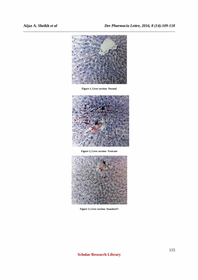
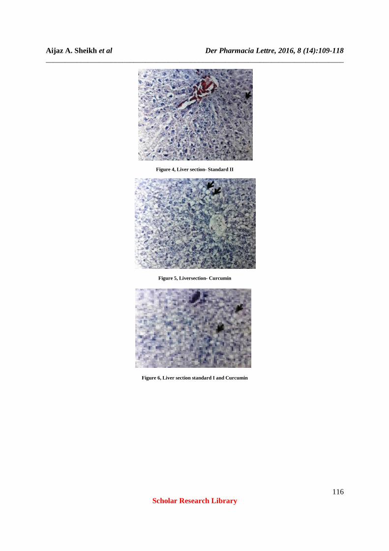
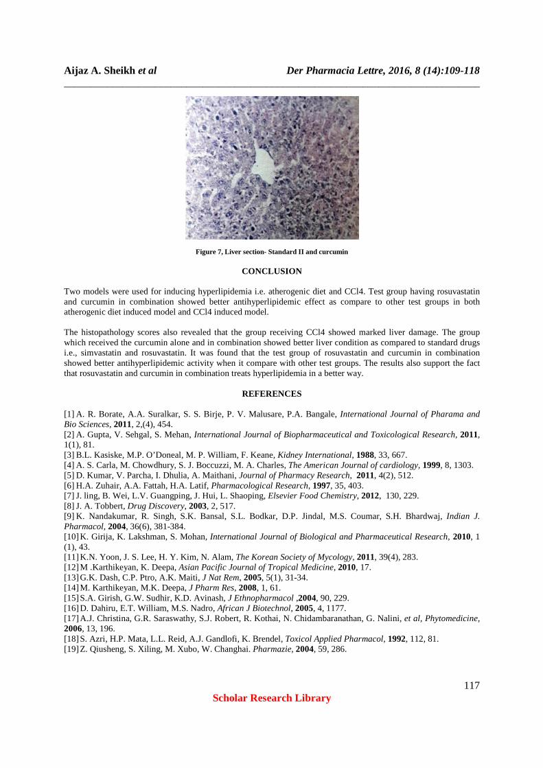
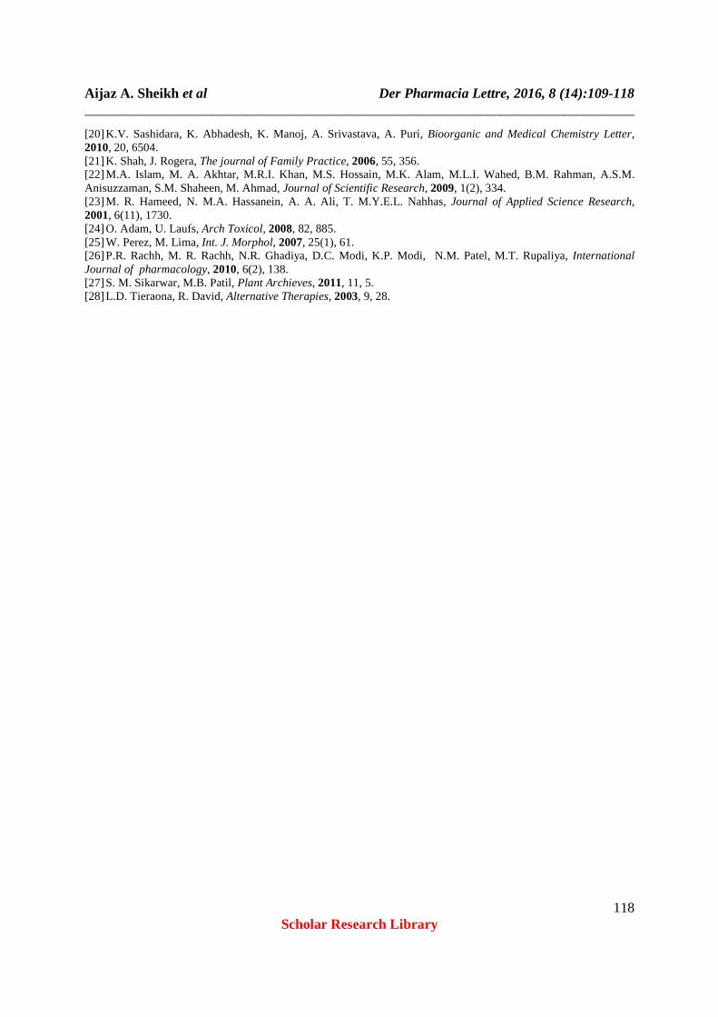
Serum total cholesterol level significantly elevated in both models i.e. AD and CCl4, as compare to normal control group. Treatment with curcumin alone and its combination with simvastatin and rosuvastatin for seven days showed marked decrease in concentration of serum cholesterol as compare to toxicant control group [21]. Serum HDL-C level decreased in both models (AD and CCl4) as compare to the normal control group. Treatment with curcumin alone and its combination with simvastatin and rosuvastatin for seven days showed marked increased in concentration of serum HDL-C as compare to toxicant control group. The serum triglyceride significantly elevated in atherogenic diet model and decreased in CCl4 model as compare to normal control group. Treatment with curcumin alone and its combination with simvastatin and rosuvastatin for seven days showed marked decrease in concentration of serum triglyceride in atherogenic diet model and increased in CCl4 model as compare to toxicant control group. Serum low density lipoprotein cholesterol significantly elevated in both models (AD and CCl4) as compare to the normal control group [22]. Treatment with curcumin alone and its combination with simvastatin and rosuvastatin for seven days showed marked decrease in concentration of serum low density lipoprotein as compare to the toxicant control group. Serum very low density lipoprotein cholesterol elevated in atherogenic diet model and decrease in CCl4 model as compare to the normal control group. Treatment with curcumin alone and its combination with simvastatin and rosuvastatin for seven days showed marked decrease in concentration of serum VLDL-C in atherogenic diet and increased in CCl4 model as compare to toxicant control group[23, 24]. Serum atherogenic index increased in both models (AD and CCl4) as compare to normal control group. Treatment with curcumin alone and its combination with simvastatin and rosuvastatin for seven days showed marked decrease in concentration of serum atherogenic index as compare to toxicant control group. Serum LDL-C/HDL-C ratio significantly elevated in both models i.e. AD and CCl4 as compare to normal control group. Treatment with curcumin alone and its combination with simvastatin and rosuvastatin for seven days showed marked decrease in concentration of serum LDL-C/HDL-C ratio as compare to toxicant control group [25]. Serum lipid per oxidation increased in both models i.e. AD and CCl4 as compare to normal control group. Treatment with curcumin alone and its combination with simvastatin and rosuvastatin for seven days showed marked decrease in concentration of serum lipid per oxidation as compare to toxicant control group. Liver of rat from normal group showed normal histological structure of hepatic lobule [26]. Liver of rat from toxicant group showed focal hepatic necrosis, hemorrhages, fibroplasias in portal triad associated with chronic cholangitis. Liver of rat from standard I group depicted hydropic degeneration of some hepatocytes [27]. In liver section of standard II group there was congestion of hepatic sinusoid observed. Hepatocellualr vacuolization was seen in liver section of rat from curcumin. Improvement in hepatocytes was noticed in liver of rat belongs to standard I and curcumin group. Finally liver of rat from standard II and curcumin group showed apparently normal hepatocytes [28]. In present study rosuvastatin and curcumin combination was found to be better as compare to other groups.
Aijaz A. Sheikh et al Der Pharmacia Lettre, 2016, 8 (14):109-118 ______________________________________________________________________________
114 Scholar Research Library
Table 7, Effect of curcumin alone and in combination on liver function parameters in AD induced hyperlipidemic model
Sr. No
Parameters Groups
Normal control
Toxicant control
Standard I
Standard II
Curcumin Curcumin + Standard I
Curcumin + Standard II
1. TC
(mg/dl) 96.28 ±1.05
244.98 ±1.74
169.41 ±1.91
153.70 ±1.31
208.34 ±2.23
131.29 ±1.50
117.34 ±1.41
2. TG (mg/dl)
81.66 ±0.68
160.62 ±1.64
124.35 ±1.37
114.68 ±1.40
137.36 ±1.27
105.78 ±1.67
94.06 ±1.33
3. HDL-C (mg/dl)
64.34 ± 0.88
27.52 ± 0.80
45.38 ± 0.77
50.53 ± 1.96
38.92 ±1.38
59.26 ± 1.59
62.21 ± 0.57
4. LDL-C (mg/dl)
31.27 ±1.62
244.55 ±2.16
145.87 ±1.16
122.36 ±1.14
195.41 ±3.65
86.47 ±2.60
64.58 ±1.51
5. VLDL-C (mg/dl)
16.33 ±0.13
32.12 ±0.32
24.86 ±0.27
22.93 ±0.28
27.47 ±0.25
21.15 ±0.33
18.80 ±0.26
6. LDL-C/ HDL-C
ratio 0.38 ±0.02
9.09 ±0.23
3.015 ±0.05
2.15 ±0.10
5.06 ±0.27
1.32 ±0.06
0.89 ±0.02
7. AI 0.140 ±0.02
7.92 ±0.20
2.5 ±0.05
1.81 ±0.05
4.40 ±0.26
0.99 ±0.05
0.62 ±0.02
8. LPO 27.71 ±0.56
65.70 ±0.26
54.91 ±0.24
50.32 ±0.34
58.11 ±0.16
38.15 ±0.81
33.7 ±0.33
Values are mean ± SEM (n=6), p<0.01 as compared to toxicant control group The statistical analysis of data was carried out by one way ANOVA followed by Dunnett's multiple comparison test.
Table 8, Effect of curcumin alone and in combination on liver function parameters in CCl4 induced hyperlipidemic model
Sr. No
Parameters
Groups
Normal control
Toxicant control
Standard I
Standard II
Curcumin Curcumin + Standard
I
Curcumin + Standard II
1 TC (mg/dl) 77.31 ±1.23
191.11 ±1.75
122.73 ±1.75
135.77 ±1.94
150.07 ±1.32
94.47 ±1.43
84.32 ±1.38
2. TG(mg/dl) 110.19 ±1.21
55.81 ±0.64
80.12 ±0.98
86.14 ±1.06
68.03 ±1.61
93.77 ±1.25
103.76 ±1.20
3. HDL-C(mg/dl) 62.31 ± 2.80
30.50 ± 2.35
44.10 ± 1.60
50.49 ±1.47
38.66 ± 1.19
54.21 ± 1.81
58.18 ± 0.93
4. LDL-C 19.38 ±1.91
228.11 ±3.22
123.07 ±1.35
98.39 ±2.08
174.82 ±2.97
65.06 ±1.99
45.59 ±1.52
5. VLDL-C(mg/dl) 22.03 ±0.24
11.16 ±0.13
16.02 ±0.19
17.13 ±0.26
13.60 ±0.32
18.59 ±0.25
20.74 ±0.24
6. LDL-C/ HDL-C
ratio 0.33 ±0.01
5.23 ±0.31
2.01 ±0.06
1.560 ±0.02
3.22 ±0.21
1.00 ±0.04
0.71 ±0.02
7. AI 0.17 ±0.01
7.93 ±0.20
2.50 ±0.05
1.82 ±0.05
4.39 ±0.24
0.93 ±0.05
0.62 ±0.02
8. LPO 26.78 ±0.80
68.30 ±0.60
41.19 ±0.51
35.61 ±0.81
52.32 ±0.72
36.55 ±0.11
33.03 ±0.49
Values are mean ± SEM (n=6), p<0.01 as compared to toxicant control group The statistical analysis of data was carried out by one way ANOVA followed by Dunnett's multiple comparison test.
Table 9, Scores of histopathological study
Group Congestion Hydropic
degeneration Necrosis
Cellular Vacuolization
Fibroplasia
Normal 00 00 00 00 00 Toxicant (CCl4) ++++ ++++ ++++ ++++ ++++ Standard-I ++ +++ ++ +++ ++ Standard –II +++ +++ ++ ++ +++ Curcumin ++ +++ ++ +++ ++ Standard-I+ Curcumin + ++ ++ + + Standard-II+Curcumin + + + ++ +
0: No abnormality detected +: Damage/ active changes up to less than 25%
++: Damage/ active changes up to less than 50% +++: Damage/ active changes up to less than 75%
++++: Damage/ active changes up to more than 75%
Aijaz A. Sheikh et al Der Pharmacia Lettre, 2016, 8 (14):109-118 ______________________________________________________________________________
115 Scholar Research Library
Figure 1, Liver section- Normal
Figure 2, Liver section- Toxicant
Figure 3, Liver section- Standard I
Aijaz A. Sheikh et al Der Pharmacia Lettre, 2016, 8 (14):109-118 ______________________________________________________________________________
116 Scholar Research Library
Figure 4, Liver section- Standard II
Figure 5, Liversection- Curcumin
Figure 6, Liver section standard I and Curcumin
Aijaz A. Sheikh et al Der Pharmacia Lettre, 2016, 8 (14):109-118 ______________________________________________________________________________
117 Scholar Research Library
Figure 7, Liver section- Standard II and curcumin
CONCLUSION
Two models were used for inducing hyperlipidemia i.e. atherogenic diet and CCl4. Test group having rosuvastatin and curcumin in combination showed better antihyperlipidemic effect as compare to other test groups in both atherogenic diet induced model and CCl4 induced model. The histopathology scores also revealed that the group receiving CCl4 showed marked liver damage. The group which received the curcumin alone and in combination showed better liver condition as compared to standard drugs i.e., simvastatin and rosuvastatin. It was found that the test group of rosuvastatin and curcumin in combination showed better antihyperlipidemic activity when it compare with other test groups. The results also support the fact that rosuvastatin and curcumin in combination treats hyperlipidemia in a better way.
REFERENCES
[1] A. R. Borate, A.A. Suralkar, S. S. Birje, P. V. Malusare, P.A. Bangale, International Journal of Pharama and Bio Sciences, 2011, 2,(4), 454. [2] A. Gupta, V. Sehgal, S. Mehan, International Journal of Biopharmaceutical and Toxicological Research, 2011, 1(1), 81. [3] B.L. Kasiske, M.P. O’Doneal, M. P. William, F. Keane, Kidney International, 1988, 33, 667. [4] A. S. Carla, M. Chowdhury, S. J. Boccuzzi, M. A. Charles, The American Journal of cardiology, 1999, 8, 1303. [5] D. Kumar, V. Parcha, I. Dhulia, A. Maithani, Journal of Pharmacy Research, 2011, 4(2), 512. [6] H.A. Zuhair, A.A. Fattah, H.A. Latif, Pharmacological Research, 1997, 35, 403. [7] J. ling, B. Wei, L.V. Guangping, J. Hui, L. Shaoping, Elsevier Food Chemistry, 2012, 130, 229. [8] J. A. Tobbert, Drug Discovery, 2003, 2, 517. [9] K. Nandakumar, R. Singh, S.K. Bansal, S.L. Bodkar, D.P. Jindal, M.S. Coumar, S.H. Bhardwaj, Indian J. Pharmacol, 2004, 36(6), 381-384. [10] K. Girija, K. Lakshman, S. Mohan, International Journal of Biological and Pharmaceutical Research, 2010, 1 (1), 43. [11] K.N. Yoon, J. S. Lee, H. Y. Kim, N. Alam, The Korean Society of Mycology, 2011, 39(4), 283. [12] M .Karthikeyan, K. Deepa, Asian Pacific Journal of Tropical Medicine, 2010, 17. [13] G.K. Dash, C.P. Ptro, A.K. Maiti, J Nat Rem, 2005, 5(1), 31-34. [14] M. Karthikeyan, M.K. Deepa, J Pharm Res, 2008, 1, 61. [15] S.A. Girish, G.W. Sudhir, K.D. Avinash, J Ethnopharmacol ,2004, 90, 229. [16] D. Dahiru, E.T. William, M.S. Nadro, African J Biotechnol, 2005, 4, 1177. [17] A.J. Christina, G.R. Saraswathy, S.J. Robert, R. Kothai, N. Chidambaranathan, G. Nalini, et al, Phytomedicine, 2006, 13, 196. [18] S. Azri, H.P. Mata, L.L. Reid, A.J. Gandlofi, K. Brendel, Toxicol Applied Pharmacol, 1992, 112, 81. [19] Z. Qiusheng, S. Xiling, M. Xubo, W. Changhai. Pharmazie, 2004, 59, 286.
Aijaz A. Sheikh et al Der Pharmacia Lettre, 2016, 8 (14):109-118 ______________________________________________________________________________
118 Scholar Research Library
[20] K.V. Sashidara, K. Abhadesh, K. Manoj, A. Srivastava, A. Puri, Bioorganic and Medical Chemistry Letter, 2010, 20, 6504. [21] K. Shah, J. Rogera, The journal of Family Practice, 2006, 55, 356. [22] M.A. Islam, M. A. Akhtar, M.R.I. Khan, M.S. Hossain, M.K. Alam, M.L.I. Wahed, B.M. Rahman, A.S.M. Anisuzzaman, S.M. Shaheen, M. Ahmad, Journal of Scientific Research, 2009, 1(2), 334. [23] M. R. Hameed, N. M.A. Hassanein, A. A. Ali, T. M.Y.E.L. Nahhas, Journal of Applied Science Research, 2001, 6(11), 1730. [24] O. Adam, U. Laufs, Arch Toxicol, 2008, 82, 885. [25] W. Perez, M. Lima, Int. J. Morphol, 2007, 25(1), 61. [26] P.R. Rachh, M. R. Rachh, N.R. Ghadiya, D.C. Modi, K.P. Modi, N.M. Patel, M.T. Rupaliya, International Journal of pharmacology, 2010, 6(2), 138. [27] S. M. Sikarwar, M.B. Patil, Plant Archieves, 2011, 11, 5. [28] L.D. Tieraona, R. David, Alternative Therapies, 2003, 9, 28.