Embed Size (px)
Citation preview
fmicb-08-00552 March 25, 2017 Time: 12:55 # 1
ORIGINAL RESEARCHpublished: 28 March 2017
doi: 10.3389/fmicb.2017.00552
Edited by:Maria Schirone,
University of Teramo, Italy
Reviewed by:Alejandro Castillo,
Texas A&M University, USASoner Soylu,
Mustafa Kemal University, Turkey
*Correspondence:Pradeep Kumar
†These authors have contributedequally to this work.
Specialty section:This article was submitted to
Food Microbiology,a section of the journal
Frontiers in Microbiology
Received: 13 January 2017Accepted: 16 March 2017Published: 28 March 2017
Citation:Bajpai VK, Shukla S, Paek WK,
Lim J, Kumar P and Na M (2017)Antibacterial Action of Jineol Isolated
from Scolopendra subspinipesmutilans against Selected Foodborne
Pathogens. Front. Microbiol. 8:552.doi: 10.3389/fmicb.2017.00552
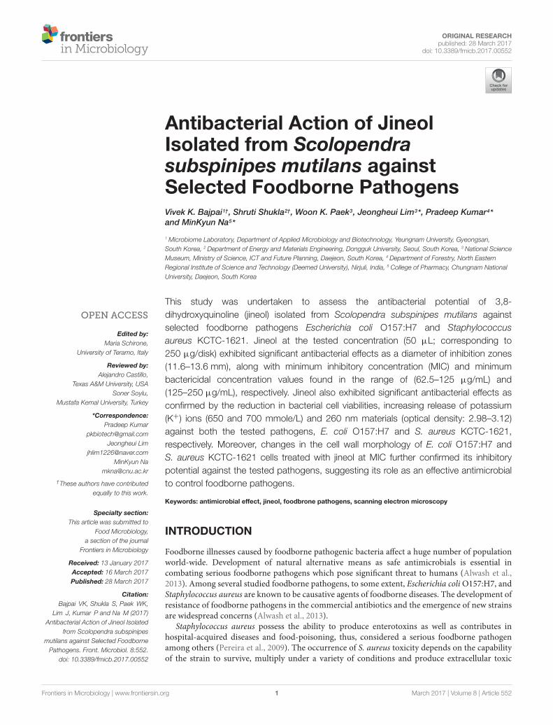
Antibacterial Action of JineolIsolated from Scolopendrasubspinipes mutilans againstSelected Foodborne PathogensVivek K. Bajpai1†, Shruti Shukla2†, Woon K. Paek3, Jeongheui Lim3*, Pradeep Kumar4*and MinKyun Na5*
1 Microbiome Laboratory, Department of Applied Microbiology and Biotechnology, Yeungnam University, Gyeongsan,South Korea, 2 Department of Energy and Materials Engineering, Dongguk University, Seoul, South Korea, 3 National ScienceMuseum, Ministry of Science, ICT and Future Planning, Daejeon, South Korea, 4 Department of Forestry, North EasternRegional Institute of Science and Technology (Deemed University), Nirjuli, India, 5 College of Pharmacy, Chungnam NationalUniversity, Daejeon, South Korea
This study was undertaken to assess the antibacterial potential of 3,8-dihydroxyquinoline (jineol) isolated from Scolopendra subspinipes mutilans againstselected foodborne pathogens Escherichia coli O157:H7 and Staphylococcusaureus KCTC-1621. Jineol at the tested concentration (50 µL; corresponding to250 µg/disk) exhibited significant antibacterial effects as a diameter of inhibition zones(11.6–13.6 mm), along with minimum inhibitory concentration (MIC) and minimumbactericidal concentration values found in the range of (62.5–125 µg/mL) and(125–250 µg/mL), respectively. Jineol also exhibited significant antibacterial effects asconfirmed by the reduction in bacterial cell viabilities, increasing release of potassium(K+) ions (650 and 700 mmole/L) and 260 nm materials (optical density: 2.98–3.12)against both the tested pathogens, E. coli O157:H7 and S. aureus KCTC-1621,respectively. Moreover, changes in the cell wall morphology of E. coli O157:H7 andS. aureus KCTC-1621 cells treated with jineol at MIC further confirmed its inhibitorypotential against the tested pathogens, suggesting its role as an effective antimicrobialto control foodborne pathogens.
Keywords: antimicrobial effect, jineol, foodbrone pathogens, scanning electron microscopy
INTRODUCTION
Foodborne illnesses caused by foodborne pathogenic bacteria affect a huge number of populationworld-wide. Development of natural alternative means as safe antimicrobials is essential incombating serious foodborne pathogens which pose significant threat to humans (Alwash et al.,2013). Among several studied foodborne pathogens, to some extent, Escherichia coli O157:H7, andStaphylococcus aureus are known to be causative agents of foodborne diseases. The development ofresistance of foodborne pathogens in the commercial antibiotics and the emergence of new strainsare widespread concerns (Alwash et al., 2013).
Staphylococcus aureus possess the ability to produce enterotoxins as well as contributes inhospital-acquired diseases and food-poisoning, thus, considered a serious foodborne pathogenamong others (Pereira et al., 2009). The occurrence of S. aureus toxicity depends on the capabilityof the strain to survive, multiply under a variety of conditions and produce extracellular toxic
Frontiers in Microbiology | www.frontiersin.org 1 March 2017 | Volume 8 | Article 552
fmicb-08-00552 March 25, 2017 Time: 12:55 # 2
Bajpai et al. Antibacterial Potential of Jineol
compounds. Contaminated food, especially undercooked groundbeef, raw milk, soft cheese, raw fruits and vegetables are majorsources of E. coli O157:H7 associated illness. E. coli O157:H7 hasthe ability to produce Shiga toxin, thereby causes bloody diarrheaand sometimes kidney failure (Du et al., 2008).
Consumers, the food industry and the regulatory authoritiesare concerned about the safety of food from the contaminationby foodborne pathogens (Al-Zorekya and Al-Taher, 2015).In the USA, foodborne pathogens have been reported tobe the cause of 75% of the foodborne disease outbreaks,involving 68% of all reported cases of foodborne illness (Scot,2003). Additionally, in Canada, a worth of approximately500 million dollars is imposed on the treatment of diseasescaused by foodborne pathogens surviving on meat or meat-products (Oussalah et al., 2007). In addition, failures inpreservation technologies to control foodborne and food spoilagepathogens have reinforced the suggestion for exploring othereffective classes of antimicrobials (Awaisheh and Ibrahim,2009; Negi, 2012). Furthermore, prevalence of synthetic andchemical additives in food and food products has urged anurgent need of application of natural preservatives to meetthe consumer acceptability (Shakiba et al., 2011; Shen et al.,2014).
Previous findings support the fact that the use of chemicalor synthetic food preservatives imposes a higher rate of healthcomplications, therefore, food processors are often looking forsafe and effective antimicrobials of natural origin for foodprotection and food preservation purposes (Taguri et al., 2004;Santas et al., 2010; Soylu et al., 2010). Also, development ofpathogen resistance to commercially available antimicrobialsevidences employment of innovative research strategies toexplore safe, and effective antimicrobial treatments (Militelloet al., 2011). Hence, the present study was designed to isolatea biologically effective 3,8-dihydroxyquinoline (jineol) from acentipede Scolopendra subspinipes mutilans, and to determineits antimicrobial potential against selected foodborne pathogenicbacteria.
MATERIALS AND METHODS
Chemicals and ReagentsThe nutrient broth (NB) medium was purchased from DifcoLtd., USA. Highly pure quality reagents and chemicals wereemployed for the test assays. Test samples of jineol were preparedin 1% dimethyl sulfoxide (DMSO) (Sigma–Aldrich, Germany).For absorbance reading, an enzyme-linked immunosorbent assay(ELISA) (Tecan, Infinite M200, Männedorf, Switzerland) wasused.
Test Foodborne PathogensStaphylococcus aureus KCTC1621 (Gram+), and E. coli O157:H7(Gram−) foodborne pathogens procured from the KoreanCollection for Type Cultures (KCTC, Korea) were used in thisstudy. For the growth and culture of strains, NB was used andcultures were incubated at 37◦C, followed by maintenance ofstrains on nutrient agar (NA) slants at 4◦C. To reactivate the
cultures, cultures were taken out and a loop-full of colonieswere inoculated in the fresh NB medium, and incubated for24 h and 37oC. Further, sub-culturing was maintained in NBmedium.
Insect MaterialDried Scolopendra subspinipes mutilans specimens werepurchased from the herbal market at Geumsan, South Korea,and identified by in-charge of the department. A voucherspecimen (CNU-INS 1408) was deposited at the PharmacognosyLaboratory of the College of Pharmacy, Chungnam NationalUniversity (Daejeon, South Korea).
Extraction, Isolation, andCharacterization of JineolJineol was isolated from specimens using a chromatographicapproach (Lee et al., 2016). Briefly, the dried ethanol (EtOH)extract (110.0 g) was suspended in water and fractionatedsuccessively with ethyl acetate (EtOAc) and then n-butanol(BuOH) to yield EtOAc-soluble (60.0 g) and n-BuOH-soluble(8.0 g) fractions, and residue (40.0 g). The EtOAc-soluble fractionwas subjected to vacuum-liquid chromatography (VLC) usinghexane-EtOAc 40:1, 20:1, 10:1, and 4:1; hexane-EtOAc-MeOH2:1:0.2; CHCl3-MeOH 6:1; CHCl3-MeOH-H2O 3:1:0.1 and thenwashed with MeOH to yield eight fractions (E1–E8). Fraction E4(8.0 g), which was obtained by eluting with hexane-EtOAc 4:1 waspartitioned with hexane and MeOH to afford jineol (60.0 mg).
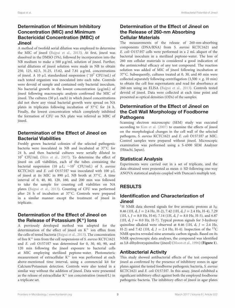
Jineol: yellowish amorphous powder; 1H NMR (300 MHz,CD3OD) δH 8.46 (1H, d, J = 2.4 Hz, H-2), 7.42 (1H, d, J = 2.4 Hz,H-4), 7.29 (1H, t, J = 8.0 Hz, H-6), 7.14 (1H, d, J = 8.0 Hz, H-5),6.87 (1H, d, J = 8.0 Hz, H-7), 13C NMR (75 MHz, CD3OD) δc154.2 (C-8), 153.0 (C-3), 142.3 (C-2), 134.8 (C-8a), 131.9 (C-4a),129.0 (C-6), 117.9 (C-5), 117.2 (C-4), 109.0 (C-7) (Moon et al.,1996).
Determination of Antibacterial Activity ofJineolA method of agar diffusion was employed to determine theantibacterial activity of jineol using Luria-Bertani (LB) agar plates(Bajpai and Kang, 2010). To make the desired concentrations (0,101, 102, 103, 104, 105, 106, 107, 108, and 109 cells/mL) of thetested strains, pathogens were cultured in NB medium followingincubation at 37◦C for 24 h and serially diluted. Further, a methodof the microbial plate-count was employed to determine theviable cell numbers. A 100 µL inoculum containing 107 CFU/mLwas poured on dried agar plates and spread uniformly using abacterial plate spreader followed by drying at room temperature.The compound was dissolved in 5% DMSO, and finally 50 µLjineol solution, corresponding to 250 µg/disk was impregnatedon a sterilized filter paper (Whatman No. 1) disk. The samesolvent used for dissolving the sample was also tested as a negativecontrol. Further, plates followed the incubation of 37◦C for 24 h,and zones of inhibition around the disks were measured toconfirm the antibacterial activity of test compound in triplicatemeasurements.
Frontiers in Microbiology | www.frontiersin.org 2 March 2017 | Volume 8 | Article 552
fmicb-08-00552 March 25, 2017 Time: 12:55 # 3
Bajpai et al. Antibacterial Potential of Jineol
Determination of Minimum InhibitoryConcentration (MIC) and MinimumBactericidal Concentration (MBC) ofJineolA method of twofold serial dilution was employed to determinethe MIC of jineol (Bajpai et al., 2013). At first, jineol wasdissolved in the DMSO (5%), followed by incorporation into theNB medium to make a 500 µg/mL solution of jineol. Further,serial dilutions of jineol solution were made in NB to obtain250, 125, 62.5, 31.25, 15.62, and 7.81 µg/mL concentrationsof jineol. A 10 µL standardized suspension (˜107 CFU/mL) ofeach tested organism was inoculated into each tube. Controlswere devoid of sample and contained only bacterial inoculum.No bacterial growth in the lowest concentration (µg/mL) ofjineol following macroscopic analysis confirmed the MIC ofjineol. The cultures (50 µL each) in which jineol concentrationsdid not show any visual bacterial growth were spread on NAplates in triplicates following incubation of 37◦C for 24 h.Finally, the lowest concentration which completely inhibitedthe formation of CFU on NA plate was referred as MBC ofjineol.
Determination of the Effect of Jineol onBacterial ViabilitiesFreshly grown bacterial colonies of the selected pathogenicbacteria were inoculated in NB and incubated at 37◦C for24 h, and then bacterial cultures were serially diluted to107 CFU/mL (Shin et al., 2007). To determine the effect ofjineol on cell viabilities, each of the tubes containing thebacterial suspension (10 µL; ∼107 CFU/mL) of S. aureusKCTC1621 and E. coli O157:H7 was inoculated with 100 µLof jineol at its MIC in 890 µL NB broth at 37◦C. A timeinterval of 0, 40, 80, 120, 160, and 200 min was followedto take the sample for counting cell viabilities on NAplates (Bajpai et al., 2013). Counting of CFU was performedafter 24 h of incubation at 37◦C. Controls were preparedin a similar manner except the treatment of jineol intriplicate.
Determination of the Effect of Jineol onthe Release of Potassium (K+) IonsA previously developed method was adopted for thedetermination of the effect of jineol on K+ ion efflux fromthe cells of tested bacteria (Bajpai et al., 2013). The concentrationof free K+ ions from the cell suspensions of S. aureus KCTC1621and E. coli O157:H7 was determined for 0, 30, 60, 90, and120 min following the jineol exposure to bacterial cellsat MIC employing sterilized peptone-water. Photometericmeasurement of extracellular K+ ion was performed at eachabove-mentioned time interval, using a commercial kit forCalcium/Potassium detection. Controls were also tested in asimilar way without the addition of jineol. Data were presentedas the release of extracellular K+ ion concentration (mmol/L) ina triplicate set.
Determination of the Effect of Jineol onthe Release of 260-nm AbsorbingCellular MaterialsThe measurements of the release of 260-nm-absorbingcomponents (DNA/RNA) from S. aureus KCTC1621 andE. coli O157:H7 cells were performed in a 2 mL aliquot of thebacterial inoculum in a sterilized peptone-water. The loss of260 nm cellular materials is considered a good indication ofthe antimicrobial efficacy of any test compound. The reactionsolution was added of MIC of jineol following incubation at37◦C. Subsequently, cultures treated at 0, 30, and 60 min werecollected separately following centrifugation (3,500 × g, 10 min)to obtain the cell free supernatants and read for absorbance at260-nm using an ELISA (Bajpai et al., 2013). Controls testeddevoid of jineol. Data were collected at each time point andpresented as optical densities (ODs) of the samples.
Determination of the Effect of Jineol onthe Cell Wall Morphology of FoodbornePathogensScanning electron microscopic (SEM) study was executedaccording to Kim et al. (2007) to examine the effects of jineolon the morphological changes in the cell wall of the selectedpathogens, S. aureus KCTC1621 and E. coli O157:H7 at MIC.Control samples were prepared without jineol. Microscopicexamination was performed using a S-4300 SEM Analyzer(Hitachi, Japan).
Statistical AnalysisExperiments were carried out in a set of triplicate, and thedata obtained were presented as mean ± SD following one-wayANOVA statistical analysis coupled with Duncan’s multiple test.
RESULTS
Identification and Characterization ofJineol1H NMR data showed signals for five aromatic protons at δH8.46 (1H, d, J = 2.4 Hz, H-2), 7.42 (1H, d, J = 2.4 Hz, H-4), 7.29(1H, t, J = 8.0 Hz, H-6), 7.14 (1H, d, J = 8.0 Hz, H-5), and 6.87(1H, d, J = 8.0 Hz, H-7). Typical proton signals for 3-hydroxyquinoline alkaloid were observed at 8.46 (1H, d, J = 2.4 Hz,H-2) and 7.42 (1H, d, J = 2.4 Hz, H-4). Inspection of the 13CNMR spectra revealed nine aromatic carbon signals. Based on itsNMR spectroscopic data analyses, the compound was identifiedas 3,8-dihydroxyquinoline (jineol) (Moon et al., 1996) (Figure 1).
Antibacterial ActivityThis study showed antibacterial effects of the test compoundjineol as confirmed by the presence of inhibitory zones in agarplates against the tested foodborne pathogenic bacteria, S. aureusKCTC1621 and E. coli O157:H7. In this assay, jineol exhibited asignificant inhibitory effect against both the employed foodbornepathogenic bacteria. The inhibitory effect of jineol in agar plates
Frontiers in Microbiology | www.frontiersin.org 3 March 2017 | Volume 8 | Article 552
fmicb-08-00552 March 25, 2017 Time: 12:55 # 4
Bajpai et al. Antibacterial Potential of Jineol
FIGURE 1 | Chemical structure of jineol isolated from Scolopendrasubspinipes mutilans.
TABLE 1 | Antibacterial activity of jineol against foodborne pathogensStaphylococcus aureus KCTC1621 and Escherichia coli O157:H7.
Pathogens Jineol
Zones of inhibitionx Susceptibility
MICy zMBC
Staphylococcus aureus KCTC1621 13.6 ± 0.2a 62.5a 125a
Escherichia coli O157:H7 11.6 ± 0.3b 125b 250b
xDiameters of inhibition zones in millimeter; yMinimum inhibitory concentration(values in µg/mL); zMinimum bactericidal concentration (values in µg/mL). Values inthe same column with different superscripts are significantly different according toDuncan’s Multiple Range Test (P < 0.05). All values were expressed as mean ± SDof three parallel measurements (n = 3). Zones of inhibition around the disk weremeasured in millimeter (mm) using a Vernier’s caliper.
was confirmed through the diameters of zones of inhibition,which were found to be 11.6–13.6 mm (Table 1). It was observedthat jineol exhibited antibacterial effects against both the testedbacterial isolates.
MIC and MBCThis assay revealed different susceptibilities of test compoundjineol against the tested foodborne pathogens as confirmed by thelow and variable MIC and MBC values. As a result, the MIC andMBC values of jineol against the tested pathogens were ranged62.5–125, and 125–250 µg/mL, respectively (Table 1). In thisassay, it was observed that jineol had inhibitory effects againstboth Gram-positive and Gram-negative bacteria.
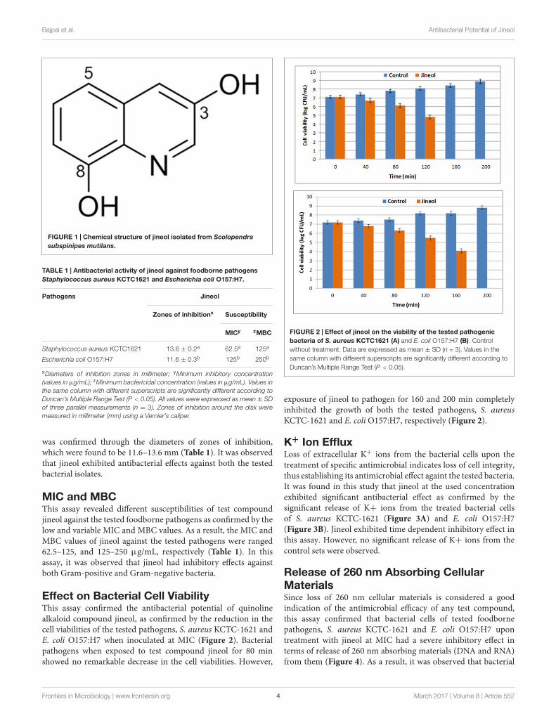
Effect on Bacterial Cell ViabilityThis assay confirmed the antibacterial potential of quinolinealkaloid compound jineol, as confirmed by the reduction in thecell viabilities of the tested pathogens, S. aureus KCTC-1621 andE. coli O157:H7 when inoculated at MIC (Figure 2). Bacterialpathogens when exposed to test compound jineol for 80 minshowed no remarkable decrease in the cell viabilities. However,
FIGURE 2 | Effect of jineol on the viability of the tested pathogenicbacteria of S. aureus KCTC1621 (A) and E. coli O157:H7 (B). Controlwithout treatment. Data are expressed as mean ± SD (n = 3). Values in thesame column with different superscripts are significantly different according toDuncan’s Multiple Range Test (P < 0.05).
exposure of jineol to pathogen for 160 and 200 min completelyinhibited the growth of both the tested pathogens, S. aureusKCTC-1621 and E. coli O157:H7, respectively (Figure 2).
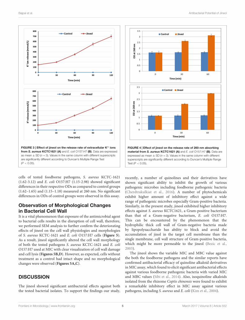
K+ Ion EffluxLoss of extracellular K+ ions from the bacterial cells upon thetreatment of specific antimicrobial indicates loss of cell integrity,thus establishing its antimicrobial effect againt the tested bacteria.It was found in this study that jineol at the used concentrationexhibited significant antibacterial effect as confirmed by thesignificant release of K+ ions from the treated bacterial cellsof S. aureus KCTC-1621 (Figure 3A) and E. coli O157:H7(Figure 3B). Jineol exhibited time dependent inhibitory effect inthis assay. However, no significant release of K+ ions from thecontrol sets were observed.
Release of 260 nm Absorbing CellularMaterialsSince loss of 260 nm cellular materials is considered a goodindication of the antimicrobial efficacy of any test compound,this assay confirmed that bacterial cells of tested foodbornepathogens, S. aureus KCTC-1621 and E. coli O157:H7 upontreatment with jineol at MIC had a severe inhibitory effect interms of release of 260 nm absorbing materials (DNA and RNA)from them (Figure 4). As a result, it was observed that bacterial
Frontiers in Microbiology | www.frontiersin.org 4 March 2017 | Volume 8 | Article 552
fmicb-08-00552 March 25, 2017 Time: 12:55 # 5
Bajpai et al. Antibacterial Potential of Jineol
FIGURE 3 | Effect of jineol on the release rate of extracellular K+ ionsfrom S. aureus KCTC1621 (A) and E. coli O157:H7 (B). Data are expressedas mean ± SD (n = 3). Values in the same column with different superscriptsare significantly different according to Duncan’s Multiple Range Test(P < 0.05).
cells of tested foodborne pathogens, S. aureus KCTC-1621(1.62-3.12) and E. coli O157:H7 (1.15-2.98) showed significantdifferences in their respective ODs as compared to control groups(1.62–1.65) and (1.15–1.18) measured at 260 nm. No significantdifferences in ODs of control groups were observed in this assay.
Observation of Morphological Changesin Bacterial Cell WallIt is a vital phenomenon that exposure of the antimicrobial agentto bacterial cells results in the disruption of cell wall, therefore,we performed SEM analysis to further confirm the deterioratingeffects of jineol on the cell wall physiologies and morphologiesof S. aureus KCTC-1621 and E. coli O157:H7 cells (Figure 5).As a result, jineol significantly altered the cell wall morphologyof both the tested pathogens S. aureus KCTC-1621 and E. coliO157:H7 used at MIC with clear visualization of cell wall damageand cell lysis (Figures 5B,D). However, as expected, cells withouttreatment as a control had intact shape and no morphologicalchanges were observed (Figures 5A,C).
DISCUSSION
The jineol showed significant antibacterial effects against boththe tested bacterial isolates. To support the findings our study,
FIGURE 4 | Effect of jineol on the release rate of 260-nm absorbingmaterial from S. aureus KCTC1621 (A) and E. coli O157:H7 (B). Data areexpressed as mean ± SD (n = 3). Values in the same column with differentsuperscripts are significantly different according to Duncan’s Multiple RangeTest (P < 0.05).
recently, a number of quinolines and their derivatives haveshown significant ability to inhibit the growth of variouspathogenic microbes including foodborne pathogenic bacteria(Cherdtrakulkiat et al., 2016). A number of phytochemicalsexhibit higher amount of inhibitory effect against a widerange of pathogenic microbes especially Gram-positive bacteria.Similarly, in the present study, jineol exhibited higher inhibitoryeffects against S. aureus KCTC1621, a Gram-positive bacteriumthan that of a Gram-negative bacterium, E. coli O157:H7.This can be encountered by the phenomenon that thehydrophilic thick cell wall of Gram-negative bacteria madeby lipopolysaccharide has ability to block and avoid theaccumulation of jinol in the target cell membrane than thesingle membrane, cell wall structure of Gram-positive bacteria,which might be more permeable to the jineol (Bezic et al.,2003).
The jineol shows the variable MIC and MBC value againstthe both the foodborne pathogens and the similar reports haveconfirmed antibacterial efficacy of quinoline alkaloid derivativesin MIC assay, which found to elicit significant antibacterial effectsagainst various foodborne pathogenic bacteria with varied MICand MBC values (Sibi et al., 2014). Also, isoquinoline alkaloidsisolated from the rhizome Coptis chinensis were found to exhibita remarkable inhibitory effect in MIC assay against variouspathogens, including S. aureus and E. coli (Kim et al., 2004).
Frontiers in Microbiology | www.frontiersin.org 5 March 2017 | Volume 8 | Article 552

fmicb-08-00552 March 25, 2017 Time: 12:55 # 6
Bajpai et al. Antibacterial Potential of Jineol
FIGURE 5 | Scanning electron microscopic (SEM) analysis of E. coli0157:H7 and S. aureus KCTC-1621 cells treated with jineol at minimuminhibitory concentration (MIC). Controls (A,C) showing a regular andsmooth surface; whereas treated cells (B,D) arrows showing disruption andcell lysis, respectively.
The exposure of jineol to pathogen for 160 and 200 mincompletely inhibited the growth of both the tested pathogens.Previous reports have confirmed the inhibitory effects of variousphytochemicals isolated from different sources on the cellviabilities of foodborne pathogenic bacteria (Rastogi et al., 2008;Bajpai and Kang, 2010).
The significant release of K+ ions from the jineol treatedbacterial cells and no release of K+ ions from the control setswere observed. Similar findings were observed when Patra et al.(2015) tested the efficacy of bio-oil on the release of extracellularK+ ions from Bacillus cereus and Listeria monocytogenes cells.Permeability of essential ions such as K+ ions is regulated by thebacterial plasma membrane where membrane chemo-structuralcomposition plays a significant role on the release of such ions,and extensive release of these ions from the target bacterial cellscould be considered as a disruptive effect of treatment agent onthe plasma membrane, hence, validating the antimicrobial role ofjineol, as also reported previously (Cox et al., 2001).
This study validated that the increasing release of 260-nmabsorbing cellular materials from the target bacterial cellsmediated by jineol had a huge influence on the release ofcellular metabolites indicating structural damage to the plasmamembrane, thereby causing cell death. Such findings on specificantimicrobials have also been confirmed previously (Farag et al.,
1989). The results clearly indicate that regulation of nucleicacid or cellular materials from the tested pathogens couldbe considered a significant indication of membrane structuraldamage. Similar to our findings, Souza et al. (2013) tested theantibacterial efficacy of essential oil components, carvacrol andthymol against foodborne pathogens, and it was noted that boththe compounds were able to cause membrane damaging effects,and eventually caused efflux of 260-nm nuclear components.
Using SEM analysis, it was confirmed that upon treatmentwith jineol, morphology of treated bacterial cells was dramaticallychanged or hampered. Similar reports on morphologicaldeteriorating effects of several phytochemicals have beenobserved previously against various foodborne pathogens (Soyluet al., 2007; Alwash et al., 2013). Such morphological andphysiological changes could be caused by the changes in theintegrity and fluidity of plasma membrane as well as changes inthe composition of the lipid profile of plasma membrane directlyhampered by the antimicrobial action of treating agent (Sikkemaet al., 1994).
CONCLUSION
This study reports isolation of a quinoline compound jineolfrom Scolopendra subspinipes mutilans which was enough ableto exhibit significant inhibitory effects against two selectedfoodborne pathogens. The findings of this study suggest thatthe antimicrobial action of jineol could be mediated through itsefficacy to alter cell membrane permeability parameters of thetested pathogens as demonstrated by the excessive release of K+ions and 260-nm absorbing materials, thus causing membranedisruption as also confirmed during SEM analysis. We concludethat jineol, exhibiting significant antibacterial activity againstselected foodborne pathogens could be potential efficacy forusing in food and pharma industries as an alternative means ofantimicrobial.
AUTHOR CONTRIBUTIONS
Author VB and SS designed performed the experiments and writethe manuscript. WP assist during experimental work. PK helps inmanuscript preparation and editing. JL and MN help in editingand finalizing the manuscript.
ACKNOWLEDGMENT
This work was supported by National Research Foundation ofKorea (2013M3A9A5047052, 2008-2004707 and 2012-0006701).
REFERENCESAlwash, M. S., Ibrahim, N., and Ahmad, W. Y. (2013). Identification and mode
of action of antibacterial components from Melastoma malabathricumLinn leaves. Am. J. Infect. Dis. 9, 46–58. doi: 10.3844/ajidsp.2013.46.58
Al-Zorekya, N. S., and Al-Taher, A. Y. (2015). Antibacterial activity of spathe fromPhoenix dactylifera L. against some food-borne pathogens. Ind. Crops Prod. 65,241–246. doi: 10.1016/j.indcrop.2014.12.014
Awaisheh, S. S., and Ibrahim, S. A. (2009). Screening of antibacterial activity oflactic acid bacteria against different pathogens found in vacuum-packaged meatproducts. Foodborne Pathog. Dis. 6, 1125–1132. doi: 10.1089/fpd.2009.0272
Frontiers in Microbiology | www.frontiersin.org 6 March 2017 | Volume 8 | Article 552

fmicb-08-00552 March 25, 2017 Time: 12:55 # 7
Bajpai et al. Antibacterial Potential of Jineol
Bajpai, V. K., and Kang, S. C. (2010). Antibacterial abietane-type diterpenoid,taxodone from Metasequoia glyptostroboides Miki ex Hu. J. Biosci. 35, 533–538.doi: 10.1007/s12038-010-0061-z
Bajpai, V. K., Sharma, A., and Baek, K. H. (2013). Antibacterial mode of actionof Cudrania tricuspidata fruit essential oil, affecting membrane permeabilityand surface characteristics of food-borne pathogens. Food Control 32, 582–590.doi: 10.1016/j.foodcont.2013.01.032
Bezic, N., Skocibusic, M., Dinkic, V., and Radonic, A. (2003). Composition andantimicrobial activity of Achillea clavennae L. essential oil. Phytother. Res. 17,1037–1040. doi: 10.1002/ptr.1290
Cherdtrakulkiat, R., Boonpangrak, S., Sinthupoom, N., Prachayasittikul, S.,Ruchirawat, S., and Prachayasittikul, V. (2016). Derivatives (halgen, nitroand amino) of 8-hydroxyquinoline with highly potent antimicrobial andantioxidant activities. Biochem. Biophys. Rep. 6, 135–141.
Cox, S. D., Mann, C. M., Markhan, J. L., Gustafson, J. E., Warmington, J. R.,and Wyllie, S. G. (2001). Determining the antimicrobial action of tea tree oil.Molecules 6, 87–91. doi: 10.3390/60100087
Du, W. X., Olsen, C. W., Avena-Bustillos, R. J., Mchugh, T. H., Levin, C. E., andFriedman, M. (2008). Antibacterial activity against E. coli O157:H7, physicalproperties, and storage stability of novel carvacrol-containing edible tomatofilms. J. Food Sci. 73, 378–383. doi: 10.1111/j.1750-3841.2008.00892.x
Farag, R. S., Daw, Z. Y., Hewedi, F. M., and El-Baroty, G. S. A. (1989).Antimicrobial activity of some Egyptian spice essential oil. J. Food Prot. 52,665–667. doi: 10.1021/JF101620C
Kim, J. E., Choi, N. H., and Kang, S. C. (2007). Anti-listerial properties of garlicshoot juice at growth and morphology of Listeria monocytogenes. Food Control18, 1198–1203. doi: 10.1016/j.foodcont.2006.07.017
Kim, S. H., Shin, D. S., Oh, M. N., Chung, S. C., Lee, J. S., and Oh, K. B. (2004).Inhibition of the bacterial surface protein anchoring transpeptidase sortase byisoquinoline alkaloids. Biosci. Biotechnol. Biochem. 68, 421–424. doi: 10.1271/bbb.68.421
Lee, W., Lee, J., Kulkarni, R., Kim, M. A., Hwang, J. S., Na, M., et al. (2016).Antithrombotic and antiplatelet activities of small-molecule alkaloids fromScolopendra subspinipes mutilans. Sci. Rep. 6:e21956. doi: 10.1038/srep21956
Militello, M., Settanni, L., Aleo, A., Mammina, C., Moschetti, G., Giammanco,G. M., et al. (2011). Chemical composition and antibacterial potentialof Artemisia arborescens L. essential oil. Curr. Microbiol. 62, 1274–1281.doi: 10.1007/s00284-010-9855-3
Moon, S. S., Cho, N., Shin, J., Seo, Y., Lee, C. O., and Choi, S. U. (1996). Jineol, acytotoxic alkaloid from the centipede Scolopendra subspinipes. J. Nat. Prod. 59,777–779. doi: 10.1021/np960188t
Negi, P. S. (2012). Plant extracts for the control of bacterial growth: efficacy,stability and safety issues for food application. Int. J. Food Microbiol. 156, 7–17.doi: 10.1016/j.ijfoodmicro.2012.03.006
Oussalah, M., Cailet, S., Soucier, I., and Iacroix, M. (2007). Inhibitory effectof selected plant essential oils on the growth of four pathogenic bacteria:E. coli O157:H7, Salmonella typhimurium, Staphylococcus aureus and Listeriamonocytogenes. Food Control 18, 414–420. doi: 10.1016/j.foodcont.2005.11.009
Patra, J. K., Hwang, H., Choi, J. W., and Baek, K. H. (2015). Bactericidal mechanismof bio-oil obtained from fast pyrolysis of Pinus densiflora against two foodbornepathogens, Bacillus cereus and Listeria monocytogenes. Foodborne Pathog. Dis.12, 529–535. doi: 10.1089/fpd.2014.1914
Pereira, V., Lopes, C., Castro, A., Silva, J., Gibbs, P., and Teixeira, P. (2009).Characterization for enterotoxin production, virulence factors, and antibioticsusceptibility of Staphylococcus aureus isolated from various foods in Portugal.Food Microbiol. 26, 278–282. doi: 10.1016/j.fm.2008.12.008
Rastogi, N., Domadia, P., Shetty, S., and Dasgupta, D. (2008). Screening of naturalphenolic compounds for potential to inhibit bacterial cell division protein FtsZ.Ind. J. Exp. Biol. 46, 783–787.
Santas, J., Almajano, M., and Carbó, R. (2010). Antimicrobial and antioxidantactivity of crude onion (Allium cepa L.) extracts. Int. J. Food Sci. Technol. 45,403–409. doi: 10.1111/j.1365-2621.2009.02169.x
Scot, E. (2003). Food safety and foodborne disease in 21st century homes. Can. J.Microbiol. 14, 277–280.
Shakiba, M., Kariminik, A., and Parsia, P. (2011). Antimicrobial activity of differentparts of Phoenix dactylifera. Int. J. Mol. Clin. Microbiol. 1, 107–111.
Shen, X., Sun, X., Xie, Q., Liu, H., Zhao, Y., Pan Hwang, Y., et al. (2014).Antimicrobial effect of blueberry (Vaccinium corymbosum L.) extracts againstthe growth of Listeria monocytogenes and Salmonella enteritidis. Food Control35, 159–165. doi: 10.1016/j.foodcont.2013.06.040
Shin, S. Y., Bajpai, V. K., Kim, H. R., and Kang, S. C. (2007). Antibacterial activityof bioconverted eicosapentaenoic (EPA) and docosahexaenoic acid (DHA)against foodborne pathogenic bacteria. Int. J. Food Microbiol. 113, 233–236.doi: 10.1016/j.ijfoodmicro.2006.05.020
Sibi, G., Venkategowda, A., and Gowda, L. (2014). Isolation and characterizationof antimicrobial alkaloids from Plumeria alba flowers against foodbornepathogens. Am. J. Life Sci. 2, 1–6.
Sikkema, J., De Bont, J. A. M., and Poolman, B. (1994). Interactions of cyclichydrocarbons with biological membranes. J. Biol. Chem. 269, 8022–8028.
Souza, E. L., Oliveira, C. E. V., Stamford, T. L. M., Conceicao, M. L., and GomesNeto, N. J. (2013). Influence of carvacrol and thymol on the physiologicalattributes, enterotoxin production and surface characteristics of Staphylococcusaureus strains isolated from foods. Braz. J. Microbiol. 44, 29–35. doi: 10.1590/S1517-83822013005000001
Soylu, E. M., Kurt, S., and Soylu, S. (2010). In vitro and in vivo antifungal activitiesof the essential oils of various plants against tomato grey mould disease agentBotrytis cinerea. Int. J. Food Microbiol. 143, 183–189. doi: 10.1016/j.ijfoodmicro.2010.08.015
Soylu, S., Yigitbas, H., Soylu, E. M., and Kurt, S. (2007). Antifungal effects ofessential oils from oregano and fennel on Sclerotinia sclerotiorum. J. Appl.Microbiol. 103, 1021–1030. doi: 10.1111/j.1365-2672.2007.03310.x
Taguri, T., Tanaka, T., and Kouno, I. (2004). Antimicrobial activity of 10 differentplant polyphenols against bacteria causing food-borne disease. Biol. Pharm.Bull. 27, 1965–1969. doi: 10.1248/bpb.27.1965
Conflict of Interest Statement: The authors declare that the research wasconducted in the absence of any commercial or financial relationships that couldbe construed as a potential conflict of interest.
Copyright © 2017 Bajpai, Shukla, Paek, Lim, Kumar and Na. This is an open-accessarticle distributed under the terms of the Creative Commons Attribution License(CC BY). The use, distribution or reproduction in other forums is permitted, providedthe original author(s) or licensor are credited and that the original publication in thisjournal is cited, in accordance with accepted academic practice. No use, distributionor reproduction is permitted which does not comply with these terms.
Frontiers in Microbiology | www.frontiersin.org 7 March 2017 | Volume 8 | Article 552