Embed Size (px)
Citation preview

1/00 medslides.com 1
Objectives
• Clinical Assessment
• Stress Testing
• Treatment
• Patient Follow Up
JACC 1999; 33, 7:2092-2197Circulation 1999;99::2829-2848
http://www.acc.org/clinical/guidelines/index.html

1/00 medslides.com 2
Definition of Angina
A pain or discomfort in the chest or adjacent areas caused by insufficient blood flow to the heart muscle.
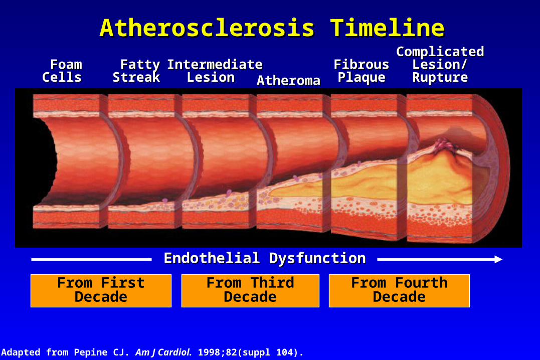
Atherosclerosis TimelineAtherosclerosis TimelineFoamFoamCells Cells
FattyFattyStreak Streak
IntermediateIntermediateLesion Lesion AtheromaAtheroma
FibrousFibrousPlaquePlaque
ComplicatedComplicatedLesion/Lesion/RuptureRupture
Adapted from Pepine CJ. Am J Cardiol. 1998;82(suppl 104).
From FirstDecade
From ThirdDecade
From FourthDecade
Endothelial DysfunctionEndothelial Dysfunction
1/00 medslides.com 4
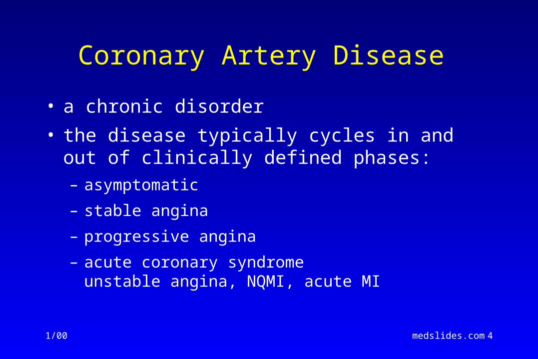
Coronary Artery Disease
• a chronic disorder
• the disease typically cycles in and out of clinically defined phases: – asymptomatic
– stable angina
– progressive angina
– acute coronary syndromeunstable angina, NQMI, acute MI
1/00 medslides.com 5
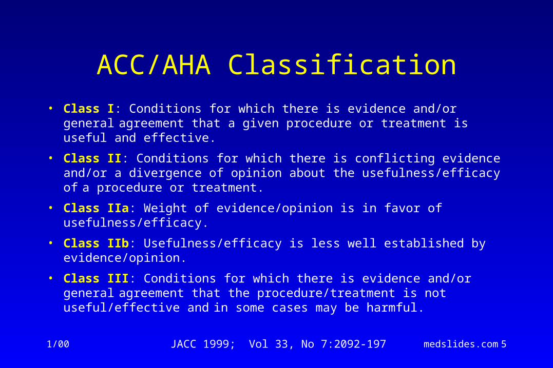
ACC/AHA Classification
• Class I: Conditions for which there is evidence and/or general
agreement that a given procedure or treatment is useful and effective.
• Class II: Conditions for which there is conflicting evidence and/or a divergence of opinion about the usefulness/efficacy of a procedure or treatment.
• Class IIa: Weight of evidence/opinion is in favor of usefulness/efficacy.
• Class IIb: Usefulness/efficacy is less well established by
evidence/opinion.
• Class III: Conditions for which there is evidence and/or general
agreement that the procedure/treatment is not useful/effective and in some cases may be harmful.
JACC 1999; Vol 33, No 7:2092-197
1/00 medslides.com 6
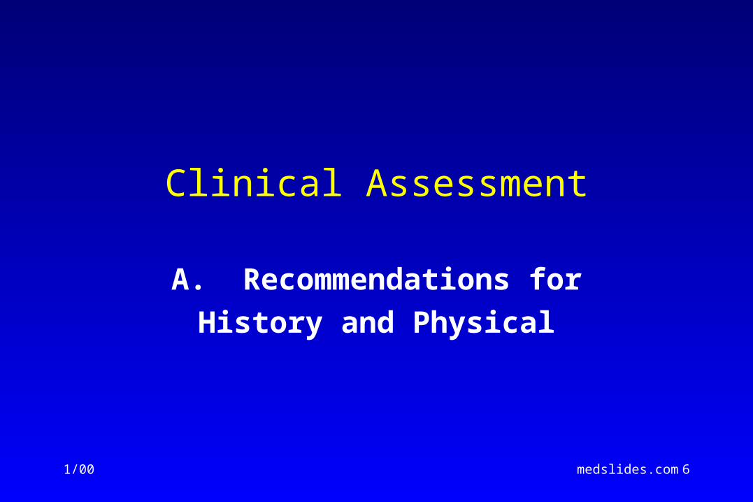
Clinical Assessment
A. Recommendations for
History and Physical
1/00 medslides.com 7
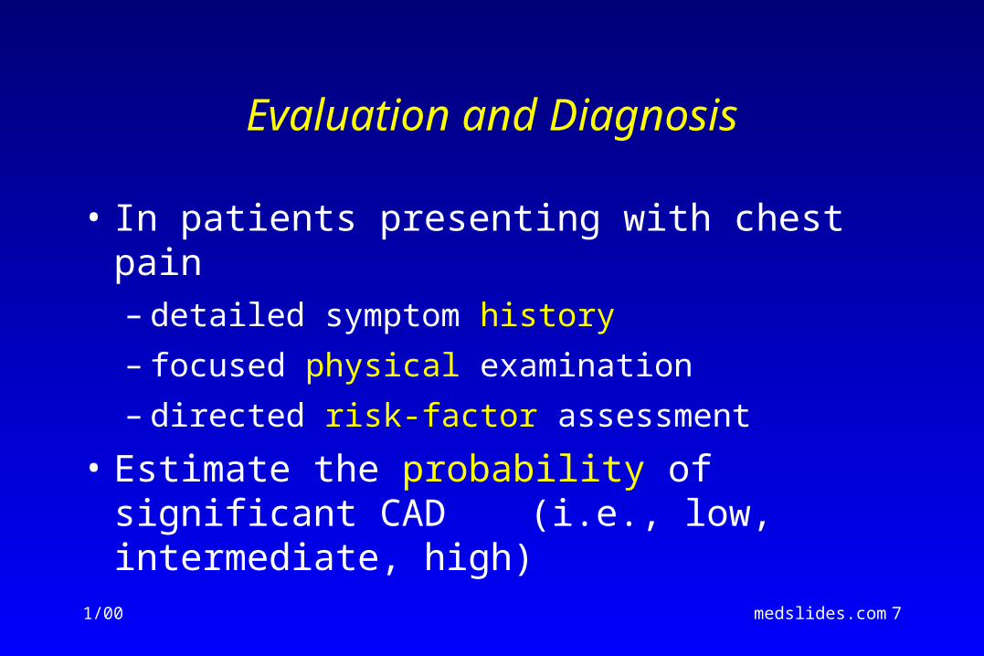
Evaluation and Diagnosis
• In patients presenting with chest pain– detailed symptom history
– focused physical examination
– directed risk-factor assessment
• Estimate the probability of significant CAD (i.e., low, intermediate, high)
1/00 medslides.com 8
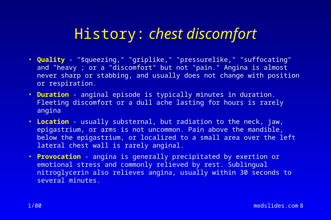
History: chest discomfort
• Quality - "squeezing," "griplike," "pressurelike," "suffocating" and "heavy”; or a "discomfort" but not "pain." Angina is almost never sharp or stabbing, and usually does not change with position or respiration.
• Duration - anginal episode is typically minutes in duration. Fleeting discomfort or a dull ache lasting for hours is rarely angina
• Location - usually substernal, but radiation to the neck, jaw, epigastrium, or arms is not uncommon. Pain above the mandible, below the epigastrium, or localized to a small area over the left lateral chest wall is rarely anginal.
• Provocation - angina is generally precipitated by exertion or emotional stress and commonly relieved by rest. Sublingual nitroglycerin also relieves angina, usually within 30 seconds to several minutes.
1/00 medslides.com 9
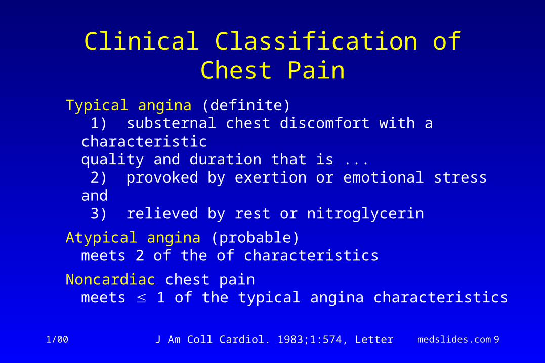
Clinical Classification of Chest Pain
Typical angina (definite) 1) substernal chest discomfort with a characteristic
quality and duration that is ... 2) provoked by exertion or emotional stress and 3) relieved by rest or nitroglycerin
Atypical angina (probable)meets 2 of the of characteristics
Noncardiac chest painmeets 1 of the typical angina characteristics
J Am Coll Cardiol. 1983;1:574, Letter
1/00 medslides.com 10
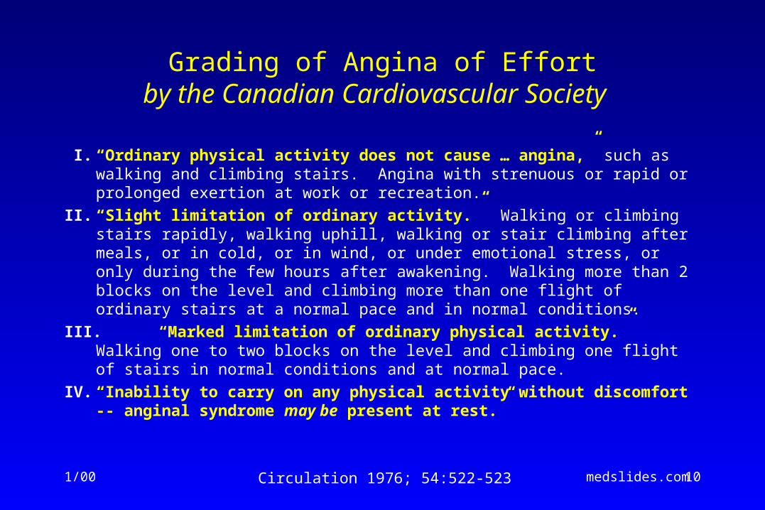
Grading of Angina of Effortby the Canadian Cardiovascular Society
I. “Ordinary physical activity does not cause … angina,” such as walking and climbing stairs. Angina with strenuous or rapid or prolonged exertion at work or recreation.
II. “Slight limitation of ordinary activity.” Walking or climbing stairs rapidly, walking uphill, walking or stair climbing after meals, or in cold, or in wind, or under emotional stress, or only during the few hours after awakening. Walking more than 2 blocks on the level and climbing more than one flight of ordinary stairs at a normal pace and in normal conditions.
III. “Marked limitation of ordinary physical activity.” Walking one to two blocks on the level and climbing one flight of stairs in normal conditions and at normal pace.
IV. “Inability to carry on any physical activity without discomfort -- anginal syndrome may be present at rest.”
Circulation 1976; 54:522-523
1/00 medslides.com 11
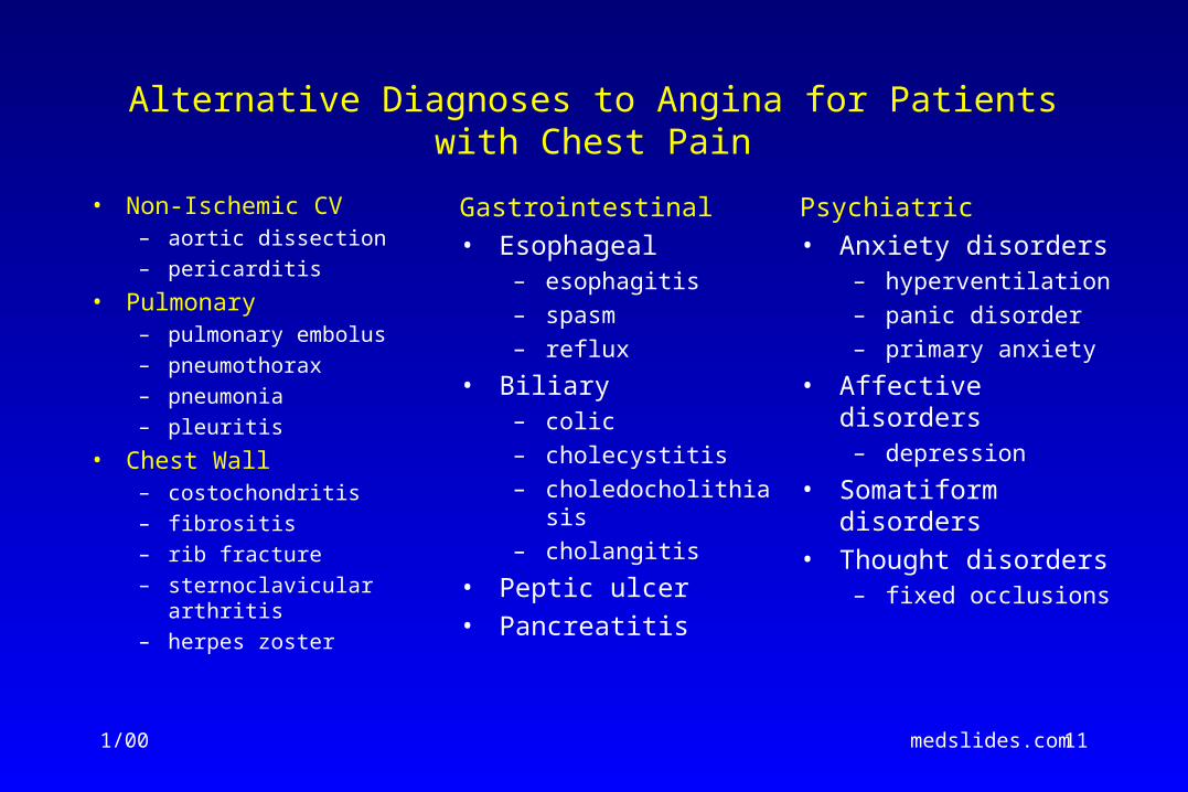
Alternative Diagnoses to Angina for Patients with Chest Pain
• Non-Ischemic CV– aortic dissection– pericarditis
• Pulmonary– pulmonary embolus– pneumothorax– pneumonia– pleuritis
• Chest Wall– costochondritis– fibrositis– rib fracture– sternoclavicular arthritis– herpes zoster
Gastrointestinal
• Esophageal– esophagitis– spasm– reflux
• Biliary– colic– cholecystitis– choledocholithiasis– cholangitis
• Peptic ulcer
• Pancreatitis
Psychiatric
• Anxiety disorders– hyperventilation– panic disorder– primary anxiety
• Affective disorders– depression
• Somatiform disorders
• Thought disorders– fixed occlusions
1/00 medslides.com 12
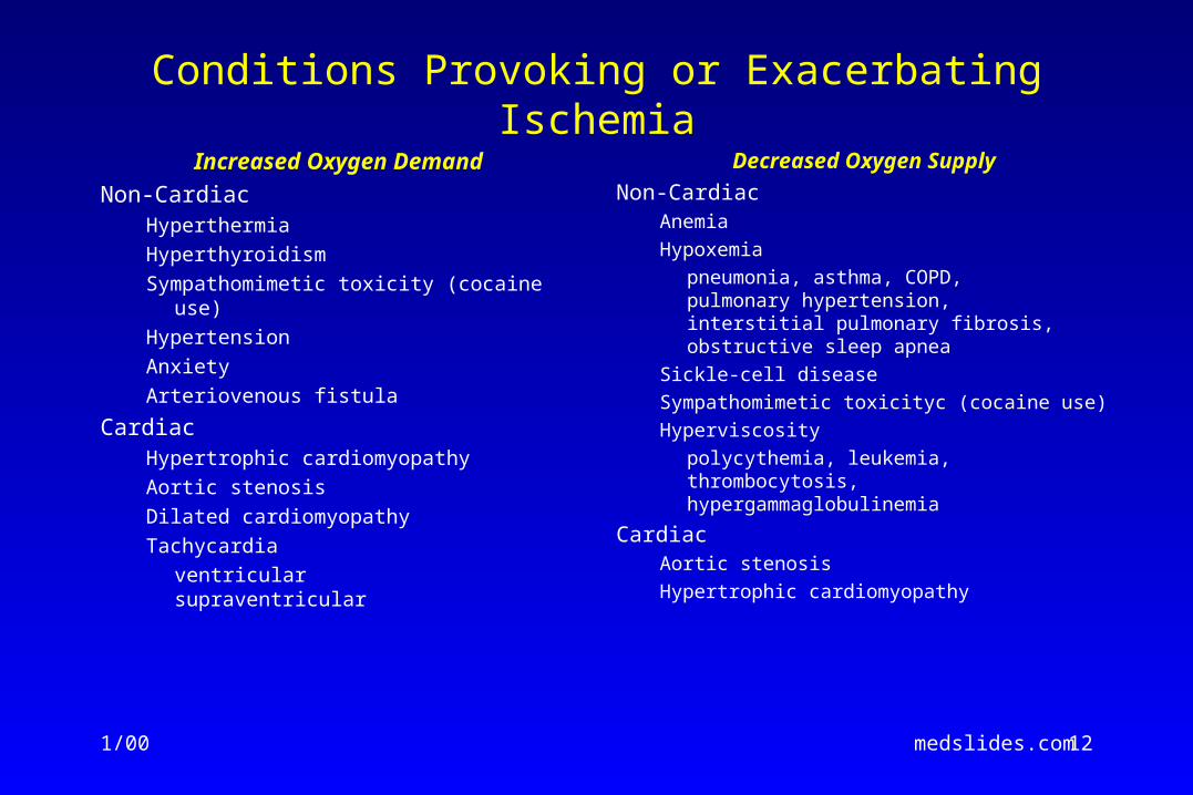
Conditions Provoking or Exacerbating Ischemia
Increased Oxygen Demand
Non-CardiacHyperthermia
Hyperthyroidism
Sympathomimetic toxicity (cocaine use)
Hypertension
Anxiety
Arteriovenous fistula
CardiacHypertrophic cardiomyopathy
Aortic stenosis
Dilated cardiomyopathy
Tachycardia
ventricularsupraventricular
Decreased Oxygen Supply
Non-CardiacAnemia
Hypoxemia
pneumonia, asthma, COPD,pulmonary hypertension,interstitial pulmonary fibrosis,obstructive sleep apnea
Sickle-cell disease
Sympathomimetic toxicityc (cocaine use)
Hyperviscosity
polycythemia, leukemia,thrombocytosis, hypergammaglobulinemia
CardiacAortic stenosis
Hypertrophic cardiomyopathy
1/00 medslides.com 13
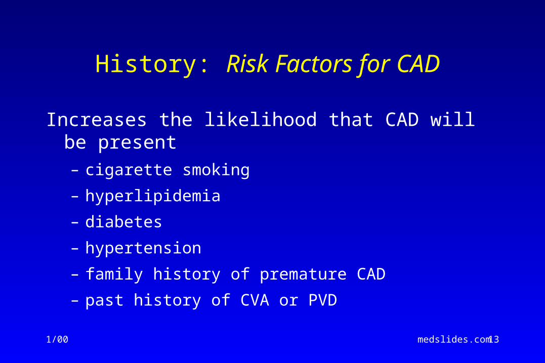
History: Risk Factors for CAD
Increases the likelihood that CAD will be present– cigarette smoking
– hyperlipidemia
– diabetes
– hypertension
– family history of premature CAD
– past history of CVA or PVD
1/00 medslides.com 14
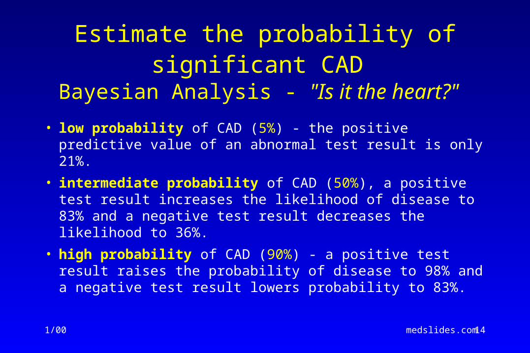
Estimate the probability of significant CAD
Bayesian Analysis - "Is it the heart?"
• low probability of CAD (5%) - the positive predictive value of an abnormal test result is only 21%.
• intermediate probability of CAD (50%), a positive test result increases the likelihood of disease to 83% and a negative test result decreases the likelihood to 36%.
• high probability of CAD (90%) - a positive test result raises the probability of disease to 98% and a negative test result lowers probability to 83%.
1/00 medslides.com 15
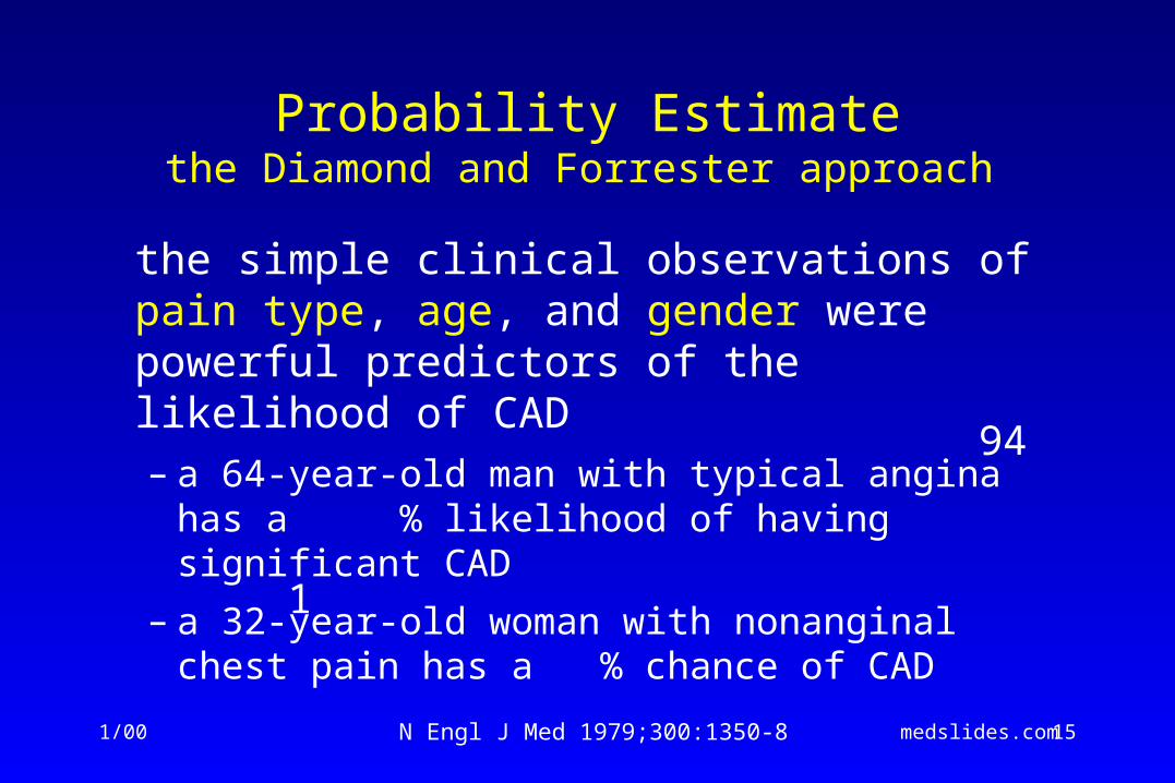
Probability Estimatethe Diamond and Forrester approach
the simple clinical observations of pain type, age, and gender were powerful predictors of the likelihood of CAD– a 64-year-old man with typical angina has a %
likelihood of having significant CAD
– a 32-year-old woman with nonanginal chest pain has a % chance of CAD
N Engl J Med 1979;300:1350-8
94
1
1/00 medslides.com 16
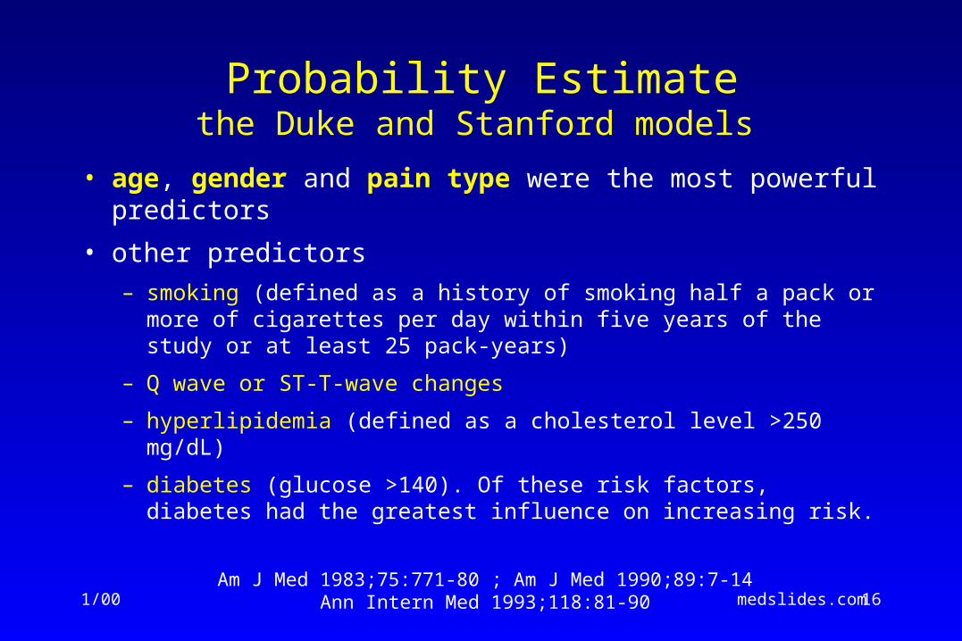
Probability Estimatethe Duke and Stanford models
• age, gender and pain type were the most powerful predictors
• other predictors
– smoking (defined as a history of smoking half a pack or more of cigarettes per day within five years of the study or at least 25 pack-years)
– Q wave or ST-T-wave changes
– hyperlipidemia (defined as a cholesterol level >250 mg/dL)
– diabetes (glucose >140). Of these risk factors, diabetes had the greatest influence on increasing risk.
Am J Med 1983;75:771-80 ; Am J Med 1990;89:7-14Ann Intern Med 1993;118:81-90
1/00 medslides.com 17
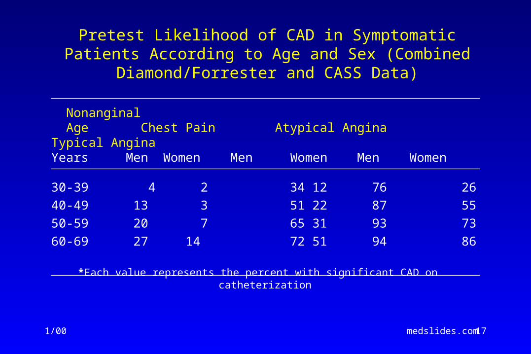
Pretest Likelihood of CAD in Symptomatic Patients According to Age and Sex (Combined Diamond/Forrester and CASS Data)
Nonanginal Age Chest Pain Atypical Angina Typical AnginaYears Men Women Men Women Men Women
30-39 4 2 34 12 76 26
40-49 13 3 51 22 87 55
50-59 20 7 65 31 93 73
60-69 27 14 72 51 94 86
*Each value represents the percent with significant CAD on catheterization
1/00 medslides.com 18
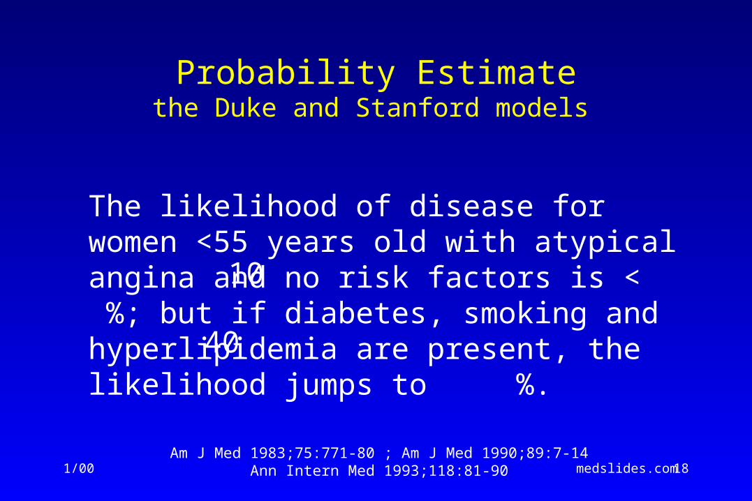
Probability Estimatethe Duke and Stanford models
The likelihood of disease for women <55 years old with atypical angina and no risk factors is < %; but if diabetes, smoking and hyperlipidemia are present, the likelihood jumps to %.
Am J Med 1983;75:771-80 ; Am J Med 1990;89:7-14Ann Intern Med 1993;118:81-90
10
40
1/00 medslides.com 19
Risk Stratification With Clinical Parameters
History• demographics such as age and gender
• coronary risk factors including hypertension, diabetes, hypercholesterolemia, smoking, peripheral vascular or arterial disease and previous MI
Physical examination
• vascular disease (abnormal fundi, decreased peripheral pulses, bruits)
• long-standing hypertension (blood pressure, abnormal fundi)
• aortic valve stenosis or idiopathic hypertrophic subaortic stenosis (systolic murmur, abnormal carotid pulse, abnormal apical pulse)
• left-heart failure (third heart sound, displaced apical impulse, bibasilar rales)
• right-heart failure (jugular venous distension, hepatomegaly, ascites, pedal edema)
1/00 medslides.com 20
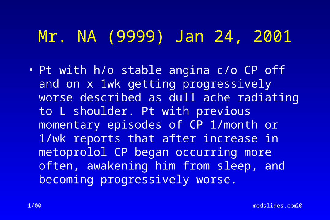
Mr. NA (9999) Jan 24, 2001
• Pt with h/o stable angina c/o CP off and on x 1wk getting progressively worse described as dull ache radiating to L shoulder. Pt with previous momentary episodes of CP 1/month or 1/wk reports that after increase in metoprolol CP began occurring more often, awakening him from sleep, and becoming progressively worse.
1/00 medslides.com 21
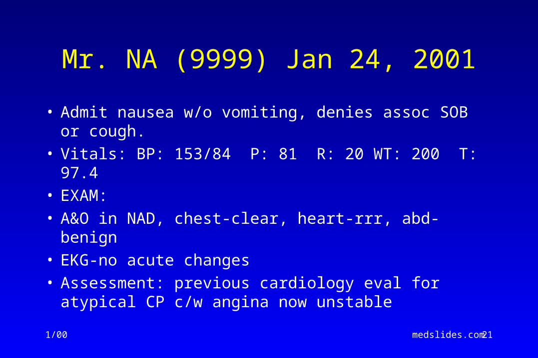
Mr. NA (9999) Jan 24, 2001
• Admit nausea w/o vomiting, denies assoc SOB or cough.
• Vitals: BP: 153/84 P: 81 R: 20 WT: 200 T: 97.4 • EXAM:• A&O in NAD, chest-clear, heart-rrr, abd-benign• EKG-no acute changes• Assessment: previous cardiology eval for atypical
CP c/w angina now unstable
1/00 medslides.com 22
Clinical Assessment
B. Recommendations for Initial
Laboratory Tests, ECG, and Chest X-Ray for Diagnosis
1/01 medslides.com 23
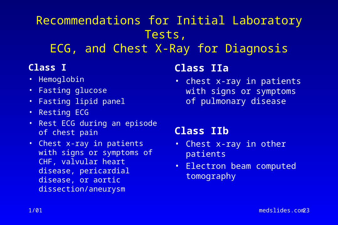
Recommendations for Initial Laboratory Tests, ECG, and Chest X-Ray for Diagnosis
Class I• Hemoglobin• Fasting glucose• Fasting lipid panel• Resting ECG• Rest ECG during an episode of
chest pain• Chest x-ray in patients with signs
or symptoms of CHF, valvular heart disease, pericardial disease, or aortic dissection/aneurysm
Class IIa• chest x-ray in patients with signs
or symptoms of pulmonary disease
Class IIb• Chest x-ray in other patients• Electron beam computed
tomography
1/00 medslides.com 24
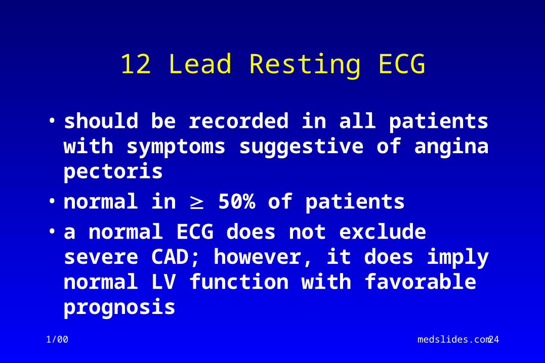
12 Lead Resting ECG
• should be recorded in all patients with symptoms suggestive of angina pectoris
• normal in 50% of patients
• a normal ECG does not exclude severe CAD; however, it does imply normal LV function with favorable prognosis
1/00 medslides.com 25Am J Cardiol 1982;49:1604-14
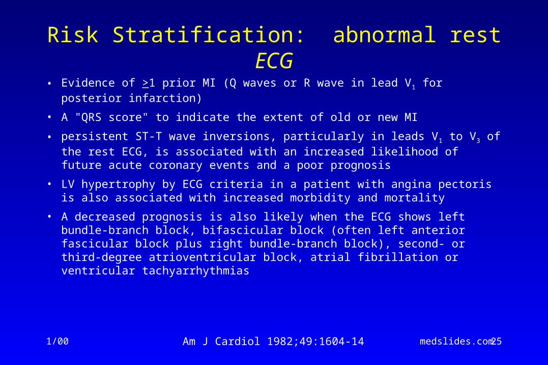
Risk Stratification: abnormal rest ECG
• Evidence of >1 prior MI (Q waves or R wave in lead V1 for posterior infarction)
• A "QRS score" to indicate the extent of old or new MI
• persistent ST-T wave inversions, particularly in leads V1 to V3 of the rest ECG, is associated with an increased likelihood of future acute coronary events and a poor prognosis
• LV hypertrophy by ECG criteria in a patient with angina pectoris is also associated with increased morbidity and mortality
• A decreased prognosis is also likely when the ECG shows left bundle-branch block, bifascicular block (often left anterior fascicular block plus right bundle-branch block), second- or third-degree atrioventricular block, atrial fibrillation or ventricular tachyarrhythmias
1/00 medslides.com 26
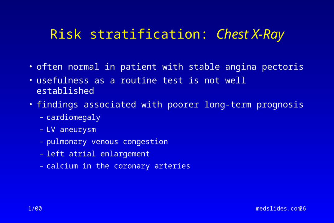
Risk stratification: Chest X-Ray
• often normal in patient with stable angina pectoris
• usefulness as a routine test is not well established
• findings associated with poorer long-term prognosis – cardiomegaly
– LV aneurysm
– pulmonary venous congestion
– left atrial enlargement
– calcium in the coronary arteries
1/00 medslides.com 27
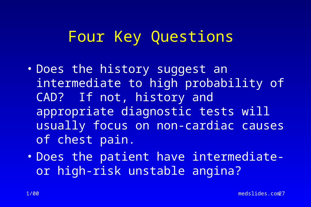
Four Key Questions
• Does the history suggest an intermediate to high probability of CAD? If not, history and appropriate diagnostic tests will usually focus on non-cardiac causes of chest pain.
• Does the patient have intermediate- or high-risk unstable angina?
1/00 medslides.com 28
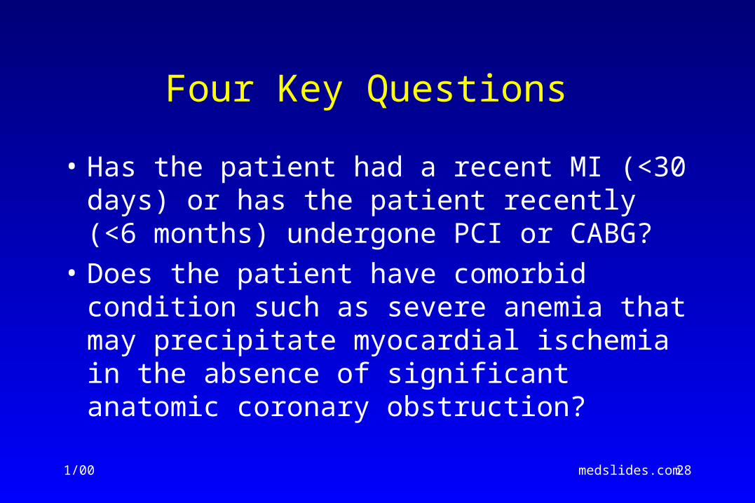
Four Key Questions
• Has the patient had a recent MI (<30 days) or has the patient recently (<6 months) undergone PCI or CABG?
• Does the patient have comorbid condition such as severe anemia that may precipitate myocardial ischemia in the absence of significant anatomic coronary obstruction?
1/00 medslides.com 29
Clinical Assessment
C. Recommendations for Echocardiography or
Radionuclide Angiography
1/00 medslides.com 30
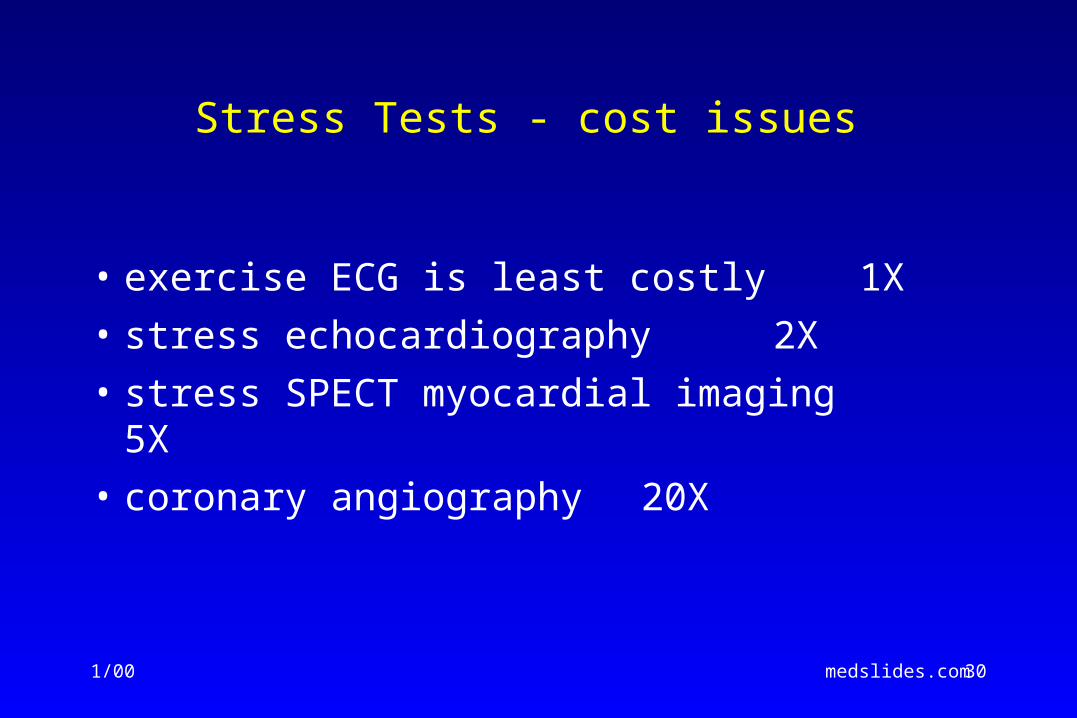
Stress Tests - cost issues
• exercise ECG is least costly 1X
• stress echocardiography 2X
• stress SPECT myocardial imaging 5X
• coronary angiography 20X
1/00 medslides.com 31
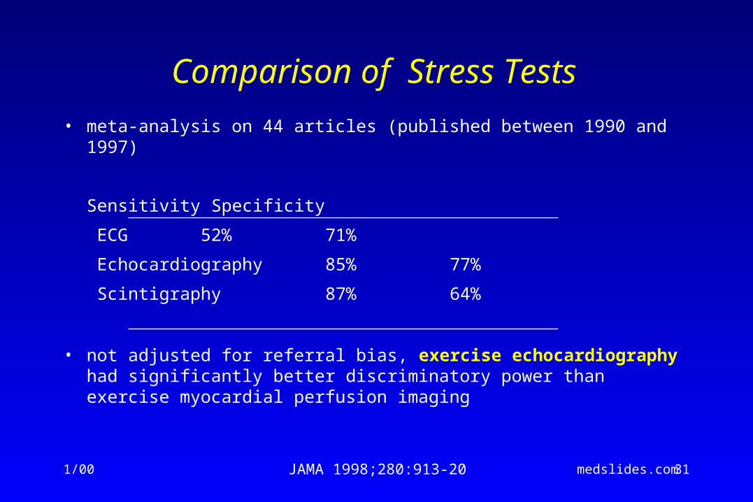
Comparison of Stress Tests
• meta-analysis on 44 articles (published between 1990 and 1997)
Sensitivity Specificity
ECG 52% 71%
Echocardiography 85% 77%
Scintigraphy 87% 64%
• not adjusted for referral bias, exercise echocardiography had significantly better discriminatory power than exercise myocardial perfusion imaging
JAMA 1998;280:913-20
1/00 medslides.com 32
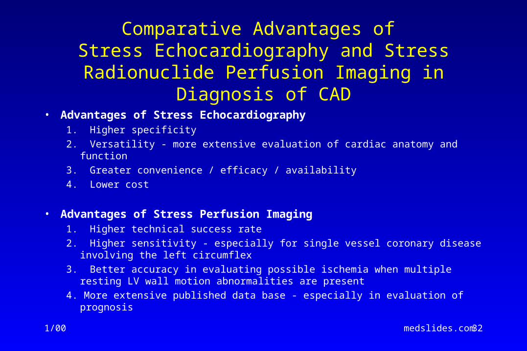
Comparative Advantages of Stress Echocardiography and Stress Radionuclide
Perfusion Imaging in Diagnosis of CAD
• Advantages of Stress Echocardiography1. Higher specificity
2. Versatility - more extensive evaluation of cardiac anatomy and function
3. Greater convenience / efficacy / availability
4. Lower cost
• Advantages of Stress Perfusion Imaging1. Higher technical success rate
2. Higher sensitivity - especially for single vessel coronary disease involving the left circumflex
3. Better accuracy in evaluating possible ischemia when multiple resting LV wall motion abnormalities are present
4. More extensive published data base - especially in evaluation of prognosis
1/00 medslides.com 33
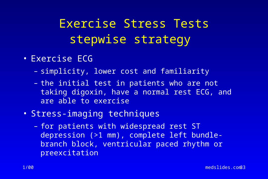
Exercise Stress Testsstepwise strategy
• Exercise ECG – simplicity, lower cost and familiarity
– the initial test in patients who are not taking digoxin, have a normal rest ECG, and are able to exercise
• Stress-imaging techniques – for patients with widespread rest ST depression (>1 mm),
complete left bundle-branch block, ventricular paced rhythm or preexcitation
1/00 medslides.com 34
Risk Stratification for Death or MI
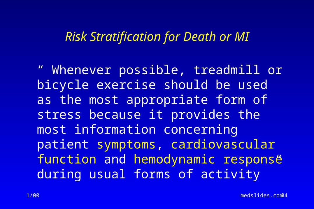
“ Whenever possible, treadmill or bicycle exercise should be used as the most appropriate form of stress because it provides the most information concerning patient symptoms, cardiovascular function and hemodynamic response during usual forms of activity ”
1/00 medslides.com 35
Prognostic Markers in Exercise Testing
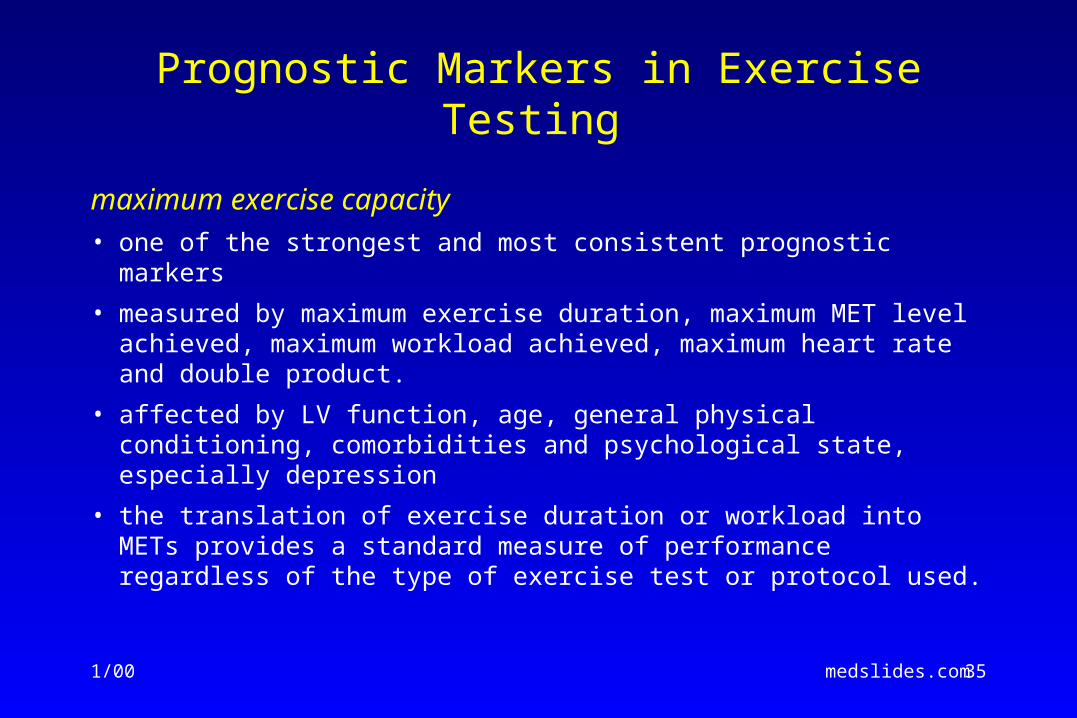
maximum exercise capacity
• one of the strongest and most consistent prognostic markers
• measured by maximum exercise duration, maximum MET level achieved, maximum workload achieved, maximum heart rate and double product.
• affected by LV function, age, general physical conditioning, comorbidities and psychological state, especially depression
• the translation of exercise duration or workload into METs provides a standard measure of performance regardless of the type of exercise test or protocol used.
1/00 medslides.com 36
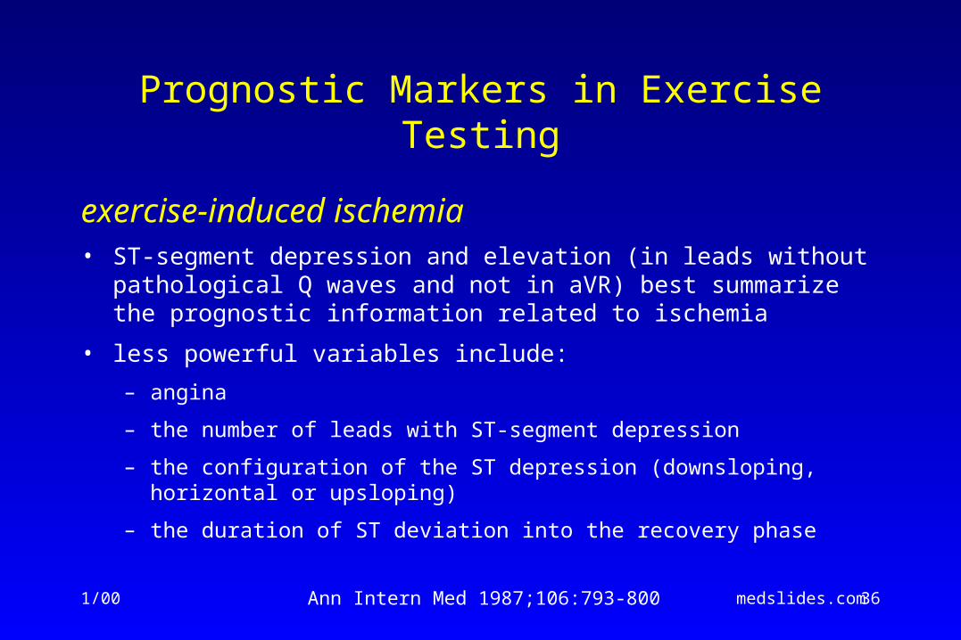
Prognostic Markers in Exercise Testing
exercise-induced ischemia• ST-segment depression and elevation (in leads without pathological Q
waves and not in aVR) best summarize the prognostic information related to ischemia
• less powerful variables include:
– angina
– the number of leads with ST-segment depression
– the configuration of the ST depression (downsloping, horizontal or upsloping)
– the duration of ST deviation into the recovery phase
Ann Intern Med 1987;106:793-800
1/00 medslides.com 37
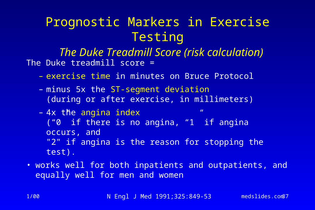
Prognostic Markers in Exercise Testing The Duke Treadmill Score (risk calculation)
The Duke treadmill score =
– exercise time in minutes on Bruce Protocol
– minus 5x the ST-segment deviation(during or after exercise, in millimeters)
– 4x the angina index(“0” if there is no angina, “1” if angina occurs, and "2" if angina is the reason for stopping the test).
• works well for both inpatients and outpatients, and equally well for men and women
N Engl J Med 1991;325:849-53
1/00 medslides.com 38
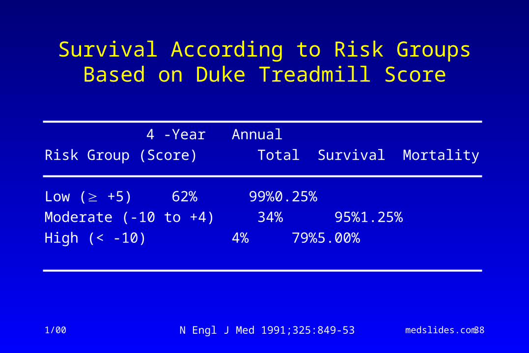
Survival According to Risk Groups Based on Duke Treadmill Score
4 -YearAnnual
Risk Group (Score) Total Survival Mortality
Low ( +5) 62% 99% 0.25%
Moderate (-10 to +4) 34% 95% 1.25%
High (< -10) 4% 79% 5.00%
N Engl J Med 1991;325:849-53
1/00 medslides.com 39
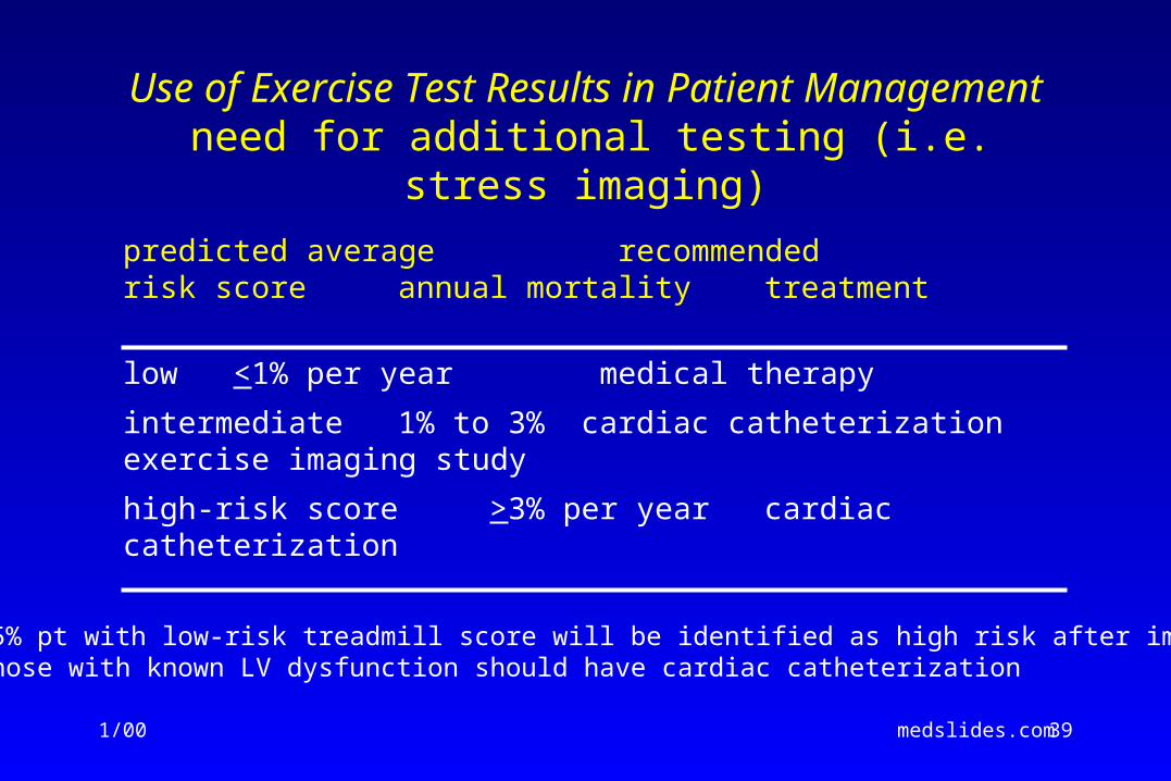
Use of Exercise Test Results in Patient Management need for additional testing (i.e. stress imaging)
predicted average recommendedrisk score annual mortality treatment
low <1% per year medical therapy
intermediate 1% to 3% cardiac catheterizationexercise imaging
study
high-risk score >3% per year cardiac catheterization* <5% pt with low-risk treadmill score will be identified as high risk after imaging
* those with known LV dysfunction should have cardiac catheterization
1/00 medslides.com 40
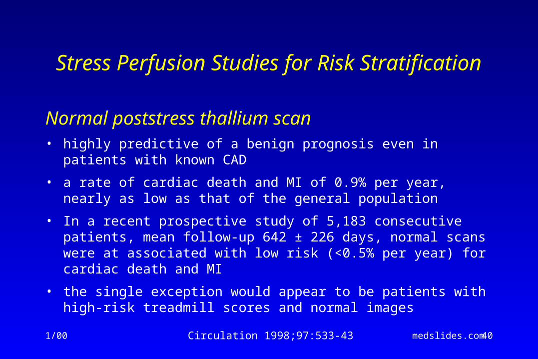
Stress Perfusion Studies for Risk Stratification
Normal poststress thallium scan
• highly predictive of a benign prognosis even in patients with known CAD
• a rate of cardiac death and MI of 0.9% per year, nearly as low as that of the general population
• In a recent prospective study of 5,183 consecutive patients, mean follow-up 642 ± 226 days, normal scans were at associated with low risk (<0.5% per year) for cardiac death and MI
• the single exception would appear to be patients with high-risk treadmill scores and normal images
Circulation 1998;97:533-43
1/00 medslides.com 41
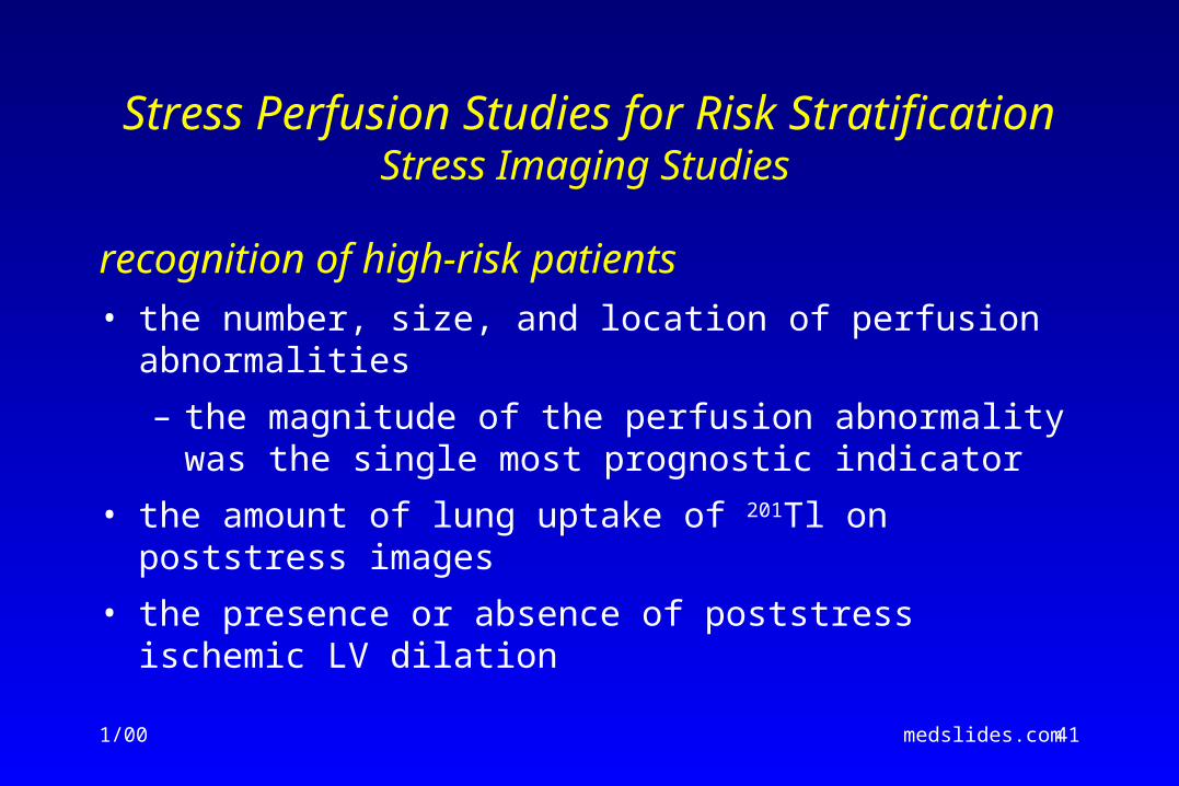
Stress Perfusion Studies for Risk StratificationStress Imaging Studies
recognition of high-risk patients
• the number, size, and location of perfusion abnormalities
– the magnitude of the perfusion abnormality was the single most prognostic indicator
• the amount of lung uptake of 201Tl on poststress images
• the presence or absence of poststress ischemic LV dilation
1/00 medslides.com 42
Application of Myocardial Perfusion Imaging to Specific Patient Subsets
• Patients With A Normal Rest ECG
• Concomitant Use Of Drugs
• Women, The Elderly, Or Obese Patients
• Left Bundle-Branch Block
• After Coronary Angiography
• After Myocardial Revascularization
• After Exercise Testing
• Stress Echocardiography for Risk Stratification
1/00 medslides.com 43
Risk Stratificationlong-term survival with CAD
• The patient's risk is usually a function of 4 types of patient characteristic: – LV functioning - ejection fraction
– anatomic extent and severity of atherosclerotic involvement of the coronary tree
– evidence of a recent coronary plaque rupture - indicator of short-term risk for cardiac death or nonfatal MI
– general health and noncoronary comorbidity
1/00 medslides.com 44
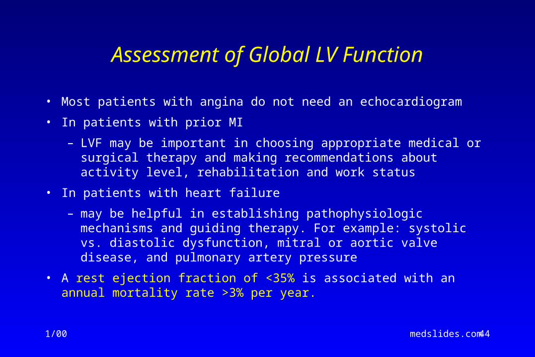
Assessment of Global LV Function
• Most patients with angina do not need an echocardiogram
• In patients with prior MI
– LVF may be important in choosing appropriate medical or surgical therapy and making recommendations about activity level, rehabilitation and work status
• In patients with heart failure
– may be helpful in establishing pathophysiologic mechanisms and guiding therapy. For example: systolic vs. diastolic dysfunction, mitral or aortic valve disease, and pulmonary artery pressure
• A rest ejection fraction of <35% is associated with an annual mortality rate >3% per year.
1/00 medslides.com 45
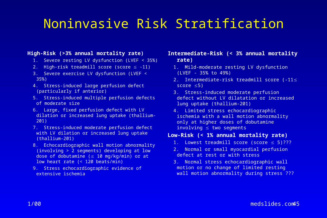
Noninvasive Risk Stratification
High-Risk (>3% annual mortality rate) 1. Severe resting LV dysfunction (LVEF < 35%)
2. High-risk treadmill score (score -11)
3. Severe exercise LV dysfunction (LVEF < 35%)
4. Stress-induced large perfusion defect (particularly if anterior)
5. Stress-induced multiple perfusion defects of moderate size
6. Large, fixed perfusion defect with LV dilation or increased lung uptake (thallium-201)
7. Stress-induced moderate perfusion defect with LV dilation or increased lung uptake (thallium-201)
8. Echocardiographic wall motion abnormality (involving > 2 segments) developing at low dose of dobutamine ( 10 mg/kg/min) or at low heart rate (< 120 beats/min)
9. Stress echocardiographic evidence of extensive ischemia
Intermediate-Risk (< 3% annual mortality rate)
1. Mild-moderate resting LV dysfunction (LVEF - 35% to 49%)
2. Intermediate-risk treadmill score (-11 score 5)
3. Stress-induced moderate perfusion defect without LV dilatation or increased lung uptake (thallium-201)
4. Limited stress echocardiographic ischemia with a wall motion abnormality only at higher doses of dobutamine involving two segments
Low-Risk (< 1% annual mortality rate) 1. Lowest treadmill score (score 5)???
2. Normal or small myocardial perfusion defect at rest or with stress
3. Normal stress echocardiographic wall motion or no change of limited resting wall motion abnormality during stress ???
1/00 medslides.com 46
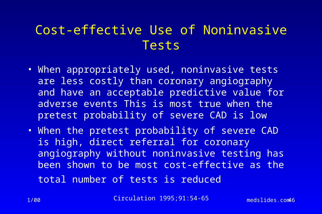
Cost-effective Use of Noninvasive Tests
• When appropriately used, noninvasive tests are less costly than coronary angiography and have an acceptable predictive value for adverse events This is most true when the pretest probability of severe CAD is low
• When the pretest probability of severe CAD is high, direct referral for coronary angiography without noninvasive testing has been shown to be most cost-effective as the total
number of tests is reduced
Circulation 1995;91:54-65
1/00 medslides.com 47
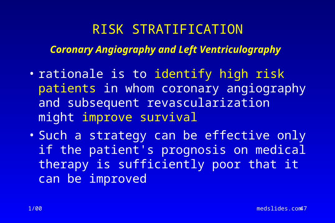
RISK STRATIFICATION
Coronary Angiography and Left Ventriculography
• rationale is to identify high risk patients in whom coronary angiography and subsequent revascularization might improve survival
• Such a strategy can be effective only if the patient's prognosis on medical therapy is sufficiently poor that it can be improved
1/00 medslides.com 48
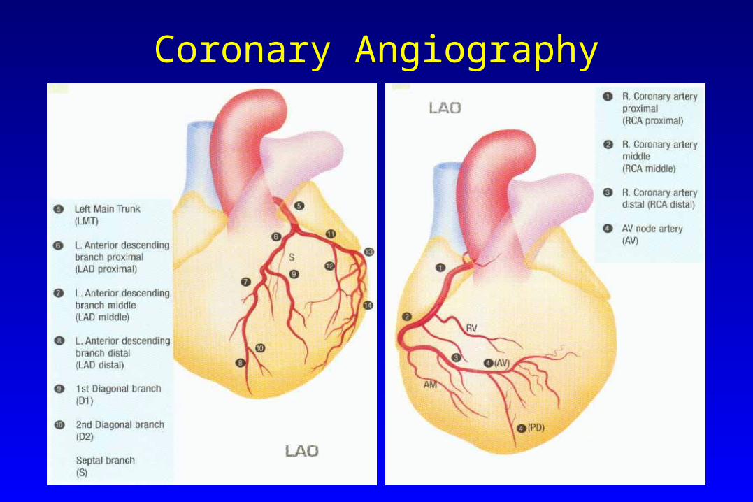
Coronary Angiography
1/00 medslides.com 49
Direct Referral For Diagnostic Coronary Angiography
• When Noninvasive Testing Is Contraindicated Or Unlikely To Be Adequate Due To Illness, Disability Or Physical Characteristics. For Example:
– coexisting chronic obstructive pulmonary disease
– noninvasive testing is abnormal but not clearly diagnostic
– patient's occupation or activity could constitute a risk to themselves or others
– a high clinical probability of severe CAD
– diabetics with paucity of symptoms of myocardial ischemia due to autonomic and sensory neuropathy
1/00 medslides.com 50
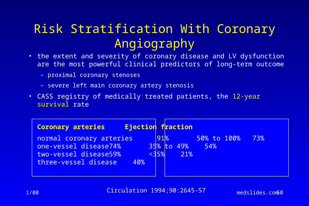
Risk Stratification With Coronary Angiography
• the extent and severity of coronary disease and LV dysfunction are the most powerful clinical predictors of long-term outcome
– proximal coronary stenoses
– severe left main coronary artery stenosis
• CASS registry of medically treated patients, the 12-year survival rate
Coronary arteries Ejection fraction
normal coronary arteries 91% 50% to 100% 73%one-vessel disease 74% 35% to 49% 54%two-vessel disease 59% <35% 21%three-vessel disease 40%
Circulation 1994;90:2645-57
1/00 medslides.com 51
Patients With Previous CABG
• progression of native CAD is not uncommon
• development of obstructive atherosclerotic vein graft lesions are prone to rapid progression and thrombotic occlusion
• low threshold for angiographic evaluation is recommended for patients who develop chronic stable angina >5 years after surgery, especially when ischemia is noninvasively documented in the distribution of a vein graft, the LAD is supplied by a vein graft, or multiple vein grafts are present
• outcome can be improved by reoperation and by percutaneous catheter-based strategies
1/00 medslides.com 52
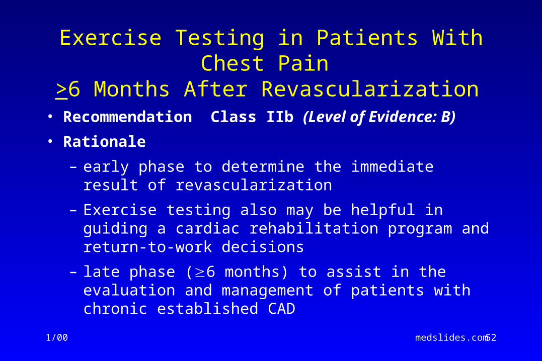
Exercise Testing in Patients With Chest Pain >6 Months After Revascularization
• Recommendation Class IIb (Level of Evidence: B)
• Rationale
– early phase to determine the immediate result of revascularization
– Exercise testing also may be helpful in guiding a cardiac rehabilitation program and return-to-work decisions
– late phase (6 months) to assist in the evaluation and management of patients with chronic established CAD
1/00 medslides.com 53
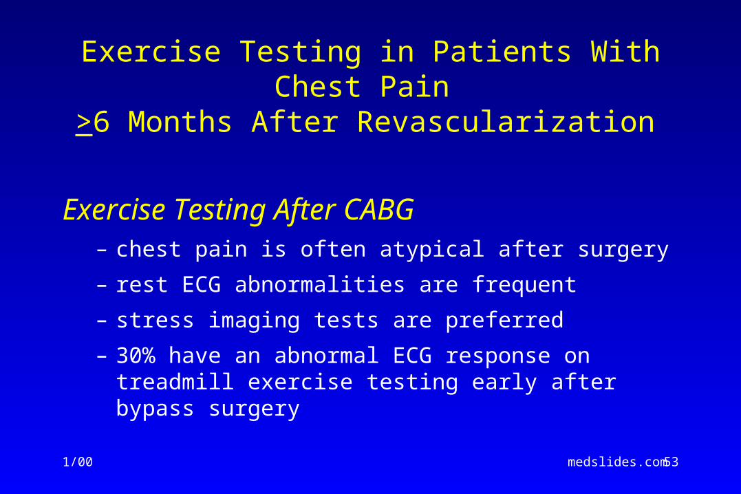
Exercise Testing in Patients With Chest Pain >6 Months After Revascularization
Exercise Testing After CABG – chest pain is often atypical after surgery
– rest ECG abnormalities are frequent
– stress imaging tests are preferred
– 30% have an abnormal ECG response on treadmill exercise testing early after bypass surgery
1/00 medslides.com 54
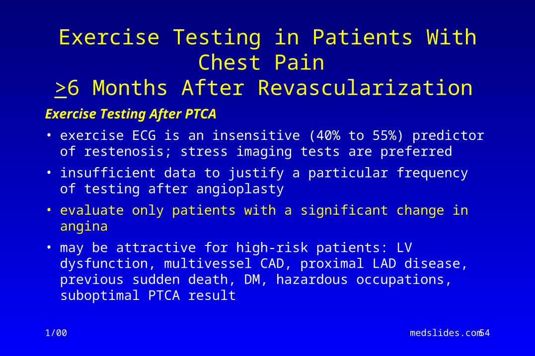
Exercise Testing in Patients With Chest Pain >6 Months After Revascularization
Exercise Testing After PTCA
• exercise ECG is an insensitive (40% to 55%) predictor of restenosis; stress imaging tests are preferred
• insufficient data to justify a particular frequency of testing after angioplasty
• evaluate only patients with a significant change in angina
• may be attractive for high-risk patients: LV dysfunction, multivessel CAD, proximal LAD disease, previous sudden death, DM, hazardous occupations, suboptimal PTCA result
1/00 medslides.com 55
Treatment
A. Recommendations for Pharmacotherapy to Prevent MI and
Death and Reduce Symptoms
1/00 medslides.com 56
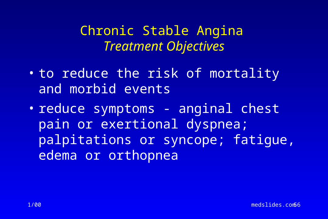
Chronic Stable Angina
Treatment Objectives
• to reduce the risk of mortality and morbid events
• reduce symptoms - anginal chest pain or exertional dyspnea; palpitations or syncope; fatigue, edema or orthopnea
1/00 medslides.com 57
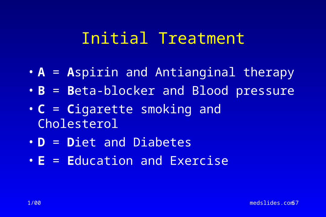
Initial Treatment
• A = Aspirin and Antianginal therapy
• B = Beta-blocker and Blood pressure
• C = Cigarette smoking and Cholesterol
• D = Diet and Diabetes
• E = Education and Exercise
1/00 medslides.com 58
Treatment
B. Pharmacotherapy
to Prevent MI and Death
1/00 medslides.com 59
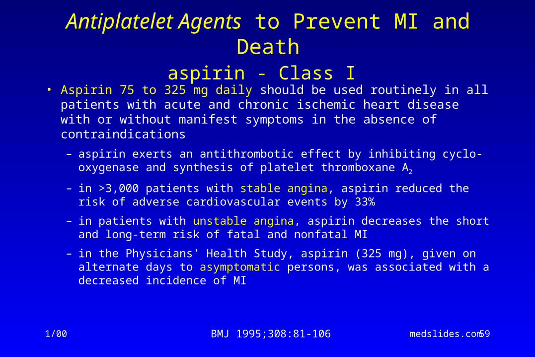
Antiplatelet Agents to Prevent MI and Deathaspirin - Class I
• Aspirin 75 to 325 mg daily should be used routinely in all patients with acute and chronic ischemic heart disease with or without manifest symptoms in the absence of contraindications
– aspirin exerts an antithrombotic effect by inhibiting cyclo-oxygenase and synthesis of platelet thromboxane A2
– in >3,000 patients with stable angina, aspirin reduced the risk of adverse cardiovascular events by 33%
– in patients with unstable angina, aspirin decreases the short and long-term risk of fatal and nonfatal MI
– in the Physicians' Health Study, aspirin (325 mg), given on alternate days to asymptomatic persons, was associated with a decreased incidence of MI
BMJ 1995;308:81-106
1/00 medslides.com 60
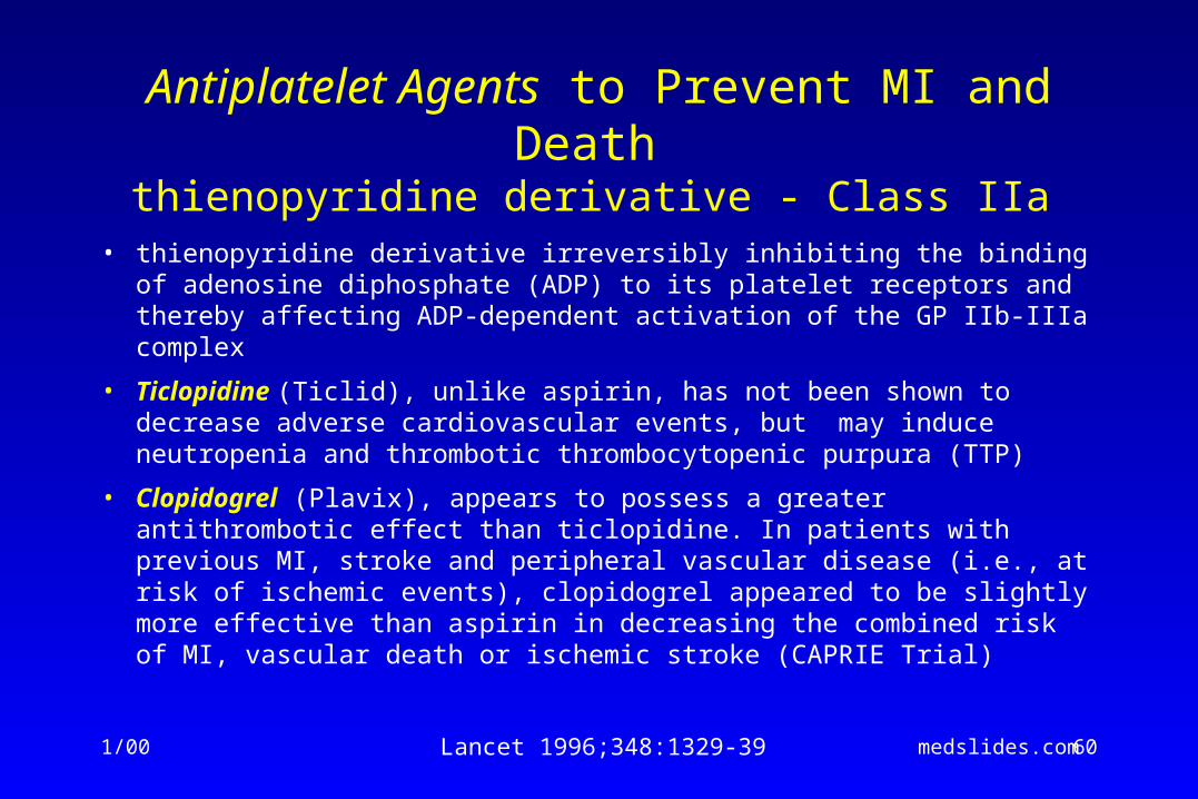
Antiplatelet Agents to Prevent MI and Death thienopyridine derivative - Class IIa
• thienopyridine derivative irreversibly inhibiting the binding of adenosine diphosphate (ADP) to its platelet receptors and thereby affecting ADP-dependent activation of the GP IIb-IIIa complex
• Ticlopidine (Ticlid), unlike aspirin, has not been shown to decrease adverse cardiovascular events, but may induce neutropenia and thrombotic thrombocytopenic purpura (TTP)
• Clopidogrel (Plavix), appears to possess a greater antithrombotic effect than ticlopidine. In patients with previous MI, stroke and peripheral vascular disease (i.e., at risk of ischemic events), clopidogrel appeared to be slightly more effective than aspirin in decreasing the combined risk of MI, vascular death or ischemic stroke (CAPRIE Trial)
Lancet 1996;348:1329-39
1/00 medslides.com 61
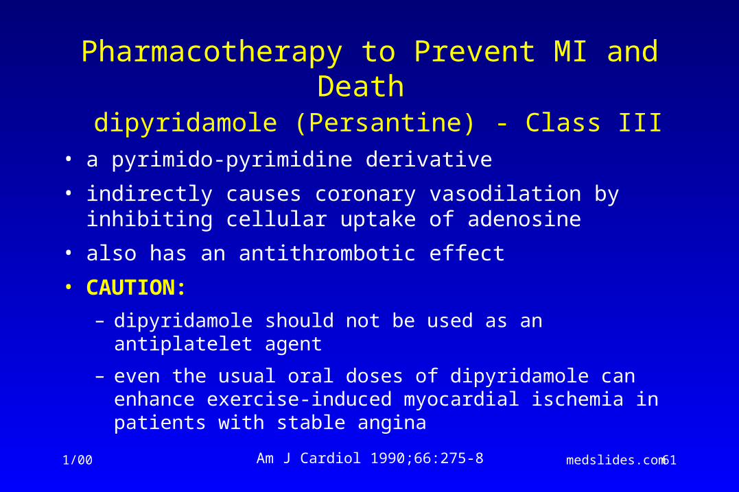
Pharmacotherapy to Prevent MI and Death dipyridamole (Persantine) - Class III
• a pyrimido-pyrimidine derivative
• indirectly causes coronary vasodilation by inhibiting cellular uptake of adenosine
• also has an antithrombotic effect
• CAUTION:
– dipyridamole should not be used as an antiplatelet agent
– even the usual oral doses of dipyridamole can enhance exercise-induced myocardial ischemia in patients with stable angina
Am J Cardiol 1990;66:275-8
1/00 medslides.com 62
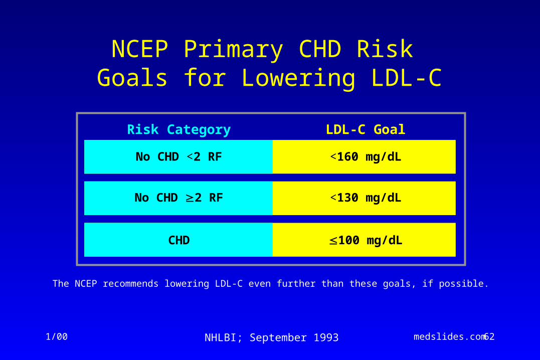
NCEP Primary CHD Risk Goals for Lowering LDL-C
LDL-C Goal
No CHD <2 RF <160 mg/dL
No CHD 2 RF <130 mg/dL
CHD 100 mg/dL
The NCEP recommends lowering LDL-C even further than these goals, if possible.
Risk Category
NHLBI; September 1993
1/00 medslides.com 63
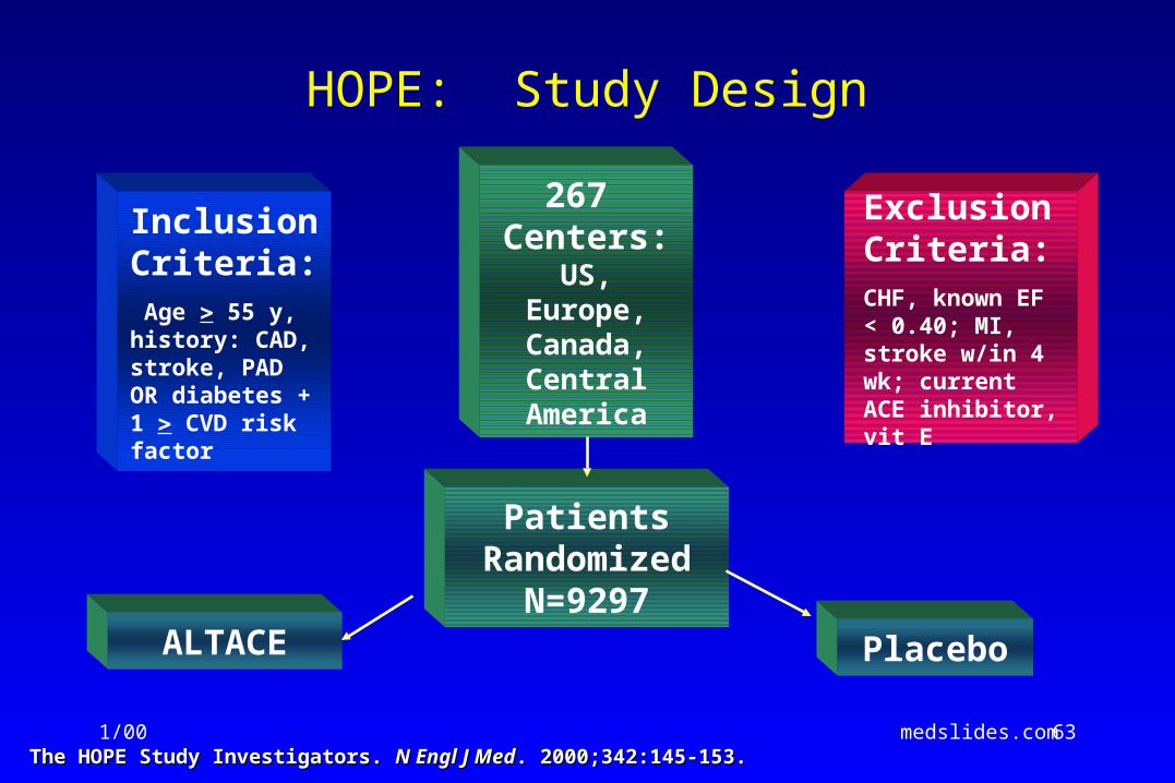
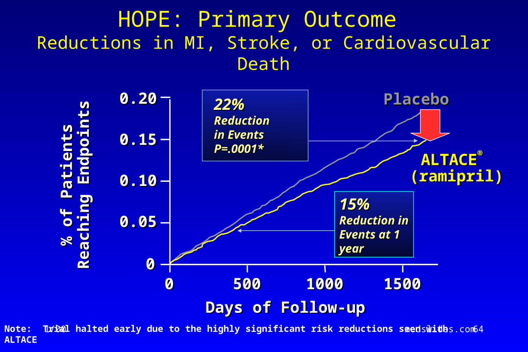
HOPE: Study Design
Inclusion Criteria: Age 55 y, history: CAD, stroke, PAD OR diabetes + 1 CVD risk factor
Exclusion Criteria: CHF, known EF < 0.40; MI, stroke w/in 4 wk; current ACE inhibitor, vit E
267 Centers:
US, Europe, Canada, Central America
ALTACE Placebo
Patients Randomized
N=9297
The HOPE Study Investigators. The HOPE Study Investigators. N Engl J MedN Engl J Med. 2000;342:145-153.. 2000;342:145-153.
1/00 medslides.com 64
HOPE: Primary Outcome Reductions in MI, Stroke, or Cardiovascular Death
Note: Trial halted early due to the highly significant risk reductions seen with ALTACE
0.200.20
0.150.15
0.100.10
0.050.05
0000 500500 10001000 15001500
22%22% ReductionReductionin Eventsin EventsP=.0001*P=.0001*
Days of Follow-upDays of Follow-up
% o
f P
atie
nts
% o
f P
atie
nts
Rea
chin
g E
nd
po
ints
Rea
chin
g E
nd
po
ints
PlaceboPlacebo
ALTACEALTACE®®
(ramipril) (ramipril)
15%15% Reduction Reduction in Events at in Events at 1 year1 year
1/00 medslides.com 65
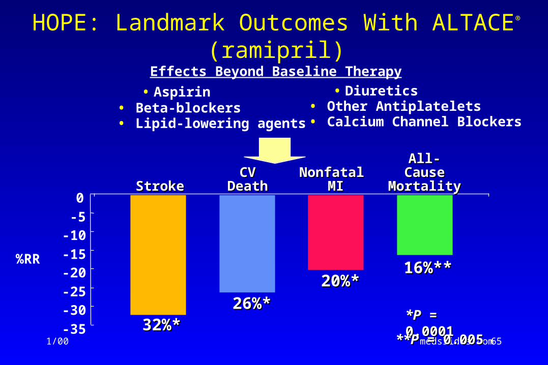
HOPE: Landmark Outcomes With ALTACE®
(ramipril)
-35
-30-25
-20-15
-10-5
0
26%*26%*
CVCVDeathDeath
Nonfatal Nonfatal MIMIStrokeStroke
32%*32%*
20%*20%*
*P*P = 0.0001 = 0.0001
16%**16%**
All-All-CauseCause
MortalityMortality
• Aspirin • Beta-blockers• Lipid-lowering agents
Effects Beyond Baseline Therapy
• Diuretics• Other Antiplatelets • Calcium Channel Blockers
%RR
**P**P = 0.005 = 0.005
1/00 medslides.com 66
Treatment
C. Pharmacotherapy to Reduce
Ischemia and Relieve Symptoms
1/00 medslides.com 67
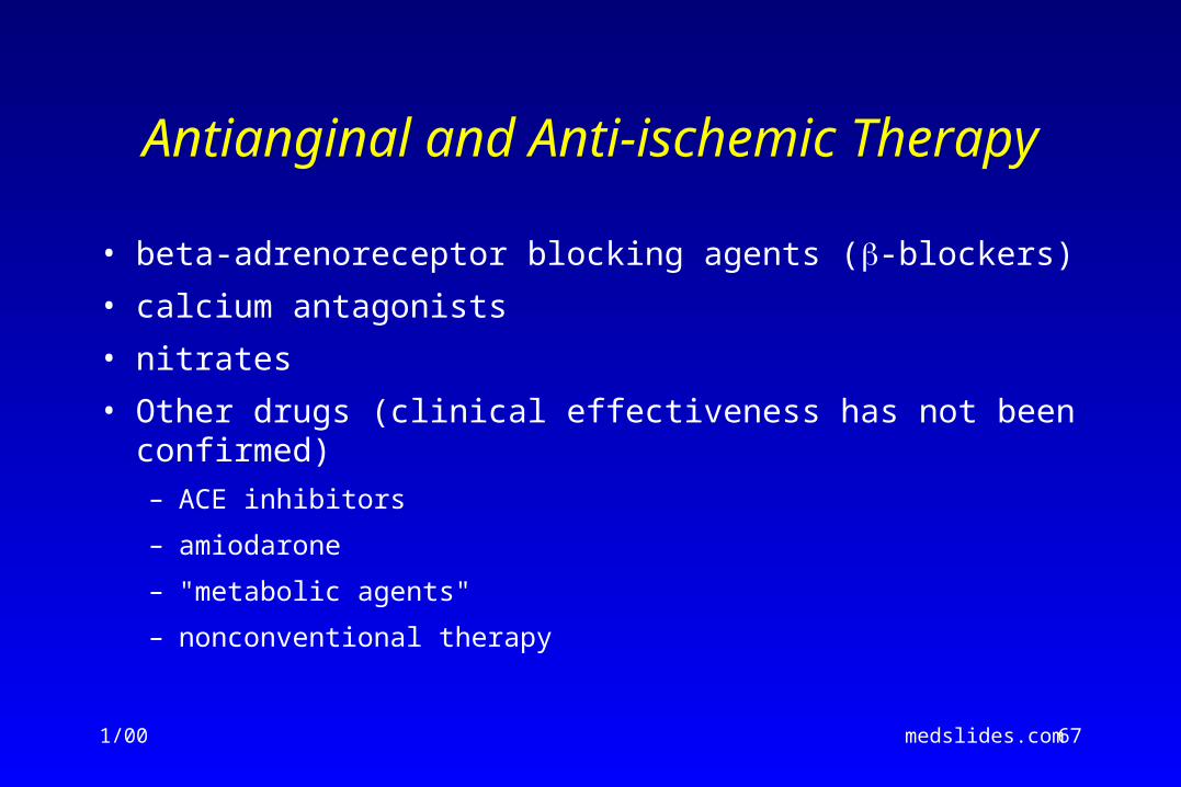
Antianginal and Anti-ischemic Therapy
• beta-adrenoreceptor blocking agents (-blockers)
• calcium antagonists
• nitrates
• Other drugs (clinical effectiveness has not been confirmed)
– ACE inhibitors
– amiodarone
– "metabolic agents"
– nonconventional therapy
1/00 medslides.com 68
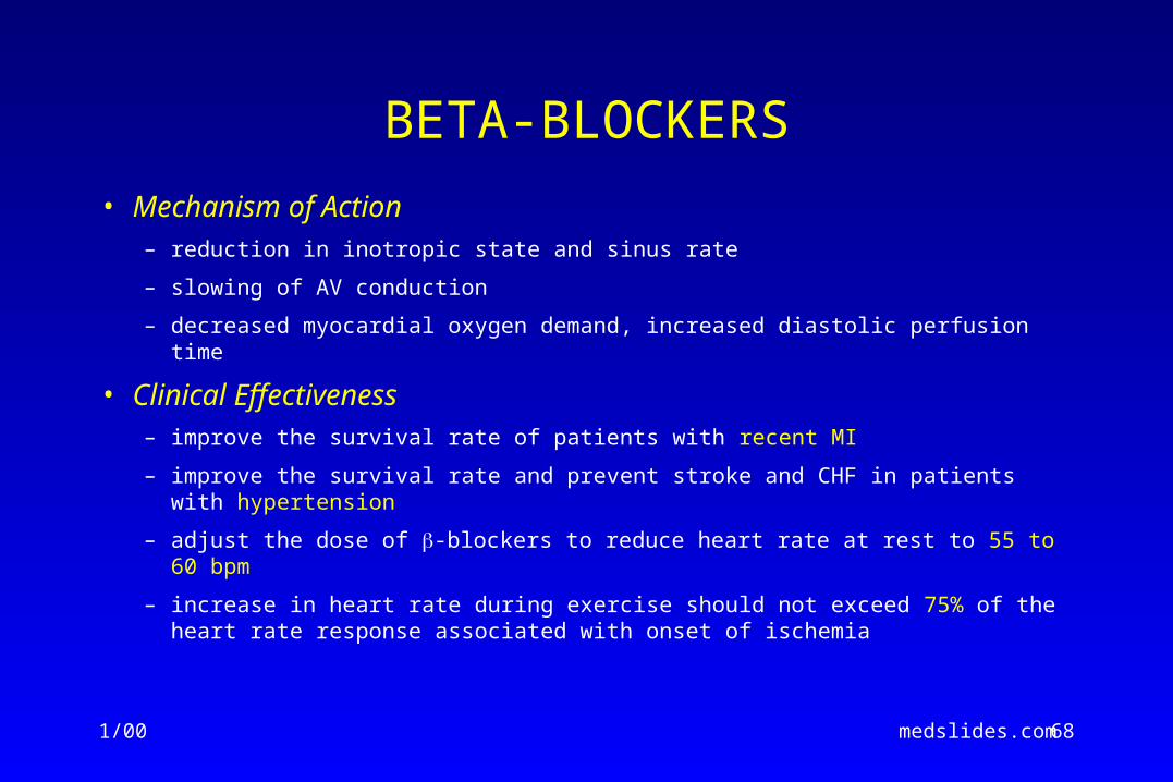
BETA-BLOCKERS
• Mechanism of Action– reduction in inotropic state and sinus rate
– slowing of AV conduction
– decreased myocardial oxygen demand, increased diastolic perfusion time
• Clinical Effectiveness– improve the survival rate of patients with recent MI
– improve the survival rate and prevent stroke and CHF in patients with hypertension
– adjust the dose of -blockers to reduce heart rate at rest to 55 to 60 bpm
– increase in heart rate during exercise should not exceed 75% of the heart rate response associated with onset of ischemia
1/00 medslides.com 69
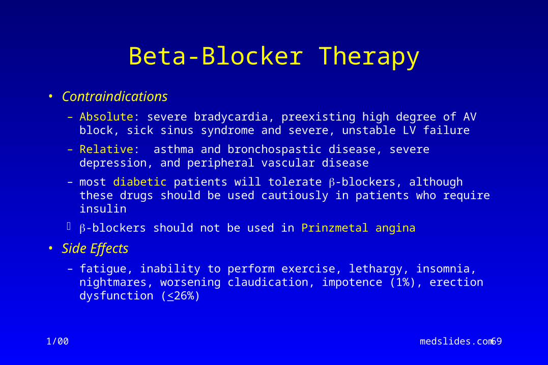
Beta-Blocker Therapy
• Contraindications
– Absolute: severe bradycardia, preexisting high degree of AV block, sick sinus syndrome and severe, unstable LV failure
– Relative: asthma and bronchospastic disease, severe depression, and peripheral vascular disease
– most diabetic patients will tolerate -blockers, although these drugs should be used cautiously in patients who require insulin
-blockers should not be used in Prinzmetal angina
• Side Effects
– fatigue, inability to perform exercise, lethargy, insomnia, nightmares, worsening claudication, impotence (1%), erection dysfunction (<26%)
1/00 medslides.com 70
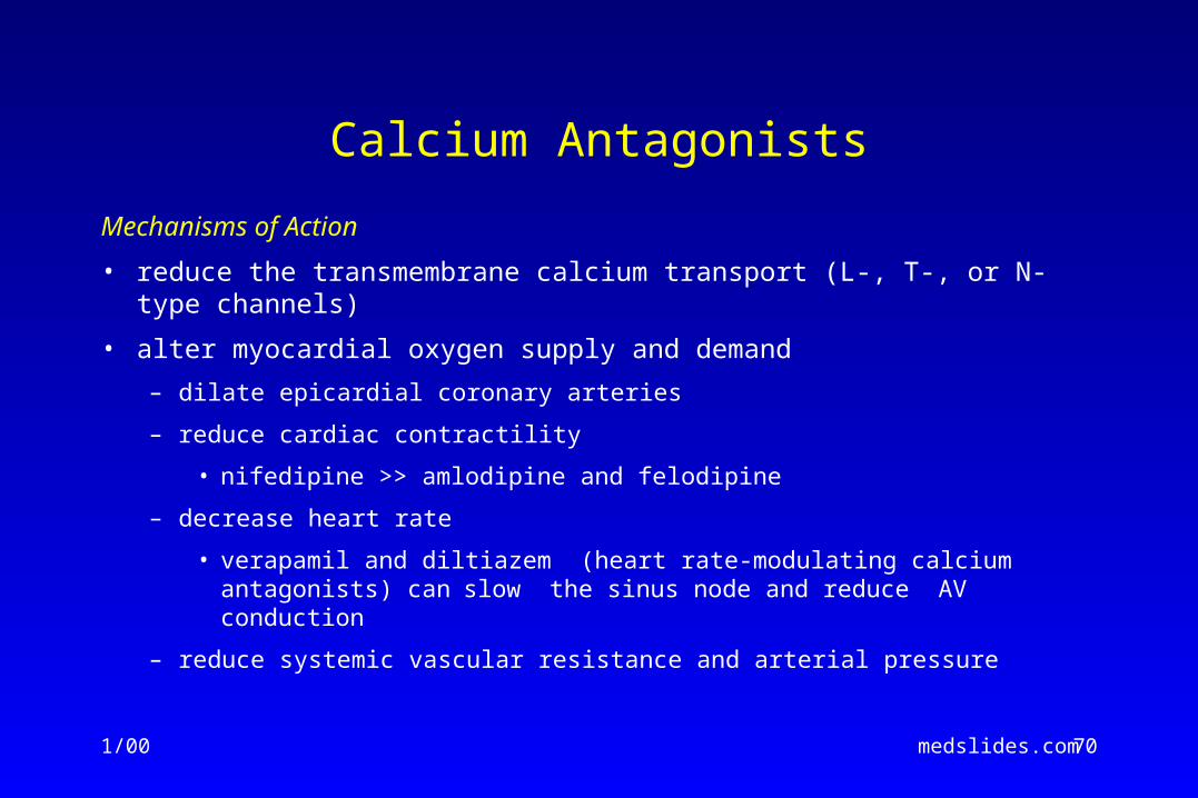
Calcium Antagonists
Mechanisms of Action
• reduce the transmembrane calcium transport (L-, T-, or N-type channels)
• alter myocardial oxygen supply and demand
– dilate epicardial coronary arteries
– reduce cardiac contractility
• nifedipine >> amlodipine and felodipine
– decrease heart rate
• verapamil and diltiazem (heart rate-modulating calcium antagonists) can slow the sinus node and reduce AV conduction
– reduce systemic vascular resistance and arterial pressure
1/00 medslides.com 71
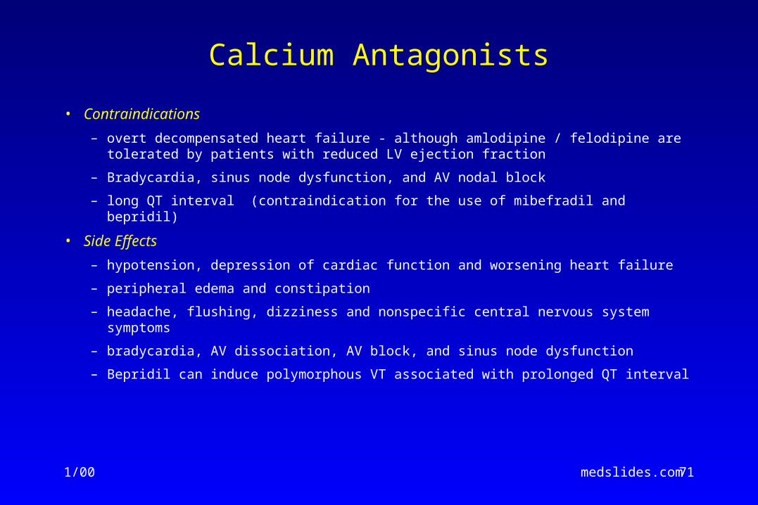
Calcium Antagonists
• Contraindications
– overt decompensated heart failure - although amlodipine / felodipine are tolerated by patients with reduced LV ejection fraction
– Bradycardia, sinus node dysfunction, and AV nodal block
– long QT interval (contraindication for the use of mibefradil and bepridil)
• Side Effects
– hypotension, depression of cardiac function and worsening heart failure
– peripheral edema and constipation
– headache, flushing, dizziness and nonspecific central nervous system symptoms
– bradycardia, AV dissociation, AV block, and sinus node dysfunction
– Bepridil can induce polymorphous VT associated with prolonged QT interval
1/00 medslides.com 72
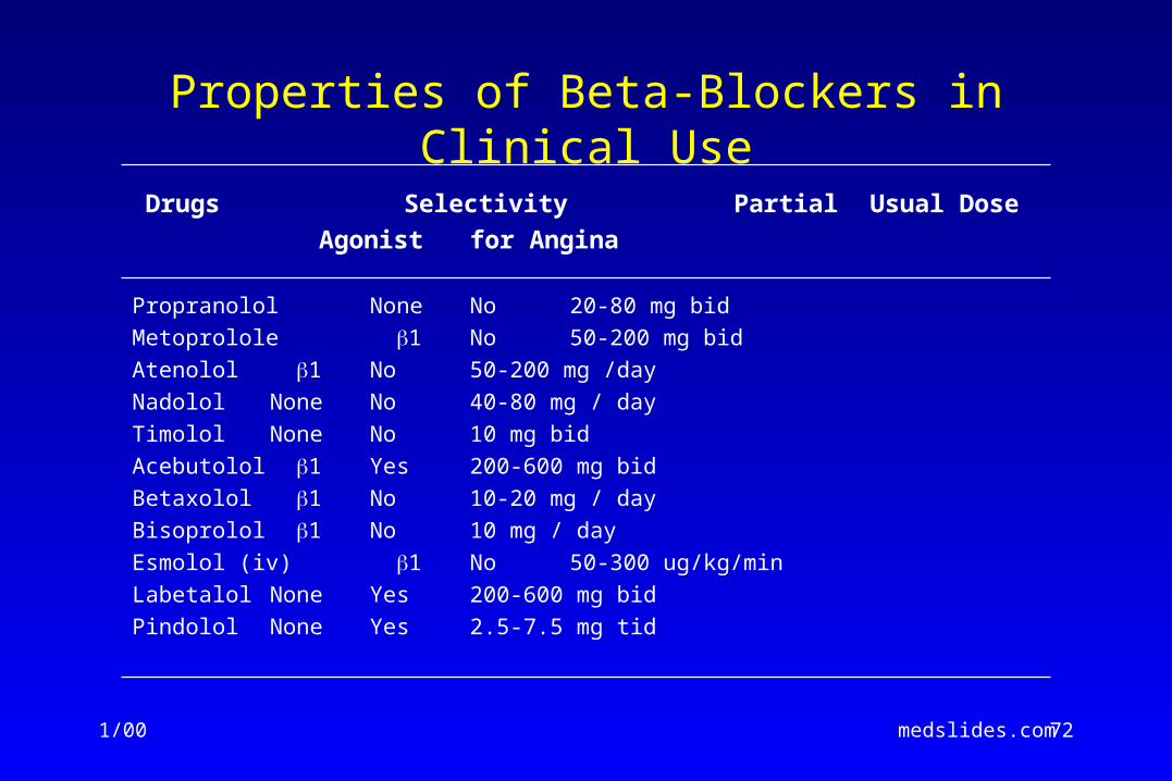
Properties of Beta-Blockers in Clinical Use
Drugs Selectivity Partial Usual Dose
Agonist for Angina
Propranolol None No 20-80 mg bid
Metoprolole 1 No 50-200 mg bid
Atenolol 1 No 50-200 mg /day
Nadolol None No 40-80 mg / day
Timolol None No 10 mg bid
Acebutolol 1 Yes 200-600 mg bid
Betaxolol 1 No 10-20 mg / day
Bisoprolol 1 No 10 mg / day
Esmolol (iv) 1 No 50-300 ug/kg/min
Labetalol None Yes 200-600 mg bid
Pindolol None Yes 2.5-7.5 mg tid
1/00 medslides.com 73
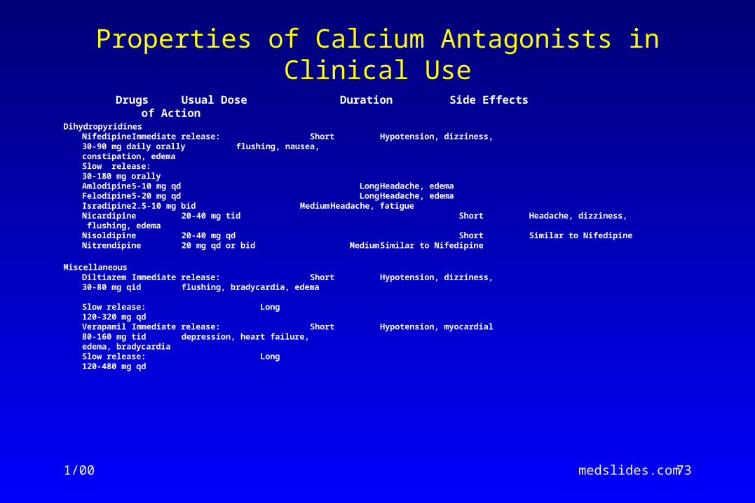
Properties of Calcium Antagonists in Clinical Use
Drugs Usual Dose Duration Side Effects of Action
DihydropyridinesNifedipine Immediate release: Short Hypotension, dizziness,
30-90 mg daily orally flushing, nausea,constipation, edema
Slow release:30-180 mg orally
Amlodipine 5-10 mg qd Long Headache, edemaFelodipine 5-20 mg qd Long Headache, edemaIsradipine 2.5-10 mg bid Medium Headache, fatigueNicardipine 20-40 mg tid Short Headache, dizziness,
flushing, edemaNisoldipine 20-40 mg qd Short Similar to NifedipineNitrendipine 20 mg qd or bid Medium Similar to Nifedipine
MiscellaneousDiltiazem Immediate release: Short Hypotension, dizziness,
30-80 mg qid flushing, bradycardia, edema
Slow release: Long120-320 mg qd
Verapamil Immediate release: Short Hypotension, myocardial80-160 mg tid depression, heart failure,
edema, bradycardiaSlow release: Long120-480 mg qd
1/00 medslides.com 74
• elimination of anginal chest pain • return to normal activities
• functional capacity of CCS class I angina
• good patient compliance - minimal side effects of therapy, cost-effective
• Goal must be modified in light of the clinical characteristics and preferences of each patient
Chronic Stable Angina
Definition of Successful Therapy
1/00 medslides.com 75
Treatment
D. Recommendations for
Treatment of Risk Factors
1/00 medslides.com 76
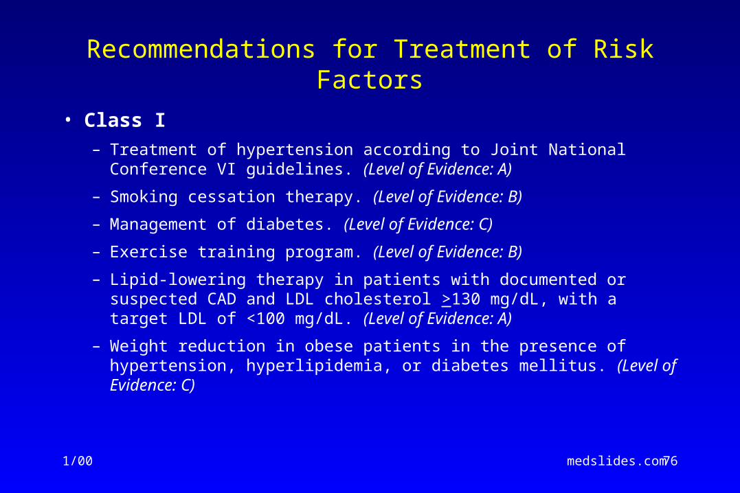
Recommendations for Treatment of Risk Factors
• Class I
– Treatment of hypertension according to Joint National Conference VI guidelines. (Level of Evidence: A)
– Smoking cessation therapy. (Level of Evidence: B)
– Management of diabetes. (Level of Evidence: C)
– Exercise training program. (Level of Evidence: B)
– Lipid-lowering therapy in patients with documented or suspected CAD and LDL cholesterol >130 mg/dL, with a target LDL of <100 mg/dL. (Level of Evidence: A)
– Weight reduction in obese patients in the presence of hypertension, hyperlipidemia, or diabetes mellitus. (Level of Evidence: C)
1/00 medslides.com 77
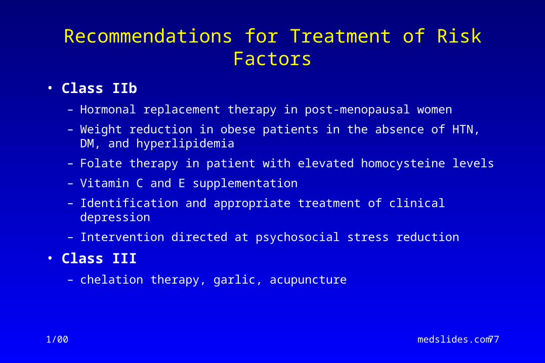
Recommendations for Treatment of Risk Factors
• Class IIb
– Hormonal replacement therapy in post-menopausal women
– Weight reduction in obese patients in the absence of HTN, DM, and hyperlipidemia
– Folate therapy in patient with elevated homocysteine levels
– Vitamin C and E supplementation
– Identification and appropriate treatment of clinical depression
– Intervention directed at psychosocial stress reduction
• Class III
– chelation therapy, garlic, acupuncture
1/00 medslides.com 78
Treatment
E. Revascularization with PCI and CABG in Patients with
Chronic Stable Angina
1/00 medslides.com 79
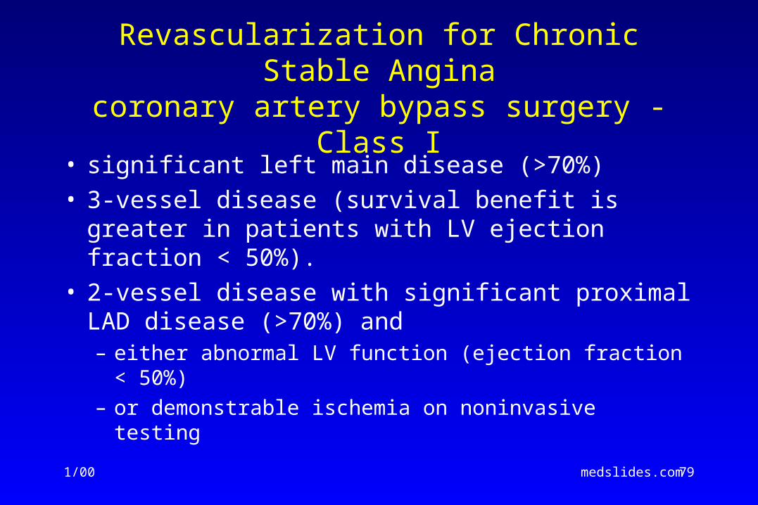
Revascularization for Chronic Stable Anginacoronary artery bypass surgery - Class I
• significant left main disease (>70%)• 3-vessel disease (survival benefit is greater in
patients with LV ejection fraction < 50%).• 2-vessel disease with significant proximal LAD
disease (>70%) and – either abnormal LV function (ejection fraction < 50%) – or demonstrable ischemia on noninvasive testing

1/00 medslides.com 80
Revascularization for Chronic Stable AnginaPCI or CABG - Class I
• PCI for 2- or 3-vessel disease with significant proximal LAD stenosis, who have anatomy suitable for catheter-based therapy, normal LV function, and who do not have treated diabetes
• PCI or CABG for 1-or two-vessel CAD without significant proximal LAD stenosis the with a large area of viable myocardium and high-risk criteria on noninvasive testing
1/00 medslides.com 81
Revascularization for Chronic Stable AnginaPCI or CABG - Class I
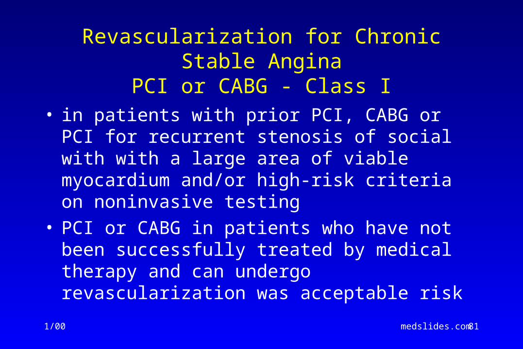
• in patients with prior PCI, CABG or PCI for recurrent stenosis of social with with a large area of viable myocardium and/or high-risk criteria on noninvasive testing
• PCI or CABG in patients who have not been successfully treated by medical therapy and can undergo revascularization was acceptable risk
1/00 medslides.com 82
Patient Follow Up
Monitoring of Symptoms and Anti-anginal Therapy
1/00 medslides.com 83
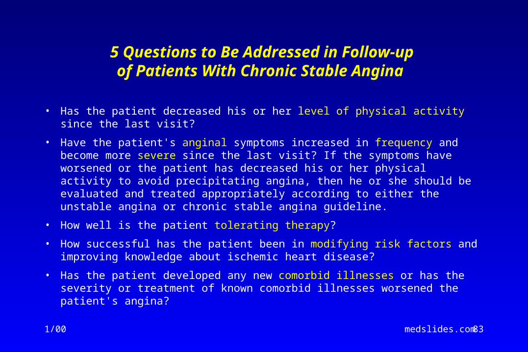
5 Questions to Be Addressed in Follow-up of Patients With Chronic Stable Angina
• Has the patient decreased his or her level of physical activity since the last visit?
• Have the patient's anginal symptoms increased in frequency and become more severe since the last visit? If the symptoms have worsened or the patient has decreased his or her physical activity to avoid precipitating angina, then he or she should be evaluated and treated appropriately according to either the unstable angina or chronic stable angina guideline.
• How well is the patient tolerating therapy?
• How successful has the patient been in modifying risk factors and improving knowledge about ischemic heart disease?
• Has the patient developed any new comorbid illnesses or has the severity or treatment of known comorbid illnesses worsened the patient's angina?
1/00 medslides.com 84
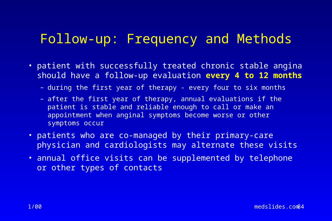
Follow-up: Frequency and Methods
• patient with successfully treated chronic stable angina should have a follow-up evaluation every 4 to 12 months
– during the first year of therapy - every four to six months
– after the first year of therapy, annual evaluations if the patient is stable and reliable enough to call or make an appointment when anginal symptoms become worse or other symptoms occur
• patients who are co-managed by their primary-care physician and cardiologists may alternate these visits
• annual office visits can be supplemented by telephone or other types of contacts
1/00 medslides.com 85
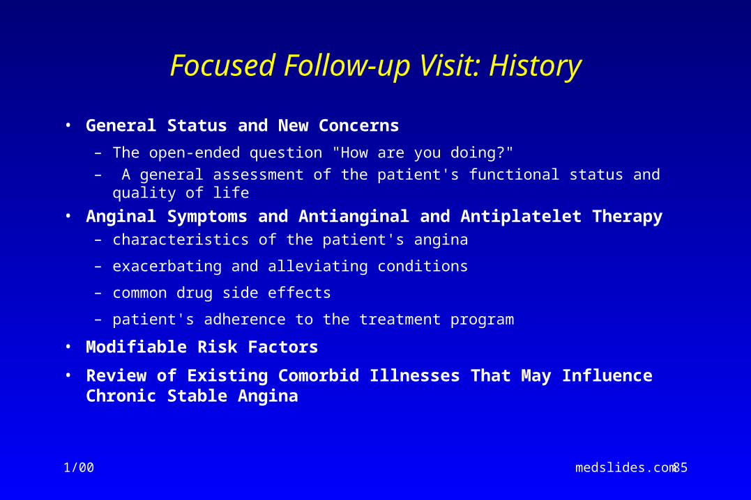
Focused Follow-up Visit: History
• General Status and New Concerns
– The open-ended question "How are you doing?"
– A general assessment of the patient's functional status and quality of life
• Anginal Symptoms and Antianginal and Antiplatelet Therapy – characteristics of the patient's angina
– exacerbating and alleviating conditions
– common drug side effects
– patient's adherence to the treatment program
• Modifiable Risk Factors
• Review of Existing Comorbid Illnesses That May Influence Chronic Stable Angina
1/00 medslides.com 86
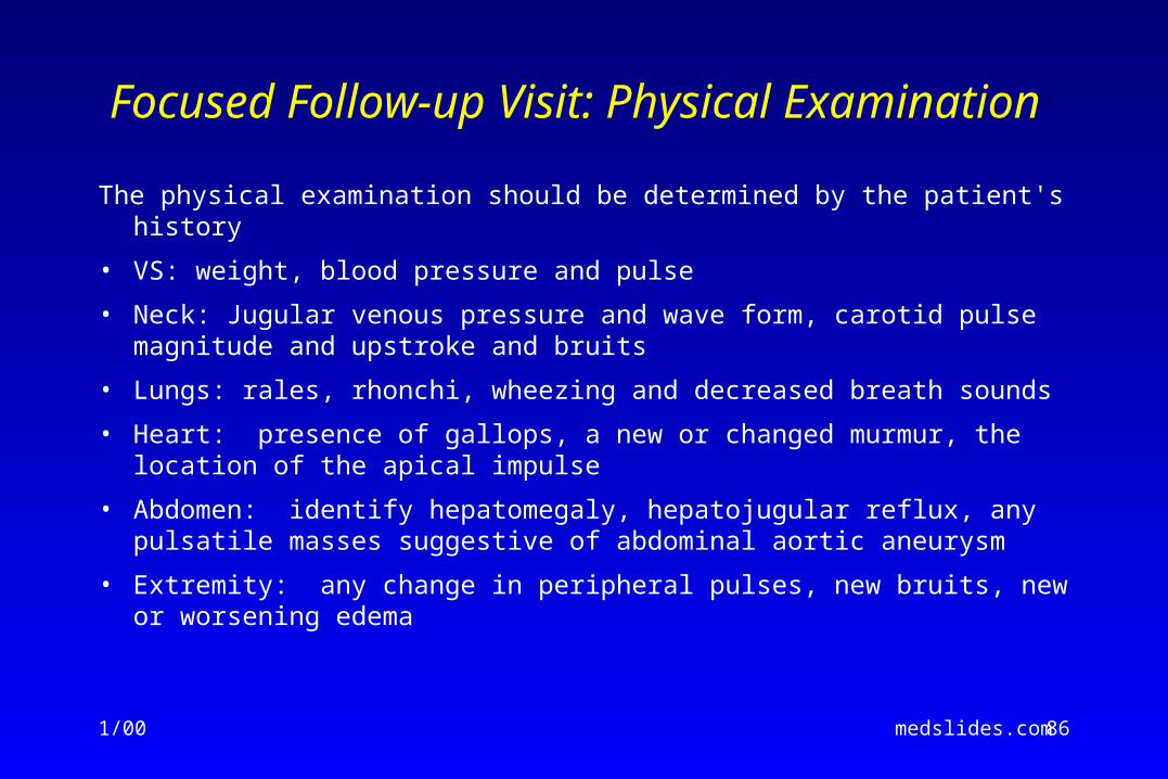
Focused Follow-up Visit: Physical Examination
The physical examination should be determined by the patient's history
• VS: weight, blood pressure and pulse
• Neck: Jugular venous pressure and wave form, carotid pulse magnitude and upstroke and bruits
• Lungs: rales, rhonchi, wheezing and decreased breath sounds
• Heart: presence of gallops, a new or changed murmur, the location of the apical impulse
• Abdomen: identify hepatomegaly, hepatojugular reflux, any pulsatile masses suggestive of abdominal aortic aneurysm
• Extremity: any change in peripheral pulses, new bruits, new or worsening edema
1/00 medslides.com 87
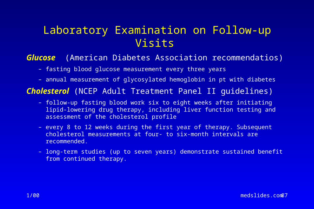
Laboratory Examination on Follow-up Visits
Glucose (American Diabetes Association recommendatios)– fasting blood glucose measurement every three years
– annual measurement of glycosylated hemoglobin in pt with diabetes
Cholesterol (NCEP Adult Treatment Panel II guidelines)– follow-up fasting blood work six to eight weeks after initiating lipid-lowering
drug therapy, including liver function testing and assessment of the cholesterol profile
– every 8 to 12 weeks during the first year of therapy. Subsequent cholesterol measurements at four- to six-month intervals are recommended.
– long-term studies (up to seven years) demonstrate sustained benefit from continued therapy.
1/00 medslides.com 88
Laboratory Examination on Follow-up Visits
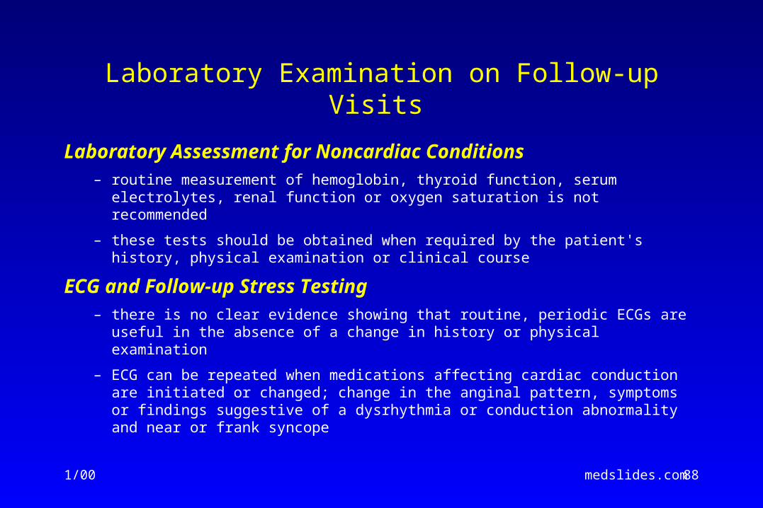
Laboratory Assessment for Noncardiac Conditions
– routine measurement of hemoglobin, thyroid function, serum electrolytes, renal function or oxygen saturation is not recommended
– these tests should be obtained when required by the patient's history, physical examination or clinical course
ECG and Follow-up Stress Testing
– there is no clear evidence showing that routine, periodic ECGs are useful in the absence of a change in history or physical examination
– ECG can be repeated when medications affecting cardiac conduction are initiated or changed; change in the anginal pattern, symptoms or findings suggestive of a dysrhythmia or conduction abnormality and near or frank syncope
1/00 medslides.com 89
Follow-up Stress Testing
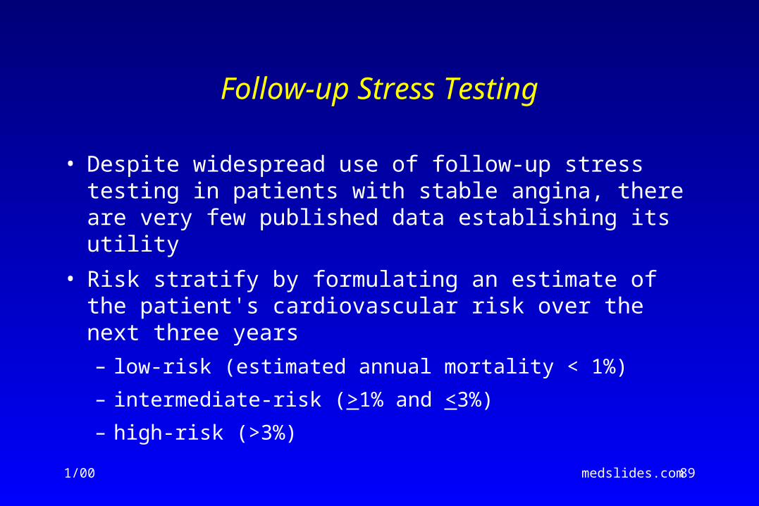
• Despite widespread use of follow-up stress testing in patients with stable angina, there are very few published data establishing its utility
• Risk stratify by formulating an estimate of the patient's cardiovascular risk over the next three years
– low-risk (estimated annual mortality < 1%)
– intermediate-risk (>1% and <3%)
– high-risk (>3%)
1/00 medslides.com 90
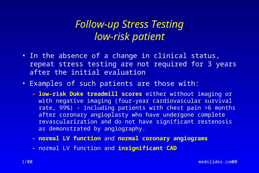
Follow-up Stress Testinglow-risk patient
• In the absence of a change in clinical status, repeat stress testing are not required for 3 years after the initial evaluation
• Examples of such patients are those with:
– low-risk Duke treadmill scores either without imaging or with negative imaging (four-year cardiovascular survival rate, 99%) - including patients with chest pain >6 months after coronary angioplasty who have undergone complete revascularization and do not have significant restenosis as demonstrated by angiography.
– normal LV function and normal coronary angiograms
– normal LV function and insignificant CAD
1/00 medslides.com 91
Follow-up Stress Testinghigh- and intermediate- risk patient
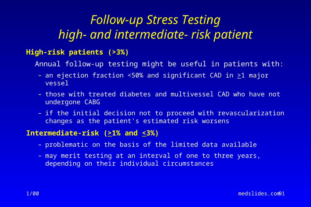
High-risk patients (>3%)
Annual follow-up testing might be useful in patients with:
– an ejection fraction <50% and significant CAD in >1 major vessel
– those with treated diabetes and multivessel CAD who have not undergone CABG
– if the initial decision not to proceed with revascularization changes as the patient's estimated risk worsens
Intermediate-risk (>1% and <3%)
– problematic on the basis of the limited data available
– may merit testing at an interval of one to three years, depending on their individual circumstances
1/00 medslides.com 92
Patient Education
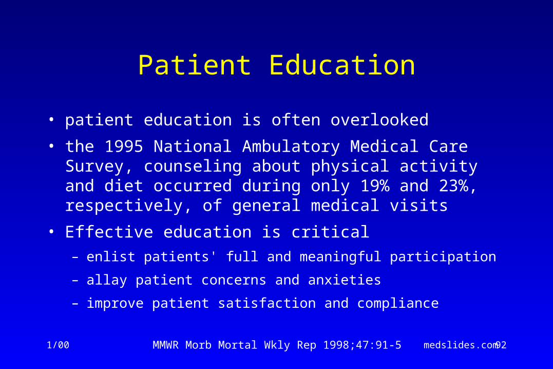
• patient education is often overlooked
• the 1995 National Ambulatory Medical Care Survey, counseling about physical activity and diet occurred during only 19% and 23%, respectively, of general medical visits
• Effective education is critical
– enlist patients' full and meaningful participation
– allay patient concerns and anxieties
– improve patient satisfaction and compliance
MMWR Morb Mortal Wkly Rep 1998;47:91-5
1/00 medslides.com 93
Principles of Patient Education
A well-designed educational programs can improve patients' knowledge and in some instances has been shown to improve outcomes
– Assess the patient's baseline understanding
– Elicit the patient's desire for information
– Use epidemiologic and clinical evidence
– Use ancillary personnel and professional when appropriate
– Use professionally prepared resources
– Develop a plan with the patient
– Involve family members in educational efforts
– Remind, repeat, and reinforce
1/00 medslides.com 94
Information for Patients General Aspects of Ischemic Heart Disease
• PATHOLOGY AND PATHOPHYSIOLOGY
– interested in varying level of detail
• RISK FACTORS
– useful to review the important known risk factors
• COMPLICATIONS – unstable angina, mi, heart failure, arrhythmia and sudden
cardiac death
1/00 medslides.com 95
Patient-Specific Information• PROGNOSIS
– useful to provide numerical estimates for risk of MI or death
• TREATMENT
– informed about their medications, including mechanisms of action, method of administration, and potentially adverse effects
• PHYSICAL ACTIVITY
– reassurance about returning to normal activities, activity limitations, and sexual relations; potentially serious consequences of using both sildenafil and nitrates within 24 h of one another
• RISK FACTOR REDUCTION
– greatest emphasis should be placed on modifiable factors
1/00 medslides.com 96
Patient-Specific Information
CONTACTING THE MEDICAL SYSTEM
• instructed about how and when to seek medical attention
• provide an action plan that covers:1) prompt use of aspirin and nitroglycerin if available2) how to access emergency medical services3) location of the nearest hospital that offers 24-h emergency
cardiovascular care
OTHER INFORMATION
• CPR training for family members is advisable
• counseling on potentially heritable condition (such as familial hypercholesterolemia) responsible for premature coronary disease.
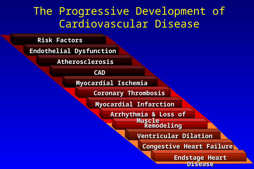
The Progressive Development of Cardiovascular Disease
Endstage Heart DiseaseEndstage Heart Disease
Congestive Heart FailureCongestive Heart Failure
Ventricular DilationVentricular Dilation
RemodelingRemodeling
Arrhythmia & Loss of MuscleArrhythmia & Loss of Muscle
Myocardial InfarctionMyocardial Infarction
Myocardial IschemiaMyocardial Ischemia
CADCAD
AtherosclerosisAtherosclerosis
Endothelial DysfunctionEndothelial Dysfunction
Risk FactorsRisk Factors
Coronary ThrombosisCoronary Thrombosis