Embed Size (px)
Citation preview
1
Chronic Stable AnginaTheodore D. Fraker, Stephan D. Fihn, and Raymond J. Gibbons
1
IntroductionClassifi cation of angina pectorisDemographics of angina pectorisPatients with new onset or changing anginal symptomsThe development of practice guidelinesAsymptomatic individualsRecommendations for the management of patients with
chronic stable angina Diagnosis
A. History and physical examination
B. Associated conditions
C. Noninvasive testing
D. Invasive testing: value of coronary angiography
Risk stratifi cation
A. Clinical evaluation
B. Noninvasive testing
C. Use of exercise test results in patient management
D. Coronary angiography and left ventriculography
Treatment
A. Pharmacologic therapy
Coronary disease risk factors and evidence that
treatment can reduce the risk for coronary
disease events
Patient follow-up: monitoring of symptoms and anti-
anginal therapy
Future issues Special consideration for women
New information on percutaneous revascularization to
be considered for the next chronic stable angina
guideline
New therapeutic agents to be considered for the next
chronic stable angina guideline
Introduction
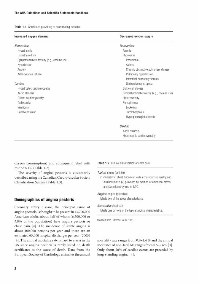
Angina pectoris is a clinical syndrome characterized by discomfort in the chest, jaw, back or arm typically aggravated by exertion or emotional stress and relieved by rest or nitroglycerin. Angina pectoris is usually associated with epicardial coronary artery disease including one or more obstructions of greater than 70%, but it can also occur in patients with valvular heart disease, hypertrophic cardiomy-opathy, or uncontrolled hypertension. Symptoms are thought to result from regional or global myo-cardial ischemia due to mismatch between myocar-dial oxygen supply and demand (Table 1.1). In women, angina pectoris can be seen in the absence of obvious epicardial coronary artery obstruction or other cardiac pathology, presumably due to coronary artery endothelial dysfunction or other factors. Chronic stable angina refers to anginal symptoms that occur daily, weekly or less frequently and are typically predictable and reproducible [1–4].
Classifi cation of angina pectoris
Chest discomfort can be described as typical angina, atypical angina or non-anginal chest pain, depend-ing upon whether or not symptoms occur with increased myocardial oxygen demand and are relieved by rest or nitroglycerin. Typical angina is usually described as a sensation of chest tightness, heaviness, pressure, burning or squeezing some-times accompanied by radiation to the inner arm, jaw, back or epigastrium. What makes the discom-fort “typical” is the predictable relationship to increased activity (implying increased myocardial
The AHA Guidelines and Scientifi c Statements Handbook,
First Edition. Edited by V Fuster. © 2009 American Heart
Association, ISBN: 9781405184632
COPYRIG
HTED M
ATERIAL
The AHA Guidelines and Scientifi c Statements Handbook
2
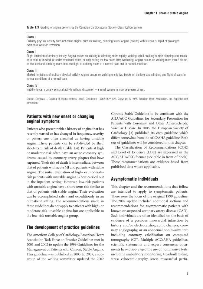
oxygen consumption) and subsequent relief with rest or NTG (Table 1.2).
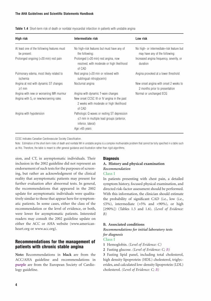
The severity of angina pectoris is customarily described using the Canadian Cardiovascular Society Classifi cation System (Table 1.3).
Demographics of angina pectoris
Coronary artery disease, the principal cause of angina pectoris, is thought to be present in 13,200,000 American adults, about half of whom (6,500,000 or 3.8% of the population) have angina pectoris or chest pain [4]. The incidence of stable angina is about 400,000 persons per year and there are an estimated 63,000 hospital discharges per year (2003) [4]. The annual mortality rate is hard to assess in the US since angina pectoris is rarely listed on death certifi cates as the cause of death. Data from the European Society of Cardiology estimates the annual
mortality rate ranges from 0.9–1.4 % and the annual incidence of non-fatal MI ranges from 0.5–2.6% [3]. Only about 20% of cardiac events are preceded by long-standing angina [4].
Table 1.1 Conditions provoking or exacerbating ischemia
Increased oxygen demand Decreased oxygen supply
Noncardiac Hyperthermia Hyperthyroidism Sympathomimetic toxicity (e.g., cocaine use) Hypertension Anxiety Arteriovenous fi stulae
Cardiac Hypertrophic cardiomyopathy Aortic stenosis Dilated cardiomyopathy Tachycardia Ventricular Supraventricular
Noncardiac Anemia Hypoxemia Pneumonia Asthma Chronic obstructive pulmonary disease Pulmonary hypertension Interstitial pulmonary fi brosis Obstructive sleep apnea Sickle cell disease Sympathomimetic toxicity (e.g., cocaine use) Hyperviscosity Polycythemia Leukemia Thrombocytosis Hypergammaglobulinemia
Cardiac Aortic stenosis Hypertrophic cardiomyopathy
Table 1.2 Clinical classifi cation of chest pain
Typical angina (defi nite) (1) Substernal chest discomfort with a characteristic quality and
duration that is (2) provoked by exertion or emotional stress and (3) relieved by rest or NTG.
Atypical angina (probable) Meets two of the above characteristics.
Noncardiac chest pain Meets one or none of the typical anginal characteristics.
Modifi ed from Diamond, IACC, 1983.
Chapter 1 Chronic Stable Angina
3
Table 1.3 Grading of angina pectoris by the Canadian Cardiovascular Society Classifi cation System
Class IOrdinary physical activity does not cause angina, such as walking, climbing stairs. Angina (occurs) with strenuous, rapid or prolonged exertion at work or recreation.
Class IISlight limitation of ordinary activity. Angina occurs on walking or climbing stairs rapidly, walking uphill, walking or stair climbing after meals, or in cold, or in wind, or under emotional stress, or only during the few hours after awakening. Angina occurs on walking more than 2 blocks on the level and climbing more than one fl ight of ordinary stairs at a normal pace and in normal condition.
Class IIIMarked limitations of ordinary physical activity. Angina occurs on walking one to two blocks on the level and climbing one fl ight of stairs in normal conditions at a normal pace.
Class IVInability to carry on any physical activity without discomfort – anginal symptoms may be present at rest.
Source: Campeau L. Grading of angina pectoris [letter]. Circulation, 1976;54:522–523. Copyright © 1976. American Heart Association. Inc. Reprinted with permission.
Patients with new onset or changing anginal symptoms
Patients who present with a history of angina that has recently started or has changed in frequency, severity or pattern are often classifi ed as having unstable angina. These patients can be subdivided by their short-term risk of death (Table 1.4). Patients at high or moderate risk often have an acute coronary syn-drome caused by coronary artery plaques that have ruptured. Their risk of death is intermediate, between that of patients with acute MI and patients with stable angina. The initial evaluation of high- or moderate-risk patients with unstable angina is best carried out in the inpatient setting. However, low-risk patients with unstable angina have a short-term risk similar to that of patients with stable angina. Their evaluation can be accomplished safely and expeditiously in an outpatient setting. The recommendations made in these guidelines do not apply to patients with high- or moderate-risk unstable angina but are applicable to the low-risk unstable angina group.
The development of practice guidelines
The American College of Cardiology/American Heart Association Task Force on Practice Guidelines met in 2001 and 2002 to update the 1999 Guidelines for the Management of Patients with Chronic Stable Angina. This guideline was published in 2003. In 2007, a sub-group of the writing committee updated the 2002
Chronic Stable Guideline to be consistent with the AHA/ACC Guidelines for Secondary Prevention for Patients with Coronary and Other Atherosclerotic Vascular Disease. In 2006, the European Society of Cardiology [3] published its own guideline which differs somewhat from the ACC/AHA guideline. Both sets of guidelines will be considered in this chapter.
The Classifi cation of Recommendations (COR) and Level of Evidence (LOE) are expressed in the ACC/AHA/ESC format (see table in front of book). These recommendations are evidence-based from published data where applicable.
Asymptomatic individuals
This chapter and the recommendations that follow are intended to apply to symptomatic patients. These were the focus of the original 1999 guideline. The 2002 update included additional sections and recommendations for asymptomatic patients with known or suspected coronary artery disease (CAD). Such individuals are often identifi ed on the basis of evidence of a previous myocardial infarction by history and/or electrocardiographic changes, coro-nary angiography, or an abnormal noninvasive test, including coronary calcifi cation on computed tomography (CT). Multiple ACC/AHA guidelines, scientifi c statements and expert consensus docu-ments have discouraged the use of noninvasive tests, including ambulatory monitoring, treadmill testing, stress echocardiography, stress myocardial perfu-
The AHA Guidelines and Scientifi c Statements Handbook
4
sion, and CT, in asymptomatic individuals. Their inclusion in the 2002 guideline did not represent an endorsement of such tests for the purposes of screen-ing, but rather an acknowledgment of the clinical reality that asymptomatic patients may present for further evaluation after abnormal tests. In general, the recommendations that appeared in the 2002 update for asymptomatic individuals were qualita-tively similar to those that appear here for symptom-atic patients. In some cases, either the class of the recommendation or the level of evidence, or both, were lower for asymptomatic patients. Interested readers may consult the 2002 guideline update on either the ACC or AHA website (www.american-heart.org or www.acc.org).
Recommendations for the management of patients with chronic stable angina
Note: Recommendations in black are from the ACC/AHA guideline and recommendations in purple are from the European Society of Cardio-logy guideline.
DiagnosisA. History and physical examinationRecommendationClass IIn patients presenting with chest pain, a detailed symptom history, focused physical examination, and directed risk-factor assessment should be performed. With this information, the clinician should estimate the probability of signifi cant CAD (i.e., low (i.e., ≤5%), intermediate (>5% and <90%), or high [≥90%]) (Tables 1.5 and 1.6). (Level of Evidence: B)
B. Associated conditionsRecommendations for initial laboratory tests for diagnosisClass I1 Hemoglobin. (Level of Evidence: C)2 Fasting glucose. (Level of Evidence: C; B)3 Fasting lipid panel, including total cholesterol, high density lipoprotein (HDL) cholesterol, triglyc-erides, and calculated low-density lipoprotein (LDL) cholesterol. (Level of Evidence: C; B)
Table 1.4 Short-term risk of death or nonfatal myocardial infarction in patients with unstable angina
High risk Intermediate risk Low risk
At least one of the following features must be present:
No high-risk features but must have any of the following:
No high- or intermediate-risk feature but may have any of the following:
Prolonged ongoing (>20 min) rest pain Prolonged (>20 min) rest angina, now resolved, with moderate or high likelihood of CAD
Increased angina frequency, severity, or duration
Pulmonary edema, most likely related to ischemia
Rest angina (>20 min or relieved with sublingual nitroglycerin)
Angina provoked at a lower threshold
Angina at rest with dynamic ST changes ≥1 mm
Nocturnal angina New onset angina with onset 2 weeks to 2 months prior to presentation
Angina with new or worsening MR murmur Angina with dynamic T-ware changes Normal or unchanged ECGAngina with S3 or new/worsening rales New onset CCSC III or IV angina in the past
2 weeks with moderate or high likelihood of CAD
Angina with hypotension Pathologic Q waves or resting ST depression ≤1 mm in multiple lead groups (anterior, inferior, lateral)
Age >65 years
CCSC indicates Canadian Cardiovascular Society Classifi cation.
Note: Estimation of the short-term risks of death and nonfatal MI in unstable angina is a complex multivariable problem that cannot be fully specifi ed in a table such
as this. Therefore, the table is meant to offer general guidance and illustration rather than rigid algorithms.
Chapter 1 Chronic Stable Angina
5
Table 1.5 Pretest likelihood of CAD in symptomatic patients according to age and sex* (combined Diamond/Forrester and CASS Data)
Nonanginal Chest pain Atypical angina Typical angina
Age (years) Men Women Men Women Men Women
30–39 4 2 34 12 76 2640-49 13 3 51 22 87 5550–59 20 7 65 31 93 7360–69 27 14 72 51 94 86
* Each value represents the percent with signifi cant CAD on catheterization.
Table 1.6 Comparing pretest likelihoods of CAD in low-risk symptomatic patients with high-risk symptomatic patients – Duke Database
Nonanginal Chest pain Atypical angina Typical angina
Age (years) Men Women Men Women Men Women
35 y 3–35 1–19 8–59 2–39 30–88 10–7845 y 9–47 2–22 21–70 5–43 51–92 20–7955 y 23–59 4–25 45–79 10–47 80–95 38–8265 y 49–69 9–29 71–86 20–51 93–97 56–84
Each value represents the percent with signifi cant CAD. The fi rst is the percentage for a low-risk, mid-decade patient without diabetes, smoking, or hyperlipidemia.
The second is that of the same age patient with diabetes, smoking, and hypelipidemia. Both high- and low-risk patients have normal resting ECGs. If ST-T-wave
changes or Q waves had been present, the likelihood of CAD would be higher in each entry of the table.
4 Full blood count including Hb and white cell count (Level of Evidence: B)5 Creatinine (Level of Evidence: C)6 Markers of myocardial damage if evaluation sug-gests clinical instability or acute coronary syndrome (Level of Evidence: A)7 Thyroid function if clinically indicated (Level of Evidence: C)
Class IIaOral glucose tolerance test (Level of Evidence: B)
Class IIb1 Hs C-reactive protein (Level of Evidence: B)2 Lipoprotein a, ApoA, and ApoB (Level of Evidence: B)3 Homocysteine (Level of Evidence: B)4 HbA1c (Level of Evidence: B)5 NT-BNP (Level of Evidence: B)
C. Noninvasive testing1. ECG/chest X-ray: Recommendations for electrocardiography, chest X-ray, or electron-beam computed tomography in the diagnosis of chronic stable anginaClass I1 A rest ECG in patients without an obvious non-cardiac cause of chest pain is recommended. (Level of Evidence: B)2 A rest ECG during an episode of chest pain is recommended. (Level of Evidence: B)3 A chest X-ray in patients with signs or symptoms of congestive heart failure (CHF), valvular heart disease, pericardial disease, or aortic dissection/aneurysm is recommended. (Level of Evidence: B)4 A resting ECG is recommended while the patient is pain-free. (Level of Evidence: C)
The AHA Guidelines and Scientifi c Statements Handbook
6
Class IIaA chest X-ray in patients with signs or symptoms of pulmonary disease is reasonable. (Level of Evidence: B)
Class IIb1 A chest X-ray in other patients may be consid-ered. (Level of Evidence: C)2 Electron-beam computed tomography may be considered. (Level of Evidence: B)3 A routine periodic ECG in the absence of clinical change may be considered. (Level of Evidence: C)
2. Recommendations for diagnosis of obstructive CAD with exercise ECG testing without an imaging modalityClass IExercise ECG is recommended in patients with an intermediate pretest probability of CAD (>5% and <90%) based on age, gender, and symptoms, includ-ing those with complete right bundle-branch block or less than 1 mm of ST depression at rest (excep-tions are listed below in classes II and III). (Level of Evidence: B) (See Tables 1.5 and 1.6).
Class IIaExercise ECG is reasonable in patients with suspected vasospastic angina. (Level of Evidence: C)
Class IIb1 Exercise ECG may be considered in patients with a high pretest probability of CAD by age, gender, and symptoms. (Level of Evidence: B)2 Exercise ECG may be considered in patients with a low pretest probability of CAD by age, gender, and symptoms. (Level of Evidence: B)3 Exercise ECG may be considered in patients taking digoxin whose ECG has less than 1 mm of baseline ST-segment depression. (Level of Evidence: B)4 Exercise ECG may be considered in patients with ECG criteria for LVH and less than 1 mm of baseline ST-segment depression. (Level of Evidence: B)5 Routine periodic exercise ECG may be reasonable in the absence of clinical change. (Level of Evidence: C)
Class III1 Exercise ECG is not recommended in patients with the following baseline ECG abnormalities.
a. Pre-excitation (Wolff–Parkinson–White) syn-drome. (Level of Evidence: B)b. Electronically paced ventricular rhythm. (Level of Evidence: B)c. More than 1 mm of ST depression at rest. (Level of Evidence: B)d. Complete left bundle-branch block. (Level of Evidence: B)
2 Exercise ECG is not recommended in patients with an established diagnosis of CAD owing to prior MI or coronary angiography; however, testing can assess functional capacity and pro-gnosis, as discussed in Section III. (Level of Evidence: B)
3. Echocardiography: Recommendations for echocardiography for diagnosis of cause of chest pain in patients with suspected chronic stable angina pectorisClass I1 Echocardiography is recommended for patients with systolic murmur suggestive of aortic stenosis or hypertrophic cardiomyopathy (Level of Evidence: C, B)2 Echocardiography is recommended for evalua-tion of extent (severity) of ischemia (e.g., LV segmental wall-motion abnormality) when the echocardiogram can be obtained during pain or within 30 min after its abatement. (Level of Evidence: C)3 Echocardiography is recommended for patients with suspected heart failure (Level of Evidence: B).4 Echocardiography is recommended for patients with prior MI (Level of Evidence: B).5 Echocardiography is recommended for patients with LBBB, Q waves or other signifi cant patho-logical changes on ECG, including electrocardio-graphic left anterior hemiblock (Level of Evidence: C).
Class IIbEchocardiography may be considered in patients with a click or murmur to diagnose mitral valve prolapse [15]. (Level of Evidence: C)
Chapter 1 Chronic Stable Angina
7
Class IIIEchocardiography is not recommended in patients with a normal ECG, no history of MI, and no signs or symptoms suggestive of heart failure, valvular heart disease, or hypertrophic cardiomyopathy. (Level of Evidence: C)
4. Stress imaging studies: echocardiographic and nuclear recommendations for cardiac stress imaging as the initial test for diagnosis in patients with chronic stable angina who are able to exerciseSee Table 1.7.Class I1 Exercise myocardial perfusion imaging or exer-cise echocardiography is recommended in patients with an intermediate pretest probability of CAD who have one of the following baseline ECG abnormalities:
a. Pre-excitation (Wolff–Parkinson–White) syn-drome. (Level of Evidence: B)b. More than 1 mm of ST depression at rest. (Level of Evidence: B)
2 Exercise myocardial perfusion imaging or exer-cise echocardiography is recommended in patients with prior revascularization (either PCI or CABG). (Level of Evidence: B)
3 Adenosine or dipyridamole myocardial perfusion imaging is recommended in patients with an inter-mediate pretest probability of CAD and one of the following baseline ECG abnormalities:
a. Electronically paced ventricular rhythm. (Level of Evidence: C)b. Left bundle-branch block. (Level of Evidence: B)
4 Exercise myocardial perfusion imaging or exer-cise echocardiography is recommended in patients with a non-conclusive exercise ECG but reason-able exercise tolerance, who do not have a high probability of signifi cant coronary disease and in whom the diagnosis is still in doubt. (Level of Evidence: B)
Class IIaExercise myocardial perfusion imaging or exercise echocardiography is reasonable in the following circumstances:
1 Patients with prior revascularization (PCI or CABG) in whom localization of ischaemia is impor-tant. (Level of evidence: B)2 As an alternative to exercise ECG in patients where facilities, costs, and personnel resources allow. (Level of evidence: B)3 As an alternative to exercise ECG in patients with a low pre-test probability of disease such as women with atypical chest pain. (Level of Evidence: B)4 To assess functional severity of intermediate lesions on coronary arteriography. (Level of Evi-dence: C)5 To localize ischaemia when planning revascular-ization options in patients who have already had arteriography. (Level of Evidence: B)6 Pharmacological stress imaging techniques [either echocardiography or perfusion] are reasonable with the same Class I indications outlined above, where local facilities favor pharmacologic rather than exer-cise stress. (Level of Evidence: B)
Class IIb1 Exercise myocardial perfusion imaging or exer-cise echocardiography may be considered in patients with a low or high probability of CAD who have one of the following baseline ECG abnormalities:
a. Pre-excitation (Wolff–Parkinson–White) syn-drome. (Level of Evidence: B)
Table 1.7 Comparative advantages of stress echocardiography and stress radionuclide perfusion imaging in diagnosis of CAD
Advantages of stress echocardiography 1. Higher specifi city 2. Versatility – more extensive evaluation of cardiac anatomy
and function 3. Greater convenience/effi cacy/availability 4. Lower cost
Advantages of stress perfusion imaging 1. Higher technical success rate 2. Higher sensitivity – especially for single vessel coronary
disease involving the left circumfl ex 3. Better accuracy in evaluating possible ischemia when multiple
resting IV wall motion abnormalities are present 4. More extensive published database – especially in evaluation
of prognosis
The AHA Guidelines and Scientifi c Statements Handbook
8
b. More than 1 mm of ST depression. (Level of Evidence: B)
2 Adenosine or dipyridamole myocardial perfusion imaging may be considered in patients with a low or high probability of CAD and one of the following baseline ECG abnormalities:
a. Electronically paced ventricular rhythm. (Level of Evidence: C)b. Left bundle-branch block. (Level of Evidence: B)
3 Exercise myocardial perfusion imaging or exer-cise echocardiography may be considered in patients with an intermediate probability of CAD who have one of the following:
a. Digoxin use with less than 1 mm ST depression on the baseline ECG. (Level of Evidence: B)b. LVH with less than 1 mm ST depression on the baseline ECG. (Level of Evidence: B)
4 Exercise myocardial perfusion imaging, exercise echocardiography, adenosine or dipyridamole myo-cardial perfusion imaging, or dobutamine echocar-diography may be considered as the initial stress test in a patient with a normal rest ECG who is not taking digoxin. (Level of Evidence: B)5 Exercise or dobutamine echocardiography may be considered in patients with left bundle-branch block. (Level of Evidence: C)
5. Recommendations for cardiac stress imaging as the initial test for diagnosis in patients with chronic stable angina who are unable to exercise(Pharmacological stress with imaging techniques [either echocardiography or perfusion] is recom-mended in the initial assessment of angina with the same Class I, IIa and IIb indications outlined above, if the patient is unable to exercise adequately.)
Class I1 Adenosine or dipyridamole myocardial perfusion imaging or dobutamine echocardiography is recom-mended in patients with an intermediate pretest probability of CAD. (Level of Evidence: B)2 Adenosine or dipyridamole stress myocardial per-fusion imaging or dobutamine echocardiography is recommended in patients with prior revascul-arization (either PCI or CABG). (Level of Evidence: B)
Class IIb1 Adenosine or dipyridamole stress myocardial per-fusion imaging or dobutamine echocardiography may be considered in patients with a low or high probability of CAD in the absence of electronically paced ventricular rhythm or left bundle-branch block. (Level of Evidence: B)2 Adenosine or dipyridamole myocardial perfusion imaging may be considered in patients with a low or a high probability of CAD and one of the following baseline ECG abnormalities:
a. Electronically paced ventricular rhythm. (Level of Evidence: C)b. Left bundle-branch block. (Level of Evidence: B)
3 Dobutamine echocardiography in patients with left bundle-branch block. (Level of Evidence: C)
6. Recommendations for ambulatory ECG for initial diagnostic assessment of anginaClass IAn ambulatory ECG is recommended for angina with suspected arrhythmia. (Level of Evidence: B)
Class IIaAn ambulatory ECG may be reasonable for sus-pected vasospastic angina. (Level of Evidence: C)
7. Recommendations for the use of CT angiography in stable anginaClass IIbCT angiography may be considered in patients with a low pre-test probability of disease, with a noncon-clusive exercise ECG or stress imaging test. (Level of Evidence: C)
D. Invasive testing: value of coronary angiographyRecommendations for coronary angiography to establish a diagnosis in patients with suspected angina, including those with known CAD who have a signifi cant change in anginal symptomsClass I1 Coronary angiography is recommended in patients with known or possible angina pectoris who have survived sudden cardiac death. (Level of Evi-dence: B)
Chapter 1 Chronic Stable Angina
9
2 Coronary angiography is recommended in patients with severe stable angina (Class 3 or greater of Canadian Cardiovascular Society Classifi -cation, with a high pre-test probability of disease, particularly if the symptoms are inadequately responding to medical treatment.) (Level of Evi-dence: B)3 Coronary angiography is recommended in patients with serious ventricular arrhythmias. (Level of Evidence: C)4 Coronary angiography is recommended in patients previously treated by myocardial revascu-larization (PCI, CABG), who develop early recur-rence of moderate or severe angina pectoris. (Level of Evidence: C)
Class IIa1 Coronary angiography is reasonable in patients with an uncertain diagnosis after noninvasive testing in whom the benefi t of a more certain diagnosis outweighs the risk and cost of coronary angiogra-phy. (Level of Evidence: C)2 Coronary angiography is reasonable in patients who cannot undergo noninvasive testing because of disability, illness, or morbid obesity. (Level of Evi-dence: C)3 Coronary angiography is reasonable in patients with an occupational requirement for a defi nitive diagnosis. (Level of Evidence: C)4 Coronary angiography is reasonable in patients who by virtue of young age at onset of symptoms, noninvasive imaging, or other clinical parameters are suspected of having a nonatherosclerotic cause for myocardial ischemia (coronary artery anomaly, Kawasaki disease, primary coronary artery dissec-tion, radiation-induced vasculopathy). (Level of Evi-dence: C)5 Coronary angiography is reasonable in patients in whom coronary artery spasm is suspected and provocative testing may be necessary. (Level of Evidence: C)6 Coronary angiography is reasonable in patients with a high pretest probability of left main or three-vessel CAD. (Level of Evidence: C)7 Coronary angiography is reasonable in patients with a high risk of restenosis after PCI, if PCI has been performed in a prognostically important site. (Level of Evidence: C)
Class IIb1 Coronary angiography may be considered in patients with recurrent hospitalization for chest pain in whom a defi nite diagnosis is judged necessary. (Level of Evidence: C)2 Coronary angiography may be considered in patients with an overriding desire for a defi nitive diagnosis and a greater-than-low probability of CAD. (Level of Evidence: C)
Class III1 Coronary angiography is not recommended in patients with signifi cant comorbidity in whom the risk of coronary arteriography outweighs the benefi t of the procedure. (Level of Evidence: C)2 Coronary angiography is not recommended in patients with an overriding personal desire for a defi nitive diagnosis and a low probability of CAD. (Level of Evidence: C)
Risk stratifi cationThe recommendations that follow are for risk strati-fi cation by clinical evaluation, including ECG and laboratory tests, in stable angina.
A. Clinical evaluationClass I1 A detailed clinical history and physical examina-tion is recommended including BMI and/or waist circumference in all patients, also including a full description of symptoms, quantifi cation of functional impairment, past medical history, and cardiovascular risk profi le. (Level of Evidence: B) (Figure 1.1).2 Resting ECG in all patients is recommended. (Level of Evidence: B)
B. Noninvasive testingRecommendations for measurement of rest LV function by echocardiography or radionuclide angiography in patients with chronic stable anginaClass I1 Echocardiography or RNA is recommended in patients with a history of prior MI, pathologic Q waves, or symptoms or signs suggestive of heart
The AHA Guidelines and Scientifi c Statements Handbook
10
failure to assess LV function. (Level of Evidence: B)2 Echocardiography is recommended in patients with a systolic murmur that suggests mitral regurgi-tation to assess its severity and etiology. (Level of Evidence: C)3 Echocardiography or RNA is recommended in patients with complex ventricular arrhythmias to assess LV function. (Level of Evidence: B)4 Resting echocardiography is recommended in patients with hypertension. (Level of Evidence: B)5 Resting echocardiography is recommended in patients with diabetes. (Level of Evidence: C)
Class IIaResting echocardiography is recommended in patients with a normal resting ECG without prior MI who are not otherwise to be considered for coro-nary arteriography. (Level of Evidence: C)
Class III1 Echocardiography or RNA is not recommended for routine periodic reassessment of stable patients for whom no new change in therapy is contem-plated. (Level of Evidence: C)2 Echocardiography or RNA is not recommended in patients with a normal ECG, no history of MI,
and no symptoms or signs suggestive of CHF. (Level of Evidence: B)
Recommendations for exercise testing risk assessment and prognosis in patients with an intermediate or high probability of CADClass I1 Exercise testing is recommended in patients undergoing initial evaluation. (Exceptions are listed below in Classes IIb and III) (Level of Evidence: B)2 Exercise testing is recommended in patients after a signifi cant change in cardiac symptoms. (Level of Evidence: C). (Tables 1.8, 1.9 and 1.10).
Class IIaExercise testing is reasonable in patients post-revascularization with a signifi cant deterioration in symptomatic status. (Level of Evidence: B)
Class IIb1 Exercise testing may be considered in patients with the following ECG abnormalities:
a. Pre-excitation (Wolff-Parkinson-White) syn-drome. (Level of Evidence: B)b. Electronically paced ventricular rhythm. (Level of Evidence: B)c. More than 1 mm of ST depression at rest. (Level of Evidence: B)d. Complete left bundle-branch block. (Level of Evidence: B)
2 Exercise testing may be considered in patients who have undergone cardiac catheterization to identify ischemia in the distribution of coronary lesion of borderline severity. (Level of Evidence: C)3 Exercise testing may be considered in post-revas-cularization patients who have a signifi cant change in anginal pattern suggestive of ischemia. (Level of Evidence: C)
Class IIIExercise testing is not recommended in patients with severe comorbidity likely to limit life expec-tancy or prevent revascularization. (Level of Evi-dence: C)
1.0
0.8
0.6
0.4
0.2
0.030 35 40 45 50 55
Age, y
60 65 70 75 80
Pre
dic
ted p
robabili
ty
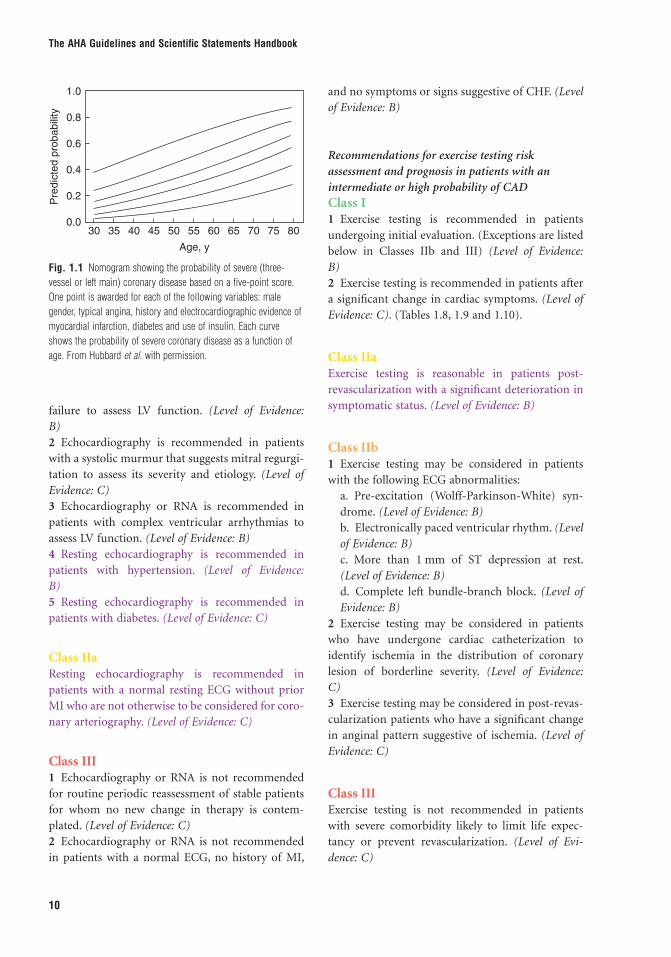
Fig. 1.1 Nomogram showing the probability of severe (three-vessel or left main) coronary disease based on a fi ve-point score. One point is awarded for each of the following variables: male gender, typical angina, history and electrocardiographic evidence of myocardial infarction, diabetes and use of insulin. Each curve shows the probability of severe coronary disease as a function of age. From Hubbard et al. with permission.
Chapter 1 Chronic Stable Angina
11
Table 1.8 Survival according to risk groups based on Duke Treadmill Scores
Risk group (score) Percentage of total Four-year survival Annual mortality (percent)
Low (≥+5) 62 0.99 0.25Moderate (−10 to +4) 34 0.95 1.25High (<−10) 4 0.79 5.0
The Duke treadmill score equals the exercise time in minutes minus (5 times the ST-segment deviation, during or after exercise, in millimeters).
Table 1.9 Noninvasive risk stratifi cation
High-risk (greater than 3% annual mortality rate) 1. Severe resting left ventricular dysfunction (LVEF < 35%) 2. High-risk treadmill score (score ≤−11) 3. Severe exercise left ventricular dysfunction (exercise LVEF < 35%) 4. Stress-induced large perfusion defect (particularly if anterior) 5. Stress-induced multiple perfusion defects of moderate size 6. Large, fi xed perfusion defect with LV dilation or increased lung uptake (thallium-201) 7. Stress-induced moderate perfusion defect with LV dilation or increased lung uptake (thallium-201) 8. Echocardiographic wall motion abnormality (involving greater than two segments) developing at low dose of dobutamine (≤10 mg/kg/
min) or at a low heart rate (<120 beats/min) 9. Stress echocardiographic evidence of extensive ischemia
Intermediate-risk (1–3% annual mortality rate) 1. Mild/moderate resting left ventricular dysfunction (LVEF = 35% to 49%) 2. Intermediate-risk treadmill score (−11 < score < 5) 3. Stress-induced moderate perfusion defect without LV dilation or increased lung intake (thallium-201) 4. Limited stress echocardiographic ischemia with a wall motion abnormality only at higher doses of dobutamine involving less than or
equal to two segments
Low-risk (less than 1% annual mortality rate) 1. Low-risk treadmill score (score ≥5) 2. Normal or small myocardial perfusion defect at rest or with stress* 3. Normal stress echocardiographic wall motion or no change of limited resting wall motion abnormalities during stress*
* Although the published data are limited, patients with these fi ndings will probably not be at low risk in the presence of either a high-risk treadmill score or severe
resting left ventricular dysfunction (LVEF < 35%).
C. Use of exercise test results in patient managementRecommendation for exercise testing in patients with chest pain 6 months or more after revascularizationClass IIbExercise testing may be considered in patients with a signifi cant change in anginal pattern suggestive of ischemia. (Level of Evidence: B)
Recommendations for cardiac stress imaging as the initial test for risk stratifi cation of patients with chronic stable angina who are able to exerciseClass I1 Exercise myocardial perfusion imaging or exer-cise echocardiography is recommended to identify the extent, severity, and location of ischemia in patients who do not have left bundle-branch block or an electronically paced ventricular rhythm and
The AHA Guidelines and Scientifi c Statements Handbook
12
who either have an abnormal rest ECG or are using digoxin. (Level of Evidence: B)2 Dipyridamole or adenosine myocardial perfusion imaging is recommended in patients with left bundle-branch block or electronically paced ven-tricular rhythm. (Level of Evidence: B)3 Exercise myocardial perfusion imaging or exer-cise echocardiography is recommended to assess the functional signifi cance of coronary lesions (if not already known) in planning PCI. (Level of Evidence: B)4 Exercise myocardial perfusion imaging or exercise echocardiography is recommended in patients with a non-conclusive exercise ECG, but intermediate or high probability of disease. (Level of Evidence: B)
Class IIa1 Exercise myocardial perfusion imaging or exer-cise echocardiography is reasonable in patients with a deterioration in symptoms post-revascularization. (Level of Evidence B)2 Exercise myocardial perfusion imaging or exer-cise echocardiography is reasonable as an altern-ative to exercise ECG in patients, in which facilities, cost, and personnel resources allow. (Level of Evi-dence: B)
3 Pharmacological stress imaging techniques [either echocardiography or perfusion] are reasonable with the same Class I indications outlined above, where local facilities favor pharmacologic rather than exer-cise stress (Level of Evidence: B)
Class IIb1 Exercise or dobutamine echocardiography may be considered in patients with left bundle-branch block. (Level of Evidence: C)2 Exercise, dipyridamole, or adenosine myocardial perfusion imaging, or exercise or dobutamine echo-cardiography may be considered as the initial test in patients who have a normal rest ECG and who are not taking digoxin. (Level of Evidence: B)
Class III1 Exercise myocardial perfusion imaging is not rec-ommended in patients with left bundle-branch block. (Level of Evidence: C)2 Exercise, dipyridamole, or adenosine myocardial perfusion imaging, or exercise or dobutamine echo-cardiography is not recommended in patients with severe comorbidity likely to limit life expecta-tion or prevent revascularization. (Level of Evi-dence: C)
Table 1.10 CAD Prognostic Index
Extent of CAD Prognostic weight (0–100) 5-Year survival rate (%)*
1-vessel disease, 75% 23 93>1-vessel disease, 50% to 74% 23 931-vessel disease, ≥95% 32 912-vessel disease 37 882-vessel disease, both ≥95% 42 861-vessel disease, ≥95% proximal LAD 48 832-vessel disease, ≥95% LAD 48 832-vessel disease, ≥95% proximal LAD 56 793-vessel disease 56 793-vessel disease, ≥95% m at least 1 63 733-vessel disease, 75% proximal LAD 67 673-vessel disease, ≥95% proximal LAD 74 59
* Assuming medical treatment only. CAD indicates coronary artery disease; LAD, left anterior descending artery. From Califf RM, Armstrong PW. Carver JR, et al:
Task Force 5. Stratifi cation of patients into high-, medium- and low-risk subgroups for purposes of risk factor management. J Am Coll Cardiol.
1996;27:964–1047.
Chapter 1 Chronic Stable Angina
13
Recommendations for cardiac stress imaging as the initial test for risk stratifi cation of patients with chronic stable angina who are unable to exerciseClass I1 Dipyridamole or adenosine myocardial perfusion imaging or dobutamine echocardiography is recom-mended to identify the extent, severity, and location of ischemia in patients who do not have left bundle-branch block or electronically paced ventricular rhythm. (Level of Evidence: B)2 Dipyridamole or adenosine myocardial perfusion imaging is recommended in patients with left bundle-branch block or electronically paced ven-tricular rhythm. (Level of Evidence: B)3 Dipyridamole or adenosine myocardial perfusion imaging or dobutamine echocardiography is recom-mended to assess the functional signifi cance of coro-nary lesions (if not already known) in planning PCI. (Level of Evidence: B)
Class IIbDobutamine echocardiography may be considered in patients with left bundle-branch block. (Level of Evidence: C)
Class IIIDipyridamole or adenosine myocardial perfusion imaging or dobutamine echocardiography is not recommended in patients with severe comorbidity likely to limit life expectation or prevent revascular-ization. (Level of Evidence: C)
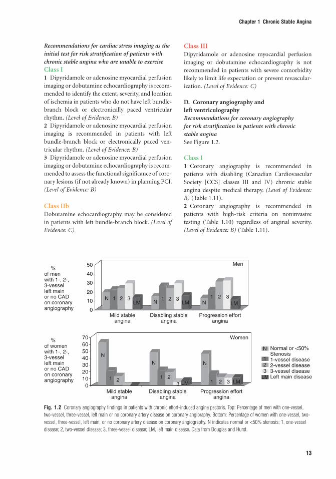
D. Coronary angiography and left ventriculographyRecommendations for coronary angiography for risk stratifi cation in patients with chronic stable anginaSee Figure 1.2.
Class I1 Coronary angiography is recommended in patients with disabling (Canadian Cardiovascular Society [CCS] classes III and IV) chronic stable angina despite medical therapy. (Level of Evidence: B) (Table 1.11).2 Coronary angiography is recommended in patients with high-risk criteria on noninvasive testing (Table 1.10) regardless of anginal severity. (Level of Evidence: B) (Table 1.11).
%of womenwith 1-, 2-,3-vesselleft mainor no CADon coronaryangiography
0
10
20
30
40
50
60
70
%of menwith 1-, 2-,3-vesselleft mainor no CADon coronaryangiography
Normal or <50%Stenosis1-vessel disease2-vessel disease3-vessel diseaseLeft main disease
0
10 N
Mild stableangina
Disabling stableangina
Progression effortangina
LM
LM
1 2 3N LM N LM
1 2 3
Men
Women
1 2 320
30
40
50
N
Mild stableangina
Disabling stableangina
Progression effortangina
1 2
N N
N
123
LM1 2
3 1 2 3 LM
Fig. 1.2 Coronary angiography fi ndings in patients with chronic effort-induced angina pectoris. Top: Percentage of men with one-vessel, two-vessel, three-vessel, left main or no coronary artery disease on coronary angioraphy. Bottom: Percentage of women with one-vessel, two-vessel, three-vessel, left main, or no coronary artery disease on coronary angiography. N indicates normal or <50% stenosis; 1, one-vessel disease; 2, two-vessel disease; 3, three-vessel disease; LM, left main disease. Data from Douglas and Hurst.
The AHA Guidelines and Scientifi c Statements Handbook
14
3 Coronary angiography is recommended in patients with angina who have survived sudden cardiac death or serious ventricular arrhythmia. (Level of Evidence: B)4 Coronary angiography is recommended in patients with angina and symptoms and signs of CHF. (Level of Evidence: C)5 Coronary angiography is recommended in patients with clinical characteristics that indicate a high likeli-hood of severe CAD. (Level of Evidence: C)6 Coronary angiography is recommended in patients with stable angina in patients who are being considered for major noncardiac surgery, especially vascular surgery (repair of aortic aneurysm, femoral bypass, carotid endarterectomy) with intermediate or high risk features on noninvasive testing. (Level of Evidence: B)
Class IIa1 Coronary angiography is reasonable in patients with signifi cant LV dysfunction (ejection fraction less than 45%), CCS class I or II angina, and demon-strable ischemia but less than high-risk criteria on noninvasive testing. (Level of Evidence: C)2 Coronary angiography is reasonable in patients with inadequate prognostic information after non-invasive testing. (Level of Evidence: C)3 Coronary angiography is reasonable in patients with a high risk of restenosis after PCI, if PCI has been performed in a prognostically important site. (Level of Evidence: C)
Class IIb1 Coronary angiography may be considered in patients with CCS class I or II angina, preserved LV function (ejection fraction greater than 45%), and less than high-risk criteria on noninvasive testing. (Level of Evidence: C)2 Coronary angiography may be considered in patients with CCS class III or IV angina, which with medical therapy improves to class I or II. (Level of Evidence: C)3 Coronary angiography may be considered in patients with CCS class I or II angina but intolerance (unacceptable side effects) to adequate medical therapy. (Level of Evidence: C)
Class III1 Coronary angiography is not recommended in patients with CCS class I or II angina who respond to medical therapy and who have no evidence of isch-emia on noninvasive testing. (Level of Evidence: C)2 Coronary angiography is not recommended in patients who prefer to avoid revascularization. (Level of Evidence: C)
Recommendations for investigation in patients with the classical triad of Syndrome XClass IA resting echocardiogram is recommended in patients with angina and normal or non-obstructed
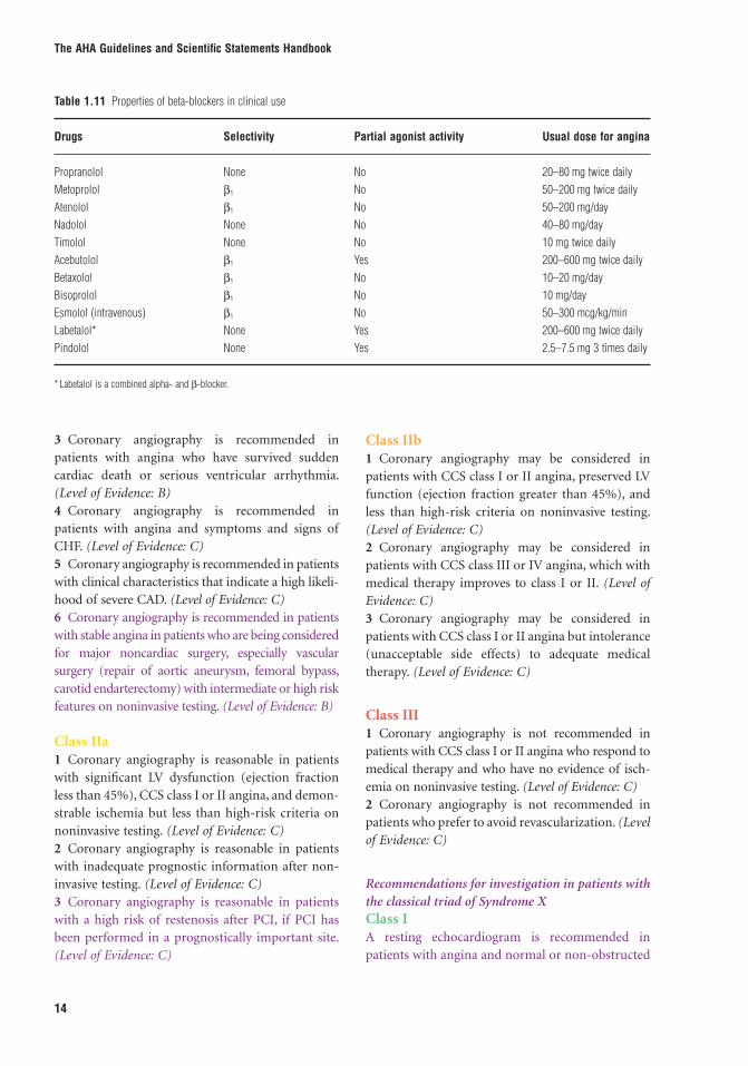
Table 1.11 Properties of beta-blockers in clinical use
Drugs Selectivity Partial agonist activity Usual dose for angina
Propranolol None No 20–80 mg twice dailyMetoprolol β1 No 50–200 mg twice dailyAtenolol β1 No 50–200 mg/dayNadolol None No 40–80 mg/dayTimolol None No 10 mg twice dailyAcebutolol β1 Yes 200–600 mg twice dailyBetaxolol β1 No 10–20 mg/dayBisoprolol β1 No 10 mg/dayEsmolol (intravenous) β1 No 50–300 mcg/kg/minLabetalol* None Yes 200–600 mg twice dailyPindolol None Yes 2.5–7.5 mg 3 times daily
* Labetalol is a combined alpha- and β-blocker.
Chapter 1 Chronic Stable Angina
15
coronary arteries to assess for presence of ventricu-lar hypertrophy and/or diastolic dysfunction. (Level of Evidence: C)
Class IIb1 Intracoronary acetylcholine is reasonable during coronary arteriography, if the arteriogram is visually normal, to assess endothelium dependent coronary fl ow reserve, and exclude vasospasm. (Level of Evi-dence: C)2 Intracoronary ultrasound, coronary fl ow reserve, or fractional fl ow reserve are reasonable measure-ments to exclude missed obstructive lesions, if angiographic appearances are suggestive of a non-obstructive lesion rather than completely normal, and stress imaging techniques identify an extensive area of ischaemia. (Level of Evidence: C)
TreatmentA. Pharmacologic therapyRecommendations for pharmacotherapy to prevent MI and death and to reduce symptomsClass I1 Aspirin should be started at 75 to 162 mg per day (75 mg per day in ESC guideline) and continued
indefi nitely in all patients unless contraindicated. (Level of Evidence: A)2 Beta-blockers as initial therapy is recommended to reduce symptoms in the absence of contraindica-tions in patients with prior MI (Level of Evidence: A) or without prior MI. (Level of Evidence: B)Test the effects of a beta-1 blocker, and titrate to full dose; consider the need for 24 h protection against ischemia. (Level of Evidence: A) (Table 1.12).3 It is benefi cial to start and continue beta-blocker therapy indefi nitely in all patients who have had MI, acute coronary syndrome, or left ventricular dys-function with or without heart failure symptoms, unless contraindicated. (Level of Evidence: A)4 ACE inhibitors should be started and continued indefi nitely in all patients with left ventricular ejec-tion fraction less than or equal to 40% and in those with hypertension, diabetes, or chronic kidney disease unless contraindicated. (Level of Evidence: A)5 ACE inhibitors should be started and continued indefi nitely in patients who are not lower risk (lower risk defi ned as those with normal left ventricular ejec-tion fraction in whom cardiovascular risk factors are well controlled and revascularization has been per-formed), unless contraindicated. (Level of Evidence: B)
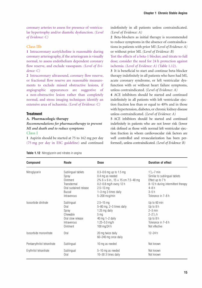
Table 1.12 Nitroglycerin and nitrates in angina
Compound Route Dose Duration of effect
Nitroglycerin Sublingual tablets 0.3–0.6 mg up to 1.5 mg 11/2–7 minSpray 0.4 mg as needed Similar to sublingual tabletsOintment 2% 6 × 6 in., 15 × 15 cm 7.5–40 mg Effect up to 7 hTransdermal 0.2–0.8 mg/h every 12 h 8–12 h during intermittent therapyOral sustained release 2.5–13 mg 4–8 hBuccal 1–3 mg 3 times daily 3–5 hIntravenous 5–200 mcg/min Tolerance in 7–8 h
Isosorbide dinitrate Sublingual 2.5–15 mg Up to 60 minOral 5–80 mg, 2–3 times daily Up to 8 hSpray 1.25 mg daily 2–3 minChewable 5 mg 2–21/2 hOral slow release 40 mg 1–2 daily Up to 8 hIntravenous 1.25–5.0 mg/h Tolerance in 7–8 hOintment 100 mg/24 h Not effective
Isosorbide mononitrate Oral 20 mg twice daily 12–24 h60–240 mg once daily
Pentaerythritol tetranitrate Sublingual 10 mg as needed Not known
Erythritol tetranitrate Sublingual 5–10 mg as needed Not knownOral 10–30 3 times daily Not known
The AHA Guidelines and Scientifi c Statements Handbook
16
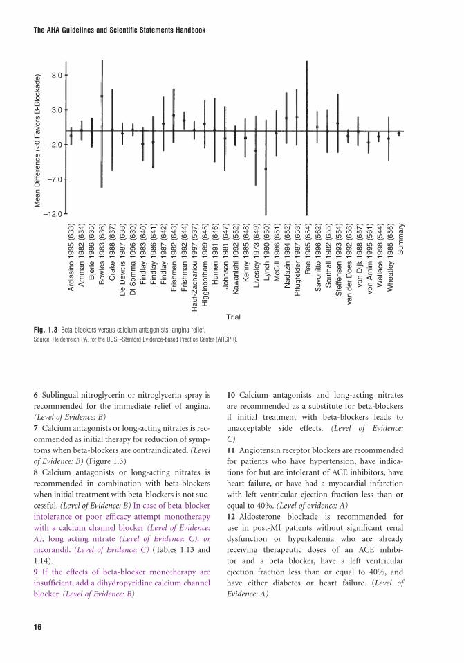
6 Sublingual nitroglycerin or nitroglycerin spray is recommended for the immediate relief of angina. (Level of Evidence: B)7 Calcium antagonists or long-acting nitrates is rec-ommended as initial therapy for reduction of symp-toms when beta-blockers are contraindicated. (Level of Evidence: B) (Figure 1.3)8 Calcium antagonists or long-acting nitrates is recommended in combination with beta-blockers when initial treatment with beta-blockers is not suc-cessful. (Level of Evidence: B) In case of beta-blocker intolerance or poor effi cacy attempt monotherapy with a calcium channel blocker (Level of Evidence: A), long acting nitrate (Level of Evidence: C), or nicorandil. (Level of Evidence: C) (Tables 1.13 and 1.14).9 If the effects of beta-blocker monotherapy are insuffi cient, add a dihydropyridine calcium channel blocker. (Level of Evidence: B)
10 Calcium antagonists and long-acting nitrates are recommended as a substitute for beta-blockers if initial treatment with beta-blockers leads to unacceptable side effects. (Level of Evidence: C)11 Angiotensin receptor blockers are recommended for patients who have hypertension, have indica-tions for but are intolerant of ACE inhibitors, have heart failure, or have had a myocardial infarction with left ventricular ejection fraction less than or equal to 40%. (Level of evidence: A)12 Aldosterone blockade is recommended for use in post-MI patients without signifi cant renal dysfunction or hyperkalemia who are already receiving therapeutic doses of an ACE inhibi-tor and a beta blocker, have a left ventricular ejection fraction less than or equal to 40%, and have either diabetes or heart failure. (Level of Evidence: A)
Ard
issin
o 1
995 (
633)
Arn
man 1
982 (
634)
Bje
rle 1
986 (
635)
Bow
les 1
983 (
636)
Cra
ke 1
988 (
637)
De D
evitiis
1987 (
638)
Di S
om
ma 1
996 (
639)
Fin
dla
y 1
983 (
640)
Fin
dla
y 1
986 (
641)
Fin
dla
y 1
987 (
642)
Frishm
an 1
982 (
643)
Frishm
an 1
992 (
644)
Hauf-
Zachariou 1
997 (
537)
Hig
gin
both
am
1989 (
645)
Hum
en 1
991 (
646)
Johnson 1
981 (
647)
Kaw
anis
hi 1992 (
552)
Kenny 1
985 (
648)
Liv
esle
y 1
973 (
649)
Lynch 1
980 (
650)
McG
ill 1
986 (
651)
Nadazin
1994 (
652)
Pflugfe
lder
1987 (
653)
Rae 1
985 (
654)
Savonitto
1996 (
562)
South
all
1982 (
655)
Ste
ffensen 1
993 (
554)
van d
er
Does 1
992 (
656)
van D
ijk 1
988 (
657)
von A
rnim
1995 (
561)
Walla
ce 1
998 (
544)
Wheatley 1
985 (
656)
Sum
mary
Trial
8.0
3.0
–2.0
–7.0
Mean D
iffe
rence (
<0 F
avors
B-B
lockade)
–12.0
Fig. 1.3 Beta-blockers versus calcium antagonists: angina relief. Source: Heidenreich PA, for the UCSF-Stanford Evidence-based Practice Center (AHCPR).
Chapter 1 Chronic Stable Angina
17
Tabl
e 1.
13 R
ecom
men
ded
drug
ther
apy
(calc
ium
ant
agon
ist v
s. be
ta-bl
ocke
r) in
pati
ents
with
ang
ina
and
asso
ciated
con
ditio
ns
Cond
ition
Reco
mm
ende
d tre
atm
ent (
and
alte
rnat
ive)
Avoi
d
Med
ical c
ondi
tions
Sy
stem
ic hy
perte
nsio
nBe
ta-bl
ocke
rs (c
alciu
m a
ntag
onist
s)
M
igra
ine
or v
ascu
lar h
eada
ches
Beta-
bloc
kers
(ver
apam
il or
dilt
iazem
)
As
thm
a or
chr
onic
obstr
uctiv
e pu
lmon
ary
dise
ase
with
bron
chos
pasm
Vera
pam
il or
dilt
iazem
Beta-
bloc
kers
Hype
rthyr
oidi
smBe
ta-bl
ocke
rs
Ra
ynau
d’s s
yndr
ome
Long
-acti
ng s
low-
relea
se c
alciu
m a
ntag
onist
sBe
ta-bl
ocke
rs
In
sulin
-dep
ende
nt d
iabete
s m
ellitu
sBe
ta-bl
ocke
rs (p
artic
ular
ly if
prio
r MI)
or lo
ng-a
cting
slo
w-re
lease
calc
ium
ant
agon
ists
Non-
insu
lin–d
epen
dent
diab
etes
mell
itus
Beta-
bloc
kers
or l
ong-
actin
g slo
w-re
lease
calc
ium
ant
agon
ists
Depr
essio
nLo
ng-a
cting
slo
w-re
lease
calc
ium
ant
agon
ists
Beta-
bloc
kers
Mild
per
iphe
ral v
ascu
lar d
iseas
eBe
ta-bl
ocke
rs o
r calc
ium
ant
agon
ists
Seve
re p
erip
hera
l vas
cular
dise
ase
with
rest
ische
mia
Calci
um a
ntag
onist
sBe
ta-bl
ocke
rs
Card
iac a
rrhyth
mias
and
con
ducti
on a
bnor
mali
ties
Si
nus
brad
ycar
dia
Long
-acti
ng s
low-
relea
se c
alciu
m a
ntag
onist
s th
at do
not
dec
reas
e he
art r
ateBe
ta-bl
ocke
rs, v
erap
amil,
dilt
iazem
Si
nus
tachy
card
ia (n
ot d
ue to
hea
rt fai
lure
)Be
ta-bl
ocke
rs
Supr
aven
tricu
lar ta
chyc
ardi
aVe
rapa
mil,
dilt
iazem
, or b
eta-b
lock
ers
At
riove
ntric
ular
blo
ckLo
ng-a
cting
slo
w-re
lease
calc
ium
ant
agon
ists
that
do n
ot s
low
A-V
cond
uctio
nBe
ta-bl
ocke
rs, v
erap
amil,
dilt
iazem
Ra
pid
atrial
fi br
illati
on (w
ith d
igita
lis)
Vera
pam
il, d
iltiaz
em, o
r beta
-blo
cker
s
Vent
ricul
ar a
rrhyth
mias
Beta-
bloc
kers
Left
vent
ricul
ar d
ysfu
nctio
n
Cong
estiv
e he
art f
ailur
e
M
ild (L
VEF
≥ 40
%)
Beta-
bloc
kers
Mod
erate
to s
ever
e (L
VEF
< 40
%)
Amlo
dipi
ne o
r felo
dipi
ne (n
itrate
s)Ve
rapa
mil,
dilt
iazem
Le
ft-sid
ed v
alvul
ar h
eart
dise
ase
Mild
aor
tic s
tenos
isBe
ta-bl
ocke
rs
Ao
rtic
insu
ffi cie
ncy
Long
-acti
ng s
low-
relea
se d
ihyd
ropy
ridin
es
M
itral
regu
rgita
tion
Long
-acti
ng s
low-
relea
se d
ihyd
ropy
ridin
es
M
itral
steno
sisBe
ta-bl
ocke
rs
Hy
pertr
ophi
c ca
rdio
myo
path
yBe
ta-bl
ocke
rs, n
on-d
ihyd
ropy
ridin
e ca
lcium
ant
agon
istNi
trates
, dih
ydro
pyrid
ine
calci
um a
ntag
onist
s
MI i
ndica
tes m
yoca
rdial
infar
ction
; LVE
F, lef
t ven
tricu
lar e
jectio
n fra
ction
.
The AHA Guidelines and Scientifi c Statements Handbook
18
13 An annual infl uenza vaccination is recom-mended for patient with cardiovascular disease. (Level of Evidence: B)14 Lipid management – see subsequent recommen-dations for treatment of risk factors.
Class IIa1 Clopidogrel is reasonable when aspirin is abso-lutely contraindicated. (Level of Evidence: B)2 Long-acting nondihydropyridine calcium antag-onists are reasonable instead of beta-blockers as initial therapy. (Level of Evidence: B)3 It is reasonable to use ACE inhibitors among lower-risk patients with mildly reduced or normal left ventricular ejection fraction in whom cardiovas-cular risk factors are well controlled and revascular-ization has been performed. (Level of Evidence: B)4 High-dose statin therapy is reasonable in high risk (>2% annual CV mortality) patients with proven coronary disease. (Level of Evidence: B)5 In cases of beta-blocker intolerance try sinus node inhibitor (Level of Evidence: B)6 If calcium channel blocker (CCB) monotherapy or combination therapy (CCB with beta-blocker) is unsuccessful, substitute the CCB with a long-acting
nitrate or nicorandil. Be careful to avoid nitrate tol-erance. (Level of Evidence C)
Class IIb1 Low-intensity anticoagulation with warfarin may be considered in addition to aspirin. Use of warfarin in conjunction with aspirin and/or clopidogrel is associated with an increased risk of bleeding and should be monitored closely. (Level of Evidence: B)2 Angiotensin receptor blockers may be considered in combination with ACE inhibitors for heart failure due to left ventricular systolic dysfunction. (Level of Evidence: B)3 Fibrate therapy may be considered in patients with low HDL and high triglycerides who have dia-betes or the metabolic syndrome. (Level of Evidence: B)4 Fibrate or nicotinic acid as adjunctive therapy to statin may be considered in patients with low HDL and high triglycerides at high risk (>2% annual CV mortality). (Level of Evidence: C)5 Metabolic agents may be used where available as add on therapy, or as substitution therapy when conventional drugs are not tolerated. (Level of Evi-dence: B)
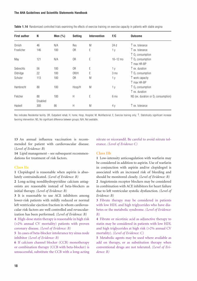
Table 1.14 Randomized controlled trials examining the effects of exercise training on exercise capacity in patients with stable angina
First author N Men (%) Setting Intervention F/C Outcome
Ornish 46 N/A Res M 24 d ↑ ex. toleranceFroelicher 146 100 OR E 1 y ↑ ex. tolerance
↑ O2 consumptionMay 121 N/A OR E 10–12 mo ↑ O2 consumption
↑ max HR-BPSebrechts 56 100 OR E 1 y ↑ ex. durationOldridge 22 100 OR/H E 3 mo ↑ O2 consumptionSchuler 113 100 OR M 1 y ↑ work capacity
↑ max HR-BPHambrecht 88 100 Hosp/H M 1 y ↑ O2 consumption
↑ ex. durationFletcher 88 100 H E 6 mo NS (ex. duration or O2 consumption)
DisabledHaskell 300 86 H M 4 y ↑ ex. tolerance
Res indicates Residential facility. OR, Outpatient rehab; H, home; Hosp, Hospital; M, Multifactorial; E, Exercise training only; ↑, Statistically signifi cant increase
favoring intervention; NS, No signifi cant difference between groups; N/A, Not available.
Chapter 1 Chronic Stable Angina
19
Class III1 Dipyridamole is not recommended. (Level of Evi-dence: B)2 Chelation therapy (intravenous infusions of eth-ylenediamine tetraacetic acid of EDTA) is not rec-ommended for the treatment of chronic angina or arteriosclerotic cardiovascular disease and may be harmful because of its potential to cause hypocalce-mia. (Level of Evidence: C)
Recommendations for pharmacological therapy to improve symptoms in patients with Syndrome XClass I1 Therapy with nitrates, beta-blockers, and calcium antagonists alone or in combination are recom-mended. (Level of Evidence: B)2 Statin therapy in patients with hyperlipidemia is recommended. (Level of Evidence: B)3 ACE-inhibition in patients with hypertension is recommended. (Level of Evidence: C)
Class IIaTrial of therapy with other anti-anginals including nicorandil and metabolic agents is reasonable. (Level of Evidence: C)
Class IIb1 Aminophylline for continued pain despite Class I measures may be considered. (Level of Evidence: C)2 Imipramine for continued pain despite Class I measures may be considered. (Level of Evidence: C)
Recommendations for pharmacological therapy of vasospastic anginaClass ITreatment with calcium antagonists and if necessary nitrates in patients whose coronary arteriogram is normal or shows only non-obstructive lesions is rec-ommended. (Level of Evidence: B)
Coronary disease risk factors and evidence that treatment can reduce the risk for coronary disease eventsRecommendations for treatment of risk factorsClass I1 Patients should initiate and/or maintain lifestyle modifi cation-weight control; increased physical
activity; moderation of alcohol consumption; limited sodium intake; and maintenance of a diet high in fresh fruits, vegetables, and low-fat dairy products. (Level of Evidence: B)2 Blood pressure control according to Joint Nation Conference VII guidelines is recommended (i.e., blood pressure less than 140/90 mm Hg or less than 130/80 mm Hg for patients with diabetes or chronic kidney disease). (Level of Evidence: A)3 For hypertensive patients with well established coronary artery disease, it is useful to add blood pressure medication as tolerated, treating initially with beta blockers and/or ACE inhibitors, with addi-tion of other drugs as needed to achieve target blood pressure. (Level of Evidence: C)4 Smoking cessation and avoidance of exposure to environmental tobacco smoke at work and home is recommended. Follow-up, referral to special pro-grams, and/or pharmacotherapy (including nicotine replacement) is recommended, as is a stepwise strat-egy for smoking cessation (Ask, Advise, Assess, Assist, Arrange). (Level of Evidence: B)5 Diabetes management should include lifestyle and pharmacotherapy measures to achieve a near-normal HbA1c. (Level of Evidence: B)6 Vigorous modifi cation of other risk factors (e.g., physical activity, weight management, blood pres-sure control, and cholesterol management) as rec-ommended should be initiated and maintained. (Level of Evidence: B)7 Physical activity of 30 to 60 minutes, 7 days per week (minimum 5 days per week) is recommended. All patients should be encouraged to obtain 30 to 60 minutes of moderate-intensity aerobic activity, such as brisk walking, on most, preferably all, days of the week, supplemented by an increase in daily activities (such as walking breaks at work, gardening, or household work). (Level of Evidence: B).8 The patient’s risk should be assessed with a physi-cal activity history. Where appropriate, an exercise test is useful to guide the exercise prescription. (Level of Evidence: B)9 Medically supervised programs (cardiac rehabili-tation) are recommended for at-risk patients (e.g., recent acute coronary syndrome or reva-scularization, heart failure). (Level of Evi-dence: B)10 Dietary therapy for all patients should include reduced intake of saturated fats (to less than 7% of
The AHA Guidelines and Scientifi c Statements Handbook
20
total calories), trans-fatty acids, and cholesterol (to less than 200 mg per day). (Level of Evidence: B)11 Daily physical activity and weight management are recommended for all patients. (Level of Evidence: B)12 Recommended lipid management includes assessment of a fasting lipid profi le. (Level of Evi-dence: A)13 LDL-C should be less than 100 mg per dL. (Level of Evidence: A)14 If baseline LDL-C is greater than or equal to 100 mg per dL, LDL-lowering drug therapy should be initiated in addition to therapeutic lifestyle changes. When LDL-lowering medications are used in high-risk or moderately high-risk persons, it is recommended that intensity of therapy be suffi cient to achieve a 30% to 40% reduction in LDL-C levels. (Level of Evidence: A)15 If on treatment LDL-C is greater than or equal to 100 mg per dL, LDL-lowering drug therapy should be intensifi ed. (Level of Evidence: A)16 If TG are 200 to 499 mg per dL, non-HDL-C should be less than 130 mg per dL. (Level of Evi-dence: A)17 BMI and waist circumference should be assessed regularly. On each patient visit, it is useful to con-sistently encourage weight maintenance/reduction through an appropriate balance of physical activity, caloric intake, and formal behavioral programs when indicated to achieve and maintain a BMI between 18.5 and 24.9 kg/m2. (Level of Evidence: B)18 If waist circumference is greater than or equal to 35 inches (89 cm) in women or greater than or equal to 40 inches (102 cm) in men it is benefi cial to initi-ate lifestyle changes and consider treatment strate-gies for metabolic syndrome as indicated. Some male patients can develop multiple metabolic risk factors when the waist circumference is only mar-ginally increased, e.g., 37 to 40 inches (94 to 102 cm). Such persons may have a strong genetic contribu-tion to insulin resistance. They should benefi t from changes in life habits, similarly to men with categori-cal increases in waist circumference. (Level of Evi-dence: B)19 The initial goal of weight loss therapy should be to gradually reduce body weight by approximately 10% from baseline. With success, further weight loss can be attempted if indicated through further assess-ment. (Level of Evidence: B)
Class IIa1 Adding plant stanol/sterols (2 g per day) and/or viscous fi ber (greater than 10 g per day) is reason-able to further lower LDL-C. (Level of Evidence: A)2 Reduction of LDL-C to less than 70 mg per dL or high-dose statin therapy is reasonable. (Level of Evi-dence: A)3 If baseline LDL-C is 70 to 100 mg per dL, it is reasonable to treat LDL-C to less than 70 mg per dL. (Level of Evidence: B)4 Further reduction of non-HDL-C to less than 100 mg per dL is reasonable.5 If TG are greater than or equal to 200 to 499 mg per dL therapeutic options to reduce non-HDL-C are:
a. niacin can be useful as a therapeutic option to reduce non-HDL-C (after LDL-C-lowering therapy) orb. fi brate therapy as a therapeutic option can be useful to reduce non-HDL-C (after LDL-C lowering therapy. (Level of Evidence: B)
6 The following lipid management strategies can be benefi cial: If LDL-C less than 70 mg per dL is the chosen target, consider drug titration to achieve this level to minimize side effects and cost. When LDL-C less than 70 mg per dL is not achievable because of high baseline LDL-C levels, it generally is possible to achieve reductions of greater than 50% in LDL-C levels by either statins or LDL-C-lowering drug combinations.(Level of Evidence: C)
Class IIb1 Folate therapy may be considered in patients with elevated homocysteine levels. (Level of Evidence: C)2 Identifi cation and appropriate treatment of clini-cal depression may be considered to improve CAD outcomes. (Level of Evidence: C)3 Intervention directed at psychosocial stress reduc-tion may be considered. (Level of Evidence: C)4 Expanding physical activity to include resistance training on 2 days per week may be reasonable. (Level of Evidence: C)5 For all patients, encouraging consumption of omega-T fatty acids in the form of fi sh or in capsule form (1 g per day) for risk reduction may be reason-able. For treatment of elevated TG, higher doses are usually necessary for risk reduction. (Level of Evi-dence: B)
Chapter 1 Chronic Stable Angina
21
Class III1 Initiation of hormone replacement therapy in postmenopausal women is not recommended for the purpose of reducing cardiovascular risk. (Level of Evidence: A)2 Vitamin C and E supplementation is not recom-mended. (Level of Evidence: A)3 Chelation therapy (intravenous infusions of eth-ylenediamine tetraacetic acid of EDTA) is not rec-ommended for the treatment of chronic angina or arteriosclerotic cardiovascular disease and may be harmful because of its potential to cause hypocalce-mia. (Level of Evidence: C)4 Garlic is not recommended. (Level of Evidence: C)5 Acupuncture is not recommended. (Level of Evi-dence: C)6 Coenzyme Q is not recommended. (Level of Evi-dence: C)
Recommendations for revascularization with PCI (or other catheter-based techniques) and CABG in patients with stable anginaClass I1 Coronary artery bypass grafting is recommended for patients with signifi cant left main coronary disease. (Level of Evidence: A)2 Coronary artery bypass grafting is recommended for patients with three-vessel disease. The survival benefi t is greater in patients with abnormal LV func-tion (ejection fraction less than 50%). (Level of Evi-dence: A)3 Coronary artery bypass grafting is recommended for patients with two-vessel disease with signifi cant proximal LAD CAD and either abnormal LV func-tion (ejection fraction less than 50%) or demon-strable ischemia on noninvasive testing. (Level of Evidence: A)4 CABG is recommended for signifi cant disease with impaired LV function and viability demon-strated by noninvasive testing. (Level of Evidence: B)5 Percutaneous coronary intervention is recom-mended for patients with two- or three-vessel disease with signifi cant proximal LAD CAD, who have anatomy suitable for catheter-based therapy and normal LV function and who do not have treated diabetes. (Level of Evidence: B)
6 Percutaneous coronary intervention or CABG is recommended for patients with one- or two-vessel CAD without signifi cant proximal LAD CAD but with a large area of viable myocardium and high-risk criteria on noninvasive testing. (Level of Evidence: B)7 Coronary artery bypass grafting is recommended for patients with one- or two-vessel CAD without signifi cant proximal LAD CAD who have survived sudden cardiac death or sustained ventricular tachy-cardia. (Level of Evidence: C)8 In patients with prior PCI, CABG or PCI is rec-ommended for recurrent stenosis associated with a large area of viable myocardium or high-risk criteria on noninvasive testing. (Level of Evidence: C)9 Percutaneous coronary intervention or CABG is recommended for patients who have not been suc-cessfully treated by medical therapy (see text) and can undergo revascularization with acceptable risk. (Level of Evidence: B)
Class IIa1 Repeat CABG is reasonable for patients with mul-tiple saphenous vein graft stenoses, especially when there is signifi cant stenosis of a graft supplying the LAD. It may be appropriate to use PCI for focal saphenous vein graft lesions or multiple stenoses in poor candidates for reoperative surgery. (Level of Evidence: C)2 Use of PCI or CABG is reasonable for patients with one- or two-vessel CAD without signifi cant proximal LAD disease but with a moderate area of viable myocardium and demonstrable ischemia on noninvasive testing. (Level of Evidence: B)3 Use of PCI or CABG is reasonable for patients with one-vessel disease with signifi cant proximal LAD disease. (Level of Evidence: B)4 CABG is reasonable for single- or two-vessel CAD without signifi cant proximal LAD stenosis in patients who have survived sudden cardiac death or sustained ventricular tachycardia. (Level of Evidence: B)5 CABG is reasonable for signifi cant three vessel disease in diabetics with reversible ischaemia on functional testing. (Level of Evidence: C)6 PCI or CABG is reasonable for patients with reversible ischaemia on functional testing and evi-dence of frequent episodes of ischaemia during daily activities. (Level of Evidence: C)
The AHA Guidelines and Scientifi c Statements Handbook
22
Class IIb1 Compared with CABG, PCI may be considered for patients with two- or three-vessel disease with signifi cant proximal LAD CAD, who have anatomy suitable for catheter-based therapy, and who have treated diabetes or abnormal LV function. (Level of Evidence: B)2 Use of PCI may be considered for patients with signifi cant left main coronary disease who are not candidates for CABG. (Level of Evidence: C)3 PCI may be considered for patients with one- or two-vessel CAD without signifi cant proximal LAD CAD who have survived sudden cardiac death or sustained ventricular tachycardia. (Level of Evidence: C)
Class III1 Use of PCI or CABG is not recommended for patients with one- or two vessel CAD without sig-nifi cant proximal LAD CAD, who have mild symp-toms that are unlikely due to myocardial ischemia, or who have not received an adequate trial of medical therapy and
a. have only a small area of viable myocardium orb. have no demonstrable ischemia on noninva-sive testing. (Level of Evidence: C)
2 Use of PCI or CABG is not recommended for patients with borderline coronary stenoses (50% to 60% diameter in locations other than the left main coronary artery) and no demonstrable ischemia on noninvasive testing. (Level of Evidence: C)3 Use of PCI or CABG is not recommended for patients with insignifi cant coronary stenosis (less than 50% diameter). (Level of Evidence: C)4 Use of PCI is not recommended in patients with signifi cant left main coronary artery disease who are candidates for CABG. (Level of Evidence: B)
Recommendations for revascularization to improve symptoms in patients with stable anginaClass I1 CABG for multi-vessel disease (MVD) technically suitable for surgical revascularization is recom-mended in patients with moderate to severe symp-toms not controlled by medical therapy, in whom
risks of surgery do not outweigh potential benefi ts. (Level of Evidence: A)2 PCI for single vessel disease technically suitable for percutaneous revascularization is recommended in patients with moderate to severe symptoms not controlled by medical therapy, in whom procedural risks do not outweigh potential benefi ts. (Level of Evidence: A)3 PCI for MVD without high risk coronary anatomy, technically suitable for percutaneous revasculariza-tion is recommended in patients with moderate to severe symptoms not controlled by medical therapy and in whom procedural risks do not outweigh potential benefi ts. (Level of Evidence: A)
Class IIa1 PCI for single vessel disease technically suit-able for percutaneous revascularization is reason-able in patients with mild to moderate symptoms which are nonetheless unacceptable to the patient, in whom procedural risks do not outweigh potential benefi ts. (Level of Evidence: A)2 CABG for single vessel disease technically suit-able for surgical revascularization is reasonable in patients with moderate to severe symptoms not controlled by medical therapy, in whom operative risk does not outweigh potential benefi t. (Level of Evidence: A)3 CABG in MVD technically suitable for surgical revascularization is reasonable in patients with mild to moderate symptoms, which are nonetheless unac-ceptable to the patient, in whom operative risk does not outweigh potential benefi t. (Level of Evidence: A)4 PCI for MVD technically suitable for percutane-ous revascularization is reasonable in patients with mild to moderate symptoms, which are nonetheless unacceptable to the patient, in whom procedural risks do not outweigh potential benefi ts. (Level of Evidence: A)
Class IIbCABG in single vessel disease technically suitable for surgical revascularization may be considered in patients with mild-to-moderate symptoms, which are nonetheless unacceptable to the patient, in whom operative risk is not greater than estimated annual mortality. (Level of Evidence: B)
Chapter 1 Chronic Stable Angina
23
Recommendations for alternative therapies for chronic stable angina in patients refractory to medical therapy who are not candidates for percutaneous intervention or surgical revascularizationClass IIaSurgical laser transmyocardial revascularization is reasonable. (Level of Evidence: A)
Class IIb1 Enhanced external counterpulsation may be con-sidered. (Level of Evidence: B)2 Spinal cord stimulation may be considered. (Level of Evidence: B)
Patient follow-up: monitoring of symptoms and anti-anginal therapyRecommendations for echocardiography, treadmill exercise testing, stress radionuclide imaging, stress echocardiography studies, and coronary angiography during patient follow-upClass I1 A chest X-ray is recommended for patients with evidence of new or worsening CHF. (Level of Evi-dence: C)2 Assessment of LV ejection fraction and segmental wall motion by echocardiography or radionuclide imaging is recommended in patients with new or worsening CHF or evidence of intervening MI by history or ECG. (Level of Evidence: C)3 Echocardiography is recommended for evidence of new or worsening valvular heart disease. (Level of Evidence: C)4 Treadmill exercise test is recommended for patients without prior revascularization who have a signifi cant change in clinical status, are able to exer-cise, and do not have any of the ECG abnormalities listed below in number 5. (Level of Evidence: C)5 Stress radionuclide imaging or stress echocar-diography procedures are recommended for patients without prior revascularization who have a signifi -cant change in clinical status and are unable to exercise or have one of the following ECG abnormalities:
a. Pre-excitation (Wolff–Parkinson–White) syn-drome. (Level of Evidence: C)
b. Electronically paced ventricular rhythm. (Level of Evidence: C)c. More than 1 mm of rest ST depression. (Level of Evidence: C)d. Complete left bundle-branch block. (Level of Evidence: C)
6 Stress radionuclide imaging or stress echocar-diography procedures are recommended for patients who have a signifi cant change in clinical status and required a stress imaging procedure on their initial evaluation because of equivocal or intermediate-risk treadmill results. (Level of Evidence: C)7 Stress radionuclide imaging or stress echocar-diography procedures are recommended for patients with prior revascularization who have a signifi cant change in clinical status. (Level of Evidence: C)8 Coronary angiography is recommended in patients with marked limitation of ordinary activity (CCS class III) despite maximal medical therapy. (Level of Evidence: C)
Class IIbAnnual treadmill exercise testing may be considered in patients who have no change in clinical status, can exercise, have none of the ECG abnormalities listed in number 5, and have an estimated annual mortal-ity rate greater than 1%. (Level of Evidence: C)
Class III1 Echocardiography or radionuclide imaging is not recommended for assessment of LV ejection frac-tion and segmental wall motion in patients with a normal ECG, no history of MI, and no evidence of CHF. (Level of Evidence: C)2 Repeat treadmill exercise testing is not recom-mended in less than three years in patients who have no change in clinical status and an estimated annual mortality rate less than 1% on their initial evalua-tion, as demonstrated by one of the following:
a. Low-risk Duke treadmill score (without imaging). (Level of Evidence: C)b. Low-risk Duke treadmill score with negative imaging. (Level of Evidence: C)c. Normal LV function and a normal coronary angiogram. (Level of Evidence: C)d. Normal LV function and insignifi cant CAD. (Level of Evidence: C)
The AHA Guidelines and Scientifi c Statements Handbook
24
3 Stress imaging or echocardiography is not recom-mended for patients who have no change in clinical status and a normal rest ECG, are not taking digoxin, are able to exercise, and did not require a stress imaging or echocardiographic procedure on their initial evaluation because of equivocal or intermedi-ate-risk treadmill results. (Level of Evidence: C)4 Repeat coronary angiography is not recom-mended in patients with no change in clinical status, no change on repeat exercise testing or stress imaging, and insignifi cant CAD on initial evalua-tion. (Level of Evidence: C)
Future issues
Since publication of these guideline recommenda-tions in 2002, important new evidence has been published. As a result of this new evidence, the next revision of the guidelines, which is currently under-way, will likely refl ect changes in the following areas:
Special consideration for womenRecent evidence, particularly from the NHLBI-sponsored Women’s Ischemic Syndrome Evaluation (WISE) Study [5,6], has suggested that traditional approaches signifi cantly underestimate the presence of obstructive CAD in women, particularly younger women. Moreover, many women without obstruc-tive disease continue to have symptoms and a poor quality of life [7,8]. Many have evidence of micro-vascular dysfunction [9]. There is growing interest in the development of gender-specifi c tools for the assessment of ischemic heart disease in women, but the evidence is not yet robust enough to support the widespread use of a new approach.
New information on percutaneous revascularization to be considered for the next chronic stable angina guidelineAs listed above, the 2002 guidelines included a Class I recommendation for PCI or CABG in symptom-atic or asymptomatic patients with “one- or two-vessel CAD . . . with high risk criteria on noninvasive testing.” A randomized trial reported in 2007 has challenged the assumption that revascularization improves patient outcomes in many patients with
multi-vessel coronary disease. The COURAGE (Clinical Outcomes Utilizing Revascularization and Aggressive Drug Evaluation) trial – the largest reported randomized clinic trial on coronary artery disease to date – enrolled 2287 patients with signifi -cant coronary artery disease and inducible ischemia. In contrast to previous trials, medical therapy in the COURAGE trial focused not only on symptomatic relief, but also risk factor reduction. Medical therapy resulted in very high rates of adherence to the rec-ommendations for blood pressure, lipid levels, exer-cise, diet, and smoking cessation that are detailed above. When added to such medical therapy, PCI did not provide any advantage for the primary end-point of death or myocardial infarction. Future revi-sions of the stable angina clinic practice guideline will consider the results of COURAGE. Although we do not want to prejudge the careful rigorous process of guideline development, it certainly seems likely that the indications for revascularization in asymp-tomatic patients, and in selected symptomatic patients, are likely to be more cautious than those listed above [10–12].
New therapeutic agents to be considered for the next chronic stable angina guidelineRanolazine is a novel therapeutic agent recently approved by the FDA for the treatment of refractory angina. It appears to reduce anginal episodes and to increase exercise tolerance without increasing car-diovascular risk despite a potential to increase the QT interval. Varenicline is a partial nicotine recep-tor agonist that shows great promise to help patients overcome addiction to smoking. Both of these agents will be thoroughly assessed by the next chronic stable angina writing group with a new guideline expected in late 2008 [13–18].
References available online at www.Wiley.com/go/AHAGuidelineHandbook.
During the production of this book this relevant AHA statement and guideline was published: ACCF/ASE/ACEP/AHA/ASNC/SCAI/SCCT/SCMR 2008 Appropriateness Criteria for Stress Echocardio-graphy, http://circ.ahajournals.org/cgi/content/full/117/11/1478.