Embed Size (px)
Citation preview

CHAPTER 10
ANATOMY OF THEMUSCULARSYSTEM
KEY TERMSantagonistfixatorinsertionlever
originpostureprime moversynergist
Survival depends on the ability to maintain a relativelyconstant internal environment. Such stability often re-quires movement of the body. For example, we must
gather and eat food, defend ourselves, seek shelter, and maketools, clothing, or other objects. Whereas many different sys-tems of the body have some role in accomplishing move-ment, it is the skeletal and muscular systems acting togetherthat actually produce most body movements. We have inves-tigated the architectural plan of the skeleton and have seenhow its firm supports and joint structures make movementpossible. However, bone and joints cannot move themselves.They must be moved by something. Our subject for now,then, is the large mass of skeletal muscle that moves theframework of the body: the muscular system (Figures 10-1and 10-2).
Movement is one of the most characteristic and easily ob-served “characteristics of life.” When we walk, talk, run,breathe, or engage in a multitude of other physical activitiesthat are under the “willed” control of the individual, we doso by the contraction of skeletal muscle.
There are more than 600 skeletal muscles in the body.Collectively, they constitute 40% to 50% of our body weight.And, together with the scaffolding provided by the skeleton,muscles also determine the form and contours of our body.
Contraction of individual muscle cells is ultimately re-sponsible for purposeful movement. In Chapter 11 the phys-iology of muscular contraction is discussed. In this prelimi-nary chapter, however, we will learn how contractile unitsare grouped into unique functioning organs—or muscles.
CHAPTER OUTLINESkeletal Muscle Structure, 281
Connective Tissue Components, 281Size, Shape, and Fiber Arrangement, 282Attachment of Muscles, 282Muscle Actions, 283Lever Systems, 283
First-class levers, 284Second-class levers, 284Third-class levers, 284
How Muscles Are Named, 285Hints on How To Deduce Muscle Actions, 286
Important Skeletal Muscles, 286Muscles of Facial Expression, 287Muscles of Mastication, 288Muscles That Move the Head, 288
Trunk Muscles, 289Muscles of the Thorax, 289Muscles of the Abdominal Wall, 289Muscles of the Back, 290Muscles of the Pelvic Floor, 290
Upper Limb Muscles, 293Muscles Acting on the Shoulder Girdle, 293Muscles That Move the Upper Arm, 294Muscles That Move the Forearm, 295Muscles That Move the Wrist, Hand, and Fingers, 295Lower Limb Muscles, 300Muscles That Move the Thigh and Lower Leg, 301Muscles That Move the Ankle and Foot, 304
Posture, 306How Posture Is Maintained, 307
Cycle of Life, 308The Big Picture, 308Case Study, 309
278

Anatomy of the Muscular System Chapter 10 279
Facial muscles
Sternocleidomastoid
Trapezius
Pectoralis major
Serratus anterior
Rectus abdominis
External abdominal oblique
Tensor fasciae latae
Vastus lateralis
Rectus femoris
Patella
Tibialis anterior
Peroneus longus
Peroneus brevis
Superior extensorretinaculum
Extensor digitorumlongus
Flexors of wristand fingers
Deltoid
Biceps brachii
Linea alba
Extensors of wristand fingers
Adductorsof thigh
Retinaculum
Sartorius
Vastus medialis
Patellar tendon
Gastrocnemius
Soleus
S
LR
I
Figure 10-1 General overview of the body musculature. Anterior view.

280 Unit 2 Support and Movement
Sternocleidomastoid
Seventh cervical vertebra
Teres minor
Teres major
Triceps brachii
Latissimus dorsi
Semitendinosus
Biceps femoris
Semimembranosus
Extensors of the wristand fingers
Hamstringgroup
Gastrocnemius
Peroneus longus
Peroneus brevis
Trapezius
Infraspinatus
External abdominaloblique
Gluteus maximus
Adductor magnus
Gracilis
Iliotibial tract
Soleus
Calcaneal tendon(Achilles tendon)
Splenius capitis
Deltoid
S
RL
I
Figure 10-2 General overview of the body musculature. Posterior view.

The manner in which muscles are grouped, the relationshipof muscles to joints, and how muscles attach to the skeletondetermine purposeful body movement. A discussion of mus-cle shape and how muscles attach to and move bones is fol-lowed by information on specific muscles and muscle groups.The chapter will end with a review of the concept of posture.
SKELETAL MUSCLE STRUCTURE
CONNECTIVE TISSUE COMPONENTSThe highly specialized skeletal muscle cells, or muscle fibers,are covered by a delicate connective tissue membrane calledthe endomysium (Figure 10-3). Groups of skeletal musclefibers, called fascicles, are then bound together by a tougherconnective tissue envelope called the perimysium. The mus-cle as a whole is covered by a coarse sheath called the epimy-sium. Because all three of these structures are continuouswith the fibrous structures that attach muscles to bones orother structures, muscles are firmly harnessed to the struc-tures they pull on during contraction. The epimysium, peri-mysium, and endomysium of a muscle, for example, may becontinuous with fibrous tissue that extends from the muscle
as a tendon, a strong tough cord continuous at its other endwith the fibrous periosteum covering a bone. Or the fibrouswrapping of a muscle may extend as a broad, flat sheet ofconnective tissue called an aponeurosis, which usuallymerges with the fibrous wrappings of another muscle. Sotough and strong are tendons and aponeuroses that they arenot often torn, even by injuries forceful enough to breakbones or tear muscles. They are, however, occasionallypulled away from bones. Fibrous connective tissue sur-rounding the muscle organ and outside the epimysium andtendon is called fascia. Fascia is a general term for the fibrousconnective tissue found under the skin and surroundingmany deeper organs, including skeletal muscles and bones.Fascia just under the skin (the hypodermis) is sometimescalled superficial fascia, and the fascia around muscles andbones is sometimes called deep fascia.
Tube-shaped structures of fibrous connective tissuecalled tendon sheaths enclose certain tendons, notably thoseof the wrist and ankle. Like the bursae, tendon sheaths havea lining of synovial membrane. Its moist, smooth surface en-ables the tendon to move easily, almost without friction, inthe tendon sheath.
Anatomy of the Muscular System Chapter 10 281
Figure 10-3 Structure of a muscle organ. Note that the connective tissue coverings, the epimysium, perimy-sium, and endomysium, are continuous with each other and with the tendon. Note also that muscle fibers areheld together by the perimysium in groups called fascicles.

SIZE, SHAPE, AND FIBER ARRANGEMENTThe structures called skeletal muscles are organs. They con-sist mainly of skeletal muscle tissue plus important connec-tive and nervous tissue components. Skeletal muscles varyconsiderably in size, shape, and arrangement of fibers. Theyrange from extremely small strands, such as the stapediusmuscle of the middle ear, to large masses, such as the mus-cles of the thigh. Some skeletal muscles are broad in shapeand some are narrow. Some are long and tapering and someare short and blunt. Some are triangular, some quadrilateral,and some irregular. Some form flat sheets and others formbulky masses.
Arrangement of fibers varies in different muscles. Insome muscles the fibers are parallel to the long axis of themuscle (Figure 10-4, A). In some they converge to a narrowattachment (Figure 10-4, B), and in some they are obliqueand pennate (Figure 10-4, C) like the feathers in an old-fashioned plume pen or bipennate (double-feathered) (Fig-ure 10-4, D). Fibers may even be curved, as in the sphinctersof the face, for example (Figure 10-4, E). The direction of the
fibers composing a muscle is significant because of its rela-tionship to function. For instance, a muscle with the bipen-nate fiber arrangement can produce a stronger contractionthan a muscle having a parallel fiber arrangement.
ATTACHMENT OF MUSCLESMost of our muscles span at least one joint and attach toboth articulating bones. When contraction occurs, one boneusually remains fixed and the other moves. The points of at-tachment are called the origin and insertion. The origin isthat point of attachment that does not move when the mus-cle contracts. Therefore the origin bone is the more station-ary of the two bones at a joint when contraction occurs. Theinsertion is the point of attachment that moves when themuscle contracts (Figure 10-5). The insertion bone thereforemoves toward the origin bone when the muscle shortens. Incase you are wondering why both bones do not move, be-cause both are pulled on by the contracting muscle, one ofthem is normally stabilized by isometric contractions ofother muscles or by certain features of its own that make itless mobile.
The terms origin and insertion provide us with usefulpoints of reference. Many muscles have multiple points oforigin or insertion. Understanding the functional relation-ship of these attachment points during muscle contractionhelps in deducing muscle actions. Attachment points of thebiceps brachii shown in Figure 10-5 help provide functionalinformation. Distal insertion on the radius of the lower armcauses flexion to occur at the elbow when contraction oc-curs. It should be realized, however, that origin and insertion
282 Unit 2 Support and Movement
A B C D E
Figure 10-4 Muscle shape and fiber arrangement. A, Parallel. B, Convergent. C, Pennate. D, Bipennate. E, Sphincter.
Figure 10-5 Attachments of a skeletal muscle. A muscle origi-nates at a relatively stable part of the skeleton (origin) and inserts atthe skeletal part that is moved when the muscle contracts (insertion).
1. Identify the connective tissue membrane that: (a) coversindividual muscle fibers, (b) surrounds groups of skeletalmuscle fibers (fascicles), and (c) covers the muscle as awhole.
2. Name the tough connective tissue cord that serves to attach amuscle to a bone.
3. Name three types of fiber arrangements seen in skeletalmuscle.

are points that may change under certain circumstances. Forexample, not only can you grasp an object above your headand pull it down, you can also pull yourself up to the object.Although origin and insertion are convenient terms, they donot always provide the necessary information to understandthe full functional potential of muscle action.
MUSCLE ACTIONSSkeletal muscles almost always act in groups rather thansingly. As a result, most movements are produced by the co-ordinated action of several muscles. Some of the muscles inthe group contract while others relax. The result is a move-ment pattern that allows for the functional classification ofmuscles or muscle groups. Several terms are used to describemuscle action during any specialized movement pattern. Theterms prime mover (agonist), antagonist, synergist, and fixatorare especially important and are discussed in the followingparagraphs. Each term suggests an important concept that isessential to an understanding of such functional muscle pat-terns as flexion, extension, abduction, adduction, and othermovements discussed in Chapter 9. The term prime moveror agonist is used to describe a muscle or group of musclesthat directly performs a specific movement. The movementproduced by a muscle acting as a prime mover is described asthe “action” or “function” of that muscle. For example, the bi-ceps brachii shown in Figure 10-5 is acting as a prime moverduring flexion of the forearm.
Antagonists are muscles that, when contracting, directlyoppose prime movers (agonists). They are relaxed while theprime mover is contracting to produce movement. Simulta-neous contraction of a prime mover and its antagonist mus-cle results in rigidity and lack of motion. The term antago-nist is perhaps unfortunate, because muscles cooperate,rather than oppose, in normal movement patterns. Antago-nists are important in providing precision and control dur-ing contraction of prime movers.
Synergists are muscles that contract at the same time asthe prime mover. They facilitate or complement primemover actions so that the prime mover produces a more ef-fective movement.
Fixator muscles generally function as joint stabilizers. Theyfrequently serve to maintain posture or balance during con-traction of prime movers acting on joints in the arms and legs.
Movement patterns are complex, and most muscles func-tion not only as prime movers but also as antagonists, syner-gists, or fixators. A prime mover in a particular movementpattern, such as flexion, may be an antagonist during exten-sion or a synergist or fixator in other types of movement.
LEVER SYSTEMSWhen a muscle shortens, the central body portion, called thebelly, contracts. The type and extent of movement is deter-mined by the load or resistance that is moved, the attach-ment of the tendinous extremities of the muscle to bone(origin and insertion), and by the particular type of joint in-volved. In almost every instance, muscles that move a part donot lie over that part. Instead, the muscle belly lies proximalto the part moved. Thus muscles that move the lower arm lieproximal to it, that is, in the upper arm.
Knowledge of lever systems is important in understand-ing muscle action. By definition, a lever is any rigid bar freeto turn about a fixed point called its fulcrum. Bones serve aslevers, and joints serve as fulcrums of these levers. A con-tracting muscle applies a pulling force on a bone lever at thepoint of the muscle’s attachment to the bone. This causes theinsertion bone to move about its joint fulcrum.
A lever system is a simple mechanical device that makesthe work of moving a weight or other load easier. Levers arecomposed of four component parts: (1) a rigid rod or bar(bone), (2) a fixed pivot, or fulcrum (F), around which therod moves (joint), (3) a load (L) or resistance that is moved,and (4) a force, or pull (P), which produces movement(muscle contraction). Figure 10-6 shows the three differenttypes of lever arrangements. All three types are found in thehuman body.
Anatomy of the Muscular System Chapter 10 283
Box 10-1 SPORTS AND FITNESS
Assessing Muscle Strength
Certified athletic trainers and other health care providersare often required to assess muscle strength in the
evaluation of athletic injuries. A basic principle of muscleaction in a lever system is called the optimum angle ofpull. An understanding of this principle is required for cor-rect assessment of muscle strength.
Generally, the optimum angle of pull for any muscle isa right angle to the long axis of the bone to which it is at-tached. When the angle of pull departs from a right angleand becomes more parallel to the long axis, the strength ofcontraction decreases dramatically. Contraction of thebrachialis muscle demonstrates this principle very well. The brachialis crosses the elbow from humerus to ulna. Inthe anatomical position the elbow is extended and the an-gle of pull of the brachialis is parallel to the long axis of theulna (see Figure 10-17, D). Contraction of the brachialis atthis angle is very inefficient. As the elbow is flexed and theangle of pull approaches a right angle, the contractionstrength of the muscle is greatly increased. Therefore totest brachialis muscle strength correctly, the forearmshould be flexed at the elbow. Understanding the optimumangle of pull for any given muscle makes a rational ap-proach to correct assessment of functional strength in thatmuscle possible.
1. Identify the point of attachment of a muscle to a bonethat: (a) does not move when the muscle contracts; (b)moves when the muscle contracts.
2. What name is used to describe a muscle that directly performsa specific movement?
3. What type of muscles helps maintain posture or balance duringcontraction of muscles acting on joints in the arms and legs?
4. Name the type of muscles that generally function as joint stabilizers.

First-Class LeversAs you can see in Figure 10-6, A, the placement of the ful-crum in a first-class lever lies between the effort, or pull (P),and the resistance, or load (W), as in a set of scales, a pair ofscissors, or a child’s seesaw. In the body the head being raisedor tipped backward on the atlas is an example of a first-classlever in action. The facial portion of the skull is the load, thejoint between the skull and atlas is the fulcrum, and the mus-cles of the back produce the pull. In the human body first-class levers are not abundant. They generally serve as leversof stability.
Second-Class LeversIn second-class levers the load lies between the fulcrum andthe joint at which the pull is exerted. The wheelbarrow is of-ten used as an example. The presence of second-class leversin the human body is a controversial issue. Some authorities
interpret the raising of the body on the toes as an exampleof this type of lever (Figure 10-6, B). In this example thepoint of contact between the toes and the ground is the ful-crum, the load is located at the ankle, and pull is exerted bythe gastrocnemius muscle through the Achilles tendon.Opening the mouth against resistance (depression of themandible) is also considered to be an example of a second-class lever.
Third-Class LeversIn a third-class lever the pull is exerted between the fulcrumand resistance or load to be moved. Flexing of the forearm atthe elbow joint is a frequently used example of this type oflever (Figure 10-6, C). Third-class levers permit rapid andextensive movement and are the most common type foundin the body. They allow insertion of a muscle very close tothe joint that it moves.
284 Unit 2 Support and Movement
A B
C
Figure 10-6 Lever classes. A, Class I: fulcrum (F) between the load (L)and force or pull (P); B, Class II: load (L) between the fulcrum (F) andforce or pull (P); C, Class III: force or pull (P) between the fulcrum (F)and the load (L). The lever rod is yellow in each.
HOW MUSCLES ARE NAMEDThe first thing you notice when you start studying the mus-cles of the body is that the names all seem very mysteriousand foreign. Of course, that results from them being essen-tially Latin words (sometimes with Greek origins). You mayalso find that from one reference to another, the same mus-cle will have slightly different names. Sometimes the differ-ence comes from the fact that in science, old terms are oftenbeing replaced by newer terms and it takes time for everyoneto catch on. With muscles, however, it is common to use ei-ther Latin or the English version of the Latin name. For ex-ample, the deltoid muscle can be correctly called deltoideus(Latin) or deltoid (Latin-based English). You can see thatthey both come from the same original name, but they arenot exactly the same word. In this edition, we have strived tokeep with the English names only.
Latin-based muscle names seem more logical and there-fore easier to learn when one understands the reasons for thenames. Many of the superficial muscles of the body shown inFigures 10-1 and 10-2 are named using one or more of thefollowing features:
• Location. Many muscles are named as a result oflocation. The brachialis (arm) muscle and gluteus(buttock) muscles are examples. Table 10-1 is a listing of some major muscles grouped by location.
• Function. The function of a muscle is frequently apart of its name. The adductor muscles of the thighadduct, or move, the leg toward the midline of thebody. Table 10-2 lists selected muscles grouped ac-cording to function.
• Shape. Shape is a descriptive feature used for naming many muscles. The deltoid (triangular)
muscle covering the shoulder is delta, or triangular,in shape.
• Direction of fibers. Muscles may be named accordingto the orientation of their fibers. The term rectusmeans straight. The fibers of the rectus abdominismuscle run straight up and down and are parallel toeach other.
• Number of heads or divisions. The number of divi-sions or heads (points of origin) may be used to namea muscle. The word part -cep means head. The biceps(two), triceps (three), and quadriceps (four) refer tomultiple heads, or points of origin. The biceps brachiiis a muscle having two heads located in the arm.
• Points of attachment. Origin and insertion pointsmay be used to name a muscle. For example, the ster-nocleidomastoid has its origin on the sternum andclavicle and inserts on the mastoid process of thetemporal bone.
• Size of muscle. The relative size of a muscle can beused to name a muscle, especially if it is compared tothe size of nearby muscles. For example, the gluteusmaximus is the largest muscle of the gluteal (Greekglautos, meaning “buttock”) region. Nearby, there is asmall gluteal muscle, gluteus minimus, and midsizegluteal muscle, gluteus medius.
Anatomy of the Muscular System Chapter 10 285
Table 10-1 Selected Muscles Grouped According to Location
Location Muscles Term Meaning
Neck Sternocleidomastoid
Back Trapezius
Latissimus dorsi
Chest Pectoralis major
Serratus anterior
Abdominal wall External oblique
Shoulder Deltoid
Upper arm Biceps brachii
Triceps brachii
Brachialis
Forearm Brachioradialis
Pronator teres
Buttocks Gluteus maximus
Gluteus minimus
Gluteus medius
Tensor fascia latae
Thigh
Anterior surface Quadriceps femoris group
Rectus femoris
Vastus lateralis
Vastus medialis
Vastus intermedius
Medial surface Gracilis
Adductor group (brevis, longus, magnus)
Posterior surface Hamstring group
Biceps femoris
Semitendinosus
Semimembranosus
Leg
Anterior surface Tibialis anterior
Posterior surface Gastrocnemius
Soleus
Pelvic floor Levator ani
Coccygeus
1. Name the four major components of any lever system.2. Identify the three types of lever systems found in the
human body and give one example of each.3. What type of lever system permits rapid and extensive move-
ment and is the most common type found in the body?4. List six criteria that may determine a muscle’s name and give
an example of a specific muscle named using each criterion.

HINTS ON HOW TO DEDUCE MUSCLE ACTIONSTo understand muscle actions, you need first to know certainanatomical facts such as which bones muscles attach to andwhich joints they pull across. Then, if you relate these struc-tural features to functional principles, you may find yourstudy of muscles more interesting and less difficult than youanticipate. Some specific suggestions for deducing muscleactions follow.
1. Start by making yourself familiar with the names,shapes, and general locations of the larger muscles,using Table 10-1 as a guide.
2. Try to deduce which bones the two ends of a muscleattach to from your knowledge of the shape andgeneral location of the muscle. For example, lookcarefully at the deltoid muscle as illustrated in Fig-ures 10-1 and 10-15. To which bones does it seem toattach? Check your answer with Table 10-10, p. 295.
3. Next, determine which bone moves when the muscleshortens. (The bone moved by a muscle’s contractionis its insertion bone; the bone that remains relativelystationary is its origin bone.) In many cases you cantell which is the insertion bone by trying to move onebone and then another. In some cases either bonemay function as the insertion bone. Although not allmuscle attachments can be deduced as readily asthose of the deltoid, they can all be learned moreeasily by using this deduction method than by relyingon rote memory alone.
4. Deduce a muscle’s actions by applying the principlethat its insertion moves toward its origin. Check yourconclusions with the text. Here, as in steps 2 and 3,the method of deduction is intended merely as aguide and is not adequate by itself for determiningmuscle actions.
5. To deduce which muscle produces a given action(instead of which action a given muscle produces, asin step 4), start by inferring the insertion bone (bonethat moves during the action). The body and originof the muscle will lie on one or more of the bonestoward which the insertion moves—often a bone, orbones, proximal to the insertion bone. Couple theseconclusions about origin and insertion with yourknowledge of muscle names and locations to deducethe muscle that produces the action.
For example, if you wish to determine the prime mover forthe action of raising the upper arms straight out to the sides,you infer that the muscle inserts on the humerus, because thisis the bone that moves. It moves toward the shoulder—that is,the clavicle and scapula—so that probably the muscle has itsorigin on these bones. Because you know that the deltoidmuscle fulfills these conditions, you conclude, and rightly so,that it is the muscle that raises the upper arms sideways.
IMPORTANT SKELETAL MUSCLESThe major skeletal muscles of the body are listed, grouped,and illustrated in the tables and figures that follow. Beginyour study with an overview of important superficial mus-
286 Unit 2 Support and Movement
Table 10-2 Selected Muscles Grouped According to Function
Part Moved Example of Flexor Example of Extensor Example of Abductor Example of Adductor
Head
Upper arm
Forearm
Hand
Thigh
Leg
Foot
Trunk
Sternocleidomastoid
Pectoralis major
With forearm supinated: biceps brachii
With forearm pronated: brachialis
With semisupination or semiprona-
tion: brachioradialis
Flexor carpi radialis and ulnaris
Palmaris longus
Iliopsoas
Rectus femoris (of quadriceps femoris
group)
Hamstrings
Tibialis anterior
Iliopsoas
Rectus abdominis
Semispinalis capitis
Trapezius
Latissimus dorsi
Triceps brachii
Extensor carpi radialis,
longus, and brevis
Extensor carpi ulnaris
Gluteus maximus
Quadriceps femoris
group
Gastrocnemius
Soleus
Erector spinae
Deltoid
Flexor carpi radialis
Gluteus medius and
gluteus minimus
Evertors
Peroneus longus
Peroneus brevis
Pectoralis major with
latissimus dorsi
Flexor carpi ulnaris
Adductor group
Invertor
Tibialis anterior

cles, shown in Figures 10-1 and 10-2. The remaining figuresin this chapter illustrate individual muscles or importantmuscle groups.
Basic information about many muscles is given in Ta-bles 10-3 to 10-15. Each table has a description of a groupof muscles that move one part of the body. The actionslisted for each muscle are those for which it is a primemover. Remember, however, that a single muscle actingalone rarely accomplishes a given action. Instead, musclesact in groups as prime movers, synergists, antagonists, andfixators to bring about movements.
MUSCLES OF FACIAL EXPRESSIONThe muscles of facial expression (Table 10-3) are uniquein that at least one of their points of attachment is to thedeep layers of the skin over the face or neck. Contractionof these muscles (Figure 10-7) produces a variety of facialexpressions.
The occipitofrontalis (ahk-SIP-it-o-front-AL-is), or epi-cranius, is in reality two muscles. One portion lies over theforehead (frontal bone); the other covers the occipital bonein back of the head. The two muscular parts, or bellies, arejoined by a connective tissue aponeurosis that covers the topof the skull. The frontal portion of the occipitofrontalisraises the eyebrows (surprise) and wrinkles the skin of theforehead horizontally. The corrugator supercilii (COR-u-GA-tor su-per-SIL-i) draws the eyebrows together, produc-ing vertical wrinkles above the nose (frowning). The orbicu-laris oculi (or-BIC-u-LAR-us OK-u-li) encircles and closesthe eye (blinking), whereas the orbicularis oris (OR-us) andbuccinator (BUK-si-NA-tor) pucker the mouth (kissing)and press the lips and cheeks against the teeth. The zygo-maticus (ZI-go-MAT-i-kus) major draws the corner of themouth upward (laughing).
Anatomy of the Muscular System Chapter 10 287
Table 10-3 Muscles of Facial Expression and of Mastication
Muscle Origin Insertion Function Nerve Supply
Muscles of Facial ExpressionOccipitofrontalis
(epicranius)
Corrugator supercilii
Orbicularis oculi
Zygomaticus major
Orbicularis oris
Buccinator
Muscles of MasticationMasseter
Temporalis
Pterygoids (lateral
and medial)
Occipital bone
Frontal bone
(superciliary ridge)
Encircles eyelid
Zygomatic bone
Encircles mouth
Maxillae
Zygomatic arch
Temporal bone
Undersurface of skull
Tissues of eyebrows
Skin of eyebrow
Angle of mouth
Skin of sides of mouth
Mandible (external surface)
Mandible
Mandible (medial surface)
Raises eyebrows, wrinkles fore-
head horizontally
Wrinkles forehead vertically
Closes eye
Laughing (elevates angle of
mouth)
Draws lips together
Permits smiling
Blowing, as in playing a trumpet
Closes jaw
Closes jaw
Grates teeth
Cranial nerve VII
Cranial nerve VII
Cranial nerve VII
Cranial nerve VII
Cranial nerve VII
Cranial nerve VII
Cranial nerve V
Cranial nerve V
Cranial nerve V
Temporalis
Occipitofrontalis (occipital portion)
Temporalis
Masseter
Buccinator
Occipitofrontalis(frontal portion)
Occipitofrontalis(frontal portion)
Corrugator supercilii
Orbicularis oculi
Orbicularis oculi
Orbicularis oculi(palpebral portion)
Buccinator
Orbicularis oris
Orbicularis oris
Masseter
Sternocleidomastoid
Zygomaticus major
CorrugatorSupercilii
S
LR
I
S
AP
I
A
B
Figure 10-7 Muscles of facial expression and mastication. A, Lateral view. B, Anterior view.
MUSCLES OF MASTICATIONThe muscles of mastication (mass-ti-KA-shun) shown inFigure 10-7 are responsible for chewing movements. Thesepowerful muscles (see Table 10-3) either elevate and retract
the mandible (masseter, mas-SE-ter, and temporalis, tem-po-RAL-is) or open and protrude it while causing sidewaysmovement (pterygoids, TER-i-goids). The pull of gravityhelps open the mandible during mastication, and the bucci-nator muscles play an important function by holding foodbetween the teeth as the mandible moves up and down andfrom side to side.
MUSCLES THAT MOVE THE HEADPaired muscles on either side of the neck are responsible forhead movements (Figure 10-8). Note the points of attach-ment and functions of important muscles in this grouplisted in Table 10-4. When both sternocleidomastoid (STER-no-KLI-do-MAS-toyd) muscles (Figure 10-7) contract at thesame time, the head is flexed on the thorax—hence the name“prayer muscle.” If only one muscle contracts, the head andface are turned to the opposite side. The broad semispinaliscapitis (sem-e-spi-NAL-is KAP-i-tis) is an extensor of thehead and helps to bend it laterally. Acting together, the sple-nius capitis (SPLE-ne-us KAP-i-tis) muscles serve as strongextensors that return the head to the upright position afterflexion. When either muscle acts alone, contraction results inrotation and tilting toward that side. The bandlike longis-simus capitis (lon-JIS-i-mus KAP-i-tis) muscles are coveredand not visible in Figure 10-8. They run from the neck ver-tebrae to the mastoid process of the temporal bone on eitherside and cause extension of the head when acting together.One contracting muscle will bend and rotate the head to-ward the contracting side.
288 Unit 2 Support and Movement
Semispinaliscapitis
Splenius capitis
Sternocleidomastoid
Trapezius
Ligamentumnuchae
S
RL
I
Table 10-4 Muscles That Move the Head
Muscle Origin Insertion Function Nerve Supply
Sternocleidomastoid
Semispinalis capitis
Splenius capitis
Longissimus capitis
Sternum
Clavicle
Vertebrae (transverse
processes of upper six
thoracic, articular
processes of lower
four cervical)
Ligamentum nuchae
Vertebrae (spinous
processes of upper
three or four thoracic)
Vertebrae (transverse
processes of upper six
thoracic, articular
processes of lower
four cervical)
Temporal bone
(mastoid process)
Occipital bone
(between supe-
rior and inferior
nuchal lines)
Temporal bone
(mastoid process)
Occipital bone
Temporal bone
(mastoid process)
Flexes head (prayer muscle)
One muscle alone, rotates head
toward opposite side; spasm of
this muscle alone or associated
with trapezius called torticollis
or wryneck
Extends head; bends it laterally
Extends head
Bends and rotates head toward
same side as contracting muscle
Extends head
Bends and rotates head toward
contracting side
Accessory nerve
First five cervical
nerves
Second, third, and
fourth cervical
nerves
Multiple innervation
Figure 10-8 Muscles that move the head. Posterior view ofmuscles of the neck and the back.

Rectus abdominis
External abdominaloblique
Rectus abdominis
(coveredby sheath)
Inguinal ligament
Linea alba
Transversusabdominis
Internal abdominal
oblique
S
LR
I
A B
TRUNK MUSCLES
MUSCLES OF THE THORAXThe muscles of the thorax are of critical importance in res-piration (discussed in Chapter 24). Note in Figure 10-9 andTable 10-5 that the internal and external intercostal (IN-ter-KOS-tal) muscles attach to the ribs at different places andtheir fibers are oriented in different directions. As a result,contraction of the external intercostals elevates and the in-ternal intercostals depress the ribs—important in thebreathing process. During inspiration the dome-shaped di-aphragm (DI-a-fram) flattens, thus increasing size and vol-ume of the thoracic cavity. As a result, air enters the lungs.
MUSCLES OF THE ABDOMINAL WALLThe muscles of the anterior and lateral abdominal wall (Fig-ure 10-10 and Table 10-6) are arranged in three layers, with
the fibers in each layer running in different directions muchlike the layers of wood in a sheet of plywood. The result is avery strong “girdle” of muscle that covers and supports theabdominal cavity and its internal organs.
Anatomy of the Muscular System Chapter 10 289
1. What is meant by the terms origin and insertion?2. Which muscle of facial expression has two parts, one
lying over the forehead and the other covering the backof the skull?
3. What group of muscles provides chewing movements?4. What is the action of the sternocleidomastoid muscle?
External intercostals
Internal intercostals
Central tendonof diaphragm
Diaphragm
1
2
3
4
5
6
7
8
9
10
S
LR
I
Table 10-5 Muscles of the Thorax
Muscle Origin Insertion Function Nerve Supply
External intercostals
Internal intercostals
Diaphragm
Rib (lower border; forward fibers)
Rib (inner surface, lower border;
backward fibers)
Lower circumference of thorax
(of rib cage)
Rib (upper border of rib
below origin)
Rib (upper border of rib
below origin)
Central tendon of
diaphragm
Elevate ribs
Depress ribs
Enlarges thorax, causing
inspiration
Intercostal nerves
Intercostal nerves
Phrenic nerves
Figure 10-9 Muscles of the thorax. Anterior view. Note relation-ship of internal and external intercostal muscles and placement of diaphragm.
Figure 10-10 Muscles of the trunk and abdominal wall. A, Anterior view showing superficial muscles. B, Anterior view showing deeper muscles.

The three layers of muscle in the anterolateral (side) ab-dominal walls are arranged as follows: the outermost layer,or external oblique; a middle layer, or internal oblique; andthe innermost layer, or transversus abdominis. In additionto these sheetlike muscles, the band-shaped (or strap-shaped) rectus abdominis muscle runs down the midline ofthe abdomen from the thorax to the pubis. In addition toprotecting the abdominal viscera, the rectus abdominisflexes the spinal column.
MUSCLES OF THE BACKConsidering the large number of us that suffer from backpain, strain, and injury either occasionally or chronically,you can imagine the importance of the back muscles tohealth and fitness. The superficial back muscles play a majorrole in moving the head and limbs, and so are listed else-where in this chapter. For now, we will concentrate on thedeep muscles of the back. These deep back muscles not onlyallow us to move our vertebral column, helping us to bend
this way and that, but also stabilize our trunk so that we canmaintain a stable posture. These muscles really get a work-out when we lift something heavy because they have to holdthe body straight while the load is trying to bend the back.
The erector spinae group consists of a number of long,thin muscles that travel all the way down our backs (Fig-ure 10-11). These muscles extend (straighten or pull back)the vertebral column and also flex the back laterally and ro-tate it a little. Even deeper than the erector spinae musclesare several additional back muscles. The interspinales andmultifides groups, for example, each connect one vertebra tothe next—also helping to extend the back and neck or flexthem to the side. Table 10-7 and Figure 10-11 summarizesome of the important deep back muscles.
MUSCLES OF THE PELVIC FLOORStructures in the pelvic cavity are supported by a reinforcedmuscular floor that guards the outlet below. The muscularpelvic floor filling the diamond-shaped outlet is called the
290 Unit 2 Support and Movement
Table 10-6 Muscles of the Abdominal Wall
Muscle Origin Insertion Function Nerve Supply
External
oblique
Internal
oblique
Transversus
abdominis
Rectus
abdominis
Quadratus
lumborum
Ribs (lower eight)
Pelvis (iliac crest and
inguinal ligament)
Lumbodorsal fascia
Ribs (lower six)
Pelvis (iliac crest, in-
guinal ligament)
Lumbodorsal fascia
Pelvis (pubic bone and
symphysis pubis)
Iliolumbar ligament;
iliac crest
Pelvis (iliac crest and
pubis by way of
inguinal ligament)
Linea alba by way of
an aponeurosis
Ribs (lower three)
Linea alba
Pubic bone
Linea alba
Ribs (costal cartilage
of fifth, sixth, and
seventh ribs)
Sternum (xiphoid
process)
Last rib; transverse
process of verte-
brae (L1-L4)
Compresses abdomen
Rotates trunk laterally
Important postural function of all ab-
dominal muscles is to pull front of
pelvis upward, thereby flattening
lumbar curve of spine; when these
muscles lose their tone, common
figure faults of protruding abdomen
and lordosis develop
Same as external oblique
Same as external oblique
Same as external oblique; because ab-
dominal muscles compress abdomi-
nal cavity, they aid in straining,
defecation, forced expiration, child-
birth, etc.; abdominal muscles are
antagonists of diaphragm, relaxing
as it contracts and vice versa
Flexes trunk
Flexes vertebral column laterally; de-
presses last rib
Lower seven intercostal
nerves and iliohypo-
gastric nerves
Last three intercostal
nerves; iliohypogas-
tric and ilioinguinal
nerves
Last five intercostal
nerves; iliohypogas-
tric and ilioinguinal
nerves
Last six intercostal
nerves
Lumbar

Anatomy of the Muscular System Chapter 10 291
S
LR
I
Figure 10-11 Muscles of the back. Posterior view showing the deeper muscles of the back.

perineum (per-i-NE-um). Passing through the floor are theanal canal and urethra in both sexes and the vagina in the female.
The two levator ani and coccygeus muscles form most ofthe pelvic floor. They stretch across the pelvic cavity like ahammock. This diamond-shaped outlet can be divided intotwo triangles by a line drawn from side to side between theischial tuberosities. The urogenital triangle is anterior(above) to this line, extending to the symphysis pubis, andthe anal triangle is posterior (behind it), ending at the coc-
cyx. Note in Figure 10-12 that structures in the urogenitaltriangle include the ischiocavernosus and bulbospongiosusmuscles associated with the penis in the male or vagina inthe female. Constriction of muscles called sphincter ure-thrae, which encircle the urethra in both sexes, helps con-trol urine flow. The anal triangle allows passage of the analcanal. The terminal portion of the canal is surrounded bythe external anal sphincter, which regulates defecation. Theorigin, insertion, function, and innervation of importantmuscles of the pelvic floor are listed in Table 10-8. The coc-
292 Unit 2 Support and Movement
Table 10-7 Muscles of the Back
Muscle Origin Insertion Function Nerve Supply
Erector spinae group
Iliocostalis group
Longissimus group
Spinalis group
Transversospinales group
Semispinalis group
Multifidus group
Rotatores group
Splenius
Interspinales group
Various regions of the
pelvis and ribs
Cervical and thoracic
vertebrae, ribs
Lower cervical or lower
thoracic/upper
lumbar vertebrae
Transverse processes of
vertebrae (T2-T11)
Transverse processes of
vertebrae; sacrum
and ilium
Transverse processes of
vertebrae
Spinous processes of
vertebrae (C7-T1 or
T3-T6)
Spinous processes of
vertebrae
Ribs and vertebra (superior to
origin)
Mastoid process, upper cervi-
cal vertebrae, or upper
lumbar vertebrae
Upper cervical or
middle/upper thoracic ver-
tebrae (superior to origin)
Spinous processes of vertebrae
(C2-T4)
Spinous processes of (next su-
perior) vertebrae
Spinous processes of (next su-
perior) vertebrae
Lateral occipital/mastoid or
transverse processes of ver-
tebrae (C1-C4)
Spinous processes (of next su-
perior vertebra)
Extends, laterally
flexes vertebral
column
Extends head,
neck, or verte-
bral column
Extends neck or
vertebral column
Extends neck or
vertebral column
Extends, rotates
vertebral column
Extends, rotates
vertebral column
Rotates, extends
neck and flexes
neck laterally
Extends back and
neck
Spinal, thoracic, or
lumbar nerves
Cervical, or thoracic
and lumber nerves
Cervical or thoracic
nerves
Cervical or thoracic
nerves
Spinal nerves
Spinal nerves
Cervical nerves
Spinal nerves
Table 10-8 Muscles of the Pelvic Floor
Muscle Origin Insertion Function Nerve Supply
Levator ani
Ischiocavernosus
Bulbospongiosus
Male
Female
Deep transverse perinei
Sphincter urethrae
Sphincter externus anii
Pubis and spine
of ischium
Ischium
Bulb of penis
Perineum
Ischium
Pubic ramus
Coccyx
Coccyx
Penis or clitoris
Perineum and bulb of penis
Base of clitoris
Central tendon (median raphe)
Central tendon (median raphe)
Central tendon (median raphe)
Together with coccygeus muscles
form floor of pelvic cavity and
support pelvic organs
Compress base of penis or clitoris
Constricts urethra and erects
penis
Erects clitoris
Support pelvic floor
Constrict urethra
Close anal canal
Pudendal nerve
Perineal nerve
Pudendal nerve
Pudendal nerve
Pudendal nerve
Pudendal nerve
Pudendal and S4

cygeus muscles lie behind the levator ani and are not visiblein Figure 10-12.
UPPER LIMB MUSCLESThe muscles of the upper limb include those acting on theshoulder or pectoral girdle and muscles located in the arm,forearm, and hand.
MUSCLES ACTING ON THE SHOULDER GIRDLEAttachment of the upper extremity to the torso is by mus-cles that have an anterior location (chest) or posterior
placement (back and neck). Six muscles (Table 10-9 andFigure 10-13) that pass from the axial skeleton to theshoulder or pectoral girdle (scapula and clavicle) serve notonly to “attach” the upper extremity to the body but do soin such a way that extensive movement is possible. Theclavicle can be elevated and depressed and moved forwardand back. The scapula is capable of even a greater variety ofmovements.
The pectoralis (pek-to-RAL-is) minor lies under thelarger pectoralis major muscle on the anterior chest wall. Ithelps “fix” the scapula against the thorax and also raises theribs during forced inspiration. Another anterior chest wallmuscle—the serratus (ser-RAY-tus) anterior—helps holdthe scapula against the thorax to prevent “winging” and is astrong abductor that is useful in pushing or punchingmovements.
The posterior muscles acting on the shoulder girdle in-clude the levator scapulae (le-VAY-tor SCAP-yoo-le), which
Anatomy of the Muscular System Chapter 10 293
Urogenitaltriangle
Analtriangle
Ischiocavernosus
Bulbospongiosus
Deep transverse perinei
Levator ani
Anus
Sphincter externus ani
Vagina
Urogenitaltriangle
Anal triangle
A
LR
P
A B
Figure 10-12 Muscles of the pelvic floor. A, Male, inferior view. B, Female, inferior view.
Table 10-9 Muscles Acting on the Shoulder Girdle
Muscle Origin Insertion Function Nerve Supply
Trapezius
Pectoralis minor
Serratus anterior
Levator scapulae
Rhomboid
Major
Minor
Occipital bone
(protuberance)
Vertebrae (cervical
and thoracic)
Ribs (second to
fifth)
Ribs (upper eight
or nine)
C1-C4 (transverse
processes)
T1-T4
C6-C7
Clavicle
Scapula (spine and
acromion)
Scapula (coracoid)
Scapula (anterior surface,
vertebral border)
Scapula (superior angle)
Scapula (medial border)
Scapula (medial border)
Raises or lowers shoulders and
shrugs them
Extends head when occiput acts as
insertion
Pulls shoulder down and forward
Pulls shoulder down and forward;
abducts and rotates it upward
Elevates and retracts scapula and
abducts neck
Retracts, rotates, fixes scapula
Retracts, rotates, elevates, and
fixes scapula
Spinal accessory; second,
third, and fourth cer-
vical nerves
Medial and lateral ante-
rior thoracic nerves
Long thoracic nerve
Dorsal scapular nerve
Dorsal scapular nerve
Dorsal scapular nerve
1. Name the skeletal muscles that produce respiratory movements.
2. Name two functions of the rectus abdominis muscle.3. What is the perineum?

elevates the scapula; the trapezius (trah-PEE-zee-us), whichis used to “shrug” the shoulders; and the rhomboideus(rom-BOID-ee-us) major and minor muscles, which serveto adduct and elevate the scapula.
MUSCLES THAT MOVE THE UPPER ARMThe shoulder is a synovial joint of the ball-and-socket type.As a result, extensive movement is possible in every plane ofmotion. Muscles that move the upper arm can be groupedaccording to function as flexors, extensors, abductors,adductors, and medial and lateral rotators (Table 10-10;
Figure 10-15). The actions listed in Table 10-10 include pri-mary actions and important secondary functions.
The deltoid (DEL-toid) is a good example of a multifunc-tion muscle. It has three groups of fibers and may act as threeseparate muscles. Contraction of anterior fibers will flex the
294 Unit 2 Support and Movement
Trapezius
Seventhcervicalvertebra
Levator scapulae
Rhomboidminor
SubscapularisTeres minor
Teres major
Pectoralis minor
Latissimus dorsi (cut)
Pectoralis minor (cut)
Subscapularis
Latissimus dorsi
Serratus anteriorRhomboidmajor
S
RL
I
S
LR
I
A B
Figure 10-13 Muscles acting on the shoulder girdle. A, Posterior view. Trapezius has been removed on theright to reveal the deeper muscles. B, Anterior view. Pectoralis major has been removed on both sides. The pec-toralis minor also has been removed on the right side.
Box 10-2 SPORTS AND FITNESS
Shoulder Joint Stability
The disparity in size between the large and nearly hemi-spheric head of the humerus and the much smaller and
shallow glenoid cavity of the scapula is of great clinical sig-nificance. Because the head of the humerus is more thantwo times larger than the shallow glenoid concavity thatreceives it, only about one quarter of the articular surfaceof the humeral head is in contact with the fossa in anygiven position of the joint. This anatomical fact helps ex-plain the inherent instability of the shoulder—our mostmobile joint. The soft tissues surrounding the shoulder,such as the joint capsule, ligaments, and adjacent muscles,provide the primary restraint against excessive motion andpotential dislocation.
Unfortunately, only a thin articular capsule surroundsthe shoulder joint. It is extremely loose and does not func-tion to keep the articulating bones of the joint in contact.This fact is obviously correlated with both the great rangeof motion (ROM) possible at this articulation and its ten-dency to dislocate as a result of athletic injury or othertrauma. The tendons of the supraspinatus, infraspinatus,teres minor, and subscapularis muscles (called the SITSmuscles) all blend with and strengthen the articular cap-sule. The musculotendinous cuff resulting from this fusionis called the rotator cuff (Figure 10-14). The rotator cuffprovides the necessary strength to help prevent anterior,superior, and posterior displacement of the humeral headduring most types of activity.
ClavicleCoracoid processAcromion process
Infraspinatus
Greater tubercle
Teres minor
Intertubercular(bicipital) groove
Humerus
Lesser tubercle
Supraspinatus
Subscapularis
S
ML
I
Figure 10-14 Rotator cuff muscles. Note the tendons of the teresminor, infraspinatus, supraspinatus, and subscapularis muscles sur-rounding the head of the humerus.

arm whereas lateral fibers abduct and posterior fibers serve asextensors. Four other muscles serve as both a structural andfunctional cap or cuff around the shoulder joint and are re-ferred to as the rotator cuff muscles (Figure 10-14). They in-clude the infraspinatus, supraspinatus, subscapularis, andteres minor.
MUSCLES THAT MOVE THE FOREARMSelected superficial and deep muscles of the upper extrem-ity are shown in Figure 10-16. Recall that most muscles act-ing on a joint lie proximal to that joint. Muscles acting di-rectly on the forearm, therefore, are found proximal to theelbow and attach the bones of the forearm (ulna and ra-dius) to the humerus or scapula above. Table 10-11 lists themuscles acting on the lower arm, giving the origin, inser-tion, function, and innervation of each. Figure 10-17 showsthe detail of attachment of several important muscles inthis group.
MUSCLES THAT MOVE THE WRIST, HAND, AND FINGERSMuscles that move the wrist, hand, and fingers can be ex-trinsic muscles or intrinsic muscles. The term extrinsicmeans from the outside and refers to muscles originating out-side of the part of the skeleton moved. Extrinsic musclesoriginating in the forearm can pull on their insertions in thewrist, hand, and fingers to move them. The term intrinsic,meaning from within, refers to muscles that are actuallywithin the part moved. Muscles that begin and end at differ-ent points within the hand can produce fine finger move-ments, for example.
Extrinsic muscles acting on the wrist, hand, and fingersare located on the anterior or the posterior surfaces of theforearm (Figure 10-18). In most instances the muscles lo-cated on the anterior surface of the forearm are flexors, andthose on the posterior surface are extensors of the wrist,hand, and fingers (Table 10-12).
Anatomy of the Muscular System Chapter 10 295
Table 10-10 Muscles That Move the Upper Arm
Muscle Origin Insertion Function Nerve Supply
Axial*Pectoralis major
Latissimus dorsi
Scapular*Deltoid
Coracobrachialis
Supraspinatus†
Teres minor†
Teres major
Infraspinatus†
Subscapularis†
*Axial muscles originate on the axial skeleton.Scapular muscles originate on the scapula.†Muscles of the rotator cuff.
Clavicle (medial half)
Sternum
Costal cartilages of true
ribs
Vertebrae (spines of lower
thoracic, lumbar, and
sacral)
Ilium (crest)
Lumbodorsal fascia
Clavicle
Scapula (spine and
acromion)
Scapula (coracoid process)
Scapula (supraspinous
fossa)
Scapula (axillary border)
Scapula (lower part,
axillary border)
Scapula (infraspinatus
border)
Scapula (subscapular fossa)
Humerus (greater
tubercle)
Humerus (intertubercular
groove)
Humerus (lateral side
about half-way down—
deltoid tubercle)
Humerus (middle third,
medial surface)
Humerus (greater
tubercle)
Humerus (greater
tubercle)
Humerus (upper part, an-
terior surface)
Humerus (greater
tubercle)
Humerus (lesser tubercle)
Flexes upper arm
Adducts upper arm an-
teriorly; draws it
across chest
Extends upper arm
Adducts upper arm
posteriorly
Abducts upper arm
Assists in flexion and ex-
tension of upper arm
Adduction; assists in
flexion and medial ro-
tation of arm
Assists in abducting arm
Rotates arm outward
Assists in extension, ad-
duction, and medial
rotation of arm
Rotates arm outward
Medial rotation
Medial and lateral ante-
rior thoracic nerves
Thoracodorsal nerve
Axillary nerve
Musculocutaneous nerve
Suprascapular nerve
Axillary nerve
Lower subscapular nerve
Suprascapular nerve
Suprascapular nerve

296 Unit 2 Support and Movement
Deltoideus (cut)
Coracobrachialis
Pectoralis major
Serratus anterior
S
LR
I
A
Deltoideus
Levator scapulae
Supraspinatus
Rhomboideus minor
Teres minor
Rhomboideus major
Infraspinatus
Teres major
Latissimus dorsi
Twelfth thoracicvertebra
Thoracolumbarfascia
External abdominal oblique
S
RL
I
B
Clavicle
Deltoid
Pectoralis major
Biceps brachii(long head)
Brachialis
Brachioradialis
Tricepsbrachii
Coracobrachialis
Teres major
Ulna
Pronator teres
Radius
Brachialis
Triceps brachii
Biceps brachii
P
AP
DP
ML
D
A B
Figure 10-15 Muscles that move the upper arm. A, Anterior view.B, Posterior view.
Figure 10-16 Muscles acting on the forearm. A, Lateral view of the right shoulder and arm. B, Anterior viewof the right shoulder and arm (deep). Deltoid and pectoralis major muscles have been removed to reveal deeperstructures.

Anatomy of the Muscular System Chapter 10 297
Table 10-11 Muscles That Move the Forearm
Muscle Origin Insertion Function Nerve Supply
FlexorsBiceps brachii
Brachialis
Brachioradialis
ExtensorTriceps brachii
PronatorsPronator teres
Pronator
quadratus
SupinatorSupinator
Scapula (supraglenoid tuberosity)
Scapula (coracoid)
Humerus (distal half, anterior
surface)
Humerus (above lateral
epicondyle)
Scapula (infraglenoid tuberosity)
Humerus (posterior surface—
lateral head above radial
groove; medial head, below)
Humerus (medial epicondyle)
Ulna (coronoid process)
Ulna (distal fourth, anterior
surface)
Humerus (lateral epicondyle)
Ulna (proximal fifth)
Radius (tubercle at
proximal end)
Ulna (front of coro-
noid process)
Radius (styloid
process)
Ulna (olecranon
process)
Radius (middle third
of lateral surface)
Radius (distal fourth,
anterior surface)
Radius (proximal
third)
Flexes supinated forearm
Supinates forearm and hand
Flexes pronated forearm
Flexes semipronated or semi-
supinated forearm; supinates
forearm and hand
Extends lower arm
Pronates and flexes forearm
Pronates forearm
Supinates forearm
Musculocutaneous
nerve
Musculocutaneous
nerve
Radial nerve
Radial nerve
Median nerve
Median nerve
Radial nerve
Coracoid process (O)Supraglenoidtuberosity (O)
Short head
Long head
Biceps brachii:
Tubercle ofradius (I)
P
ML
D
Coronoid processof ulna (I)
Brachialis
Humerus,distal half (O)
P
ML
D
A
DInfraglenoid tubercle (O)
Posterior surfaceof humerus (O);lateralintermuscularseptum
Triceps brachii:
Long head
Lateral (short head)
Medial head
Olecranonprocessof ulna (I)
P
LM
D
B
Coracoid process (O)
Coracobrachialis
Medial surface of humerus (I)
Medial epicondyle of humerus (O)
Pronator teres
Lateral surface ofradius (I)
P
ML
D
C
Figure 10-17 Muscles acting on the forearm. A, Biceps brachii. B, Triceps brachii. C, Coracobrachialis andpronator teres. D, Brachialis. O, Origin; I, insertion.
A number of intrinsic muscles are responsible for precisemovements of the hand and fingers. Examples include thelumbrical (LUM-bri-kal) and interosseous (in-ter-OS-ee-us)muscles, which originate from and fill the spaces between themetacarpal bones and then insert on the phalanges of the fin-
gers. As a group, the intrinsic muscles abduct and adduct thefingers and aid in flexing them. Eight additional musclesserve the thumb, enabling it to be placed in opposition to thefingers in tasks requiring grasping and manipulation. The op-ponens pollicis (o-PO-nenz POL-i-cis) is a particularly im-
298 Unit 2 Support and Movement
Pronator teres
Radius
Opponens pollicis(deep)
Flexor pollicisbrevis (superficial)
Medial epicondyle of humerus
Flexor carpi radialis
Palmaris longus
Flexor carpi ulnaris
Ulna
Abductor digiti minimi
Flexor digiti minimi
P
ML
D
Pronator quadratus
Supinator
Radius
Lateral epicondyle of humerus
Ulna
Flexor digitorumprofundus
Palmarinterosseus
P
ML
D
A
C
Brachioradialis
Flexor digitorumsuperficialis
P
ML
D
Medial epicondyleof humerus
Extensor carpi ulnaris (cut)
Cut tendonsof extensordigitorum
Extensor pollicis brevis
Extensor pollicis longus
Abductor pollicislongus
Extensor carpi radialis longus
Supinator (deep)
Extensor digitorum (cut and reflected)
Extensor carpi radialis brevis
P
LM
D
B
D
Figure 10-18 Muscles of the forearm. A, Anterior view shows right forearm (superficial). Brachioradialis musclehas been removed. B, Anterior view shows right forearm (deeper than A). Pronator teres, flexor carpi radialis andulnaris, and palmaris longus muscles have been removed. C, Anterior view shows right forearm (deeper than A orB). Brachioradialis, pronator teres, flexor carpi radialis and ulnaris, palmaris longus, and flexor digitorum superfi-cialis muscles have been removed. D, Posterior view shows deep muscles of the right forearm. Extensor digitorum,extensor digiti minimi, and extensor carpi ulnaris muscles have been cut to reveal deeper muscles.

Anatomy of the Muscular System Chapter 10 299
Table 10-12 Muscles That Move the Wrist, Hand, and Fingers
Muscle Origin Insertion Function Nerve Supply
ExtrinsicFlexor carpi radialis
Palmaris longus
Flexor carpi ulnaris
Extensor carpi
radialis longus
Extensor carpi
radialis brevis
Extensor carpi
ulnaris
Flexor digitorum
profundus
Flexor digitorum
superficialis
Extensor digitorum
IntrinsicOpponens pollicis
Abductor pollicis
brevis
Adductor pollicis
Flexor pollicis brevis
Abductor digiti
minimi
Flexor digiti minimi
brevis
Opponens digiti
minimi
Interosseous (palmar
and dorsal)
Lumbricales
Humerus (medial
epicondyle)
Humerus (medial
epicondyle)
Humerus (medial
epicondyle)
Ulna (proximal two
thirds)
Humerus (ridge above
lateral epicondyle)
Humerus (lateral
epicondyle)
Humerus (lateral
epicondyle)
Ulna (proximal three
fourths)
Ulna (anterior surface)
Humerus (medial
epicondyle)
Radius
Ulna (coronoid
process)
Humerus (lateral
epicondyle)
Trapezium
Trapezium
Second and third
metacarpals
Trapezoid
Capitate
Flexor retinaculum
Pisiform
Hamate
Hamate
Flexor retinaculum
Metacarpals
Tendons of flexor dig-
itorum profundus
Second metacarpal (base of)
Fascia of palm
Pisiform bone
Third, fourth, and fifth
metacarpals
Second metacarpal (base of)
Second, third metacarpals
(bases of)
Fifth metacarpal (base of)
Distal phalanges (fingers
2 to 5)
Tendons of fingers
Phalanges (fingers 2 to 5)
Thumb metacarpal
Proximal phalanx of thumb
Proximal phalanx of thumb
Proximal phalanx of thumb
Proximal phalanx of fifth
finger (base of)
Proximal and middle
phalanx of fifth finger
Fifth metacarpal
Proximal phalanges
Phalanges (2 to 5)
Flexes hand
Flexes forearm
Flexes hand
Flexes hand
Adducts hand
Extends hand
Abducts hand (moves
toward thumb side when
hand supinated)
Extends hand
Extends hand
Adducts hand (moves
toward little finger side
when hand supinated)
Flexes distal interpha-
langeal joints
Flexes fingers
Extends fingers
Opposes thumb to fingers
Abducts thumb
Adducts thumb
Flexes thumb
Abducts fifth finger
Flexes fifth finger
Flexes fifth finger
Opposes fifth finger slightly
Adducts second, fourth,
fifth fingers (palmar)
Abducts second, third,
fourth fingers (dorsal)
Flexes proximal phalanges
(2 to 5)
Extends middle and distal
phalanges (2 to 5)
Median nerve
Median nerve
Ulnar nerve
Radial nerve
Radial nerve
Radial nerve
Median and ulnar
nerves
Median nerve
Radial nerve
Median nerve
Median nerve
Ulnar nerve
Median and ulnar
nerves
Ulnar nerve
Ulnar nerve
Ulnar nerve
Ulnar nerve
Median nerve (pha-
langes 2 and 3)
Ulnar nerve (pha-
langes 4 and 5)

portant thumb muscle. It allows the thumb to be drawnacross the palm to touch the tip of any finger—a criticalmovement for many manipulative-type activities. Figure 10-19shows the placement and points of attachment for various in-dividual extrinsic muscles acting on the wrist, hand, and fin-gers. Figure 10-20 shows a detailed illustration of many of theintrinsic muscles of the hand.
LOWER LIMB MUSCLESThe musculature, bony structure, and joints of the pelvicgirdle and lower extremity function in locomotion andmaintenance of stability. Powerful muscles at the back of thehip, at the front of the thigh, and at the back of the leg alsoserve to raise the full body weight from a sitting to a stand-ing position. The muscles of the lower limb include thoseacting on the hip or pelvic girdle, as well as muscles locatedin the thigh, leg, and foot. Unlike the highly mobile shouldergirdle, the pelvic girdle is essentially fixed. Therefore ourstudy of muscles in the lower extremity begins with thosearising from the pelvic girdle and passing to the femur, pro-ducing their effects at the hip joint by moving the thigh.
300 Unit 2 Support and Movement
Brachio-radialis
Pronatorquadratus
Palmarislongus
Flexor carpiradialis
Flexorpollicislongus
Flexorcarpiulnaris Flexor
digitorumsublimis
Flexordigitorum
profundus
P
ML
D
Figure 10-19 Some muscles of the anterior aspect of the right forearm.
Flexor retinaculum
Abductor digiti minimi
Flexor digiti minimi brevis
Opponens digiti minimi
Hypothenareminence
LumbricalesFlexor digitorumsuperficialis tendon
LFD
Abductor pollicisbrevis (cut)
Opponens pollicis
Flexor pollicis brevis
Thenareminence
Adductor pollicis
Interosseous(palmar)
Interosseous(dorsal)
L
FD
FD
FD FD
L L L P
ML
D
Figure 10-20 Intrinsic muscles of the hand. Anterior (palmar) view.
1. What are the functions of the deltoid muscle?2. What is the function of the biceps brachii muscle?3. Distinguish extrinsic from intrinsic muscles of the hand
and wrist.

MUSCLES THAT MOVE THE THIGH AND LOWER LEGTable 10-13 identifies muscles that move the thigh andlists the origin, insertion, function, and nerve supply ofeach (Figure 10-21). Refer to Figures 10-1 and 10-2 andFigures 10-21 through 10-24, which show individual mus-cles, as you study the information provided in the table.Muscles acting on the thigh can be divided into threegroups: (1) muscles crossing the front of the hip, (2) thethree gluteal (GLOO-tee-al) muscles and the tensor fas-ciae latae (TEN-sor FASH-ee LAT-tee), and (3) the thighadductors.
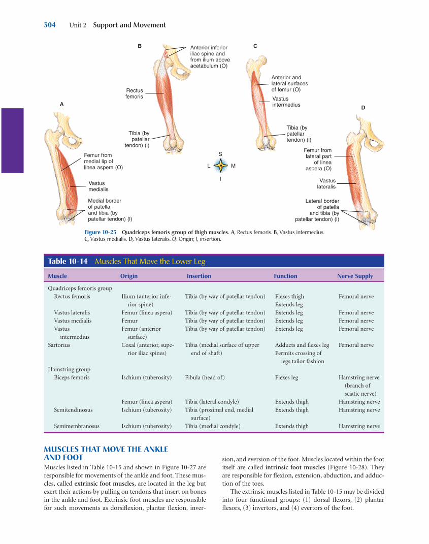
Table 10-14 identifies muscles that move the lower leg.Again, see Figures 10-1 and 10-2 and refer to Figures 10-25and 10-26 as you study the table.
Anatomy of the Muscular System Chapter 10 301
Box 10-3 HEALTH MATTERS
Carpal Tunnel Syndrome
The carpal tunnel. The median nerve and muscles that flex the fingers pass through a concavity in thewrist called the carpal tunnel.
Tendons of flexorsof fingers
Median nerve
Carpaltunnel
Tendon sheath Carpal bones
Some epidemiologists specialize in the field of occupa-tional health, the study of health matters related to work
or the workplace. Many problems seen by occupationalhealth experts are caused by repetitive motions of the wristsor other joints. Word processors (typists) and meat cutters,for example, are at risk of developing conditions caused byrepetitive motion injuries.
One common problem often caused by such repetitivemotion is tenosynovitis (ten-o-sin-o-VYE-tis)— inflamma-tion of a tendon sheath. Tenosynovitis can be painful, andthe swelling characteristic of this condition can limit move-ment in affected parts of the body. For example, swelling ofthe tendon sheath around tendons in an area of the wristknown as the carpal tunnel can limit movement of the
wrist, hand, and fingers. The figure shows the relative po-sitions of the tendon sheath and median nerve within thecarpal tunnel. If this swelling, or any other lesion in thecarpal tunnel, presses on the median nerve, a conditioncalled carpal tunnel syndrome may result. Because themedian nerve connects to the palm and radial side (thumbside) of the hand, carpal tunnel syndrome is characterizedby weakness, pain, and tingling in this part of the hand.The pain and tingling may also radiate to the forearm andshoulder. Prolonged or severe cases of carpal tunnel syn-drome may be relieved by injection of antiinflammatoryagents. A permanent cure is sometimes accomplished bysurgical cutting or removal of the swollen tissue pressingon the median nerve.
Bodies of twelfth thoracic and all
lumbar vertebrae (O)Psoas major
Psoas minor
Iliacus
Femur (l)(lesser trochanter)
S
LR
I
Figure 10-21 Iliopsoas muscle (iliacus, psoas major, and psoasminor muscles). O, Origin; I, insertion.
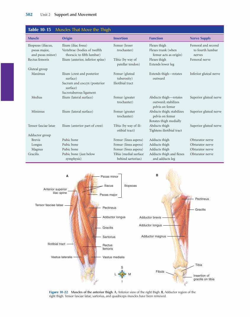
302 Unit 2 Support and Movement
Table 10-13 Muscles That Move the Thigh
Muscle Origin Insertion Function Nerve Supply
Iliopsoas (iliacus,
psoas major,
and psoas minor)
Rectus femoris
Gluteal group
Maximus
Medius
Minimus
Tensor fasciae latae
Adductor group
Brevis
Longus
Magnus
Gracilis
Ilium (iliac fossa)
Vertebrae (bodies of twelfth
thoracic to fifth lumbar)
Ilium (anterior, inferior spine)
Ilium (crest and posterior
surface)
Sacrum and coccyx (posterior
surface)
Sacrotuberous ligament
Ilium (lateral surface)
Ilium (lateral surface)
Ilium (anterior part of crest)
Pubic bone
Pubic bone
Pubic bone
Pubic bone (just below
symphysis)
Femur (lesser
trochanter)
Tibia (by way of
patellar tendon)
Femur (gluteal
tuberosity)
Iliotibial tract
Femur (greater
trochanter)
Femur (greater
trochanter)
Tibia (by way of ili-
otibial tract)
Femur (linea aspera)
Femur (linea aspera)
Femur (linea aspera)
Tibia (medial surface
behind sartorius)
Flexes thigh
Flexes trunk (when
femur acts as origin)
Flexes thigh
Extends lower leg
Extends thigh—rotates
outward
Abducts thigh—rotates
outward; stabilizes
pelvis on femur
Abducts thigh; stabilizes
pelvis on femur
Rotates thigh medially
Abducts thigh
Tightens iliotibial tract
Adducts thigh
Adducts thigh
Adducts thigh
Adducts thigh and flexes
and adducts leg
Femoral and second
to fourth lumbar
nerves
Femoral nerve
Inferior gluteal nerve
Superior gluteal nerve
Superior gluteal nerve
Superior gluteal nerve
Obturator nerve
Obturator nerve
Obturator nerve
Obturator nerve
Anterior superioriliac spine
Tensor fasciae latae
Iliotibial tract
Vastus lateralis
Psoas minor
Iliacus
Psoas major
Pectineus
Adductor longus
Gracilis
Sartorius
Rectusfemoris
Vastus medialis
Iliopsoas
S
ML
I
Adductor brevis
Adductor longus
Adductor magnus
FibulaInsertion of gracilis on tibia
Tibia
Gracilis
Pectineus
A B
Figure 10-22 Muscles of the anterior thigh. A, Anterior view of the right thigh. B, Adductor region of theright thigh. Tensor fasciae latae, sartorius, and quadriceps muscles have been removed.

Anatomy of the Muscular System Chapter 10 303
Pubic bone(anterior surfacebelow origin of adductor longus) (O)
Adductor brevis
Femur (upperhalf of linea aspera) (l) Femur (l)
(middle third of linea aspera)
Pubic bone (below symphysis) (O)
Adductor longus
Gracilis
Tibia (l)Pubic bone (superior ramus) (O)
Pectineus
Femoral shaft(below lessertrochanter) (l)
Adductor magnus
Anterior view
Adductormagnus
Femur (adductor tubercle) (l)
Femur (linea aspera) (l)
Pubic archand outer part
of inferior surfaceof ischial tuberosity (O)
Posterior view
S
ML
I
S
ML
I
S
ML
I
S
LM
I
S
ML
I
Figure 10-23 Muscles that adduct the thigh. O, Origin; I, insertion.
Greater trochanter (l)
Gluteus medius
Ilium, posterior surfaceof iliac crest (O)
Crest and posteriorsurface of ilium,sacrum, coccyx
and sacrotuberousligament (O)
Gluteus maximus
Gluteal tuberosityof femur and ilio-
tibial tract (l)
Gluteal surfaceof ilium (O)
Gluteus minimus
Greater trochanter of femur (l)
S
LM
I
A CB
Figure 10-24 Gluteal muscles. A, Gluteus maximus. B, Gluteus minimus. C, Gluteus medius.
MUSCLES THAT MOVE THE ANKLE AND FOOTMuscles listed in Table 10-15 and shown in Figure 10-27 areresponsible for movements of the ankle and foot. These mus-cles, called extrinsic foot muscles, are located in the leg butexert their actions by pulling on tendons that insert on bonesin the ankle and foot. Extrinsic foot muscles are responsiblefor such movements as dorsiflexion, plantar flexion, inver-
sion, and eversion of the foot. Muscles located within the footitself are called intrinsic foot muscles (Figure 10-28). Theyare responsible for flexion, extension, abduction, and adduc-tion of the toes.
The extrinsic muscles listed in Table 10-15 may be dividedinto four functional groups: (1) dorsal flexors, (2) plantarflexors, (3) invertors, and (4) evertors of the foot.
304 Unit 2 Support and Movement
Table 10-14 Muscles That Move the Lower Leg
Muscle Origin Insertion Function Nerve Supply
Quadriceps femoris group
Rectus femoris
Vastus lateralis
Vastus medialis
Vastus
intermedius
Sartorius
Hamstring group
Biceps femoris
Semitendinosus
Semimembranosus
Ilium (anterior infe-
rior spine)
Femur (linea aspera)
Femur
Femur (anterior
surface)
Coxal (anterior, supe-
rior iliac spines)
Ischium (tuberosity)
Femur (linea aspera)
Ischium (tuberosity)
Ischium (tuberosity)
Tibia (by way of patellar tendon)
Tibia (by way of patellar tendon)
Tibia (by way of patellar tendon)
Tibia (by way of patellar tendon)
Tibia (medial surface of upper
end of shaft)
Fibula (head of)
Tibia (lateral condyle)
Tibia (proximal end, medial
surface)
Tibia (medial condyle)
Flexes thigh
Extends leg
Extends leg
Extends leg
Extends leg
Adducts and flexes leg
Permits crossing of
legs tailor fashion
Flexes leg
Extends thigh
Extends thigh
Extends thigh
Femoral nerve
Femoral nerve
Femoral nerve
Femoral nerve
Femoral nerve
Hamstring nerve
(branch of
sciatic nerve)
Hamstring nerve
Hamstring nerve
Hamstring nerve
B
Femur from medial lip of linea aspera (O)
Vastus medialis
Medial border of patellaand tibia (bypatellar tendon) (l)
Rectus femoris
Tibia (bypatellar
tendon) (l)
Anterior inferioriliac spine and from ilium aboveacetabulum (O)
Anterior and lateral surfacesof femur (O)
Vastus intermedius
Tibia (bypatellartendon) (l)
Femur from lateral part
of linea aspera (O)
Vastus lateralis
Lateral borderof patella
and tibia (bypatellar tendon) (l)
AD
S
ML
I
C
Figure 10-25 Quadriceps femoris group of thigh muscles. A, Rectus femoris. B, Vastus intermedius. C, Vastus medialis. D, Vastus lateralis. O, Origin; I, insertion.

Anatomy of the Muscular System Chapter 10 305
Table 10-15 Muscles That Move the Foot
Muscle Origin Insertion Function Nerve Supply
ExtrinsicTibialis anterior
Gastrocnemius
Soleus
Peroneus longus
Peroneus brevis
Peroneus tertius
Extensor digitorum
longus
IntrinsicLumbricales
Flexor digiti minimi
brevis
Flexor hallucis brevis
Flexor digitorum brevis
Abductor digiti minimi
Abductor hallucis
Tibia (lateral condyle of
upper body)
Femur (condyles)
Tibia (underneath
gastrocnemius)
Fibula
Tibia (lateral condyle)
Fibula (head and shaft)
Fibula (lower two
thirds of lateral
surface of shaft)
Fibula (distal third)
Tibia (lateral condyle)
Fibula (anterior surface)
Tendons of flexor digi-
torum longus
Fifth metatarsal
Cuboid
Medial and lateral
cuneiform
Calcaneous
Plantar fascia
Calcaneous
Calcaneous
Tarsal (first cuneiform)
Metatarsal (base of first)
Tarsal (calcaneus by way
of Achilles tendon)
Tarsal (calcaneus by way
of Achilles tendon)
First cuneiform
Base of first metatarsal
Fifth metatarsal (tubercle,
dorsal surface)
Fourth and fifth meta-
tarsals (bases of)
Second and third pha-
langes (four lateral toes)
Phalanges (2 to 5)
Proximal phalanx of
fifth toe
Proximal phalanx of first
(great) toe
Middle phalanges of toes
(2 to 5)
Proximal phalanx of fifth
(small) toe
First (great) toe
Flexes foot
Inverts foot
Extends foot
Flexes lower leg
Extends foot (plantar
flexion)
Extends foot (plantar
flexion)
Everts foot
Everts foot
Flexes foot
Flexes foot
Everts foot
Dorsiflexion of foot; ex-
tension of toes
Flex proximal phalanges
Extend middle and
distal phalanges
Flexes fifth (small) toe
Flexes first (great) toe
Flexes toes two through
five
Abducts fifth (small) toe
Flexes fifth toe
Abducts first (great) toe
Common and deep
peroneal nerves
Tibial nerve (branch
of sciatic nerve)
Tibial nerve
Common peroneal
nerve
Superficial peroneal
nerve
Deep peroneal nerve
Deep peroneal nerve
Lateral and medial
plantar nerve
Lateral plantar nerve
Medial and lateral
plantar nerve
Medial plantar nerve
Lateral plantar nerve
Medial plantar nerve
Tuberosity of ischium (long head) (O)
Biceps femoris(long head)
Linea asperaof femur(short head) (O)
Biceps femoris(short head)
Lateral condyleof tibia and head of fibula (l)
Tuberosityof ischium (O)
Semitendinosus
Medial condyleof tibia (l)
Medial surface oftibia (l)
Semimembranosus
Tuberosity of ischium (O)
S
LM
I
A B C
Figure 10-26 Hamstring group of thigh muscles. A, Biceps femoris. B, Semitendinosus. C, Semimembra-nosus. O, Origin; I, insertion.

The superficial muscles located on the posterior surfaceof the leg form the bulging “calf.” The common tendon ofthe gastrocnemius (gas-trok-NEE-me-us) and soleus iscalled the calcaneal, or Achilles, tendon. It inserts into thecalcaneus, or heel bone. By acting together, these musclesserve as powerful flexors (plantar flexion) of the foot.
Dorsal flexors of the foot, located on the anterior surfaceof the leg, include the tibialis (tib-ee-AL-is) anterior, per-oneus tertius (per-o-NEE-us TER-shus), and extensor digi-torum longus. In addition to functioning as a dorsiflexor ofthe foot the extensor digitorum longus also everts the footand extends the toes.
POSTUREWe have already discussed the major role of muscles inmovement and heat production. We shall now turn our at-tention to a third way in which muscles serve the body as awhole—that of maintaining the posture of the body. Let usconsider a few aspects of this important function.
The term posture means simply maintaining optimalbody position. “Good posture” means many things. It meansbody alignment that most favors function; it means positionthat requires the least muscular work to maintain, whichputs the least strain on muscles, ligaments, and bones; oftenit means keeping the body’s center of gravity over its base.Good posture in the standing position, for example, meanshead and chest held high; chin, abdomen, and buttockspulled in; knees bent slightly; and feet placed firmly on theground about 6 inches (15 cm) apart. Good posture in a sit-ting position varies with the position that one is trying tomaintain. Good posture during exercise, such as riding a
306 Unit 2 Support and Movement
Gastrocnemius
Soleus
Tendon ofperoneus
longus (cut)
Peroneuslongus (cut)
Tibialisanterior
Extensordigitorumlongus
Peroneusbrevis
Peroneustertius
Soleus
Peroneuslongus
Extensor digitorum
longus
Peroneusbrevis
Peroneustertius
Gastrocnemius
Tibialisanterior
Soleus
Tibia
Two heads of gastrocnemius
Gastrocnemius
Soleus
Calcanealtendon(Achillestendon)
Calcaneus
S
ML
I
S
LM
I
S
AP
I
A B C
Figure 10-27 Superficial muscles of the leg. A, Anterior view. B, Posterior view. C, Lateral view.
Lumbricales
Flexor digitiminimi brevis
Abductor digiti minimi
Flexor hallucisbrevis
Flexor digitorumbrevis
Abductor hallucis
Plantar aponeurosis(cut)
A
ML
P
Figure 10-28 Intrinsic muscles of the foot. Inferior (plantar) view.
1. Name the three gluteal muscles.2. What is the function of the gastrocnemius muscle?

horse or dribbling a basketball, means moving or tensingdifferent parts of the body frequently to avoid falling.
HOW POSTURE IS MAINTAINEDGravity pulls on the various parts of the body at all times,and because bones are too irregularly shaped to balancethemselves on each other, the only way the body can beheld in any particular position is for muscles to exert a con-tinual pull on bones in the opposite direction from gravityor other forces that pull on body parts. When the body is ina standing position, gravity tends to pull the head andtrunk forward and downward; muscles (head and trunk ex-tensors) must therefore pull backward and upward onthem. For instance, gravity pulls the lower jaw downward;muscles must pull upward on it. Muscles exert this pullagainst gravity by virtue of their property of tonicity.Tonicity, or muscle tone, literally means tension and refersto the continuous, low level of sustained contraction main-tained by all skeletal muscles. Because tonicity is absentduring deep sleep, muscle pull does not then counteractthe pull of gravity. Hence, we cannot sleep standing up. In-
terestingly, astronauts in the low-gravity conditions ofspace station missions can sleep in a standing position, aslong as they are secured inside special sleeping bags on thewall of the space station.
Many structures other than muscles and bones play a partin the maintenance of posture. The nervous system is re-sponsible for the existence of muscle tone and also regulatesand coordinates the amount of pull exerted by the individ-ual muscles. The respiratory, digestive, circulatory, excretory,and endocrine systems all contribute something toward theability of muscles to maintain posture. This is one of manyexamples of the important principle that all body functionsare interdependent.
The importance of posture can perhaps be best evaluatedby considering some of the effects of poor posture. Poor pos-ture throws more work on muscles to counteract the pull ofgravity and therefore leads to fatigue more quickly thangood posture. Poor posture puts more strain on ligaments. Itputs abnormal strains on bones and may eventually producedeformities. It also interferes with various functions such asrespiration, heart action, and digestion.
Anatomy of the Muscular System Chapter 10 307
Box 10-4 HEALTH MATTERS
Intramuscular Injections
Many drugs are administered by intramuscular injection.If the amount to be injected is 2 ml or less, the deltoid
muscle is often selected as the site of injection. Note that inpart A of this figure the needle is inserted into the muscleabout two fingers’ breadth below the acromion process of thescapula and lateral to the tip of the acromion. If the amountof medication to be injected is 2 to 3 ml, the gluteal areashown in part B of the figure, is often used. Injections aremade into the gluteus medius muscle near the center of the
upper outer quadrant, as shown in the illustration. Anothertechnique of locating the proper injection site is to draw animaginary diagonal line from a point of reference on the backof the bony pelvis (posterior superior iliac spine) to thegreater trochanter of the femur. The injection is given aboutthree fingers’ breadth above and one third of the way downthe line. It is important to avoid the sciatic nerve and the su-perior gluteal blood vessels during the injection. Proper tech-nique requires a knowledge of the underlying anatomy.
Trapezius
Acromionprocess of scapula
Deltoid
Greatertrochanter
Gluteus medius
IliaccrestSuperior gluteal artery and vein
Posterior superior iliac spine
Sciatic nerve
Gluteus maximus
A
B
Intramuscular injection sites. A, Deltoid injection site for small doses. B, Gluteal injection site for largerdoses.
A B

308 Unit 2 Support and Movement
Acting together, the muscular, skeletal, and nervous sys-tems permit us to move in a coordinated and controlled
way. However, it is the contraction, or shortening, of musclesthat ultimately provides the actual movement necessary forphysical activity. Dramatic changes occur in the muscular sys-tem throughout the cycle of life. Muscle cells may increase ordecrease in size and ability to shorten most effectively at dif-ferent periods in life. In addition to age-related changes,many pathological conditions occurring at different ages mayalso affect the muscular system.
Because of the functional interdependence of the muscu-loskeletal and nervous systems, life cycle changes affectingthe muscles often manifest in other components of the func-tional unit. During infancy and childhood the ability to coor-dinate and control the strength of muscle contraction permits
a sequential series of development steps to occur. A develop-ing youngster learns to hold the head up, roll over, sit up,stand alone, and then walk and run as developmentalchanges permit better control and coordination of muscularcontraction.
Degenerative changes associated with advancing age of-ten result in replacement of muscle cell volume with non-functional connective tissue. As a result, the strength of mus-cular contraction diminishes. Recent findings show that muchif not all of this age-related decrease in muscle strength actu-ally results from disuse atrophy (see Box 11-6 in Chapter 11)and thus may be avoidable. Pathological conditions associ-ated with specific age ranges can also affect one or morecomponents of the functional unit that permits us to movesmoothly and effortlessly.
CYCLE OF LIFE
Muscular System
As you read through this chapter, what struck you mostwas probably the large number of individual muscles and
their many actions. Although learning the names, locations,origins, insertions, and other details of the major muscles is aworthwhile endeavor, such an activity can cause you to losesight of the “big picture.” Step back from the details a momentto appreciate the fact that the muscles work as coordinatedteams of biological engines that move the various compo-nents of the flexible skeleton.
As a matter of fact, in this chapter you learned that the fi-brous wrappings of each muscle are continuous with its ten-dons, which in turn are continuous with the fibrous structureof the bone to which they are attached. Thus we can see thatthe muscular and skeletal systems are in essence a singlestructure. This fact is very important in seeing a “big picture”comprising many individual muscles and bones. The entireskeletomuscular system, as it is often called, is actually a sin-
gle, continuous structure that comprises a coordinated, dy-namic framework for the body.
Taking another step back, an even bigger picture comesinto focus. Other systems of the body must play a role in theactions of the skeletomuscular system. For example, the ner-vous system senses changes in body position and degrees ofmovement—permitting integration of feedback loops that ul-timately regulate the muscular contractions that maintainposture and produce movements. The cardiovascular systemmaintains blood flow in the muscles, and the urinary and res-piratory systems rid the body of wastes produced in the mus-cles. The respiratory and digestive systems bring in oxygenand nutrients necessary for muscle function.
The picture is still not complete, however. We will see evenmore when Chapter 11 continues the story of muscle func-tion by delving into the details of how each muscle works asan engine to drive the movement of the body.
THE BIG PICTURE
Skeletal Muscles and the Whole Body

Anatomy of the Muscular System Chapter 10 309
B. It will decrease the swelling associated with the injury.
C. It will remind Ms. Rider to avoid heavy lifting us-ing this extremity and thus decrease the likelihoodof reinjury to the area.
D. It will decrease the repetitive motions of the wristand prevent continuous injury.
3. During the routine examination, Ms. Rider is foundto have a slight separation of muscle when she isasked to lift her head off the examination tablewhile in a supine position. Which muscle do you expect to show this separation based on the abovedescription?A. External obliqueB. Internal obliqueC. Rectus abdominisD. Transversus abdominis
4. During the physical examination, the physician tests Ms. Rider’s right brachialis muscle strength.Which one of the following correctly describes this procedure?A. The forearm should be flexed at the elbow.B. The forearm should be extended at the elbow.C. Ms. Rider is asked to extend her elbow and push
against a hard surface.D. Ms. Rider is asked to lift a heavy object above her
head.
Patricia Rider, age 42, came to the health clinic com-plaining of pain in her right hand and fingers, espe-
cially at night, for the past 2 months. She works as asewing machine operator at a local factory making men’spants. Ms. Rider thinks of herself as being in good health.She walks daily and follows a low-fat diet. Family historyis negative for cancer, arthritis, heart disease, and diabetes.Physical examination reflects a positive Tinel’s sign (tin-gling sensation radiating from the wrist to the hand) withgentle tapping on the inside of the right wrist with a re-flex hammer.
1. After a complete physical examination, Ms. Rider is diagnosed with carpal tunnel syndrome. Which ofthe following nerves is usually involved with this diagnosis? A. FemoralB. MedianC. Ulna D. Radial
2. The physician has ordered that Ms. Rider wear a rightwrist splint held in place by an Ace bandage. Which oneof the following is the BEST rationale for this treatment?A. It will stabilize the joint, thus reducing the pain
in this area at night, while allowing Ms. Rider toremove the splint and continue her work as asewing machine operator during the day.
CASE STUDY
INTRODUCTION (Figures 10-1 and 10-2)
A. There are more than 600 skeletal muscles in the bodyB. From 40% to 50% of our body weight is skeletal muscleC. Muscles, along with the skeleton, determine the form
and contour of our body
SKELETAL MUSCLE STRUCTURE (Figure 10-3)
A. Connective tissue components1. Endomysium—delicate connective tissue membrane
that covers specialized skeletal muscle fibers2. Perimysium—tough connective tissue binding to-
gether fascicles3. Epimysium—coarse sheath covering the muscle as a
whole4. These three fibrous components may become a ten-
don or an aponeurosisB. Size, shape, and fiber arrangement (Figure 10-4)
1. Skeletal muscles vary considerably in size, shape, andfiber arrangement
2. Size—range from extremely small to large masses3. Shape—variety of shapes, such as broad, narrow,
long, tapering, short, blunt, triangular, quadrilateral,irregular, flat sheets, or bulky masses
4. Arrangement—variety of arrangements, such as paral-lel to long axis, converge to a narrow attachment,oblique, pennate, bipennate, or curved; the direction offibers is significant due to its relationship to function
C. Attachment of muscles (Figure 10-5)1. Origin—point of attachment that does not move
when the muscle contracts2. Insertion—point of attachment that moves when the
muscle contractsD. Muscle actions
1. Most movements are produced by the coordinatedaction of several muscles; some muscles in the groupcontract while others relaxa. Prime mover (agonist)—a muscle or group
of muscles that directly performs a specific movement
b. Antagonist—muscles that, when contracting, di-rectly oppose prime movers; relax while primemover (agonist) is contracting to produce movement; provide precision and control during contraction of prime movers
c. Synergists—muscles that contract at the sametime as the prime movers; they facilitate prime
CHAPTER SUMMARY

310 Unit 2 Support and Movement
mover actions to produce a more efficientmovement
d. Fixator muscles—joint stabilizersE. Lever systems
1. In the human body bones serve as levers and jointsserve as fulcrums; contracting muscle applies apulling force on a bone lever at the point of the mus-cle’s attachment to the bone, causing the insertionbone to move about its joint fulcrum
2. Lever system—composed of four component parts(Figure 10-6)a. Rigid bar (bone)b. Fulcrum (F) around which the rod moves
(joint)c. Load (L) that is movedd. Pull (P) that produces movement (muscle
contraction)3. First-class levers
a. Fulcrum lies between the pull and the loadb. Not abundant in the human body; serve as levers
of stability4. Second-class levers
a. Load lies between the fulcrum and the joint atwhich the pull is exerted
b. Presence of these levers in the human body is acontroversial issue
5. Third-class leversa. Pull is exerted between the fulcrum and loadb. Permit rapid and extensive movementc. Most common type of lever found in the body
HOW MUSCLES ARE NAMEDA. Muscle names can be in Latin or English (this book uses
English)B. Muscles are named using one or more of the following
features:1. Location, function, shape2. Direction of fibers—named according to fiber
orientation3. Number of heads or divisions4. Points of attachment—origin and insertion points5. Relative size—small, medium, or large
C. Hints on how to deduce muscle actions
IMPORTANT SKELETAL MUSCLESA. Muscles of facial expression—unique in that at least
one point of attachment is to the deep layers of theskin over the face or neck (Figure 10-7; Table 10-3)
B. Muscles of mastication—responsible for chewing move-ments (Figure 10-7; Table 10-3)
C. Muscles that move the head—paired muscles on eitherside of the neck are responsible for head movements(Figure 10-8; Table 10-4)
TRUNK MUSCLESA. Muscles of the thorax—critical importance in respira-
tion (Figure 10-9; Table 10-5)
B. Muscles of the abdominal wall—arranged in threelayers, with fibers in each layer running in different di-rections to increase strength (Figure 10-10; Table 10-6)
C. Muscles of the back—bend or stabilize the back (Fig-ure 10-11; Table 10-7)
D. Muscles of the pelvic floor—support the structures inthe pelvic cavity (Figure 10-12; Table 10-8)
UPPER LIMB MUSCLESA. Muscles acting on the shoulder girdle—muscles that
attach the upper extremity to the torso are located ante-riorly (chest) or posteriorly (back and neck); thesemuscles also allow extensive movement (Figure 10-13;Table 10-9)
B. Muscles that move the upper arm—the shoulder is asynovial joint allowing extensive movement in everyplane of motion (Figure 10-15; Table 10-10)
C. Muscles that move the forearm—found proximally to the elbow and attach to the ulna and radius (Fig-ures 10-16 and 10-17; Table 10-11)
D. Muscles that move the wrist, hand, and fingers—these muscles are located on the anterior or posteriorsurfaces of the forearm (Figures 10-18 through 10-20;Table 10-12)
LOWER LIMB MUSCLESA. The pelvic girdle and lower extremity function in loco-
motion and maintenance of stabilityB. Muscles that move the thigh and lower leg (Figures 10-
1, 10-2, 10-21 through 10-26; Tables 10-13 and 10-14)C. Muscles that move the ankle and foot (Figures 10-27
and 10-28; Table 10-15)1. Extrinsic foot muscles are located in the leg and exert
their actions by pulling on tendons that insert onbones in the ankle and foot; responsible for dorsiflex-ion, plantar flexion, inversion, and eversion
2. Intrinsic foot muscles are located within the foot; re-sponsible for flexion, extension, abduction, and ad-duction of the toes
POSTUREA. Maintaining the posture of the body is one of the major
roles muscles playB. “Good posture”—body alignment that most favors
function, requires the least muscular work to maintain,keeping the body’s center of gravity over its base
C. How posture is maintained1. Muscles exert a continual pull on bones in the oppo-
site direction from gravity2. Structures other than muscle and bones have a role in
maintaining posturea. Nervous system—responsible for the existence
of muscle tone and also regulation and coordina-tion of the amount of pull exerted by individualmuscles
b. Respiratory, digestive, excretory, and endocrinesystems all contribute to maintain posture
Anatomy of the Muscular System Chapter 10 311
CYCLE OF LIFE: MUSCULAR SYSTEMA. Muscle cells—increase or decrease in number, size, and
ability to shorten at different periodsB. Pathological conditions at different periods may affect
the muscular systemC. Life cycle changes—manifested in other components of
functional unit:1. Infancy and childhood—coordination and control-
ling of muscle contraction permits sequential devel-opment steps
D. Degenerative changes of advancing age result in re-placement of muscle cells with nonfunctional connec-tive tissue1. Diminished strength
7. Name the main muscles that flex, extend, abduct, andadduct the thigh; that flex and extend the lower leg andthigh; that flex and extend the foot.
8. Name the main muscles that flex, extend, abduct, andadduct the head.
9. Name the main muscles that move the abdominal wall;that move the chest wall.
1. Define the terms endomysium, perimysium, andepimysium.
2. Identify and describe the most common type of leversystem found in the body.
3. Give an example of a muscle named by: location, func-tion, shape, fiber direction, number of heads, points ofattachment.
4. Name the main muscles of the back, chest, abdomen,neck, shoulder, upper arm, lower arm, thigh, buttocks,leg, and pelvic floor.
5. Name the main muscles that flex, extend, abduct, andadduct the upper arm; that raise and lower the shoulder.
6. Name the main muscles that flex and extend the lowerarm; that flex and extend the wrist and hand.
REVIEW QUESTIONS
1. Identify the muscles of facial expression. What musclespermit smiling and frowning?
2. How do the origin and insertion of a muscle relate toeach other in regard to actual movement?
3. When the biceps brachii contracts, the elbow flexes.When the triceps brachii contracts, the elbow extends.Explain the role of both muscles in terms of agonistand antagonist in both of these movements.
4. Can you describe how posture is maintained?5. Describe the clinical significance regarding the differ-
ence in size between the large head of the humerus andthe small and shallow glenoid cavity of the scapula.
6. If a typist complained of weakness, pain, and tinglingin the palm and thumb side of the hand, what type ofproblem might he or she be experiencing? Explainspecifically what was happening to cause this discomfort.
7. Baseball players, particularly pitchers, often incurrotator cuff injuries. List the muscles that make up the rotator cuff and explain the importance of thesemuscles and their role in joint stability.
CRITICAL THINKING QUESTIONS