Embed Size (px)
Citation preview
Anatomy
Chapter 6 – The Muscular System
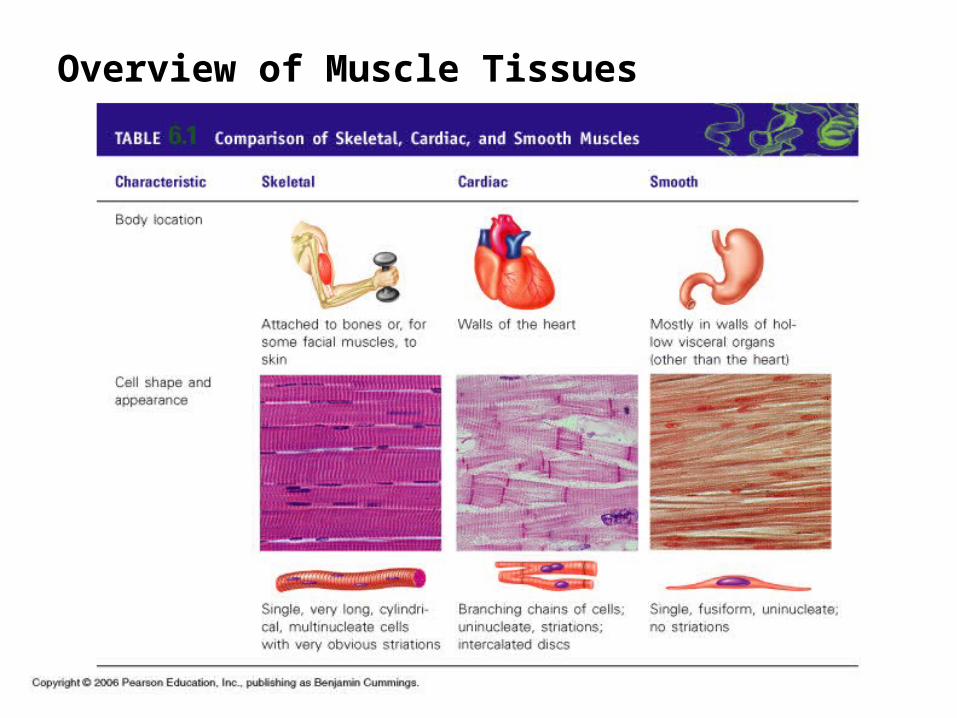
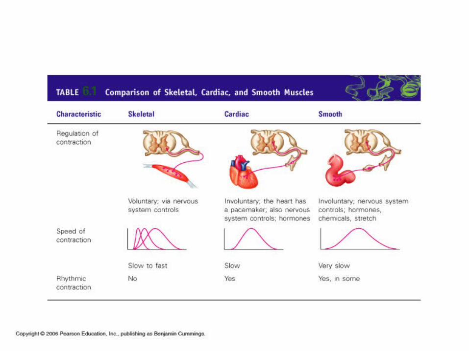
Overview of Muscle Tissues
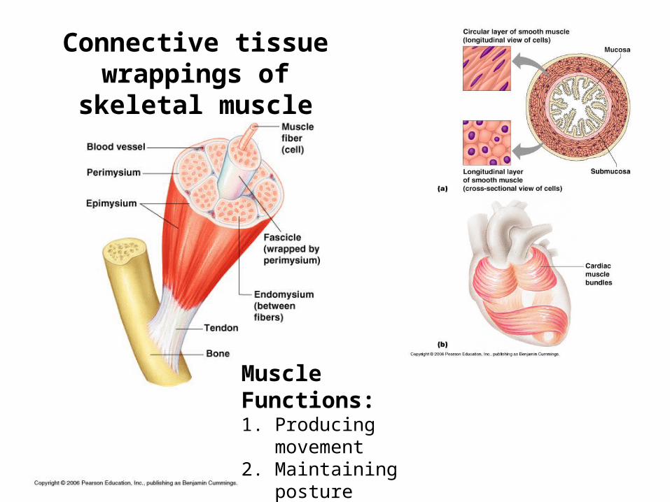
Connective tissue wrappings of skeletal muscle
Muscle Functions:1. Producing movement2. Maintaining posture3. Stabilizing joints4. Generating heat
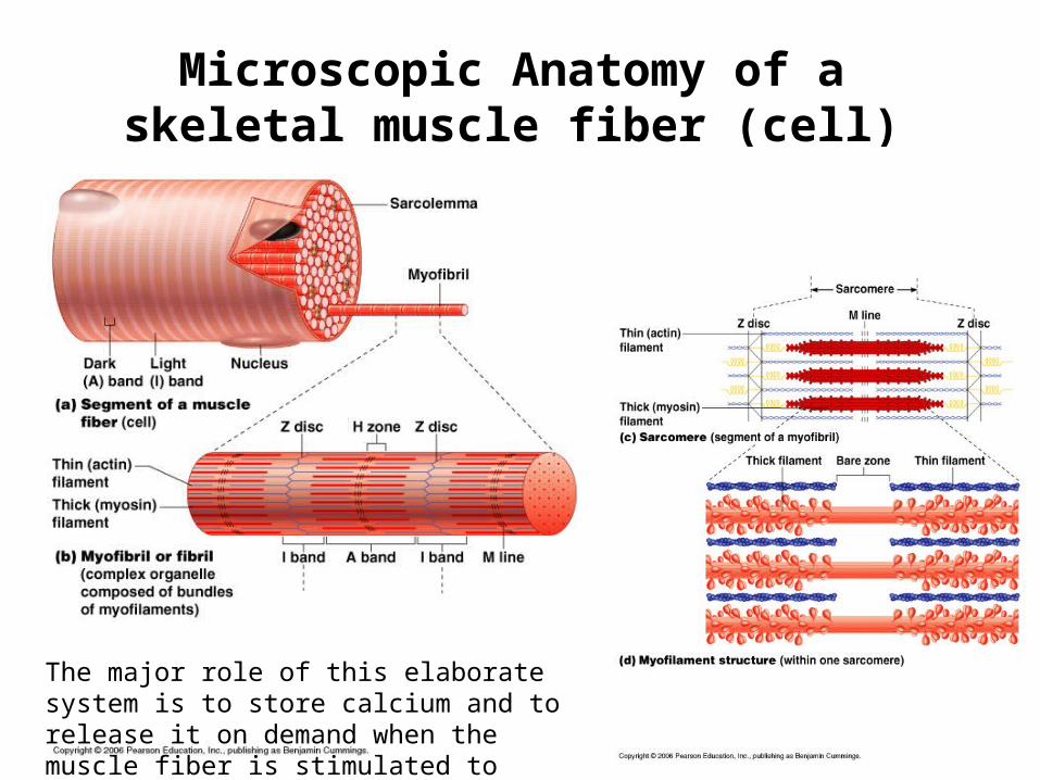
Microscopic Anatomy of a skeletal muscle fiber (cell)
The major role of this elaborate system is to store calcium and to release it on demand when the muscle fiber is stimulated to contract.
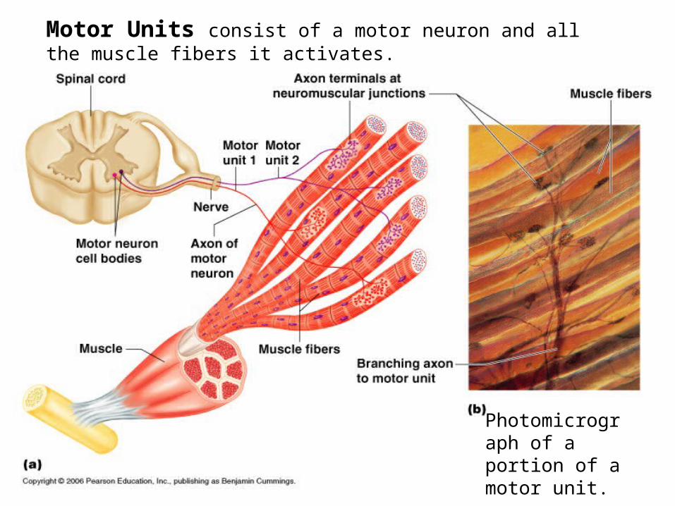
Motor Units consist of a motor neuron and all the muscle fibers it activates.
Photomicrograph of a portion of a motor unit.
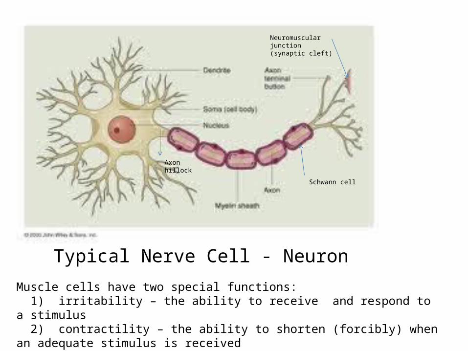
Schwann cell
Axon hillock
Neuromuscular junction (synaptic cleft)
Typical Nerve Cell - Neuron
Muscle cells have two special functions: 1) irritability – the ability to receive and respond to a stimulus 2) contractility – the ability to shorten (forcibly) when an adequate stimulus is received
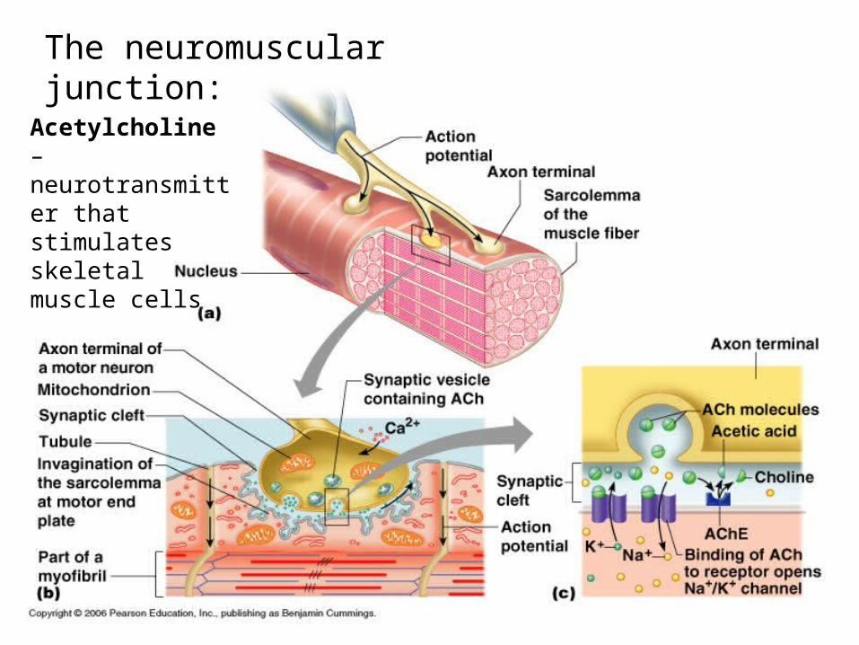
The neuromuscular junction:
Acetylcholine – neurotransmitter that stimulates skeletal muscle cells
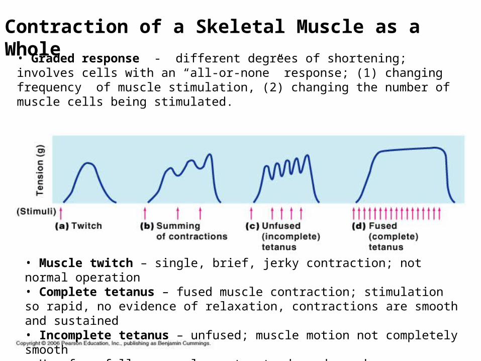
Contraction of a Skeletal Muscle as a Whole
• Graded response - different degrees of shortening; involves cells with an “all-or-none” response; (1) changing frequency of muscle stimulation, (2) changing the number of muscle cells being stimulated.
• Muscle twitch – single, brief, jerky contraction; not normal operation• Complete tetanus – fused muscle contraction; stimulation so rapid, no evidence of relaxation, contractions are smooth and sustained• Incomplete tetanus – unfused; muscle motion not completely smooth• How forcefully a muscle contracts depends on how many cells are being stimulated; the hand that caresses, can also deliver a stinging slap
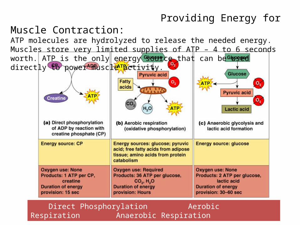
Providing Energy for Muscle Contraction: ATP molecules are hydrolyzed to release the needed energy. Muscles store very limited supplies of ATP – 4 to 6 seconds worth. ATP is the only energy source that can be used directly to power muscle activity.
Direct Phosphorylation Aerobic Respiration Anaerobic Respiration
Types of Muscle Contractions: Isotonic – “same tone” or tension; muscle shortens, movement
occurs (knee bend, smiling). Isometric – muscles do not shorten; “skidding you heels”,
pushing against the wall, wall sits.
Muscle fatigue and oxygen debt – exercising for prolonged times depletes ATP in the muscle; cannot be contracted even with stimulation; true muscle fatigue results in the muscle quitting, rarely occurs because most of us stop long before it happens – marathon runners.
Muscle tone – muscle remains firm, healthy, ready for action; continuous partial contractions. Resistance – isometric exercises, muscles pitted against immovable object.
Exercise – “Use it or lose it”; strength, stamina, endurance.
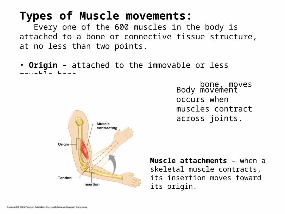
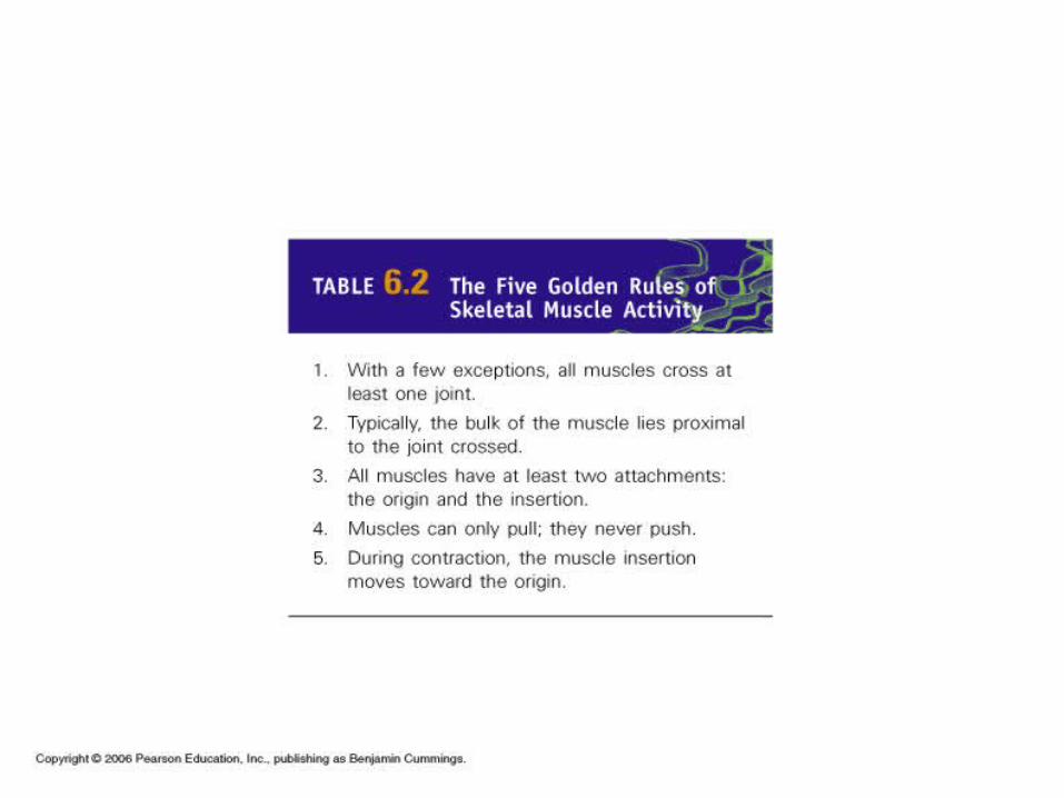
Types of Muscle movements: Every one of the 600 muscles in the body is attached to a bone or connective tissue structure, at no less than two points.
• Origin – attached to the immovable or less movable bone • Insertion – attached to the movable bone, moves toward the origin
Body movement occurs when muscles contract across joints.
Muscle attachments – when a skeletal muscle contracts, its insertion moves toward its origin.
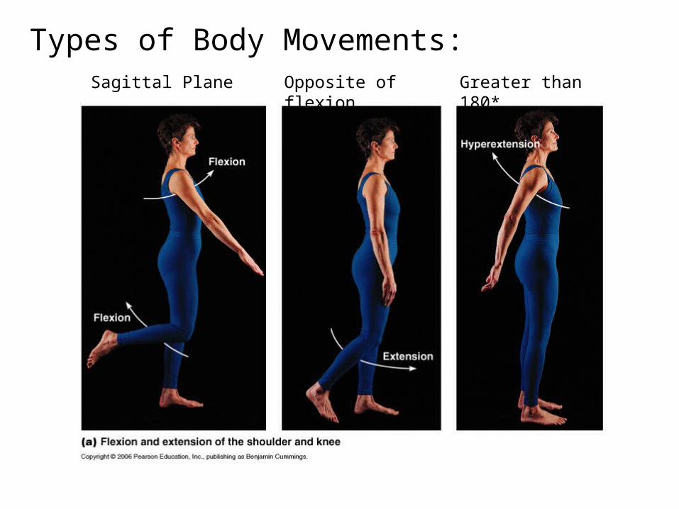
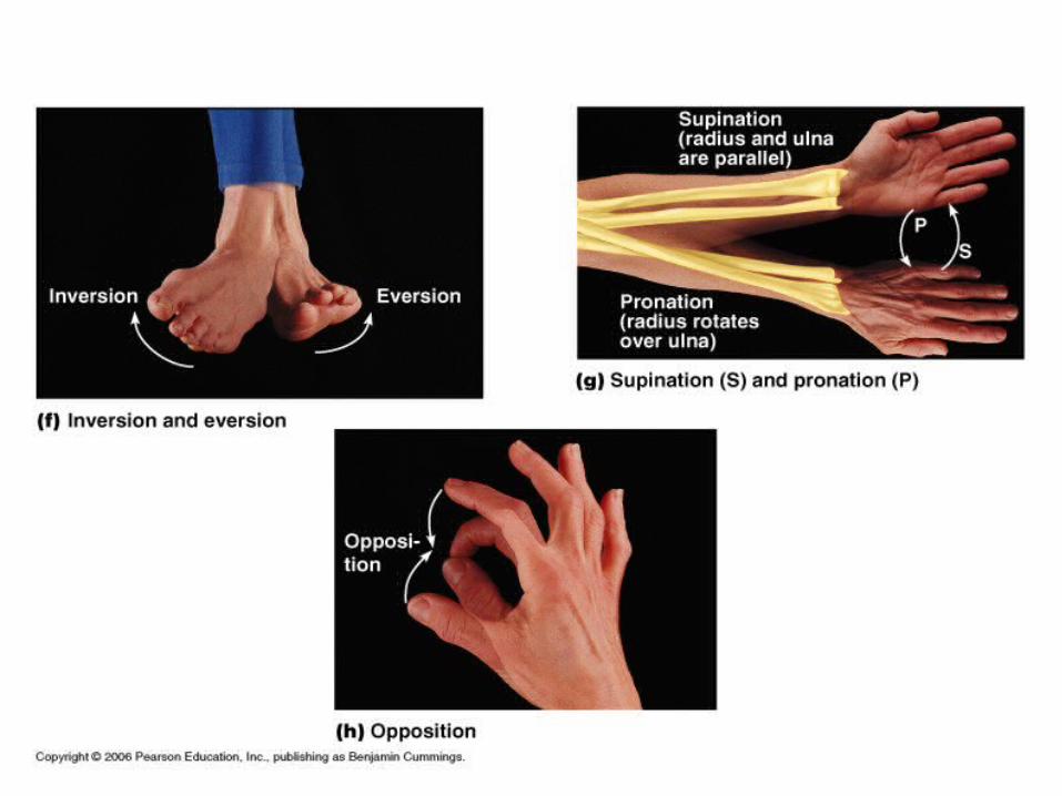
Types of Body Movements:Sagittal Plane Opposite of flexion Greater than 180*
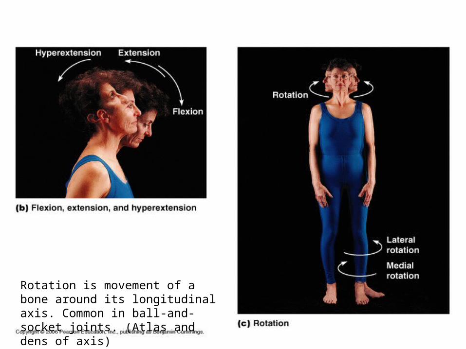
Rotation is movement of a bone around its longitudinal axis. Common in ball-and-socket joints. (Atlas and dens of axis)
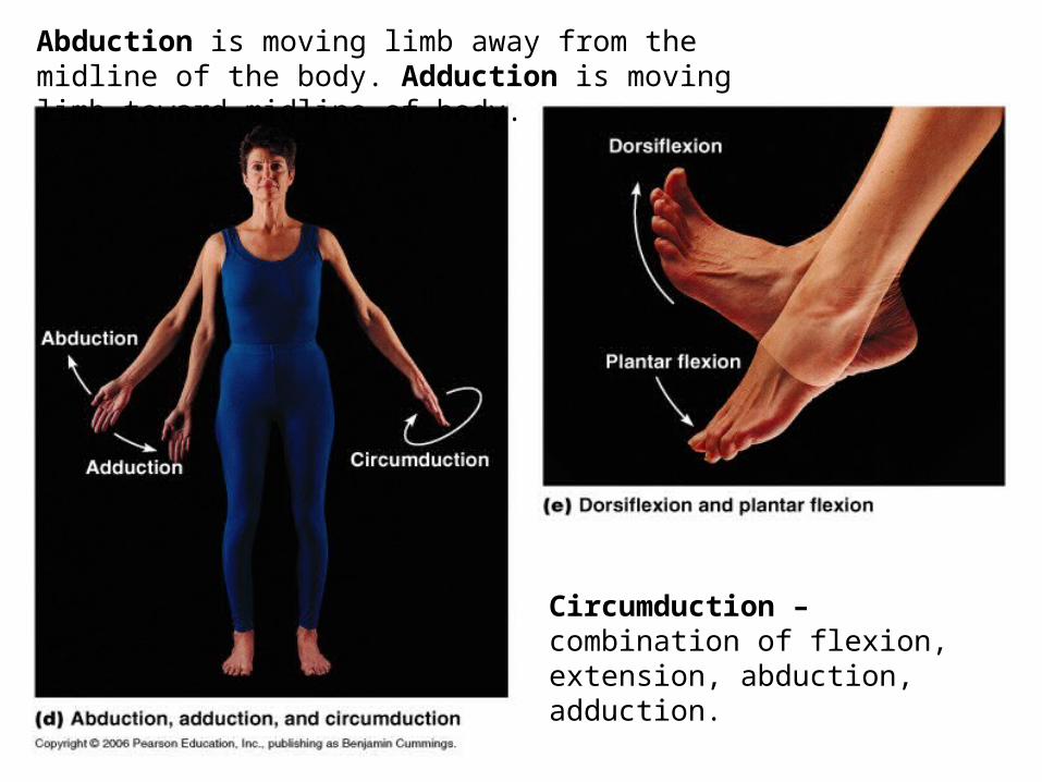
Abduction is moving limb away from the midline of the body. Adduction is moving limb toward midline of body.
Circumduction – combination of flexion, extension, abduction, adduction.
Interactions of Skeletal Muscles in the Body: Muscles cannot push – they only pull as they contract.
Movement is most often the result of the activity of two or more muscles acting together or against each other.
o Prime mover – the muscle with the major responsibility for causing a particular movement (biceps)
o Antagonist – muscle that oppose or reverse a movement (triceps)
o Synergist – help prime movers by producing the same movement or reducing undesirable movements (stabilizers)
o Fixators – specialized synergists – stabilize the origin of prime movers so all tension can be used to move the insertion bone
Naming Skeletal Muscles: Direction of muscles – reference to imaginary line; rectus – straight, oblique – running at an angle
Relative size – gluteus maximus – Large; gluteus minimus – small, longus – long
Location of muscle – bone associated with, frontalis, temporalis, occipitalis
Number of origins – biceps, triceps, quadriceps
location of the muscles origin and insertion – sternocleidomastoid
Shape of muscle – deltoid is triangular, trapezius
Action of muscle – adductor magus
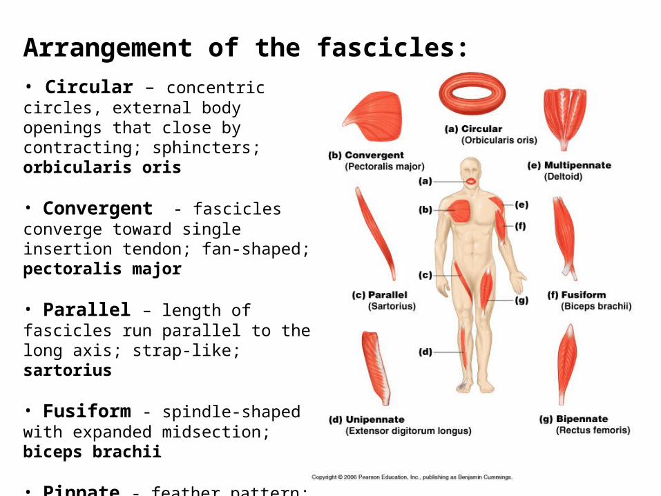
Arrangement of the fascicles:• Circular – concentric circles, external body openings that close by contracting; sphincters; orbicularis oris
• Convergent - fascicles converge toward single insertion tendon; fan-shaped; pectoralis major
• Parallel – length of fascicles run parallel to the long axis; strap-like; sartorius
• Fusiform - spindle-shaped with expanded midsection; biceps brachii
• Pinnate - feather pattern; unipennate , bipennate, multipennate
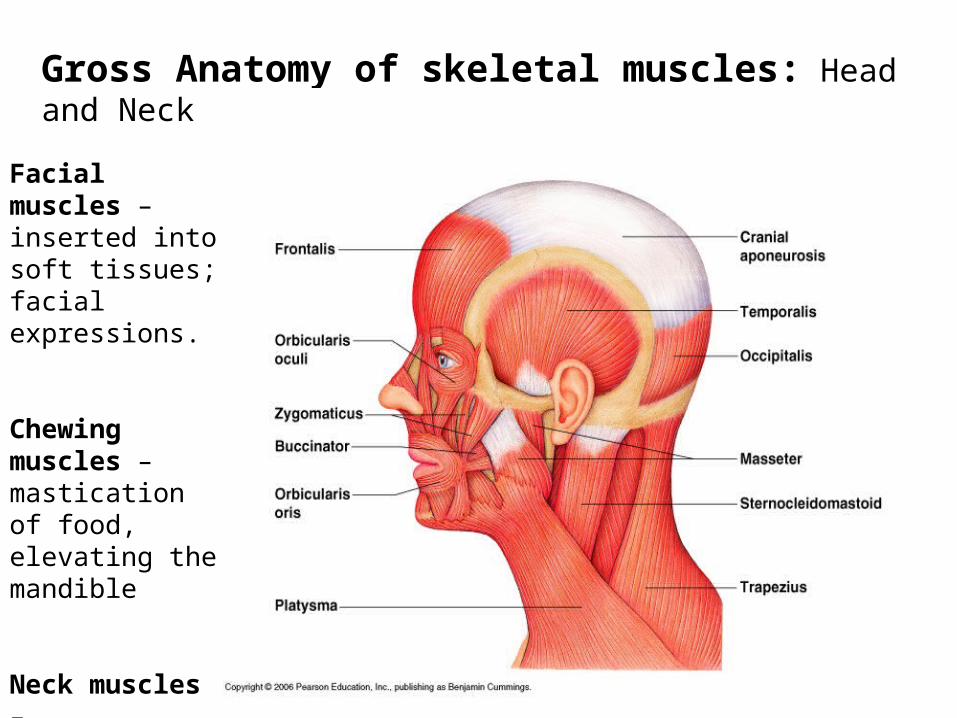
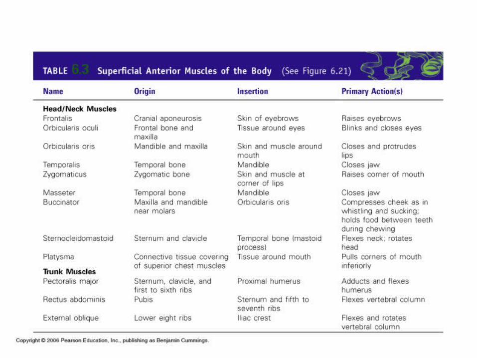
Gross Anatomy of skeletal muscles: Head and Neck
Facial muscles – inserted into soft tissues; facial expressions.
Chewing muscles –mastication of food, elevating the mandible
Neck muscles – move the head and shoulder girdle
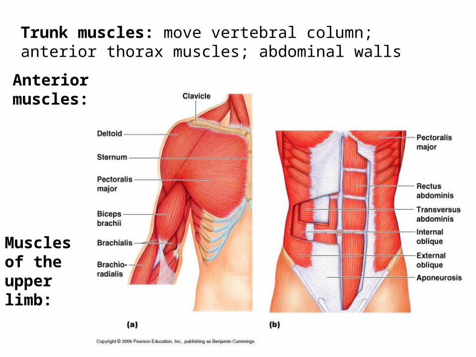
Trunk muscles: move vertebral column; anterior thorax muscles; abdominal walls
Anterior muscles:
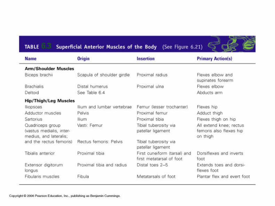
Muscles of the upper limb:
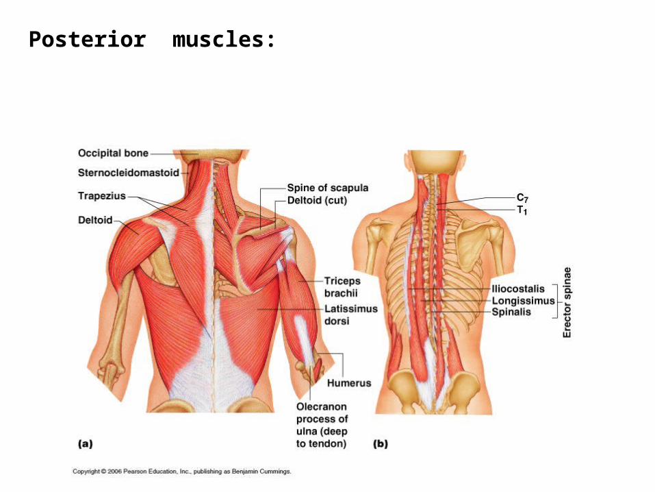
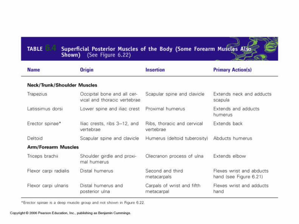
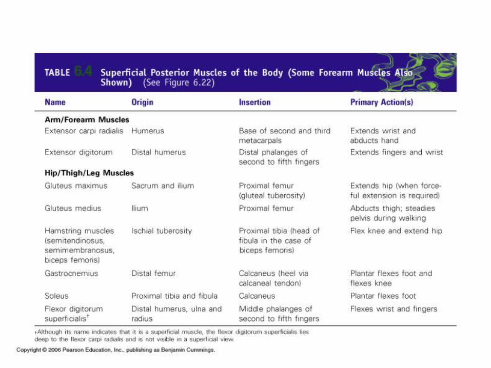
Posterior muscles:
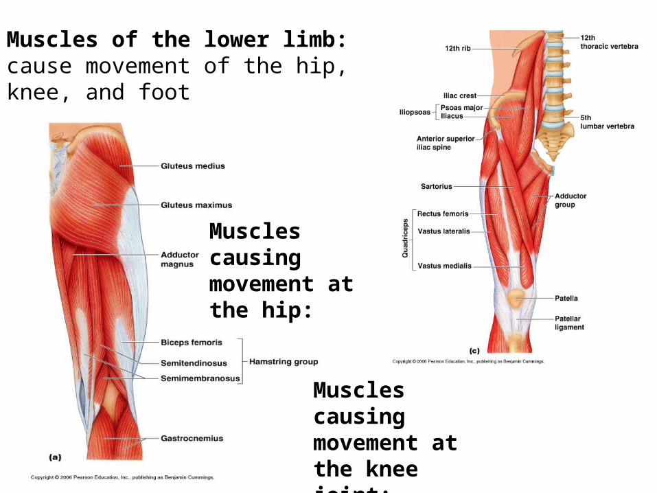
Muscles of the lower limb: cause movement of the hip, knee, and foot
Muscles causing movement at the hip:
Muscles causing movement at the knee joint:
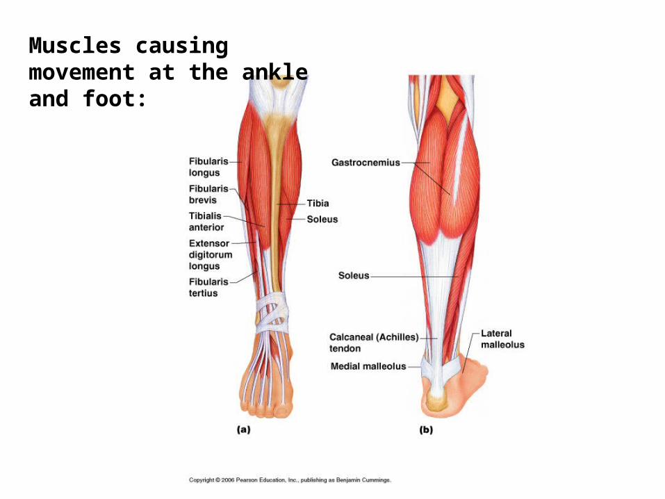
Muscles causing movement at the ankle and foot:
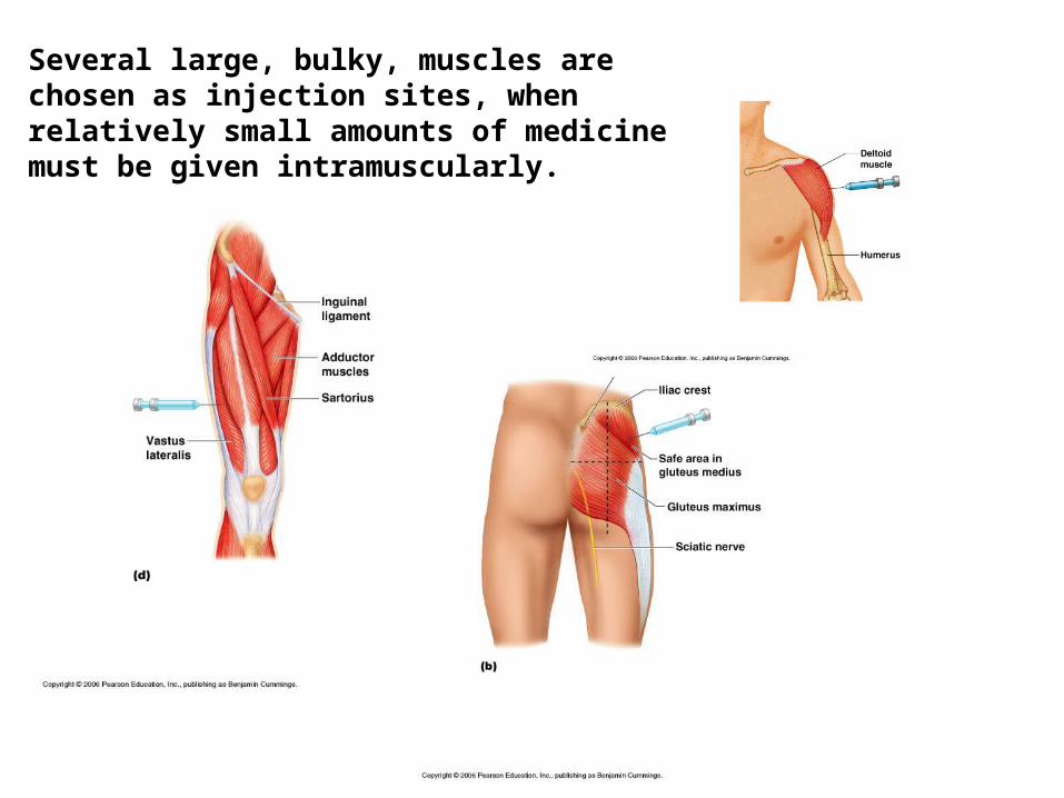
Several large, bulky, muscles are chosen as injection sites, when relatively small amounts of medicine must be given intramuscularly.
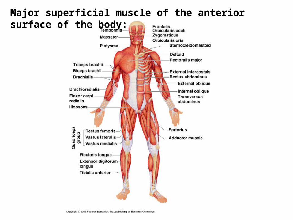
Major superficial muscle of the anterior surface of the body:
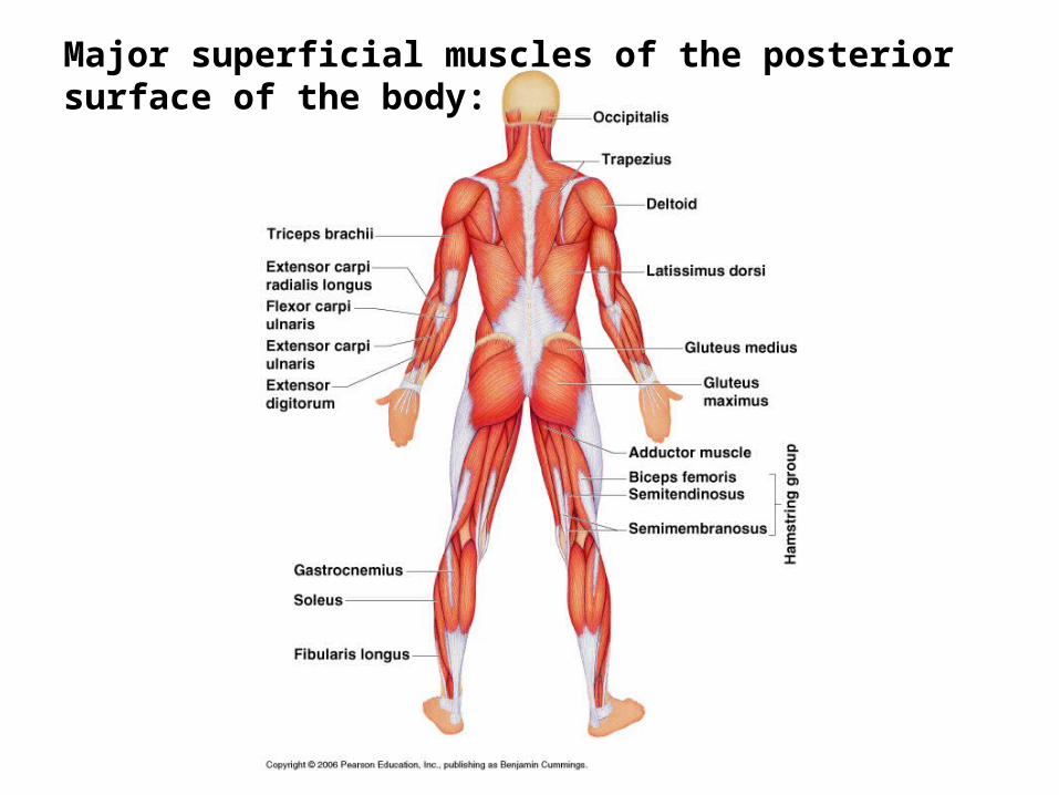
Major superficial muscles of the posterior surface of the body: