Embed Size (px)
DESCRIPTION
Anatomy of the Cardiovascular System. Cardiovascular System. Also circulatory system Consists of: the heart , arteries , veins , capil laries. Heart. Four chamber muscular organ Comparable to the size of a closed fist Located in the mediastinum. Heart. Coverings of the Heart. - PowerPoint PPT Presentation
Citation preview

Anatomy of the Cardiovascular
System

Cardiovascular System
• Also circulatory system• Consists of: the heart, arteries, veins,
capillaries

Heart
• Four chamber muscular organ• Comparable to the size of a closed
fist• Located in the mediastinum

Heart

Coverings of the Heart• Pericardium – loose fitting sac
surrounding the heart– Fibrous pericardium – tough, loose-
fitting, inelastic– Serous pericardium
• Parietal layer: lines the inside of the fibrous pericardium
• Visceral layer: adheres to outside of the heart
– Pericardial space: between parietal and visceral layer• Filled with 10-15mL of pericardial fluid• Decreases friction

Walls of the Heart• Epicardium – outer layer
– Epicardium = serous pericardium
• Myocardium – thick, contractile layer composed of cardiac muscle cells
• Endocaridium – interior of cardiac wall

Walls of the Heart

Chambers of the Heart
• Atria – two superior chambers– “Receiving chambers”– Blood from veins enters atria
• Ventricles – two inferior chambers– “pumping chambers”– Separated by interventricular septum

Valves of the Heart
• Permit blood flow in one direction during circulation
• Atrioventricular valves (AV valves)– Also cuspid valves– Between atria and ventricles
• Semilunar (SL valves)– Between ventricles and vessles

Chambers & Valves
Trace the blood flow through the heart

Blood Supply to the Heart• After traveling through the capillaries
of the heart, blood empties into the R atrium via the coronary sinus

Conduction System of the Heart
• Four structures composed of modified cardiac muscle
• Sinoatrial Node (SA Node)– Pacemaker of the heart– 100s of cells in the R atrium near the
opening of the superior vena cava
• Atrioventricular Node (AV Node)– Left lower border of R atrium
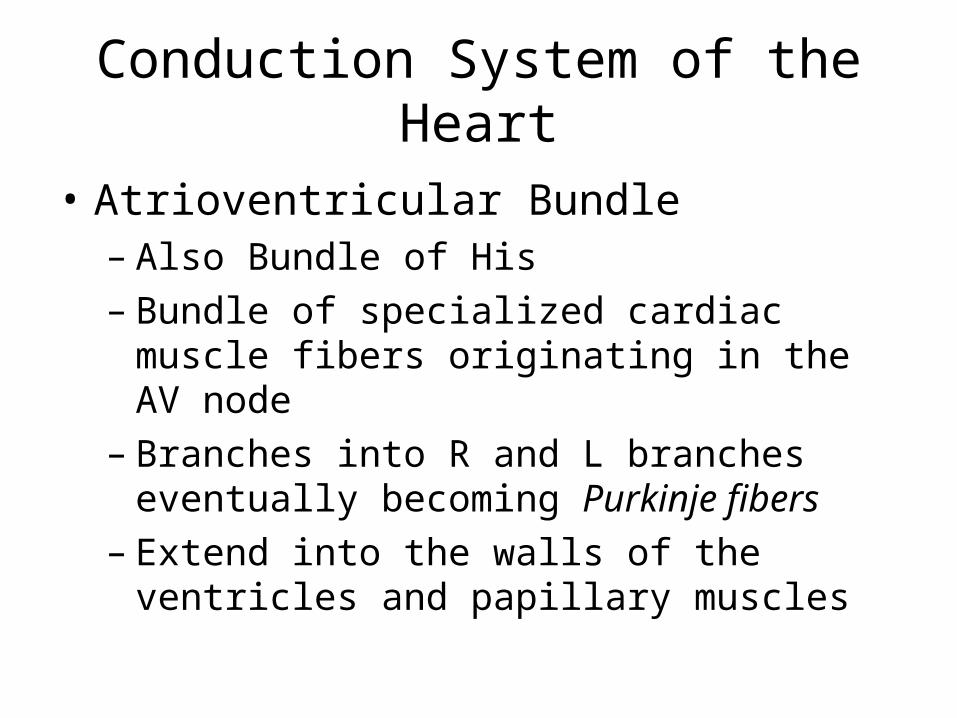
Conduction System of the Heart
• Atrioventricular Bundle– Also Bundle of His– Bundle of specialized cardiac muscle
fibers originating in the AV node– Branches into R and L branches
eventually becoming Purkinje fibers– Extend into the walls of the ventricles
and papillary muscles

Types of Blood Vessels
• Artery – carries oxygenated blood away from the heart– Arteriole: small artery– Precapillary sphincters: regulate the
blood flow into capillaries

Types of Blood Vessels
• Vein – carries unoxygenated blood towards the heart– Great ability to stretch (capacitance)– Function as reservoirs: blood pools in
the valves then is pushed forward from the pumping pressure
– Venules: small vein

Types of Blood Vessels

Types of Blood Vessels
• Capillaries – arterial system switches to venous system– “primary exchange vessels”– Transport materials to and from the cells– Speed of blood flow decreases to
increase contact time

Types of Blood Vessels

Structure of Blood Vessels
• Tunica adventitia - outermost layer– Fibrous connective tissue– Holds vessels open; prevents tearing of
vessels walls during body movements– Larger in veins than arteries
• Tunica media – middle layer– Smooth muscle and elastic CT– Helps vessels constrict and dilate– Larger in arteries
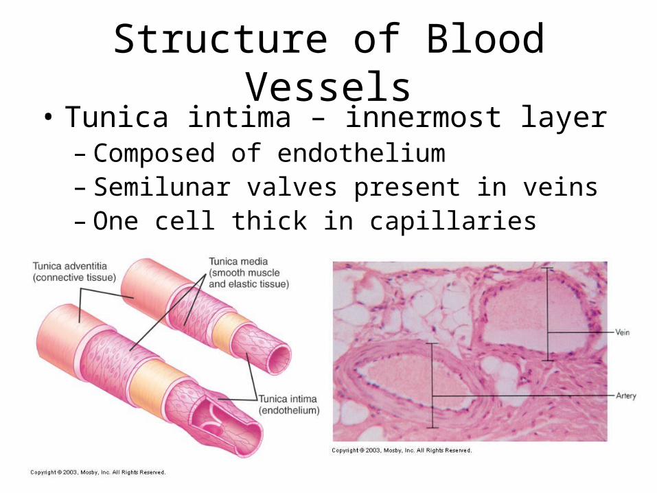
Structure of Blood Vessels• Tunica intima – innermost layer
– Composed of endothelium– Semilunar valves present in veins– One cell thick in capillaries

Circulatory Routes
• Systemic Circulation – blood flow from the L ventricle to the body & back to the R atrium
• Pulmonary Circulation – blood flow from the R ventricle to the lungs and back to the L atrium
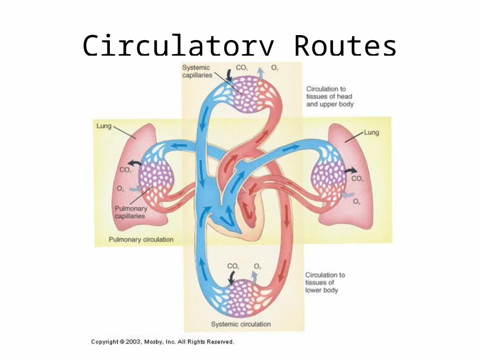
Circulatory Routes

Aorta

Systemic Arteries
• Arch of aorta• Subclavian (L and
R)• Brachiocephalic• common carotid (L
and R)• Axillary (L and R)• Brachial (L and R)• Radial• Ulnar
• Abdominal aorta• Common iliac• External iliac• Femoral• Popliteal• Posterior tibial• Anterior tibial• Dorsal pedis


Systemic Veins
• Superior vena cava• Inferior vena cava• External jugular• Internal jugular• Brachiocephalic (L
and R)• Subclavian (L and
R)• Cephalic• axillary
• Basilic• Median basilic• Median cubital• Common iliac• External iliac• Femoral• Popliteal• Great saphenous• Small saphenous


Fetal Circulation
• Two umbilical arteries carry blood to the placenta
• The placenta allows for exchange of oxygen and nutrients from the mother. Maternal and fetal blood do NOT mix.
• Umbilical vein returns oxygenated blood and enters fetus via the umbilicus
• Foramen ovale – hole btwn the R and L atria– Allows for blood to bypass the R ventricle and
pulmonary circulation

Changes After Birth
• Umbilical vein become round ligament• Umbilical arteries become umbilical
ligaments• Foramen ovale closes after first few
breaths– Full closure may take up to 9 months
• Ductus arteriosus contracts as soon as respirations begin– Become fibrous cord

Pericardium Disorders• Pericarditis – inflammation of the heart
– Causes: trauma, viral or bacteria infection, tumor– Edema causes visceral and parietal layers to rub
together = chest pain– Pus or blood build up in pericardial space– S/S
• Pain with respirations or coughing, dyspnea, restlessness
– Complications: Pericardial Effusion, Cardiac Tamponade
– Treatment: • Antibiotics, pain meds, antiinflammatory meds,
pericardiocentesis (Cardiac Tamponade)

Cardiac Tamponade

Heart Valve Disorders
• General Principles:– Congenital defect: decreased pumping
efficiency– Incompetent valve leak: allows backflow
into previous chamber– Stenosed valves: narrowed valve;
slowing blood from out of chamber

Heart Valve Disorders
• Mitral Valve Prolapse (MVP)– Flaps of mitral valve extend back into L
atrium causes leaking– Mostly genetic basis– 1 in 20 people– S/S: most asymptomatic; chest pain,
fatigue– Treatment: valvuloplasty

Mitral Valve Prolapse
Heart Valve Disorders
• Aortic Regurgitation– Blood leaks back into L ventricle during
ejection into the aorta– Volume overload in L ventricle,
hypertrophy, dilation of L ventricle– Complications: myocaridal ischemia– Treatment: valvuloplasty
Myocardium Disorders
• Atherosclerosis – Type of arteriosclerosis– Lipids build up on the inside of vessel
walls calcify vessels hard & brittle– Risk factors: cigarette smoking, high
fat/cholesterol diet, hypertension

Atherosclerosis

Myocardium Disorders• Myocardial Infarction
– “Heart Attack”– Coronary thrombosis: clot– Coronary embolism: mobilized clot– Occlude coronary artery heart tissue
deprived of oxygen cell death– S/S:
• Angina pectoris – severe chest pain resulting from inadequate oxygen to myocardium
– Treatment: Coronary Bypass Surgery• Veins are harvested from other areas of the
body and used to bypass obstructions


Myocardium Disorders• Congestive Heart Failure (CHF)
– “Left-sided Heart Failure”– Inability of the L ventricle to pump blood
efficiently– Causes: myocardial infarction– S/S: decreased pumping pressure in
systemic circulation; retained fluids• Can lead to congestion in pulmonary
circulation pulmonary edema right-sided heart failure
– Treatment: heart transplant

Congestive Heart Failure

Myocardium Disorders
• Coronary Artery Disease (CAD)– Leading cause of death in US– General term to describe decreased
blood flow to myocardium & associated side effects

Disorders of the Arteries
• Arteriosclerosis– Arteries become occluded, weak and
hardened– Complications: ischemia, necrosis, gangrene– Risk factors: age, diabetes, high
fat/cholesterol diet, hypertension, smoking– Treatment: vasodilators, angioplasty, stent
placement, bypass surgery– Complications: aneurysm

Angioplasty

Disorders of Veins
• Varicose Veins– Enlarged veins caused by pooling– Results in varicosities or varices (“spider
veins”)– Risk factors: standing for long periods
• Semilunar valves widen more pooling
– Treatment: compression stockings, surgical removal

Varicose Veins

Disorders of Veins• Phlebitis – vein inflammation
– Causes: irritation by IV catheter
• Thrombophlebitis– Deep vein thrombosis (DVT)– Phlebitis caused by a clot– S/S
• Pain, redness, swelling
– Complications• Pulmonary embolism

DVT

Pulmonary Embolism

Venous Stasis Ulcers
• Result of chronic vein insufficiency
• Lack of oxygen to peripheral tissues
• Elevate leg & apply pressure
• Irregular edges• “Aching” pain

Patent Ductus Arteriosus (PDA)

Atrial Septal Defect (ASD)

Ventricular Septal Defect (VSD)

Atrioventricular Septal Defect (AVSD)

Tetralogy of Fallot (TOF)

Transposition of the Great Vessels (TGV)

Coarcatation of the Aorta (CoA)

Ebstein’s Anomaly

Pulmonary Atresia

Truncus Arteriosus

Hypoplastic Left Heart Syndrome