Embed Size (px)
Citation preview
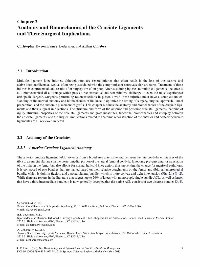
17G.C. Fanelli (ed.), The Multiple Ligament Injured Knee: A Practical Guide to Management, DOI 10.1007/978-0-387-49289-6_2, © Springer Science+Business Media New York 2013
2.1 Introduction
Multiple ligament knee injuries, although rare, are severe injuries that often result in the loss of the passive and active knee stabilizers as well as often being associated with the compromise of neurovascular structures. Treatment of these injuries is controversial, and results after surgery are often poor. After sustaining injuries to multiple ligaments, the knee is at a biomechanical disadvantage which poses a reconstructive and rehabilitative challenge to even the most experienced orthopedic surgeon. Surgeons performing reconstructions in patients with these injuries must have a complete under-standing of the normal anatomy and biomechanics of the knee to optimize the timing of surgery, surgical approach, tunnel preparation, and the anatomic placement of grafts. This chapter outlines the anatomy and biomechanics of the cruciate liga-ments and their surgical implications. The structure and form of the anterior and posterior cruciate ligaments, patterns of injury, structural properties of the cruciate ligaments and graft substitutes, functional biomechanics and interplay between the cruciate ligaments, and the surgical implications related to anatomic reconstruction of the anterior and posterior cruciate ligaments are all reviewed in detail.
2.2 Anatomy of the Cruciates
2.2.1 Anterior Cruciate Ligament Anatomy
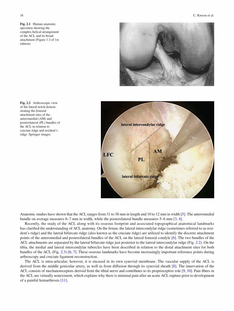
The anterior cruciate ligament (ACL) extends from a broad area anterior to and between the intercondylar eminences of the tibia to a semicircular area on the posteromedial portion of the lateral femoral condyle. It not only prevents anterior translation of the tibia on the femur but also allows for normal helicoid knee action, thus preventing the chance for meniscal pathology. It is composed of two bundles that are named based on their relative attachments on the femur and tibia: an anteromedial bundle, which is tight in fl exion, and a posterolateral bundle, which is more convex and tight in extension (Fig. 2.1 ) [ 1, 2 ] . While there are reports in the literature that suggest up to 26% of knees with microscopic single bundle ACLs as well as knees that have a third intermediate bundle, it is now generally accepted that the native ACL consists of two discrete bundles [ 3, 4 ] .
Chapter 2 Anatomy and Biomechanics of the Cruciate Ligaments and Their Surgical Implications
Christopher Kweon , Evan S. Lederman , and Anikar Chhabra
C. Kweon , M.D. (*) Banner Good Samaritan Orthopaedic Residency , 901 E. Willetta Street, 2nd fl oor , Phoenix , AZ 85006 , USA e-mail: [email protected]
E. S. Lederman , M.D. Sports Medicine Division, Orthopedic Surgery Department , The Orthopedic Clinic Association, Banner Good Samaritan Medical Center , 2222 E. Highland Avenue, #300 , Phoenix , AZ 85016 , USA e-mail: [email protected]
A. Chhabra , M.D., M.S. Arizona State University, Sports Medicine, Banner Good Samaritan, Mayo Clinic Arizona, The Orthopedic Clinic Association , 2222 E. Highland Avenue, #300 , Phoenix , AZ 85016 , USA e-mail: [email protected]
18 C. Kweon et al.
Anatomic studies have shown that the ACL ranges from 31 to 38 mm in length and 10 to 12 mm in width [ 5 ] . The anteromedial bundle on average measures 6–7 mm in width, while the posterolateral bundle measures 5–6 mm [ 3, 4 ] .
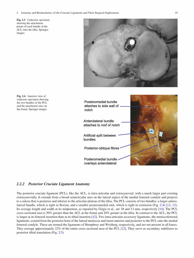
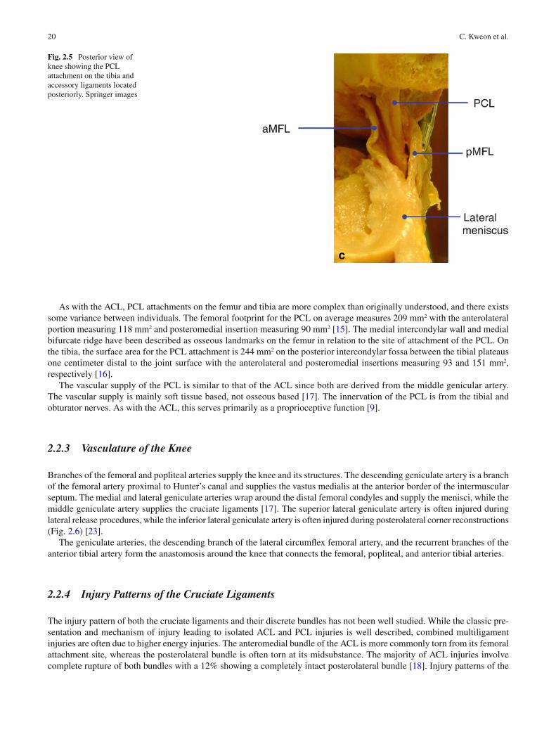
Recently, the study of the ACL along with its osseous footprint and associated topographical anatomical landmarks has clari fi ed the understanding of ACL anatomy. On the femur, the lateral intercondylar ridge (sometimes referred to as resi-dent’s ridge) and the lateral bifurcate ridge (also known as the cruciate ridge) are utilized to identify the discrete attachment points of the anteromedial and posterolateral bundles of the ACL on the lateral femoral condyle [ 6 ] . The two bundles of the ACL attachments are separated by the lateral bifurcate ridge just posterior to the lateral intercondylar ridge (Fig. 2.2 ). On the tibia, the medial and lateral intercondylar tubercles have been described in relation to the distal attachment sites for both bundles of the ACL (Fig. 2.3 ) [ 6, 7 ] . These osseous landmarks have become increasingly important reference points during arthroscopy and cruciate ligament reconstruction.
The ACL is intra-articular; however, it is encased in its own synovial membrane. The vascular supply of the ACL is derived from the middle genicular artery, as well as from diffusion through its synovial sheath [ 8 ] . The innervation of the ACL consists of mechanoreceptors derived from the tibial nerve and contributes to its proprioceptive role [ 9, 10 ] . Pain fi bers in the ACL are virtually nonexistent, which explains why there is minimal pain after an acute ACL rupture prior to development of a painful hemarthrosis [ 11 ] .
Fig. 2.1 Human anatomic specimen showing the complex helical arrangement of the ACL and its broad attachment (Figure 1.3 of 1st edition)
Fig. 2.2 Arthroscopic view of the lateral notch demon-strating the femoral attachment sites of the anteromedial (AM) and posterolateral (PL) bundles of the ACL in relation to cruciate ridge and resident’s ridge. Springer images
192 Anatomy and Biomechanics of the Cruciate Ligaments and Their Surgical Implications
2.2.2 Posterior Cruciate Ligament Anatomy
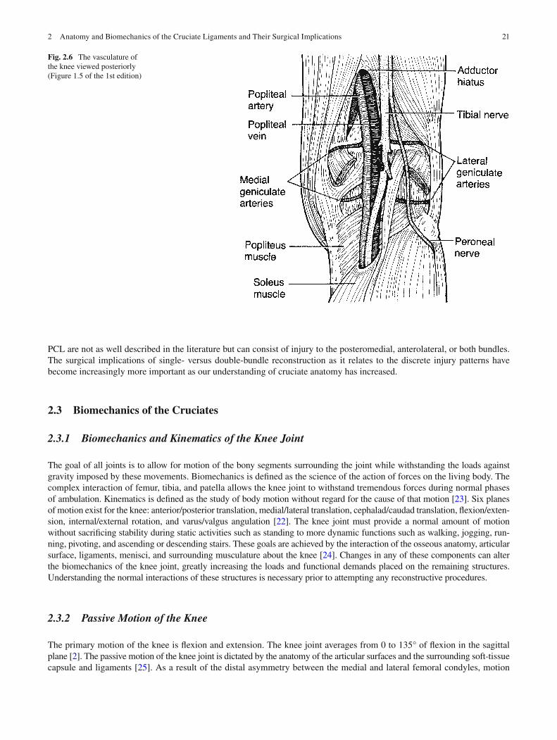
The posterior cruciate ligament (PCL), like the ACL, is intra-articular and extrasynovial, with a much larger part existing extrasynovially. It extends from a broad semicircular area on the lateral aspect of the medial femoral condyle and projects to a sulcus that is posterior and inferior to the articular plateau of the tibia. The PCL consists of two bundles: a larger antero-lateral bundle, which is tight in fl exion, and a smaller posteromedial unit, which is tight in extension (Fig. 2.4 ) [ 12, 13 ] . Its average length and width at its midportion, as reported by Girgis et al., are 38 and 13 mm, respectively [ 14 ] . The PCL cross-sectional area is 50% greater than the ACL at the femur and 20% greater at the tibia. In contrast to the ACL, the PCL is larger at its femoral insertion than at its tibial insertion [ 12 ] . Two intra-articular accessory ligaments, the meniscofemoral ligaments, extend from the posterior horn of the lateral meniscus and insert anterior and posterior to the PCL onto the medial femoral condyle. These are termed the ligaments of Humphrey and Wrisberg, respectively, and are not present in all knees. They average approximately 22% of the entire cross-sectional area of the PCL [ 12 ] . They serve as secondary stabilizers to posterior tibial translation (Fig. 2.5 ).
Fig. 2.3 Cadaveric specimen showing the attachment points of each bundle of the ACL onto the tibia. Springer images
Fig. 2.4 Anterior view of cadaveric specimen showing the two bundles of the PCL and the attachment sites on the femur. Springer images
20 C. Kweon et al.
As with the ACL, PCL attachments on the femur and tibia are more complex than originally understood, and there exists some variance between individuals. The femoral footprint for the PCL on average measures 209 mm 2 with the anterolateral portion measuring 118 mm 2 and posteromedial insertion measuring 90 mm 2 [ 15 ] . The medial intercondylar wall and medial bifurcate ridge have been described as osseous landmarks on the femur in relation to the site of attachment of the PCL. On the tibia, the surface area for the PCL attachment is 244 mm 2 on the posterior intercondylar fossa between the tibial plateaus one centimeter distal to the joint surface with the anterolateral and posteromedial insertions measuring 93 and 151 mm 2 , respectively [ 16 ] .
The vascular supply of the PCL is similar to that of the ACL since both are derived from the middle genicular artery. The vascular supply is mainly soft tissue based, not osseous based [ 17 ] . The innervation of the PCL is from the tibial and obturator nerves. As with the ACL, this serves primarily as a proprioceptive function [ 9 ] .
2.2.3 Vasculature of the Knee
Branches of the femoral and popliteal arteries supply the knee and its structures. The descending geniculate artery is a branch of the femoral artery proximal to Hunter’s canal and supplies the vastus medialis at the anterior border of the intermuscular septum. The medial and lateral geniculate arteries wrap around the distal femoral condyles and supply the menisci, while the middle geniculate artery supplies the cruciate ligaments [ 17 ] . The superior lateral geniculate artery is often injured during lateral release procedures, while the inferior lateral geniculate artery is often injured during posterolateral corner reconstructions (Fig. 2.6 ) [ 23 ] .
The geniculate arteries, the descending branch of the lateral circum fl ex femoral artery, and the recurrent branches of the anterior tibial artery form the anastomosis around the knee that connects the femoral, popliteal, and anterior tibial arteries.
2.2.4 Injury Patterns of the Cruciate Ligaments
The injury pattern of both the cruciate ligaments and their discrete bundles has not been well studied. While the classic pre-sentation and mechanism of injury leading to isolated ACL and PCL injuries is well described, combined multiligament injuries are often due to higher energy injuries. The anteromedial bundle of the ACL is more commonly torn from its femoral attachment site, whereas the posterolateral bundle is often torn at its midsubstance. The majority of ACL injuries involve complete rupture of both bundles with a 12% showing a completely intact posterolateral bundle [ 18 ] . Injury patterns of the
Fig. 2.5 Posterior view of knee showing the PCL attachment on the tibia and accessory ligaments located posteriorly. Springer images
212 Anatomy and Biomechanics of the Cruciate Ligaments and Their Surgical Implications
PCL are not as well described in the literature but can consist of injury to the posteromedial, anterolateral, or both bundles. The surgical implications of single- versus double-bundle reconstruction as it relates to the discrete injury patterns have become increasingly more important as our understanding of cruciate anatomy has increased.
2.3 Biomechanics of the Cruciates
2.3.1 Biomechanics and Kinematics of the Knee Joint
The goal of all joints is to allow for motion of the bony segments surrounding the joint while withstanding the loads against gravity imposed by these movements. Biomechanics is de fi ned as the science of the action of forces on the living body. The complex interaction of femur, tibia, and patella allows the knee joint to withstand tremendous forces during normal phases of ambulation. Kinematics is de fi ned as the study of body motion without regard for the cause of that motion [ 23 ] . Six planes of motion exist for the knee: anterior/posterior translation, medial/lateral translation, cephalad/caudad translation, fl exion/exten-sion, internal/external rotation, and varus/valgus angulation [ 22 ] . The knee joint must provide a normal amount of motion without sacri fi cing stability during static activities such as standing to more dynamic functions such as walking, jogging, run-ning, pivoting, and ascending or descending stairs. These goals are achieved by the interaction of the osseous anatomy, articular surface, ligaments, menisci, and surrounding musculature about the knee [ 24 ] . Changes in any of these components can alter the biomechanics of the knee joint, greatly increasing the loads and functional demands placed on the remaining structures. Understanding the normal interactions of these structures is necessary prior to attempting any reconstructive procedures.
2.3.2 Passive Motion of the Knee
The primary motion of the knee is fl exion and extension. The knee joint averages from 0 to 135° of fl exion in the sagittal plane [ 2 ] . The passive motion of the knee joint is dictated by the anatomy of the articular surfaces and the surrounding soft-tissue capsule and ligaments [ 25 ] . As a result of the distal asymmetry between the medial and lateral femoral condyles, motion
Fig. 2.6 The vasculature of the knee viewed posteriorly (Figure 1.5 of the 1st edition)
22 C. Kweon et al.
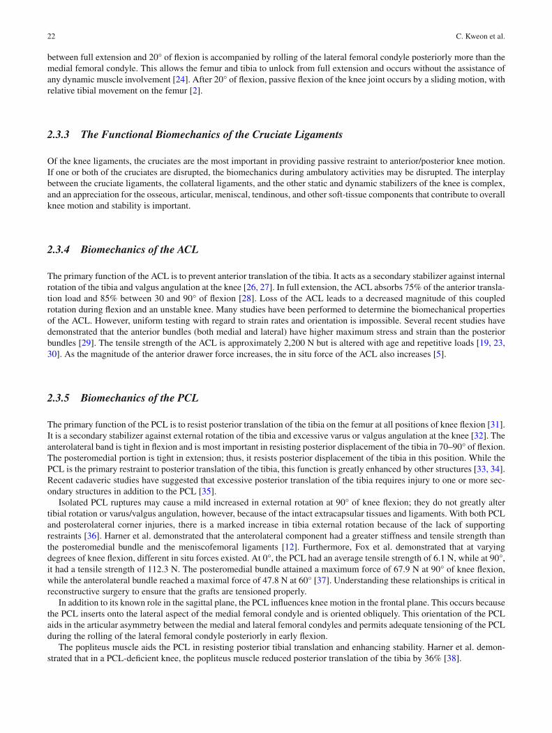
between full extension and 20° of fl exion is accompanied by rolling of the lateral femoral condyle posteriorly more than the medial femoral condyle. This allows the femur and tibia to unlock from full extension and occurs without the assistance of any dynamic muscle involvement [ 24 ] . After 20° of fl exion, passive fl exion of the knee joint occurs by a sliding motion, with relative tibial movement on the femur [ 2 ] .
2.3.3 The Functional Biomechanics of the Cruciate Ligaments
Of the knee ligaments, the cruciates are the most important in providing passive restraint to anterior/posterior knee motion. If one or both of the cruciates are disrupted, the biomechanics during ambulatory activities may be disrupted. The interplay between the cruciate ligaments, the collateral ligaments, and the other static and dynamic stabilizers of the knee is complex, and an appreciation for the osseous, articular, meniscal, tendinous, and other soft-tissue components that contribute to overall knee motion and stability is important.
2.3.4 Biomechanics of the ACL
The primary function of the ACL is to prevent anterior translation of the tibia. It acts as a secondary stabilizer against internal rotation of the tibia and valgus angulation at the knee [ 26, 27 ] . In full extension, the ACL absorbs 75% of the anterior transla-tion load and 85% between 30 and 90° of fl exion [ 28 ] . Loss of the ACL leads to a decreased magnitude of this coupled rotation during fl exion and an unstable knee. Many studies have been performed to determine the biomechanical properties of the ACL. However, uniform testing with regard to strain rates and orientation is impossible. Several recent studies have demonstrated that the anterior bundles (both medial and lateral) have higher maximum stress and strain than the posterior bundles [ 29 ] . The tensile strength of the ACL is approximately 2,200 N but is altered with age and repetitive loads [ 19, 23, 30 ] . As the magnitude of the anterior drawer force increases, the in situ force of the ACL also increases [ 5 ] .
2.3.5 Biomechanics of the PCL
The primary function of the PCL is to resist posterior translation of the tibia on the femur at all positions of knee fl exion [ 31 ] . It is a secondary stabilizer against external rotation of the tibia and excessive varus or valgus angulation at the knee [ 32 ] . The anterolateral band is tight in fl exion and is most important in resisting posterior displacement of the tibia in 70–90° of fl exion. The posteromedial portion is tight in extension; thus, it resists posterior displacement of the tibia in this position. While the PCL is the primary restraint to posterior translation of the tibia, this function is greatly enhanced by other structures [ 33, 34 ] . Recent cadaveric studies have suggested that excessive posterior translation of the tibia requires injury to one or more sec-ondary structures in addition to the PCL [ 35 ] .
Isolated PCL ruptures may cause a mild increased in external rotation at 90° of knee fl exion; they do not greatly alter tibial rotation or varus/valgus angulation, however, because of the intact extracapsular tissues and ligaments. With both PCL and posterolateral corner injuries, there is a marked increase in tibia external rotation because of the lack of supporting restraints [ 36 ] . Harner et al. demonstrated that the anterolateral component had a greater stiffness and tensile strength than the posteromedial bundle and the meniscofemoral ligaments [ 12 ] . Furthermore, Fox et al. demonstrated that at varying degrees of knee fl exion, different in situ forces existed. At 0°, the PCL had an average tensile strength of 6.1 N, while at 90°, it had a tensile strength of 112.3 N. The posteromedial bundle attained a maximum force of 67.9 N at 90° of knee fl exion, while the anterolateral bundle reached a maximal force of 47.8 N at 60° [ 37 ] . Understanding these relationships is critical in reconstructive surgery to ensure that the grafts are tensioned properly.
In addition to its known role in the sagittal plane, the PCL in fl uences knee motion in the frontal plane. This occurs because the PCL inserts onto the lateral aspect of the medial femoral condyle and is oriented obliquely. This orientation of the PCL aids in the articular asymmetry between the medial and lateral femoral condyles and permits adequate tensioning of the PCL during the rolling of the lateral femoral condyle posteriorly in early fl exion.
The popliteus muscle aids the PCL in resisting posterior tibial translation and enhancing stability. Harner et al. demon-strated that in a PCL-de fi cient knee, the popliteus muscle reduced posterior translation of the tibia by 36% [ 38 ] .
232 Anatomy and Biomechanics of the Cruciate Ligaments and Their Surgical Implications
2.3.6 The Interplay of the Cruciate Ligaments
The complex interaction between ACL and PCL at varying degrees of fl exion and extension helps account for the dynamic stability of the knee joint. The length and tension of the ACL and the PCL change during fl exion and extension owing to their asymmetric insertion sites. In full extension, the ACL is taut, while the PCL is relatively lax. When a person is standing with the knee in hyperextension, the joint is passively stable, with little need for muscular support. As the knee fl exes, the postero-lateral portion of the ACL becomes lax, while the PCL tightens, especially the anterolateral bundle. Stability is more tenuous between 20 and 50° of fl exion since neither cruciate ligament is very taut. The change in the orientation of the ACL and PCL fi bers during knee fl exion allows for dynamic stability in the sagittal plane. With increasing fl exion, the ACL changes from a vertical position to a more horizontal orientation in relation to the joint line. The PCL’s orientation is opposite to the ACL’s during fl exion and extension.
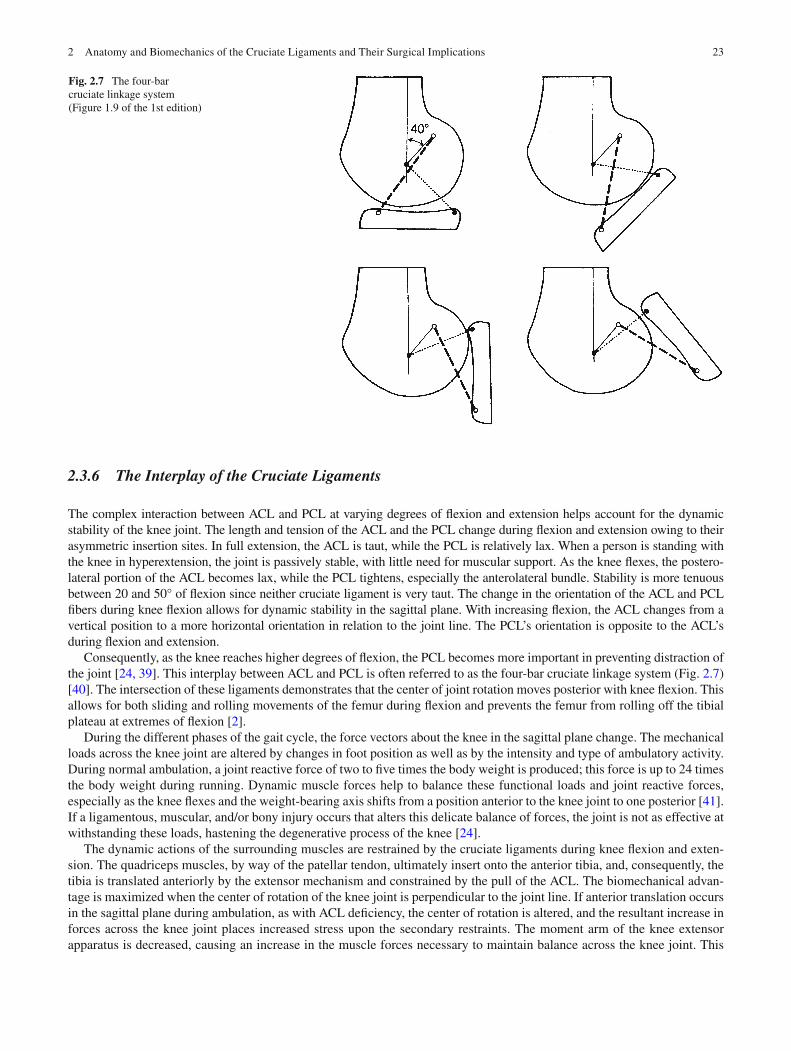
Consequently, as the knee reaches higher degrees of fl exion, the PCL becomes more important in preventing distraction of the joint [ 24, 39 ] . This interplay between ACL and PCL is often referred to as the four-bar cruciate linkage system (Fig. 2.7 ) [ 40 ] . The intersection of these ligaments demonstrates that the center of joint rotation moves posterior with knee fl exion. This allows for both sliding and rolling movements of the femur during fl exion and prevents the femur from rolling off the tibial plateau at extremes of fl exion [ 2 ] .
During the different phases of the gait cycle, the force vectors about the knee in the sagittal plane change. The mechanical loads across the knee joint are altered by changes in foot position as well as by the intensity and type of ambulatory activity. During normal ambulation, a joint reactive force of two to fi ve times the body weight is produced; this force is up to 24 times the body weight during running. Dynamic muscle forces help to balance these functional loads and joint reactive forces, especially as the knee fl exes and the weight-bearing axis shifts from a position anterior to the knee joint to one posterior [ 41 ] . If a ligamentous, muscular, and/or bony injury occurs that alters this delicate balance of forces, the joint is not as effective at withstanding these loads, hastening the degenerative process of the knee [ 24 ] .
The dynamic actions of the surrounding muscles are restrained by the cruciate ligaments during knee fl exion and exten-sion. The quadriceps muscles, by way of the patellar tendon, ultimately insert onto the anterior tibia, and, consequently, the tibia is translated anteriorly by the extensor mechanism and constrained by the pull of the ACL. The biomechanical advan-tage is maximized when the center of rotation of the knee joint is perpendicular to the joint line. If anterior translation occurs in the sagittal plane during ambulation, as with ACL de fi ciency, the center of rotation is altered, and the resultant increase in forces across the knee joint places increased stress upon the secondary restraints. The moment arm of the knee extensor apparatus is decreased, causing an increase in the muscle forces necessary to maintain balance across the knee joint. This
Fig. 2.7 The four-bar cruciate linkage system (Figure 1.9 of the 1st edition)
24 C. Kweon et al.
leads to an increase in joint reactive forces and, ultimately, stressed or injured supporting structures [ 42 ] . In an ACL-de fi cient knee, increased stress is placed on the secondary restraints of anterior translation, including the menisci and the surrounding soft-tissue capsule. When the quadriceps become atrophied after an ACL rupture, the extensor pull on the tibia lessens, decreasing the stresses placed on the secondary stabilizers.
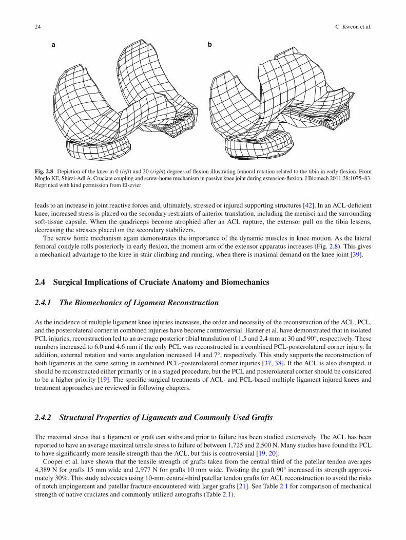
The screw home mechanism again demonstrates the importance of the dynamic muscles in knee motion. As the lateral femoral condyle rolls posteriorly in early fl exion, the moment arm of the extensor apparatus increases (Fig. 2.8 ). This gives a mechanical advantage to the knee in stair climbing and running, when there is maximal demand on the knee joint [ 39 ] .
2.4 Surgical Implications of Cruciate Anatomy and Biomechanics
2.4.1 The Biomechanics of Ligament Reconstruction
As the incidence of multiple ligament knee injuries increases, the order and necessity of the reconstruction of the ACL, PCL, and the posterolateral corner in combined injuries have become controversial. Harner et al. have demonstrated that in isolated PCL injuries, reconstruction led to an average posterior tibial translation of 1.5 and 2.4 mm at 30 and 90°, respectively. These numbers increased to 6.0 and 4.6 mm if the only PCL was reconstructed in a combined PCL-posterolateral corner injury. In addition, external rotation and varus angulation increased 14 and 7°, respectively. This study supports the reconstruction of both ligaments at the same setting in combined PCL-posterolateral corner injuries [ 37, 38 ] . If the ACL is also disrupted, it should be reconstructed either primarily or in a staged procedure, but the PCL and posterolateral corner should be considered to be a higher priority [ 19 ] . The speci fi c surgical treatments of ACL- and PCL-based multiple ligament injured knees and treatment approaches are reviewed in following chapters.
2.4.2 Structural Properties of Ligaments and Commonly Used Grafts
The maximal stress that a ligament or graft can withstand prior to failure has been studied extensively. The ACL has been reported to have an average maximal tensile stress to failure of between 1,725 and 2,500 N. Many studies have found the PCL to have signi fi cantly more tensile strength than the ACL, but this is controversial [ 19, 20 ] .
Cooper et al. have shown that the tensile strength of grafts taken from the central third of the patellar tendon averages 4,389 N for grafts 15 mm wide and 2,977 N for grafts 10 mm wide. Twisting the graft 90° increased its strength approxi-mately 30%. This study advocates using 10-mm central-third patellar tendon grafts for ACL reconstruction to avoid the risks of notch impingement and patellar fracture encountered with larger grafts [ 21 ] . See Table 2.1 for comparison of mechanical strength of native cruciates and commonly utilized autografts (Table 2.1 ).
a b
Fig. 2.8 Depiction of the knee in 0 ( left ) and 30 ( right ) degrees of fl exion illustrating femoral rotation related to the tibia in early fl exion. From Moglo KE, Shirzi-Adl A. Cruciate coupling and screw-home mechanism in passive knee joint during extension- fl exion. J Biomech 2011;38:1075–83. Reprinted with kind permission from Elsevier
252 Anatomy and Biomechanics of the Cruciate Ligaments and Their Surgical Implications
Over time, wear and degeneration cause ligaments and grafts to decrease in strength. This has been demonstrated in multiple studies by means of ACL and graft tensile tests. The biologic effects of aging, maturation, and immobilization may also affect the viscoelastic properties of a ligament or graft, leading to a decrease in biomechanical strength [ 22 ] .
2.4.3 Graft Tensioning
Cruciate anatomy has many surgical implications related to graft tensioning during ACL and PCL reconstruction. High amounts of tension through the graft can result in poor results after surgery due to excessive wear through the tunnels, impaired vascularity, and restricted range of motion [ 43– 49 ] . Too little tension may result in continued postoperative laxity of the knee. Generally, most surgeons will statically precondition the graft on the back table and/or cyclically precondition the graft in the knee prior to fi nal fi xation. Graft tensioning during cruciate reconstruction is also heavily dependent on tunnel placement. The importance of accurate tunnel placement in single- or double-bundle reconstructions or in revision recon-struction situations of the ACL and PCL cannot be understated.
2.4.4 Tunnel Placement for Cruciate Reconstruction
Cadaver and computed tomography studies have lead to a different understanding of cruciate ligament anatomy and relation-ships, osseous landmarks, and anatomical reference points for accurate placement of grafts and tunnels during ACL and PCL reconstructions [ 6, 15, 16 ] . The existence of two discrete attachment points of each of the bundles for both the ACL and PCL is now well understood and has brought to focus the surgical implications of reconstructing injured cruciate ligaments anatomically and the resultant functional outcome from surgery.
An abundance of studies have demonstrated the varying effects that tunnel placement and orientation or the addition of a second tunnel has on ACL or PCL graft tension [ 50– 52 ] . Historically, the most common technical mistake has been to place both femoral and tibial tunnels too far anteriorly. With newer cadaveric and radiologic studies that have clari fi ed the anatomic relationships between the ACL, PCL, and their corresponding bony sites of attachment, the subtleties of accurate tunnel placement during reconstruction are clearer.
Efforts have been made recently to reconstruct both the anterior and PCLs more anatomically utilizing double-bundle techniques and creating multiple tunnels when reconstructing multiple ligament injured knees. These techniques are increasing in use and have been well described [ 6, 7, 26 ] . However, drilling of multiple tunnels for double-bundle recon-struction is technically demanding and requires good patient selection and technical skill to avoid complications related to its use. Continued clinical outcome studies are currently underway to further assess the ef fi cacy and safety of anatomic reconstructions of the cruciate ligaments utilizing double-bundle techniques.
2.5 Conclusion
Knee dislocations are severe injuries because they may result in disruption of multiple ligaments, surrounding musculature, and neurovascular structures [ 53 ] . Diagnosis and acute treatment can be dif fi cult, and the varying techniques that are utilized to reconstruct the anterior and PCLs can be controversial. These injuries, owing to ligamentous disruption and surrounding soft-tissue damage, may lead to a biomechanical disadvantage of the knee joint prior to or after reconstruction attempts are made.
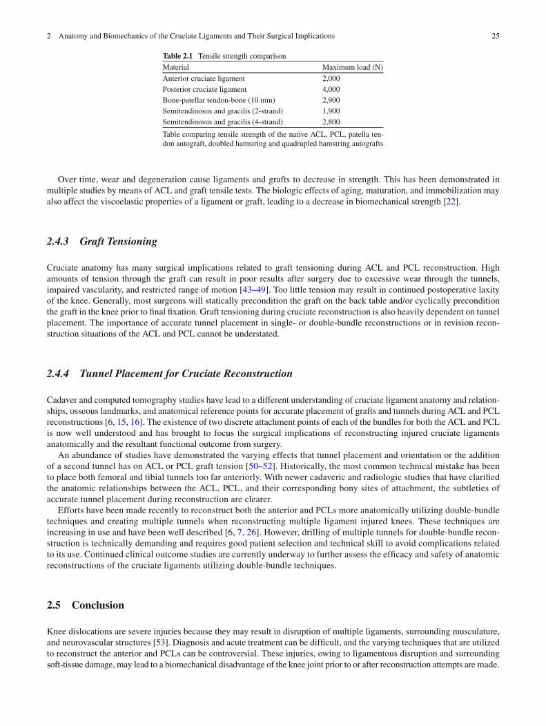
Table 2.1 Tensile strength comparison
Material Maximum load (N)
Anterior cruciate ligament 2,000 Posterior cruciate ligament 4,000 Bone-patellar tendon-bone (10 mm) 2,900 Semitendinosus and gracilis (2-strand) 1,900 Semitendinosus and gracilis (4-strand) 2,800
Table comparing tensile strength of the native ACL, PCL, patella ten-don autograft, doubled hamstring and quadrupled hamstring autografts
26 C. Kweon et al.
To prevent abnormal translations and angulations in the reconstructed knee, surgeons performing reconstructions in patients with multiple ligament injuries must have a complete understanding of the normal anatomy and biomechanics of the entire knee as well as the anterior and PCLs. This knowledge should help optimize the timing of surgery, the order of ligamentous reconstruction, the anatomic placement of grafts, and the rehabilitation of the surrounding musculature.
References
1. Feagin JA. Isolated anterior cruciate injury. In: Feagin JA, editor. The cruciate ligaments. New York: Churchill Livingstone; 1988. p. 15–23. 2. Fu FH, Harner CD, Johnson DL, Miller MD, Woo SLY. Biomechanics of knee ligaments: basic concepts and clinical application. J Bone Joint
Surg Am. 1993;75:1716–27. 3. Harner CD, Baek GH, Vogrin TM. Quantitative analysis of human cruciate ligament insertions. Arthroscopy. 1999;15:741–9. 4. Takahashi M, Doi M, Abe M, Suzuki D, Nagano A. Anatomical study of the femoral and tibial insertions of the anteromedial and posterolateral
bundles of human anterior cruciate ligament. Am J Sports Med. 2006;34:787–92. 5. Smith BA, Livesay GA, Woo SL. Biology and biomechanics of the anterior cruciate ligament. Clin Sports Med. 1993;12:637–70. 6. Forsythe B, Kopf S, Wong AK, et al. The location of femoral and tibial tunnels in anatomic double-bundle anterior cruciate ligament recon-
struction analyzed by three-dimensional computed tomography models. J Bone Joint Surg Am. 2010;92:1418–26. 7. Chhabra A, Starman JS, Ferretti M, Vidal AF, Zantop T, Fu FH. Anatomic, radiographic, biomechanical, and kinematic evaluation of the anterior
cruciate ligament and its two functional bundles. J Bone Joint Surg Am. 2006;88:2–10. 8. Arnoczky SP. Anatomy of the anterior cruciate ligament. Clin Orthop. 1983;172:19–25. 9. Kennedy JC, Alexander IJ, Hayes KC. Nerve supply of the human knee and its functional importance. Am J Sports Med. 1982;10:329–35. 10. Biedert RM, Stauffer E, Friederich NF. Occurrence of free nerve endings in the soft tissue of the knee joint. A histologic investigation. Am J Sports
Med. 1992;20:430–3. 11. Shutte MJ, Dabezies EJ, Zimney ML, et al. Neural anatomy of the human anterior cruciate ligament. J Bone Joint Surg Am. 1987;69:243–7. 12. Harner CD, Xerogeanes JW, Livesay GA, et al. The human posterior cruciate ligament complex: an interdisciplinary study. Am J Sports Med.
1995;23:736–45. 13. Johnson CJ, Bach BR. Current concepts review. Posterior cruciate ligament. Am J Knee Surg. 1990;3:143–53. 14. Girgis FG, Marshall JL, Al Monajem ARS. The cruciate ligaments of the knee joint. Anatomic, functional, and experimental analysis. Clin
Orthop. 1975;106:216–31. 15. Lopes OV, Ferretti M, Shen W, Ekdahl M, Smolinski P, Fu FH. Topography of the femoral attachment of the posterior cruciate ligament. J Bone
Joint Surg Am. 2008;90:249–55. 16. Tajima G, Nozaki M, Iriuchishima T, et al. Morphology of the tibial insertion of the posterior cruciate ligament. J Bone Joint Surg Am.
2009;91:859–66. 17. Vladimirov B. Arterial sources of blood supply in the knee joint in man. Acta Med. 1968;47:1–10. 18. Zantop T, Brucker PU, Vidal A, Zelle BA, Fu FH. Intraarticular rupture pattern of the ACL. Clin Orthop Relat Res. 2007;454:48–53. 19. Miller MD, Cooper DE, Warner JJP. Review of sports medicine and arthroscopy. Philadelphia: WB Saunders; 1995. p. 3–71. 20. Prietto MP, Bain JR, Stonebrook SN, Settlage RA. Tensile strength of the human posterior cruciate ligament (PCL). Trans Orthop Res Soc.
1988;13:195. 21. Cooper DE, Deng XH, Burstein AL, Warren RF. The strength of the central third patellar tendon graft, a biomechanical study. Am J Sports
Med. 1993;21:818–23. 22. Woo SLY, Debski RE, Withrow JD, Janaushek MA. Biomechanics of knee ligaments. Am J Sports Med. 1999;27:533–43. 23. Miller MD. Sports medicine. In: Miller MD, editor. Review of orthopaedics. 3rd ed. Philadelphia: WB Saunders; 2000. p. 195–240. 24. Andriacchi TP. Knee joint, anatomy and biomechanics. In: Pellicci PM, Tria AJ, Garvin KL, editors. Orthopaedic knowledge update. Hip and
Knee reconstruction 2. Rosemont, IL: American Academy of Orthopaedic Surgeons; 2000. p. 239–49. 25. Lafortune MA, Cavanaugh PR, Sommer III HJ, Kalenak A. Three-dimensional kinematics of the human knee during walking. J Biomech.
1992;25:347–57. 26. Buoncristiani AM, Tjoumakaris FP, Starman JS, Ferretti M, Fu FH. Anatomic double-bundle anterior cruciate ligament reconstruction.
Arthroscopy. 2006;22:1000–6. 27. Sakane M, Fox RJ, Woo SL-Y, Livesay GA, Li G, Fu FH. In situ forces in the anterior cruciate ligament and its bundles in response to anterior
tibial loads. J Orthop Res. 1997;15:285–93. 28. Butler DL, Noyes FR, Grood ES. Ligamentous restraints to anterior-posterior drawer in the human knee. A biomechanical study. J Bone Joint
Surg Am. 1980;62:259–70. 29. Butler DL, Guan Y, Kay MD, et al. Location-dependent variations in the material properties of anterior cruciate ligament subunits. Trans
Orthop Res Soc. 1991;16:234. 30. Woo SLY, Adams DJ. The tensile properties of the human anterior cruciate ligament and ACL graft tissues. In: Daniel DM, Akeson WH,
O’Conner JJ, editors. Knee ligaments, structure, function, injury, and repair. New York: Raven Press; 1990. p. 279–89. 31. Gollehon DL, Torzilli PA, Warren RF. The role of posterolateral and cruciate ligaments in the stability of the human knee. A biomechanical
study. J Bone Joint Surg Am. 1987;69:233–42. 32. Grood ES, Stowers SF, Noyes FR. Limits of movement in the human knee. Effect of sectioning the posterior cruciate ligament and posterolateral
structures. J Bone Joint Surg Am. 1988;70:88–97. 33. Amis AA, Bull AM, Gupte CM, Hijazi I, Race A, Robinson JR. Biomechanics of the PCL and related structures: posterolateral, posteromedial
and meniscofemoral ligaments. Knee Surg Sports Traumatol Arthrosc. 2003;11:271–81. 34. Covey DC, Sapega AA. Anatomy and function of the posterior cruciate ligament. Clin Sports Med. 1994;13:509–18.
272 Anatomy and Biomechanics of the Cruciate Ligaments and Their Surgical Implications
35. Schulz MS, Steenlage ES, Russe K, Strobel MJ. Distribution of posterior tibial displacement in knees with posterior cruciate ligament tears. J Bone Joint Surg Am. 2007;89:332–8.
36. Covey DC, Sapega AA. Current concepts review. Injuries of the posterior cruciate ligament. J Bone Joint Surg Am. 1993;75:1376–86. 37. Fox RJ, Harner CD, Sakane M, Carlin GJ, Woo SLY. Determination of the in situ forces in the human posterior cruciate ligament using robotic
technology, a cadaveric study. Am J Sports Med. 1998;26:395–401. 38. Harner CD, Hoher J, Vogrin TM, Carlin GJ, Woo SLY. The effects of a popliteus muscle load on in situ forces of the posterior cruciate ligament
and on knee kinematics. Am J Sports Med. 1998;26:669–73. 39. Burstein AH, Wright TM. Biomechanics. In: Insall JN, Windsor RE, Scott WN, Kelly MA, Aglietti P, editors. Surgery of the knee. 2nd ed.
New York: Churchill Livingstone; 1993. p. 43–62. 40. Muller WD. Kinematics. In: Muller W, editor. The knee, form, function, and ligament reconstruction. New York: Springer; 1983. p. 8–28. 41. Morrison JB. The mechanics of the knee joint in relation to normal walking. J Biomech. 1970;3:51. 42. Elftman H. The forces exerted by the ground in walking. Arb Physiol. 1939;10:485. 43. Graf BK, Henry J, Rothenberg M, Vanderby R. Anterior cruciate ligament reconstruction with patellar tendon. An ex vivo study of wear-related
damage and failure at the femoral tunnel. Am J Sports Med. 1994;22:131–5. 44. Katsuragi R, Yasuda K, Tsujino J, Keira M, Kaneda K. The effect of nonphysiologically high initial tension on the mechanical properties of in
situ frozen anterior cruciate ligament in a canine model. Am J Sports Med. 2000;28:47–56. 45. Yoshiya S, Andrish JT, Manley MT, Bauer TW. Graft tension in anterior cruciate ligament reconstruction. An in vivo study in dogs. Am J
Sports Med. 1987;15:464–70. 46. Amis AA. Anterior cruciate ligament replacement. Knee stability and the effects of implants. J Bone Joint Surg Br. 1989;71:819–24. 47. Bylski-Austrow DI, Grood ES, Hefzy MS, Holden JP, Butler DL. Anterior cruciate ligament replacements: a mechanical study of femoral
attachment location, fl exion angle at tensioning, and initial tension. J Orthop Res. 1990;8:522–31. 48. Gertel TH, Lew WD, Lewis JL, Stewart NJ, Hunter RE. Effect of anterior cruciate ligament graft tensioning direction, magnitude, and fl exion
angle on knee biomechanics. Am J Sports Med. 1993;21:572–81. 49. Melby 3rd A, Noble JS, Askew MJ, Boom AA, Hurst FW. The effects of graft tensioning on the laxity and kinematics of the anterior cruciate
ligament reconstructed knee. Arthroscopy. 1991;7:257–66. 50. Cyril FB, Jackson DW. Current concepts review—the science of reconstruction of the anterior cruciate ligament. J Bone Joint Surg Am.
1997;79:1556–76. 51. Simmons R, Howell SM, Hull ML. Effect of the angle of the femoral and tibial tunnels in the coronal plane and incremental excision of the
posterior cruciate ligament on tension of an anterior cruciate ligament graft: an in vitro study. J Bone Joint Surg Am. 2003;85:1018–29. 52. Shearn JT, Grood ES, Noyes FR, Levy MS. Two-bundle posterior cruciate ligament reconstruction: how bundle tension depends on femoral
placement. J Bone Joint Surg Am. 2004;86:1262–70. 53. Meyers M, Harvey JJ. Traumatic dislocations of the knee joint: a study of eighteen cases. J Bone Joint Surg Am. 1971;53:16–29.

http://www.springer.com/978-0-387-49287-2