Embed Size (px)
Citation preview

Anatomy 32
Chapter 2
Cells: The Living Units


I. Introduction to the cell- The cell is the smallest living unit. All living organisms are composed of cells
a. Robert Hooke first observed a cell when microscopes were invented in the 1600’s. Matthias Schleidena nd Theodor Schawn declared that all living things are made up of cells. Rudolf Virchow concluded that cells arise from other living cells.
b. All of these observations contributed to the Cell Theory=
all living things are made up of cells, the smallest unit of life is the cell, cells arise from other living cells by
cell division.

c. Organelles are discrete specialized structures that assist in cell function and structure. In this chapter you will learn about the function of cell organelles and how they assist the cell to perform its overall function.

II. The plasma membrane- A thin flexible structure that separates the cell’s internal environment from its surroundings. It controls what enters and leaves the cell. Other cell organelles are enclosed by a membrane structure very similar to the plasma membrane.

a. Structure- A phospholipid bilayer with proteins and cholesterol molecules used to stabilized the membrane.
1. The phospholipids layer creates hydrophilic and hydrophobic regions that controls what crosses the membrane (semi-permeable membrane.)

2. Carbohydrates (sugars) may be attached to proteins and form glycoproteins, or attach to lipids and form glycolipids. Carbohydrates are found on the outer surface of the membrane and serve as receptors or markers.

3. Integral proteins cross through the membrane and may be used as transport channels. Peripheral proteins adhere to the underside of the membrane and stabilize it.


b. Function- The membrane protects the cells internal structures and controls transport
1. The side of the membrane that faces externally presents receptors and markers unique to each kind of cell.
2. The membrane controls what molecules cros it based on size, polarity, solubility, and receptor signals.
3. The membrane expels objects out of the cell by exocytosis or surrounds them and brings them in by endocytosis through either phagocytosis or pinocytosis.
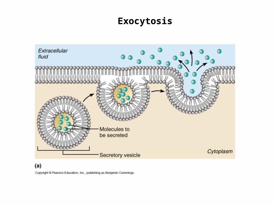
Exocytosis

Endocytosis


Receptor Mediated Endocytosis

III. The cytoplasm- the area within the cell that contains organelles between the plasma membrane and the boundary of the nucleus. a. Cytosol- a jelly-like substance that fills the cell and suspends
the organelles
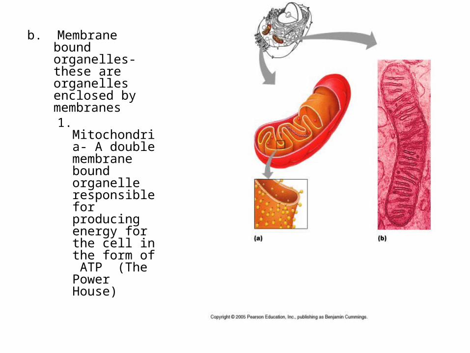
b. Membrane bound organelles- these are organelles enclosed by membranes
1. Mitochondria- A double membrane bound organelle responsible for producing energy for the cell in the form of ATP (The Power House)

2. Endoplasmic Reticulum- A single membrane folded over into different compartment, usually exists close to the nucleus (The Assembly Site)
1. Smooth ER- no ribosomes on surface, processes fats, toxins, hormones
2. Rough ER- has surface ribosomes, site of protein synthesis
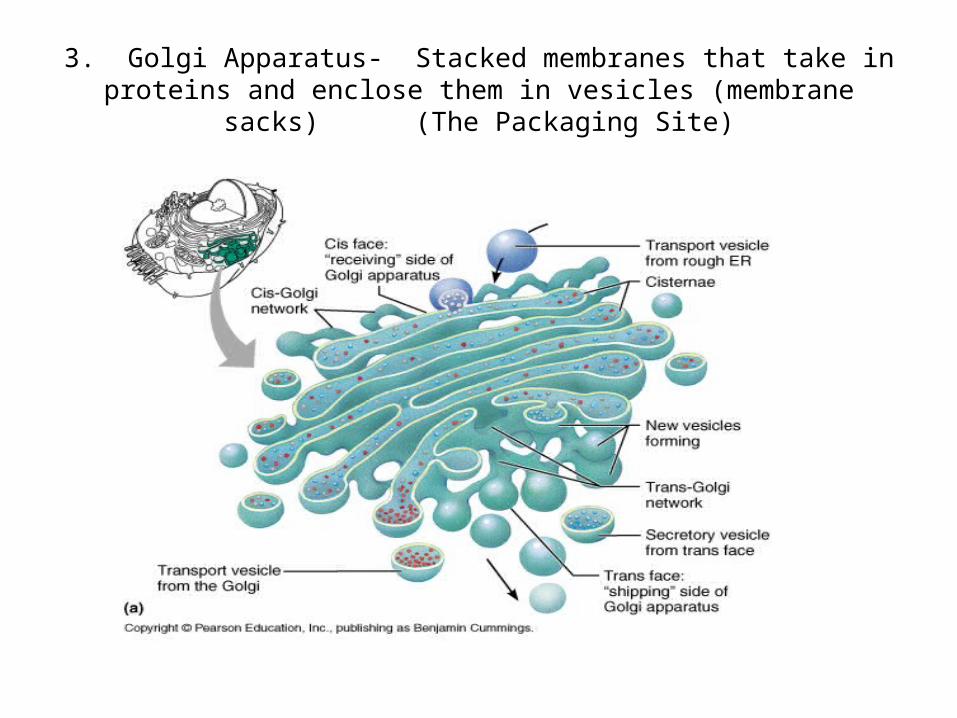
3. Golgi Apparatus- Stacked membranes that take in proteins and enclose them in vesicles (membrane sacks) (The Packaging Site)

Collaboration between organelles. Some proteins remain in the cell and some are excytosed.

4. Lysosomes- vesicles containing lytic or digestive enzymes that break down cell debri and foreign substances in the cell, like bacteria
( Demo-lition crew/ security)

5. Peroxisomes- vesicles like lysosomes but filled with enzymes that fight free radicals and break down poisons ( Neutralizers)

C. Non-membrane bound organelles- typically protein structures not surrounded by a membrane that perform a specific function.
1. Ribosome- protein units that specialize in protein production, may be free floating or attach to Rough ER. (Assembly workers)


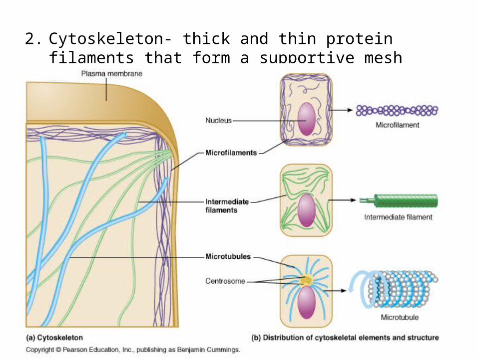
2. Cytoskeleton- thick and thin protein filaments that form a supportive mesh


3. Centrioles- protein fibers involved in cell division (Reproduc-tive machinery)

• FACTORY DRAWING-
IV The nucleus- Located at the center of the cell, it hold the instructions for life (genetic material) it is know as the “brain” of the cell.
a. Nuclear Envelope -a double membrane layer with pores that allow transport into and out of the nucleus.
b. Chromatin- DNA strands loosely wrapped around histone protein. Under the microscope it make the nucleus look cloudy. DNA is readable and accessible in this form and thus most of the time the cell contains chromatin
c. Chromosomes- DNA strands super coiled to form dense discrete structures. These are seen during cell division and resemble an X. Humans have 46 chromosomes. Chromatin condenses to form chromosomes.
d. Nucleoli – Region within the nucleus where ribosome production takes place


V The Cell Life Cycle- Stages involving cell division, preparation for cell division, and normal cell function.
a. Interphase- The majority of the time the cell is interphase. At this time it grows, performs its normal cell function, and prepares for cell division. A cell in interphase seen under the microscope shows a distinct cloudy nucleus filled with chromatin.
1. G1 (growth 1)- after a cell is created it enters this stage and performs its specialized function
2. S (synthesis)- DNA is copied so there are two sets of chromosomes
3. G2 (growth 2)- cell continues to grow and prepare for cell division


B. Cell Division- This process occurs when cells need to replace dead cells, heal damaged tissue, during growth and development. Some cells never divide once formed such as muscle cells and nerve cells
1. Mitosis- the division of the cell’s nucleus describe in four stages that may take about 2 hours to complete.1. Prophase- the nuclear envelope breaks down, DNA
condenses from chromatin to chromosomes, mitotic spindle forms
2. Metaphase- mitotic spindle lines the chromosomes at the equatorial plate
3. Anaphase- chromosome strands (sister chromatids) are pulled apart to opposite poles of the cell
4. Telophase- the cell forms a cleavage furrow, nuclear envelopes begins to form and chromosomes return to chromatin
2. Cytokinesis- the division of the entire cell into two cells.


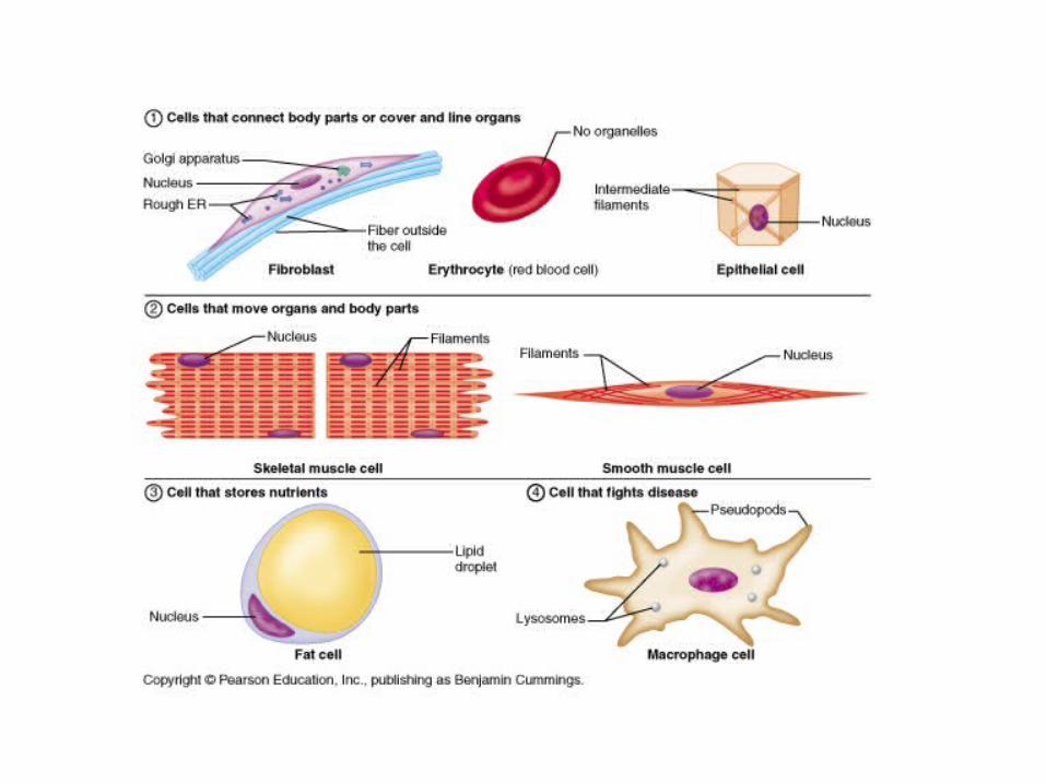
VI. Cellular Diversity- Although all the cells in your body hold exactly the same genetic information they do not have the same function or structure. Activation of specific genes causes the cell to become specialized or differentiated.
a. Cells that connect body parts or line organsb. Cells that move organs and body partsc. Cells that store nutrientsd. Cells that fight diseasee. Cells that gather information and control body
functionsf. Cells for reproduction


VII Developmental Aspects of cells All humans originated from a single cell. This cell is
formed when the egg and sperm unite, it’s called the zygote. All other cells are produced from this cell, thus every cell in our body contains the exact same DNA. As the embryo develops the cells become specialized, a process known as cell differentiation. Their structural differences reflect their functional difference. A group of specialized cells forms a type of tissue and each tissue type will have a different function.
a. Youth – before birth the fetus develops all the organs and systems necessary for a functional body. After birth the infant continues to mature and cells divide for growth. By adulthood cell division reduces to only occur during repair of tissues
b. Aging- there are several theories that try to explain the cause of aging. The end result is the same: cell greatly decrease division rate and dead cells are replaced at a slower rate, tissue mass is lost, and weakening occurs.

• Free radical- molecules ( primarily oxygen) that have an unpaired electron and are highly reactive. They build up and damage the cells. Antioxidants such as vitamin E and C are used to reduce radicals
• Mitochondrial Theory- the mitochondria produces free radicals as a result of cellular respiration/ high metabolism. Those that eat less or have a slow metabolism live longer because less radicals are produced
• Genetic Theory- our genes have a pre-determined life span for cells indicating that it is a normal part of human development
• Telomeres- sections of DNA that do not contain genes but influence the amount of times a cell divides. As the cells divide the number of telomeres decreases. An enzyme called telomerase, which adds telomeres, is found in excess in cancer cells and may increase cell division.

Cancer• VIII. Cancer• Cancer results from a genetic change in the cell that causes
it to divide rapidly. The genetic change is caused by a mutation instigated by biological factors, UV rays, exposure to carcinogens, or viruses. The rapid division does not allow for proper cell differentiation and function and results in mass formation. This is called a tumor.
• Tumors may be malignant or benign. A malignant tumor has the ability to metastasize- meaning that the cancerous cells spread through the body. The cells enter either the circulatory or lymphatic system, travel through the body, and re-establish a new tumor at a different location.
• Cancer is treated with either the surgical removal of the tumor, radiation, chemotherapy. These treatments kill healthy cells as well as cancerous cells and cause multiple side effects. New therapies are more specific at targeting the cancer and causing less severe side effects.
• READ PG 46-47


![[PPT]Cell Structure & Function - Iredell-Statesville Schools / · Web view Cell Theory All living things are made up of cells. Cells are the smallest working units of all living things](https://img.pdfslide.us/doc/110x75/5aa4d86e7f8b9a517d8c79ca/pptcell-structure-function-iredell-statesville-schools-view-cell-theory.jpg)