Embed Size (px)
Citation preview
INTRODUCTION
Chronic traumatic encephalopathy (CTE) is a progressive de-generative disease and progressive tauopathy of the brain caused by sequelae of head trauma and is common in athletes or patients with recurrent brain trauma such as boxers, professional soccer players, professional wrestlers, epilepsy patients, head bangers and domestic violence victims [1-6]. The name CTE was created in 1949 by Critchley. Critchley has defined this entity as a punch-drunk syndrome [7, 8]. McKee et al. had made a pathological cri-
teria and progressive pathological stages for CTE, that have been used for decades for the evaluation of neuropathology of this dis-ease (Table 1) [3, 7]. It is a progressive global atrophy of the brain including the cerebral hemispheres, medial temporal lobe, thala-mus, mammillary bodies and brainstem, with ventriculomegaly and fenestration of cavum septum pellucidum. For decades CTE’s neuropathology has been well established [1, 3, 5, 7, 9]. Micro-scopically, extensive p-tau immunoreactive neurofibrillary tangles (NFTs) and dot-like and thread-like neurites are patchy and irreg-ularly distributed throughout the brain. The difference in NFT in other types of tauopathy and CTE is the distribution pattern of tau protein; 1) the significant involvement of the depth of the cerebral sulci, 2) the remarkable involvement of the periventricular, peri-vascular and subpial area, and 3) tau-immunoreactive astrocytes throughout the brain are exclusively found in CTE [6]. There has been a possibility of child and adolescent onset traumatic brain
An Autopsy Proven Child Onset Chronic Traumatic Encephalopathy
Kyuho Lee1, Seong-Ik Kim1, Yujin Lee1, Jae Kyung Won1,2 and Sung-Hye Park1,2,3*1Department of Pathology, Seoul National University Hospital, College of Medicine, Seoul 03080,
2Brain Bank, Seoul National University Hospital, College of Medicine, Seoul 03080, 3Neuroscience Research Institute, Seoul National University, Seoul 03080, Korea
https://doi.org/10.5607/en.2017.26.3.172Exp Neurobiol. 2017 Jun;26(3):172-177.pISSN 1226-2560 • eISSN 2093-8144
Case Report
Here we present an autopsy case of chronic traumatic encephalopathy (CTE) in a 36-year-old man. He had a history of febrile sei-zures at the age of four and was severely demented at age 10 when he was admitted to a mental hospital. He had suffered repetitive self-harm, such as frequent banging of the head on the wall in his hospital record, but he had no clear history between the ages of four and ten. Autopsy revealed global cerebral atrophy, including the basal ganglia, thalamus, hippocampus, amygdala, mammilary bodies and lateral geniculate bodies. This case showed typical pathological features of CTE. Phosphorylated tau (p-tau)-positive neu-rofibrillary tangles (NFTs) and neuropil threads (NT) we are widely distributed in the brain, especially in the depth of the cerebral sulci. NFT and NT were also found in the basal ganglia, thalamus, amygdala and brainstem. Scanty β-amyloid deposits were found in the motor and sensory cortices, but α-synuclein was completely negative in the brain. This example showed that CTE can occur in young ages and that even children can experience CTE dementia.
Key words: Pathology, Traumatic encephalopathy, chronic, children, dementia
Received April 4, 2017, Revised June 7, 2017,Accepted June 8, 2017
*To whom correspondence should be addressed.TEL: 82-2-2072-3090, FAX: 82-2-765-5600e-mail: [email protected]
Copyright © Experimental Neurobiology 2017.www.enjournal.org
This is an Open Access article distributed under the terms of the Creative Commons Attribution Non-Commercial License (http://creativecommons.org/licenses/by-nc/4.0) which permits unrestricted non-commercial use, distribution, and reproduction in any medium, provided the original work is properly cited.
173www.enjournal.orghttps://doi.org/10.5607/en.2017.26.3.172
CTE, Autopsy
injury or chronic recurrent concussion. However, so far there was no autopsy-proven childhood-onset CTE cases [2, 10]. We report a CTE autopsy case in a young man who died at the age of 36. He suffered severe dementia since he was 10 years old and had a his-tory of febrile convulsions when he was 4 years old.
CASE REPORT
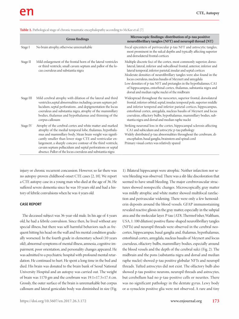
The deceased subject was 36-year-old male. In his age of 4 years old, he had a febrile convulsion. Since then, he lived without any special illness, but there was self-harmful behaviors such as fre-quent hitting his head on the wall and his mental condition gradu-ally worsened. In the fourth grade in elementary school (10 years old), abnormal symptoms of mental illness, amnesia, cognitive im-pairment, poor orientation, and personality changes appeared. He was admitted to a psychiatric hospital with profound mental retar-dation. He continued to hurt. He spent a long time in the bed and died. His brain was donated to the brain bank of Seoul National University Hospital and an autopsy was carried out. The weight of brain was 1170 gm and the cerebrum was 19.1×17.5×17.4 cm. Grossly, the outer surface of the brain is unremarkable but corpus callosum and lateral geniculate body was diminished in size (Fig.
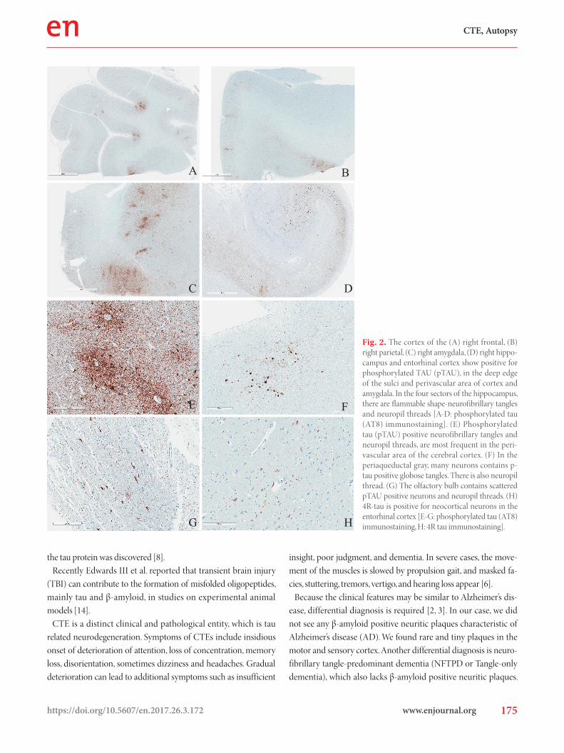
1). Bilateral hippocampi were atrophic. Neither infarction nor se-vere bleeding was observed. There was a slit-like discoloration that seemed to have small bleeding. The major cerebrovascular struc-tures showed nonspecific changes. Microscopically, gray matter was mildly atrophic and white matter showed multifocal rarefac-tion and perivascular widening. There were only a few hemosid-erin deposits around the blood vessels. GFAP immunostaining revealed reactive gliosis in the gray matter, especially in the subpial area and the molecular layer. P-tau (AT8, ThermoFisher, Waltham, USA, 1: 100 dilution) positive flame-shaped neurofibrillary tangles (NFTs) and neuropil threads were observed in the cerebral neo-cortex, hippocampus, basal ganglia and, thalamus, hypothalamus, entorhinal cortex, amygdala, nucleus basalis of Meynert and locus coeruleus, olfactory bulbs, mammillary bodies, especially around the blood vessels and the depth of the cerebral sulci (Fig. 2). The midbrain and the pons (substantia nigra and dorsal and median raphe nuclei) showed p-tau positive globular NFTs and neuropil threads. Tufted astrocytes did not exist. The olfactory bulb also showed p-tau positive neurons, neuropil threads and astrocytes, but cerebellum had no p-tau-positive cells or neurites. There was no significant pathology in the dentate gyrus. Lewy body or α-synuclein positive glia were not observed. A rare and tiny
Table 1. Pathological stage of chronic traumatic encephalopathy according to McKee et al. [7]
Gross findingsMicroscopic findings: distribution of p-tau positive
neurofibrillary tangles (NFT) and neuropil thread (NT)
Stage I No brain atrophy, otherwise unremarkable Focal epicenters of perivascular p-tau NFT and astrocytic tangles, most prominent in the sulcal depths and typically affecting superior and dorsolateral frontal cortices
Stage II Mild enlargement of the frontal horn of the lateral ventricles or third ventricle, small cavum septum and pallor of the lo-cus coeruleus and substantia nigra
Multiple discrete foci of the cortex, most commonly superior, dorso-lateral, lateral, inferior and subcallosal frontal, anterior, inferior and lateral temporal, inferior parietal, insular and septal cortices
Moderate densities of neurofibrillary tangles were also found in the locus coeruleus, nucleus basalis of Meynert and amygdala
Low densities of p-tau NFT and pretangles in the hypothalamus, CA1 of hippocampus, entorhinal cortex, thalamus, substantia nigra and dorsal and median raphe nuclei of the midbrain
Stage III Mild cerebral atrophy with dilation of the lateral and third ventricles,septal abnormalities including cavum septum pel-lucidum, septal perforations , and depigmentation the locus coeruleus and substantia nigra, atrophy of the mammillary bodies, thalamus and hypothalamus and thinning of the corpus callosum
Widespread throughout the neocortex, superior frontal, dorsolateral frontal, inferior orbital, septal, insular, temporal pole, superior middle and inferior temporal and inferior parietal cortices, hippocampus, entorhinal cortex, amygdala, nucleus basalis of Meynert and locus coeruleus, olfactory bulbs, hypothalamus, mammillary bodies, sub-stantia nigra and dorsal and median raphe nuclei
Stage IV Atrophy of the cerebral cortex and white matter and marked atrophy of the medial temporal lobe, thalamus, hypothala-mus and mammillary body, Mean brain weight was signifi-cantly smaller than lower stage CTE and ventricular en-largement, a sharply concave contour of the third ventricle, cavum septum pellucidum and septal perforations or septal absence. Pallor of the locus coeruleus and substantia nigra
Striking neuronal loss in the cortex, hippocampal sclerosis affecting CA1 and subiculum and astrocytic p-tau pathology
Widely distributed p-tau abnormalities throughout the cerebrum, di-encephalon, basal ganglia, brainstem and spinal cord
Primary visual cortex was relatively spared

174 www.enjournal.org https://doi.org/10.5607/en.2017.26.3.172
Kyuho Lee, et al.
β-amyloid positive diffuse plaques were present in the motor and the sensory cortices. 4 repeat (4R) tau (Millipore, Ontario, Canada, X100) immunostaining revealed positivity in the perikarya of the neurons and neuropil threads, but their number was smaller than the number of the p-tau positive neurons and neuropil threads (Fig. 2). There was no TDP43 positive abnormal neurons. These find-ings were consistent with the pathology of the CTE.
DISCUSSION
CTE is a long-term neurological and neuropathologic sequelae associated with repetitive brain injury to athletes, epileptics, head bangers and the victims of abuse [5, 6, 9]. The exact incidence of CTE caused by repetitive head injury is unknown, but McKee et al. speculated that it will be much higher than we expect [6]. So far there was no autopsy-proven childhood-onset CTE cases in English literature. However, children and adolescents may have traumatic brain injury or chronic recurrent concussion [2, 10].
The long-term effect of repetitive concussion of boxers and other athletes showed a unique pattern of pathology first described in 1973 by Corsellis et al. [11] It is characterized by irregularly distrib-uted NFTs and NTs with phosphorylated and 4R tau protein accu-mulation especially in the depth of cerebral sulci and perivascular area [1, 3, 5, 9]. Ghajari et al. reported the computerized modelling images of CTE [12]. The neuropathological diagnostic criteria and progressive staging pathology from I-IV were made in 2013 by McKee et al. [7] Grossly, CTE pathology involves atrophy of the ce-rebral cortex, especially the frontal and temporal lobes, diencepha-
lon and mammillary bodies, and cavum septum pellucidum or septal fenestrations [7]. Microscopically, CTE is characterized by p-tau and 4R-tau positive neurofibrillary, neurites, and astrocytic tangles, which are patchy distributed around small blood vessels, and are preferentially distributed in the depth around the cerebral sulci. In well-established disease, tau pathology is most prominent in the frontal and temporal lobes, hippocampus, amygdala, and entorhinal cortex [7]. Abnormal accumulation of malformed TAR DNA-binding protein 43 (TDP-43) is also observed in CTE and is associated with CTE symptoms [3].
According to McKee et al., brain is not atrophic and the medial temporal lobe is preserved from p-tau pathology until stage 2. In stage 3, the amygdala, hippocampus, and entorhinal cortex show substantial p-tau pathology in addition to throughout the neo-cortex [3, 7]. Our case also showed all the unique neuropatholo-gies mentioned above. With McKee’s stage, our case is compatible with stage 3, because NFTs present widespread throughout the cortex, especially at the depth of the cerebral sulci, hippocampus, entorhinal cortex, amygdala, nucleus basalis of Meynert and locus coeruleus, olfactory bulbs, hypothalamus, mammillary bodies, substantia nigra and dorsal and median raphe nuclei as well as mild cerebral atrophy and mild ventriculomegaly (Fig. 2).
Martland reported “punch drunk” in 1928, but the reported patient’s brain pathology was different from that of CTE. Because the autopsy showed only punctate hemorrhage due to concussion injury [13]. In that paper, there was no mention about tau-positive neurofibrillary tangles. Critchley used the term CTE in 1949 as a punch drunk syndrome of traumatic brain injury, but it was before
A B
C D
Fig. 1. (A-D) Grossly, there are no significant lesions on the exterior and cut surfaces of the brain. (C) Mamillary and lateral geniculate bodies look atrophic. (C, D) There are slit-like punctate discolorations, here and there, that suggests small bleeding.
175www.enjournal.orghttps://doi.org/10.5607/en.2017.26.3.172
CTE, Autopsy
the tau protein was discovered [8].Recently Edwards III et al. reported that transient brain injury
(TBI) can contribute to the formation of misfolded oligopeptides, mainly tau and β-amyloid, in studies on experimental animal models [14].
CTE is a distinct clinical and pathological entity, which is tau related neurodegeneration. Symptoms of CTEs include insidious onset of deterioration of attention, loss of concentration, memory loss, disorientation, sometimes dizziness and headaches. Gradual deterioration can lead to additional symptoms such as insufficient
insight, poor judgment, and dementia. In severe cases, the move-ment of the muscles is slowed by propulsion gait, and masked fa-cies, stuttering, tremors, vertigo, and hearing loss appear [6].
Because the clinical features may be similar to Alzheimer’s dis-ease, differential diagnosis is required [2, 3]. In our case, we did not see any β-amyloid positive neuritic plaques characteristic of Alzheimer’s disease (AD). We found rare and tiny plaques in the motor and sensory cortex. Another differential diagnosis is neuro-fibrillary tangle-predominant dementia (NFTPD or Tangle-only dementia), which also lacks β-amyloid positive neuritic plaques.
G H
E F
A B
C D
Fig. 2. The cortex of the (A) right frontal, (B) right parietal, (C) right amygdala, (D) right hippo-campus and entorhinal cortex show positive for phosphorylated TAU (pTAU), in the deep edge of the sulci and perivascular area of cortex and amygdala. In the four sectors of the hippocampus, there are flammable shape-neurofibrillary tangles and neuropil threads [A-D: phosphorylated tau (AT8) immunostaining]. (E) Phosphorylated tau (pTAU) positive neurofibrillary tangles and neuropil threads, are most frequent in the peri-vascular area of the cerebral cortex. (F) In the periaqueductal gray, many neurons contains p-tau positive globose tangles. There is also neuropil thread. (G) The olfactory bulb contains scattered pTAU positive neurons and neuropil threads. (H) 4R-tau is positive for neocortical neurons in the entorhinal cortex [E-G: phosphorylated tau (AT8) immunostaining, H: 4R tau immunostaining].
176 www.enjournal.org https://doi.org/10.5607/en.2017.26.3.172
Kyuho Lee, et al.
NFTPD is clinically and pathologically different from ours. NFT-PD is a late-onset progressive dementia with a shorter duration than AD and occurs in the elderly with an average age of 79.7 years [15]. Pathologically, the NFT distribution pattern of p-tau protein in NFTPD is similar to the distribution, spread and severity of AD. However, it does not prefer localization to the perivascular, peri-ventricular and subpial regions or the depth of the cerebral sulci [15]. P-tau positive astrocytic tangles are not present in AD. And the disease that causes similar symptoms in children is lysosomal storage disease like Gangliosidosis type II and neuronal ceroid lipofuscinosis. However, in our case, there was no lipid vacuoliza-tion, severe neuronal loss and diffuse astrogliosis with macrophage accumulation which is characteristic of this disease. So our case does not fit into these diseases.
Studies of clinicopathological correlation, such as the Under-standing Neurologic Injury and Traumatic Encephalopathy (UNITE) Study, can help identify sensitive clinical features of CTE pathology [16]. Future improvements for CTE diagnosis require prospective studies including neuropsychological tests using im-aging and fluid biomarkers.
The problem of our case is that there is no 6-year history be-tween febrile seizures and dementia. However, symptoms of CTE, such as mental retardation and behavioral problems, was already appeared when he was admitted to a mental hospital for symp-toms in his age of 10 and showed patterns of self-harm. He lived in bed for a long time and died at the age of 36. Autopsy of our case revealed a typical pathology of CTE.
Here we report a case of CTE that was proven by autopsy. We report this case because it is interesting that CTE can be diagnosed only by autopsy without a clear history of repeated brain damage. This example shows that CTE can occur at young age and that even children can experience CTE dementia.
ACKNOWLEDGEMENTS
This work was funded by the Korean government-sponsored Korea Brain Bank network.
REFERENCES
1. Noy S, Krawitz S, Del Bigio MR (2016) Chronic traumatic encephalopathy-like abnormalities in a routine neuropathol-ogy service. J Neuropathol Exp Neurol 75:1145-1154.
2. Grinberg LT, Anghinah R, Nascimento CF, Amaro E, Leite RP, Martin Mda G, Naslavsky MS, Takada LT, Filho WJ, Pasqual-ucci CA, Nitrini R (2016) Chronic traumatic encephalopathy presenting as Alzheimer’s disease in a retired soccer player. J
Alzheimers Dis 54:169-174.3. McKee AC, Stein TD, Kiernan PT, Alvarez VE (2015) The
neuropathology of chronic traumatic encephalopathy. Brain Pathol 25:350-364.
4. Montenigro PH, Baugh CM, Daneshvar DH, Mez J, Budson AE, Au R, Katz DI, Cantu RC, Stern RA (2014) Clinical sub-types of chronic traumatic encephalopathy: literature review and proposed research diagnostic criteria for traumatic en-cephalopathy syndrome. Alzheimers Res Ther 6:68.
5. Lakis N, Corona RJ, Toshkezi G, Chin LS (2013) Chronic traumatic encephalopathy - neuropathology in athletes and war veterans. Neurol Res 35:290-299.
6. McKee AC, Cantu RC, Nowinski CJ, Hedley-Whyte ET, Ga-vett BE, Budson AE, Santini VE, Lee HS, Kubilus CA, Stern RA (2009) Chronic traumatic encephalopathy in athletes: progressive tauopathy after repetitive head injury. J Neuro-pathol Exp Neurol 68:709-735.
7. McKee AC, Stern RA, Nowinski CJ, Stein TD, Alvarez VE, Daneshvar DH, Lee HS, Wojtowicz SM, Hall G, Baugh CM, Riley DO, Kubilus CA, Cormier KA, Jacobs MA, Martin BR, Abraham CR, Ikezu T, Reichard RR, Wolozin BL, Budson AE, Goldstein LE, Kowall NW, Cantu RC (2013) The spectrum of disease in chronic traumatic encephalopathy. Brain 136:43-64.
8. Critchley M (1949) Punch-drunk syndromes: the chronic traumatic encephalopathy of boxers. In: Neuro-chirurgie: hommage à Clovis Vincent (Vincent C, ed), pp 131. Maloine, Paris.
9. Zhang J, Teng Z, Song Y, Hu M, Chen C (2015) Inhibition of monoacylglycerol lipase prevents chronic traumatic enceph-alopathy-like neuropathology in a mouse model of repetitive mild closed head injury. J Cereb Blood Flow Metab 35:443-453.
10. Field M, Collins MW, Lovell MR, Maroon J (2003) Does age play a role in recovery from sports-related concussion? A comparison of high school and collegiate athletes. J Pediatr 142:546-553.
11. Corsellis JA, Bruton CJ, Freeman-Browne D (1973) The af-termath of boxing. Psychol Med 3:270-303.
12. Ghajari M, Hellyer PJ, Sharp DJ (2017) Computational modelling of traumatic brain injury predicts the location of chronic traumatic encephalopathy pathology. Brain 140:333-343.
13. Martland HS (1928) Punch drunk. J Am Med Assoc 91:1103-1107.
14. Edwards G 3rd, Moreno-Gonzalez I, Soto C (2017) Amyloid-beta and tau pathology following repetitive mild traumatic
177www.enjournal.orghttps://doi.org/10.5607/en.2017.26.3.172
CTE, Autopsy
brain injury. Biochem Biophys Res Commun 483:1137-1142.15. Jellinger KA, Attems J (2007) Neurofibrillary tangle-predom-
inant dementia: comparison with classical Alzheimer disease. Acta Neuropathol 113:107-117.
16. Mez J, Solomon TM, Daneshvar DH, Murphy L, Kiernan PT, Montenigro PH, Kriegel J, Abdolmohammadi B, Fry B, Babcock KJ, Adams JW, Bourlas AP, Papadopoulos Z, McHale
L, Ardaugh BM, Martin BR, Dixon D, Nowinski CJ, Chais-son C, Alvarez VE, Tripodis Y, Stein TD, Goldstein LE, Katz DI, Kowall NW, Cantu RC, Stern RA, McKee AC (2015) As-sessing clinicopathological correlation in chronic traumatic encephalopathy: rationale and methods for the UNITE study. Alzheimers Res Ther 7:62.