Embed Size (px)
Citation preview
1108 JONCAS AND PAvUANIs: DIABRHCEA AND VOMITING
DIARRHCEA AND VOMITINGIN INFANCY AND CHILDHOOD:VIRAL STUDIES*
J. JONCAS, M.D.t and V. PAVILANIS, M.D.,jMontreal
THE POSSIBILITY of a viral etiology for the self-limited episodes of diarrhoea and vomiting inchildren, as well as in adults, has been postulatedfor a long time. Until a few years ago, there wasno evidence to support this assumption.With the introduction of tissue culture methods
in virus research, many workers were soon able toestablish a link between certain viruses and out-breaks of diarrhoea and vomiting occurring sporad-ically or in small epidemics. The present workoffers the results of viral studies carried out oninfants and children seen in ho;spital with thissyndrome from July 1958 to May 1959.Among the most valuable contributions in this
field of research, Eichenwald et al.,' in 1958,clearly established the causal relationship ofECHO type 18 with an outbreak of diarrhoea inpremature and newborn babies at the Cornell Uni-versity Medical Center in New York. Ramos-Alvarez and Sabin,2 in 1958, reported their findingson a series of cases collected throughout two con-secutive summers and stressed the importance ofthe ECHO viruses as a cause of diarrhoea; theyalso mentioned the possible significance of adeno-viruses in this regard, having found three of suchviruses in the test group (using only monkeykidney tissue cultures) as compared with none inthe control group.A report of great interest (for purposes of com-
parison) appeared in the British literature in De-cember 1958. Sommerville,3 in a one-year survey,found 75 enteroviruses in 338 rectal swabs fromchildren with diarrhoea (an average of 22%v), and17 in a control group of 115 children with res-piratory infections. The incidence of polio andCoxsackie viruses in the two groups was com-parable, but the proportion of ECHO viruses wasgreater in the test group, although the differencewas not statistically significant. The presence ofonly one adenovirus was mentioned. Up to now,from the previous reports and reports from otherworkers, it appears that ECHO viruses (2, 7, 8, 10,11, 12, 14, 18, 19, 20) have been associated withacute episodes of diarrhoea and vomiting.
Finally, in adults a filtrable agent, which cannotbe isolated on tissue cultures, has been shown tocause diarrhoea in several successive volunteers onwhom stool filtrates were used, and to induce im-munity.4 One strain is called "F.S."5 because of thepresence of fever in the clinical picture, the other
This work has been partially aided by a grant from theDepartment of Health of the Province of Quebec (Federal-Provincial Public Health Research Grant).tFellow of the National Research Council, Institute of Micro-biology and Hygiene of the University of Montreal, andMontreal Children's Hospital.tHead of Virus Section, Institute of Microbiology andHygiene of the University of Montreal.
Canad. M. A. J.May 28,1960, vol. 82
the "Marcy strain",6 from an afebrile form of non-bacterial gastroenteritis. There are no comparablestudies in children.
MATERIAL AND METHODSFrom July 1958 to May 1959, rectal swabs were
obtained from 74 children with diarrhoea andvomiting and from 62 controls in the MontrealChildren's Hospital. In the diarrhoea group, bloodspecimens were also taken during the acute andconvalescent phase. The rectal swabs chosen wereall negative for a bacterial pathogen (Shigella,Salmonella and pathogenic coliforms). Cases ofdiarrhoea in which a cause other than viral couldbe suspected were also eliminated, i.e. diarrhoeaaccompanying parenteral infections known to pro-duce diarrhoea (mastoiditis in infants for example),malabsorption syndromes, chemical intoxications,food poisoning, etc.The case accepted for study was one of diarrhoea
and vomiting as the major complaint, with or with-out fever, and/or symptoms of upper respiratoryinfection; the course of illness was usually oneweek or less. Most of these cases were in-patients;only eight swabs were taken in the outdoor de-partment of the hospital. The control swabs wereobtained in another so-called "clean ward" frompatients admitted recently for investigation of somechronic problem; occasionally, owing to temporaryshortage of these patients, ones with pneumoniawere used. A few controls were taken at the out-door department. These controls were matched forage and time of sampling. The history and physicalexamination as well as the white blood celland differential counts were noted in each instance.The rectal swabs were processed according to
the method of Alvarez and Sabin.2 The rectal swabsused were the same as for the bacteriology depart-ment of the hospital. Immediately after samplingthe swabs were stored for a maximum period of48 hours in the freezer compartment of an ordinaryrefrigerator. They were then collected and trans-ferred to the freezer of the Institute of Micro-biology at -200 F., with 5 c.c. of Hank's solutionadded. On the day of the test the samples werethawed slowly at refrigerator temperature or morerapidly in cold water; 0.5 c.c. of an antibioticsolution was added in order to yield concentrationsof 2000 units of penicillin per c.c., 2 mg. of strepto-mycin per c.c. and 150 units of nystatin (Myco-statin) per c.c. The pH was then brought to 8 byuse of sodium bicarbonate 4.4% solution. After astorage period of 30 minutes in the refrigerator,six or eight tubes of tissue cultures (equal numberof HeLa and monkey kidney) were inoculated with0.5 c.c. of material each. The matched control caseswere inoculated the same day on the same lot oftubes. At least two passages were made, theaverage number of passages being four. In orderto detect, on occasion, a cytopathogenic agentoriginating from the tissue culture itself, serialpassage of the non-inoculated control tubes was
Canad. M. A. J.May 28, 1960, vol. 82 JONCAS AND PAVILANIS: DIARRHCEA AND VOMITING 1109
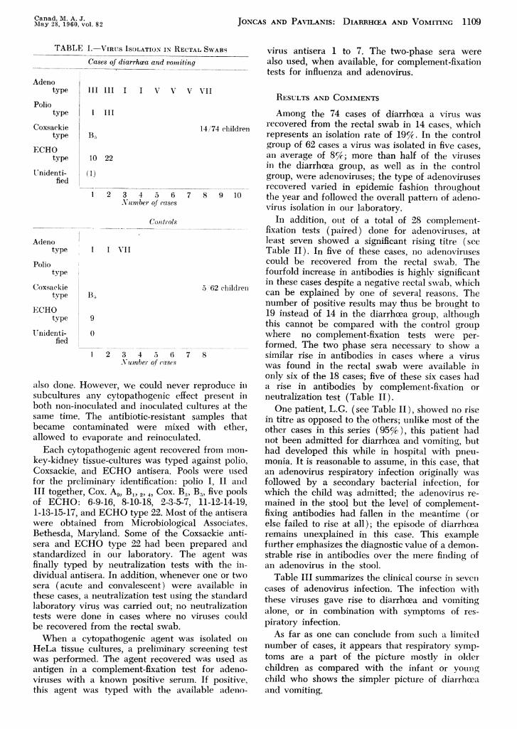
TABLE I. VIRUS ISOLATION IN RECTAL SWABS
Cases of diarrh.ea and vomitinq
Adenotype
Poliotype
Coxsackietype
ECHOtype
Unidenti-fied
Adenotype
Poliotype
Coxsaekietype
ECHOtype
UTnidenti-fied
III III I I V
I III
B,
10 22
(1)
1 2 3 4 5 6Number of cases
Conltrols
I I VrII
9
0
2 3 4 5 6
.Number of (ases
V V V'II
7
7
also done. However, we could never rEstubcultures any cytopathogenic effectboth non-inoculated and inoculated cullsame time. The antibiotic-resistant sabecame contaminated were mixed x
allowed to evaporate and reinoculated.Each cytopathogenic agent recovered
key-kidney tissue-cultures was typed agCoxsackie, and ECHO antisera. Poolsfor the preliminary identification: poliIII together, Cox. A, B1, 2, 4. Cox. B, Bof ECHO: 6-9-16, 8-10-18, 2-3-5-7, :1-13-15-17, and ECHO type 22. Most ofwere obtained from MicrobiologicalBethesda, Maryland. Some of the Cox~sera and ECHO type 22 had been prstandardized in our laboratory. Thefinally typed by neutralization tests vtdividual antisera. In addition, wheneversera (acute and convalescent) were athese cases, a neutralization test using t]laboratory virus was carried out; no ne
tests were done in cases where no viibe recovered from the rectal swab.When a cytopathogenic agent was
HeLa tissue cultures, a preliminary sCI
was performed. The agent recovered Mantigen in a complement-fixation testviruses with a known positive serum.
this agent was typed with the availa
virus antisera 1 to 7. The two-phase sera werealso used, when available, for complement-fixationtests for influenza and adenovirus.
RESULTS AND CO.MMENTSAmong the 74 cases of diarrhoea a virus was
14/74 children recovered from the rectal swab in 14 cases, whichrepresents an isolation rate of 19%-. In the controlgroup of 62 cases a virus was isolated in five cases,an average of 8Cc; more than half of the virusesin the diarrhoea group, as well as in the controlgroup, were adenoviruses; the type of adenovirusesrecovered varied in epidemic fashion throughout
8 9 10 the year and followed the overall pattern of adeno-virtus isolation in our laboratory.
In addition, out of a total of 28 complement-fixation tests (paired) done for adenoviruses, atleast seven showed a significant rising titre (seeTable II). In five of these cases, no adenoviruisescould be recovered from the rectal swab. Thefourfold increase in antibodies is highly significantin these cases despite a negative rectal swab, which
5 62 chlildreon l .can be explained by one of several reasons. Thenumber of positive results may thus be brought to19 instead of 14 in the diarrhoea group, althoughthis cannot be compared with the control groupwhere no complement-fixation tests were per-formed. The two phase sera necessary to show a
8 similar rise in antibodies in cases where a viruswas found in the rectal swab were available inonly six of the 18 cases; five of these six cases had
?produce in a rise in antibodies by complement-fixation orpresent in neutralization test (Table II).
tures at the One patient, L.G. (see Table II), showed no riseLmples that in titre as opposed to the others; unlike most of thewith ether, other cases in this series (95%), this patient had
not been admitted for diarrhboea and vomiting, butfrom mon- had developed this while in hospital with pneu-
rainst polio, monia. It is reasonable to assume, in this case, thatwere used an adenovirus respiratory infection originally was
o I, II and followed by a secondary bacterial infection, for,, five pools which the child was admitted; the adenovirus re-11-12-14-19, mained in the stool but the level of complement-the antisera fixing antibodies had fallen in the meantime (orAssociates, else failed to rise at all); the episode of diarrhoeasackie anti- remains unexplained in this case. This exampleepared and further emphasizes the diagnostic value of a demon-agent was strable rise in antibodies over the mere finding ofvith the in- ain adenovirus in the stool.Ione or two Table III summarizes the clinical course in seventvailable in cases of adenovirus infection. The infection withhe standard these viruses gave rise to diarrhoea and vomitingutralzation alone or in combination with symptoms of res-ruses couild .
piratory infection.isolated oni As far as one can conclude from such a limitedreening test number of cases, it appears that respiratory symp-vas used as toms are a part of the picture mostly in olderfor adeno- children as compared with the infant or youingIf positive, child who shows the simpler picture of diarrhoeable adeno- and vomiting.
1110 JONCAS AND PAVILANIS: DIARRHCEA AND VOMITING Canad. M. A. J.May 28, 1960, vol. 82
TABLE II.-DATA ON CASES WITH POSITIVE RESULTS IN CHRONOLOGICAL ORDER
Day Antibodies against1 st day rectal homologous virus
of swab Type of virusGroup Name Age illntess taken isolated Day Titre W.B.C. Differential count
C: R ** 9.42 Mne A11P.. 1 F'C'iI0 0 - - Auz. 4- 5000 PO18. 1050 Lvmphs. 3800
6 0 C. F. Adeno47 1/325 1/128 C. F. Adeno19 1/1284 0
41 1/32
7 0 C. F. Adeno28 1/1285 0
7 0 C. F. Adeno14 1/2567 I 0- II 0
III 0
4 1/1615 1/646 0 C. F. Adeno20 1/1288 1/128 C. F. Adeno
17 1/1285 I 0
II 0III 1 12
9 I 0II 0III 1/32
5 1 /32 C. F. Adeno8 1/64
7 1/8 C. F. Adeno18 1/64
6 0 C. F. Adeno22 1/32
N.T.3 0 k C. F. AdenoDied within 2 days
10 0 C. F. Adeno
-2 0 C. F. Adeno6 1/128
11 0 C. F. Adeno39 06 0 C. F. Adeno
Aug. 13-10,600 Polys. 2000 Lymphs. 8600
Not done
Aug. 20-12,900 Polys. 3500 Lymphs. 9000
Not done
Aug. 23-6600
Aug. 27-11,000
Sept. 15-8300
Sept. 22-6900
Oct. 10-6700
Oct. 18-7700Oct. 21-4300Oct. 27-13,300
Nov. 3-9300
Nov. 14-6300
Nov. 23-11,800
Dec. 4-5200
Dec. 10-9100
Dec. 10-11,700Dec. 27-11,400Feb. 9-10,400Feb. 19-7100Feb. 17-5900
Polys. 2376
Polys. 4100
Polys. 4980
Polys. 2413
Polys. 2211
Polys. 5544Polys. 1806Polys. 8500
Polys. 6696
Polys. 1890
Polys. 9300
Polys. 1508
Polys. 1638
Polys. 4400Polys. 5300Polys. 6200Polys. 3000Polys. 1121
Lymphs. 4224
Lymphs. 6800
Lymphs. 3071
Lymphs. 4200
Lymphs. 4355
Lymphs. 2079Lymphs. 2408Lymphs. 4400
Lymphs. 2418
Lymphs. 4284
Lymphs. 2300
Lymphs. 3380
Lymnphs. 7371
Lymphs. 6700Lymphs. 5800Lymphs. 4000Lymphs. 3300Lymphs. 4661
Feb. 23-9300 Polys. 7068 Lymphs. 1953Feb. 24-4700 Polys. 2538 Lymphs. 1974March 10-10,100 Polys. 2400 Lymphs. 7500
March 21-5000 Not done
March 19-20,000 Polys. 14,000 Lymphs. 3000
March 20-7700 Polys. 3542 Lymphs. 3850March 27-15,800 Polys. 4600 Lymphs. 9400
C-Control cases.I) a V-Diarrhuea and voiniting.
C. F. Adeno-Colnplelnent-fixation test.N.T.-Neutralization test.
*-Details of clinical findings in Table III.
**-Details of clinical findings in text.
Another patient, D.R., whose rectal swab yield-ed an adenovirus type I, deserves special comment,mainly because the post-mortem findings are avail-able. This patient died after four days of illness,of which two were spent in hospital. She was
markedly dehydrated on admission with a fever of102°F.; physical examination was otherwise nega-tive. She was rehydrated by intravenous therapy;diarrhoeea persisted until her demise. Her onlysibling, aged eight months, developed explosivediarrhoea a few days later and was admittedpromptly: his hospital course was uneventful; no
viruses or bacterial pathogens could be isolatedfrom his rectal swab. Unfortunately no bloodsamples could be obtained from this child for a
complement-fixation test.At post-mortem examination, aseptic arterial and
venous thrombi and arterial emboli were found inthe brain and lungs. Aseptic thrombi were alsopresent under the mitral and tricuspid valve leaf-lets, presumably the site of origin of the emboli.The intestinal wall showed no ulcerations or othergross alterations. Microscopically there was hyper-trophy of the Peyer's plaques with lymphoid hy-
perplasia. Samples of brain, heart and spleen were
processed and inoculated into tissue cultures, butno viruses could be isolated.
Despite the absence of antibodies in the firstserum, there is no conclusive evidence that thediarrhoea was the consequence of an adenovirusinfection. It is, however, a reasonable assumption,since no neutralizing antibodies could be detectedin the first serum; a rise in titre for adenovirusesin the sibling would have constituted an adequateproof.
In view of the somewhat unexpectedly high per-
centage of adenovirus isolations, we were interest-ed in appraising the frequency of diarrhcea as a
symptom in these infections over a larger series.With this in mind we collected from other sources
in our laboratory 15 cases of adenovirus infectionproven by complement-fixation test either aloneor in combination with the isolation of an adeno-virus. To these we added seven cases from thisseries, using the same criterion. As far as one can
judge from the hospital record of these patientsand checking back with the parents, taking intoaccount the possible effect of antibiotics among
AIug. 8
Aug. 8
Aug. 11
Aug. 1 7
? Aug. '?
Autg. 19
Aug. 23
Sept. 10
Sept. 16
Oct.
Oct.
Oct.
Oct.
7
16
20
30
) and V
D and V
I) and V
C
1) and V
O and V
I) and V
D) and V
1) and V
D and V
D and V
1) and V
1) an(l V
C
1) and V
C
1) and V
C
D and V
I) and V
1) and V
D and V
D and V
D and VD and V
J.L.V.*
P.Gl.
L.O.**
C:l.M.**
G.B.*
D.T.**
L.P.*
V.N.**
B.C.$*
P.MCC.*
P.Gr.
P.B.**
S.P.
D.L.
R.h-.*
F.M.
R.C.*
C.G
D.C.
D.R.**
L.S.
S.M.*
L.G.**
B.T.B.H.
12 mos.
2 yrs.
2 mos.
3 yrs.
13 mos.
12 mos.
2 yrs.
212 yrs.
18 n1oS.
S5 yrs.
6 yrs.
2Y yrs.
17 mos.
22 nlos.
18 nios.
712 1i0os.1 II0o.
12 1110S.
8 1110S.
20 IIIOS.
4 mos.
10 yrs.
2 maos.
3 imos.4 mos.
6
5
4
7
S
7
7
4
6
8
9
7
6
4
6
3
10
4
7
62
ECHO 22
Coxsackie B.,
ECHO 10
Adeno III
Polio I
Coxsackie B5
Adeno III
Polio III
Adeno I
Adeno VII
Adeno I
Adeno I
Adeno I
Adeno V
Adeno VII
Adeno VAdeno V
Nov. 10
Nov. 21
Nov. 28
? No\-. ?
Dec. 17
Feb. 8
Feb. 1 2
Feb. 2 1
Marclh 7
MIarch 18
Marclh 20
MarCh 21M%Iarch 29
r, k, 'ri ki tr
Canad. M. A. J.May 28, 1960, vol. 82 JONCAS AND PAVILANIS: DIARRHCEA AND VOMITING 1111
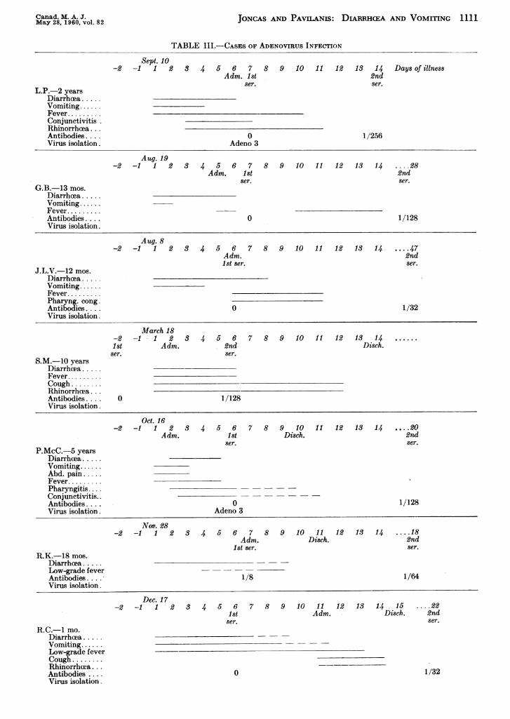
TABLE III.-CASES oF ADENOVIRUS INFECTION
Sept. 10-2 -1 1 2 3 4 5 6 7 8 9 10 11 12 13 14 Days of illness
Adm. 1st 2ndser. ser.
L.P.-2 yearsDiarrh ea.Vomiting.Fever.ConjunctivitisRhinorrheea. . .Antibodies.... 0 1/256Virus isolation. Adeno 3
Aug. 19-2 -1 1 2 3 4 5 6 7 8 9 10 11 12 13 14 ... .28
Adm. 1st 2ndser. ser.
G.B.-13 mos.Diarrhoea.Vomiting.Fever.Antibodies.... 0 1/128Virus isolation.
Aug. 8-2 -1 1 2 3 4 5 6 7 8 9 10 11 12 13 14 ... .47
Adm. 2nd1st ser. ser.
J.L.V.-12 mos.Diarrhoea.Vomiting.Fever.Pharyng. cong.Antibodies.... 0 1/32Virus isolation.
March 18-2 -1 1 2 3 4 5 6 7 8 9 10 11 12 13 14 ......
1st Adm. 2nd Disch.ser. ser.
S.M.-10 yearsDiarrhea.Fever.Cough.Rhinorrheea. . .Antibodies.... O 1/128Virus isolation.
Oct. 16-2 -1 1 2 3 4 5 6 7 8 9 10 11 12 13 14 ... .20
Adm. 1st Disch. 2ndser. ser.
P.McC.-5 yearsDiarrhoea.Vomiting.Abd. pain.Fever.Pharyngitis....Conjunctivitis..Antibodies... . O 1/128Virus isolation. Adeno 3
Nov. 28-2 -1 1 2 3 4 5 6 7 8 9 10 11 12 13 14 ... .18
Adm. Disch. 2ndIst ser. ser.
R.K.-18 mos.Diarrhcea.Low-grade fever - --Antibodies... . 1/8 1/64Virus isolation.
R.C.-1 mo.Diarrhcea .....Vomiting......Low-grade feverCough ........Rhinorrhcea. . .Antibodies ...Virus isolation.
Dec. 17-2 -1 1 2 3 4 5 6 7 8 9 10 11 12 13 14 15 .... 22
Ist Adm. Disch. 2ndser. ser.
0 1/32
1112 JONCAS AND PAVILANIS: DIARRHCEA AND VOMI
other factors in producing this symptom, diarrhkeawas a relatively conspicuous symptom in 11 out of22 cases, an average of 50%. This had already beensuspected by Tyrrell et al.,7 who stated: "Thenormal patterns of clinical infection [with adeno-viruses] in childhood have yet to be discovered....Perhaps, in some cases, alimentary-tract symptomsdominate the picture. . . In the epidemic weobserved, we think the gastro-intestinal symptomswere due to the virus."The complement-fixation test for influenza car-
ried out in 27 cases of diarrhoea and vomiting wasconsistently negative. This test was performedmainly to see if there was any scientific basis forthe common diagnosis of "intestinal flu" made incases of this nature during the winter.
In view of some resemblance between influenzaand the respiratory syndrome caused by the adeno-virus infection, one is tempted to associate thisvirus, rather than the influenza virus, with a res-piratorv-enteric type of illness. Several findings canbe put forward in favour of this hypothesis. In1954, a report by Goodall8 on "gastric flu" or"4cwinter vomiting disease" in adults also gave uni-formly negative results for influenza by comple-ment-fixation test. In the present series, adeno-viruses exclusively were isolated in the winter andspring periods. We were also impressed in thisstudy by the occurrence of diarrhoea with or with-out respiratory symptoms in the father or motheror both parents in three of our patients with diar-rhoea and vomiting from whom an adenovirus wasisolated. One of these, P. McC., had a fourfoldincrease in antibodies. In none of our other positivecases did we record a similar finding on reviewingthe family history. It is also of great interest in thisregard that a definite respiratory-enteric type ofillness occurred as the result of proven adenovirusinfection in a boy aged 5 years, P. McC., and inainother aged 10 years, S. M.-the two positive casesin this series over the 3-year age level.The number of enteroviruses isolated is too small
to permit any general conclusion. However, sincea rising titre in antibodies was demonstrated byneutralization test in the majority of the cases ofthe diarrhoea group, a few clinical observationsmay be wvorthy of note.
V.N., from whom a polio virus type I wasisolated, is a 2/2-year-old child who had a seven-day episode of low-grade fever, diarrhoea andvomiting, plus vague abdominal pain. His coursewas benign and he did not require any intravenoustherapy. His first serum showed no antibodiesagainst any of the polio strains.
P.B. had a similar seven-day illness, whichstarted like a simple cold with vomiting for threedays and two semi-liquid stools; a low-grade feverwas present in the first few days. The finding ofa questionable stiff neck in this case failed to leadto the real etiology and was interpreted as men-inigismus due to gastro-enteritis; the lumbar punc-tuire examination was negative. A polioviris type
Canad. M. A. J.May 28. 1960, vol. 82
III was isolated from the rectal swab, and a slightrising titre for polio III and none for the othertypes could be detected within five days.
B.C., in whom a Coxsackie B5 virus was found,showed a biphasic type of illness: two episodes offever, vomiting and diarrhcea that were five daysapart. A rising titre was demonstrated by neutral-ization test.The control patient, G.M., who had the other
Coxsackie B5 vinis, had had a three-week episodeof harsh cough and anorexia, with intermittentvomiting and fever that ended one week beforeher visit to the outpatient clinic, but she had hadno diarrhaea.
D.T., aged 12 months, was admitted to hospitalin August, with a clinical picture of cough, redthroat and temperature up to 1020 F. at the onset,followed shortly by vomiting and watery greenstools. The one-week duration of illness was rela-tively mild although intravenous rehydration wasnecessary. An ECHO type 10 virus was isolatedfrom her rectal swab, and her first serum, the onlyone available, showed no detectable antibodies toECHO type 10.
L.O., two months old, had a five-day episode ofuncomplicated afebrile diarrhbea and vomiting,with mild dehydration, for which she was admittedin August. She was given fluids intravenously fora very short period, and rehydration was thencarried out orally. An ECHO type 22 virus wasisolated from her rectal swab, and a definite risingtitre was demonstrable in the convalescent serum.The control patient (aged 23 months) with an
ECHO 9 virus infection was admitted in Augustfor investigation and control of petit mal seizures.After a few days in hospital, she developed anunexplained fever to 102' F. which slowly droppedwithin three days. She had no diarrhoea. One sib-ling at home, aged 9, had severe headache andunexplained fever for a few days at the same time.
In all the positive cases (Table II) the whiteblood cell count deserves attention. With practic-ally no exceptions it was between 5000 and 10,000with a definite predominance of lymphocytes ofmore than could be expected from the age of thechild in most cases. In adenovirus infections atransient polynucleosis seemed to occur at timesin the early phase, similar to the one described inpoliomyelitis.Table I discloses the tvpe of virus recovered
from rectal swabs in the two groups of children.Although the number of viruses isolated in thediarrhoea group is larger than in the control group,no specific type of virus can be found significantlyassociated with the syndrome of diarrhaea andvomiting. Adenoviruses type III and V were foundexclusively in the diarrhoea group, but the numberisolated is too small to be of statistical significance.
In the light of recent work and the present find-ings (especially from their correlation with theclinical picture) it appears that diarrhoea is a vari-able symptom that follows infection from several
Canad. M. A. J. JONCAS AND PAVILANIS: DIARRHCA AND VOMITING 1113May 28, 1960, vol. 82
different agents. The occurrence of diarrhoea andits importance seems dependent on several factors,including mainly the type of agent, the host, hisage, and status of immunity. One gathers the im-pression that this syndrome of diarrhoea andvomiting will finally be added to the list of possiblemanifestations of an infection with any one of theseviruses, just as, one after the other, aseptic menin-gitis, pleurodynia, myocarditis, encephalitis, andeven paralytic disease or summer "grippe" havebeen proved to result from apparently the sameCoxsackie B infection.The low isolation rate of enteroviruses in this
series as compared with previous series 2-3 is difficultto explain. Technical difficulties are probably themost important factors. However, the prevalence ofadenoviruses in this region as a cause of infectionmanifested by diarrhoea and vomiting alone, orother combination of symptoms,9-10 may accountfor this low isolation rate of enteroviruses. Adeno-viruses, on the other hand, are less likely to bepresent in rectal swabs than in other more suitablespecimens for isolation, such as throat washings.This may possibly explain why in spite of the prev-alence of these viruses the overall isolation rate inthis series is still quite low. In fact, five cases ofadenovirus infection with simple diarrhoea andvomiting in this series were picked up by the com-plement-fixation tests only, despite more carefulattempts at isolating the virus from the rectal swab.It is probable that many more would have shown arising titre for adenoviruses had sera been availablein all cases.
SUMMARYOver a period of 10 months from July 1958 to May
1959, 14 viruses were isolated from rectal swabs ofa group of 74 infants and children with acute self-limited episodes of diarrhoea and vomiting, as com-pared with five viruses in a control group of 62 chil-dren. At least five additional cases of viral infectionwere discovered by the complement-fixation test in thediarrhcea group.
In over half of the positive cases in the test group,as well as in the control group, the agents were adeno-viruses; in the winter and spring periods only adeno-viruses were isolated.
These findings point to a prevalence of adenovirusinfections probably greater in this region during 1958-59 than reported elsewhere. The use of HeLa tissuecultures as well as monkey kidney also partly explainsthis difference, even though there was often evidenceof the presence of adenovirus in both media.
It appears that diarrhcea from viral infection inchildren is only a symptom of variable frequency de-pendent on several factors-the type of agent, the host,his age, status of immunity, etc. Most viruses isolatedso far from rectal swabs seem able to produce diarrhoeaand vomiting as part of their infectious process. Theirability to do so varies apparently from one agent tothe other. It is estimated from our data that theaverage incidence of diarrhoea with adenovirus infec-tion is in the range of 50%. However, significant varia-tions from this may probably be found in selective agegroups, as well as under the influence of other factors.
The overall low isolation rate in this series leads us,in spite of a few reasonable explanations that are dis-cussed, to seek an additional explanation in thepossible existence of non-bacterial agents similar tothose described in adults46 and as yet not viable intissue cultures.
REFERENCES
1. EICHE:NWALD, H. F. et al.: J. A. M. A., 166: 1563, 1958.2. RAMOS-ALVAREZ, M. AND SABIN, A. B.: Ibid., 167: 147,
1958.3. SOMMERVILLE, R. G.: Lancet, 2: 1347, 1958.4. GORDON, I., PATTERSON, P. R. AND WHITNEY, E.: J. Clin.
Invest., 35: 200, 1956.5. JORDAN, W. S., JR., GORDON, I. AND DORRANCE, W. R.:
J. Exper. Med., 98: 461, 1953.6. GORDON, I., INGRAHAM, H. S. AND KORNS, R. F.: Ibid.,
86: 409, 1947.7. TYRRELL, D. A. J., BALDUCCI, D. AND ZAIMAN, T. E.:
Lancet, 2: 1326, 1956.8. GOODALL, J. F.: Brit. M. J., 1: 197, 1954.9. CHANY, C. et al.: Amt. J. Hyg., 67: 367, 1958.
10. JORDAN, W. S., JR. et al.: Ibid., 64: 336, 1956.11. HODGES, R. G. et al.: Ibid., 64: 349, 1956.12. KJELLEN, L., ZETTERBERG, B. AND SVEDMYR, A.: Acta
pxdsat., 46: 561, 1957.
2900 Mount Royal Blvd.,Montreal.
RESUMELes presentes recherches furent entreprises dans l'in-
tention de decouvrir un agent viral en relation avec lesepisodes sporadiques ou epidemiques de diarrhee et vomis-sements observes au cours d'une annee dans un hopital pourenfants et de determiner dans la mesure du possible letableau clinique correspondant 'a chaque type de virus.De juillet 1958 'a mai 1959, des echantillons de selles
(ecouvillons) furent preleves chez 74 enfants souffrantde diarrhee ainsi que chez 62 enfants te6moins du mermeage, a l'hopital Montreal Children's; ces echantillons furentchoisis de facon 'a eliminer les diarrhees d'origine bacte-rienne ainsi que les diarrhees d'apparence non infectieuse.Dans les cas de diarrhee, des echantillons de sang furentaussi preleves a la phase aigue et a la phase de conva-lescence. Les echantillons furent traites, "a de legeres modi-fications pres, selon la methode de Alvarez et Sabin (telleque decrite dans le J. A. M. A., 167: 147, 1958) et en-semences sur cultures de tissus HeLa et reins de singe.
Tout agent cytopathogenique fut type contre les serumsconnus: Polio, Coxackie A9 et B 1 'a 5, ECHO 1 'a 19inclusivement, sauf le 4, plus ECHO 22, et Adeno 1 'a 7inclusivement. Les epreuves de fixation du complementpour influenza et adenovirus furent aussi faites sur lesdeux serums, dans 28 cas de diarrhee. Les r6sultats ob-tenus sont resumes dans les Tableaux I, II et III.
I1 convient de souligner en tout premier lieu ici, l'impor-tance des adenovirus; ils constituerent plus de la moitiedes isolements dans les deux groupes d'enfants (diarrheeet temoins). Grace au test de fixation du complement, cinqcas additionnels d'infection "a adenovirus furent decouverts.Ce fut le seul virus isole tout au cours de l'annee avec unefrequence a peu pres constante et le seul 'a etre retrouvechez les enfants de plus de trois ans, ainsi que le seulvirus isole durant la periode hiver-printemps.
Plusieurs constatations mentionnees dans cet article invi-tent a rejeter sur ce virus, plut&t que sur celui de l'influenza,la responsabilit6 de ce que plusieurs appellent sans fonde-ment scientifique la "grippe intestinale". La plupart desvirus isoles des selles jusqu'a present, semblent capablesde declencher des episodes de diarrhee et vomissements,de sorte qu'il faut apparemment considerer ces manifesta-tions comme des symptomes dont la frequence varie sousl'influence de plusieurs facteurs, l'agent en cause, l'hote,son atge, son etat d'immunite, etc. A titre d'exemple tirede nos resultats au laboratoire de diagnostic des virus, Lafrequence de ces symptomes en association at une infectionat adenovirus est de 50% au momns.Le nombre restreint de virus isoles au cours de la pre-
sente etude est decevant, en d6pit de plusieurs explicationsvalables, telles que la prevalence ici d'adenovirus, la diffi-culte relative d'isolement de ces virusal partir des selleset autres difficultastecniques, on est tent6, d'offrir enguise d'explication supplementaire, l'existence d'autresagents, (demontree chez l'adulte) mais qui ne seralent pasviab)les en culture de tissus. J.J.