Embed Size (px)
Citation preview
Biochemical Pharmacology, Vol. 53, pp. 1179-l 185, 1997. Copyright 0 1997 Elsevier Science Inc.
ISSN 0006-2952/97/$17.00 -I- 0.00 PI1 s0006-2952(97)00099-3
ELSEVIER
Alteration by Flutamide of Neutrophil Response to Stimulation
IMPLICATIONS FOR TISSUE INJURY
Radhih Srinivasun,” John P. Buchweit~,f and Paticia E. Guney*kt$§ DEPARTMENTS OF *MEDICINE AND ~PH~~co~~Y AND TOXKOL~Y, AND THE $NATIONAL Foon SAWS AND
TOXICOLOGY QNTER, MICHIGAN STATE UNIVERSIIY, EAST LANSING, MI, U.S.A.
ABSTRACT. When activated, inflammatory cells such as polymorphonuclear leukocytes (PMNs) can da- mage isolated hepatocytes in vitro. These studies were performed to determine if flutamide activates PMNs. Flutamide (Eulexin) is an orally active, nonsteroidal antiandrogen that can cause liver injury associated with inflammation. Activation of PMNs was assessed from the production of superoxide anion and ihe release of myeloperoxidase in the presence or absence of flutamide and phorbol myristate acetate (PMA) or f-methionyl-
leucyl-phenylalanine (fmlp). In addition, hepatocytes were cocultured with PMNs stimulated with PMA or fmlp in the presence or absence of flutamide, and cytotoxicity to hepatocytes was evaluated from the release of ala- nine aminotransferase into the medium. Flutamide alone did not stimulate the generation of superoxide anion
by PMNs but potentiated its production in response to PMA. At lower concentrations of flutamide (i.e. 25 PM), there was a tendency toward increased release of myeloperoxidase, whereas at higher concentrations (i.e. 75-100 FM) flutamide inhibired degranulation in response to fmlp. In coculture with hepatocytes, PMNs exposed to either flutamide, fmlp, or PMA alone caused a significant increase in release of alanine aminotransferase.
Hepatocellular toxicity caused by PMNs incubated with flutamide and PMA was additive and was not affected by the addition of superoxide dismutase and catalase. Flutamide had no significant effect on fmlp-induced injury in cocultures. These data indicate that flutamide alters the activation of PMNs by subsequent stimuli in vitro. In addition, in the presence of flutamide, minor PMN-mediated injury to isolated hepatocytes was observ- ed. BIOCHEM PHARMACOL 53;8:1179-1185, 1997. @ 1997 Elsevier Science Inc.
KEY WORDS. nonsteroidal antiandrogen; superoxide; myeloperoxidase; PMA; fmlp; hepatotoxicity
Flutamide { Eulexin; 2-methyl-N-[4-nitro-3-( trifluoro-
methyl)phenyl~ProPanamide) is a nonsteroidal, orally ac-
tive antiandrogen. It exerts its antiandrogenic action by
inhibiting androgen uptake and/or by inhibiting nuclear
binding of androgen in target tissues. Flutamide is used in association with castration or luteinizing hormone releasing
factor agonists for advanced prostate cancer. It is also used in combination with oral contraceptives for the treatment of hirsutism and benign prostatic hyperplasia. Its clinical use has resulted in the occurrence of hepatitis, with a re-
ported incidence of 0.36% [l]. Flutamide-induced hepatitis is associated with cholestasis, and neutrophils and lympho- cytes have been observed in areas of portal necrosis [ 1,
(i Corresponding author: Dr. Patricia E. Ganey, Department of Pharma- cology and Toxicology, Michigan State University, B440 Life Sciences Building, East Lansing, Ml 48824. Tel. (517) 432-1761; FAX (517) 353- 8915; E-mail: g~ey~ilot.msu.edu
‘1 Abbreuiutions: ALT, alanine aminotransferase; fmlp, f-methionyl- leucyl-phenylalanine; HC, hepatocyte; LDH, lactate dehydrogenase; MPO, myeloperoxidase; O,-, superoxide anion; PBS, phosphate-buffered saline; PMA, phorbol myristate acetate; PMN, polymorphonuclear leuko- cyte; and SOD, superoxide dismutase.
Received 8 July 1996; accepted 7 November 1996.
21. Fulminant hepatitis as well as liver failure have occurred
j3]. Although the mechanism of Autamide-induced liver
damage is unknown, some cases have been associated with peripheral eosinophilia and neutropenia, suggesting an in-
volvement of the immune system [4, 51. A role for inflammatory cells such as neutrophils
(Pins”) in some animal models of tissue injury has been established. For example, PMNs play a causal role in liver
injury due to exposure to bacterial endotoxin [6] or to the cholestatic agent a-naphthylisothiocyanate [7]. The mechanisms by which PMNs contribute to liver injury in these models are poorly understood. One event likely to be common to all models of PMN,dependent damage is the activation of PMNs [8, 91. Activated PMNs play an impor- tant role in host defense against pathogens in part through the generation of 02- and release of mediators such as proteolytic enzymes and arachidonic acid metabolites. Un- der some circumstances, however, these same functions may contribute to tissue injury. Activated PMNs are cyto-
toxic to isolated hepatic parenchymal cells [S, 91 and en- dothelial cells in vitro [lo]. Since hepatic PMN accumula- tion is associated with liver lesions resulting from flutamide administration, the purpose of this study was to determine
1180 R. Srinivasan et al.
if flutamide activates isolated PMNs and to test whether this activation leads to hepatocyte injury in vitro.
MATERIALS AND METHODS
All animals were used in accordance with the guidelines of the Michigan State All University Committee for Animal Use and Care.
Isoladcm of PMNS
Glycogen-elicited PMNs were isolated from the peritoneum of male, Sprague-Dawley, retired breeder rats (Charles River Laboratories, Portage, MI) as described previously [ll]. Briefly, 30 mL of 1% glycogen in sterile saline were injected into the peritoneum of rats anesthetized with di- ethyl ether. Four hours later, the rats were anesthetized again with diethyl ether and decapitated. The peritoneum was rinsed with 30 mL of 0.1 M PBS containing 1 U/mL heparin. The rinse solution was filtered through gauze and spun for 7 min at 500 g. Red blood cells were lysed with 0.15 M NH&l, and the PMNs were washed twice with PBS. PMNs were resuspended in Hanks’ balanced salt so- lution (HBSS) for studies of PMN function or in Williams’ medium E (Gibco, Grand Island, NY) for coculture studies. The percentage of viable PMNs was routinely >95%.
Measurement of Oxygen Radical Production by PMIVs
O,- generation by PMNs in the absence or presence of flutamide (Eulexin; a gift of the Schering Corp., Kenil- worth, NJ) and/or PMA; (Sigma Chemical Co., St. Louis, MO) was measured spectrophotometrically as the SOD- sensitive reduction of ferricytochrome c [12]. PMNs (2 x 10⁶) were incubated with various concentrations of flu- tamide (0, 5, 25, 50, 75, or 100 FM) or its vehicle, HBSS, at 37” for 5 min followed by incubation with various con- centrations of PMA (0,2, or 20 ng/mL) for an additional 10 min at 37”. For each condition, two samples were included: to one sample SOD (85 U/mL) was added before incuba- tion, and to the other sample SOD was added after incu- bation. O,- produced during this 15smin period was deter- mined from the difference in absorbance at 550 nm of the cell/free supematant fluids from these two samples using an extinction coefficient of 18.5 mM_’ cm-‘.
Measurement of PMN Degranukxtion
Release of the azurophilic granular constituent MPO, used as a marker of PMN degranulation, in the absence or pres- ence of flutamide was measured spectrophotometrically by the method of Henson et al. [13]. PMNs (2 x 10⁶) were preincubated with cytochalasin B (5 kg/mL) for 10 min at room temperature, and then were incubated with various concentrations of flutamide (0,5,25,50, 75, or 100 PM) in the absence or presence of 0, 10, 50, or 100 nM fmlp (Sigma) for 15 min at 37”. Cytochalasin B was used to
promote release of granular constituents into the extracel- lular environment upon PMN stimulation [14, 151. MPO released during this 15-min period was expressed as nano- moles per minute per lo6 PMNs using an extinction coef- ficient of 7.5 mM_’ cm-‘.
Determination of Viability of PMNs
Viability of PMNs was determined from the release of LDH into the medium [16]. At the end of each experiment, PMNs were spun in a centrifuge, and the activity of LDH in the cell-free supematant fluid was measured spectrophoto- metrically from the oxidation of NADH, using pyruvate as substrate. In addition, untreated PMNs (2 x 10⁶) were lysed with Triton X-100 and sonication, then spun in a centri- fuge, and the LDH activity in the cell-free supematant fluid was used to estimate total cellular LDH. The activity of LDH in the medium was expressed as a percentage of the total releasable LDH.
Isolation of HCs
HCs were isolated from male, Sprague-Dawley rats (Crl:CD BR (SD) VAF/plus; Charles River Laboratories), weighing 125-150 g according to the method of Seglen [ 171 as modified by Klaunig et al. [18]. Briefly, rats were anes- thetized with sodium pentobarbital (50 mg/kg i.p.), and the portal vein was cannulated. The liver was perfused with approximately 200 mL of Ca”-, Mg”-free HBSS (39”) followed by perfusion with 250 mL of collagenase type B (0.5 mg/mL; Boehringer-Mannheim Biochemicals, India- napolis, IN). The resulting digest was filtered through gauze and spun at 50 g for 2 min. The HCs were resuspended in Williams’ medium E containing 10% fetal bovine serum (Intergen, Purchase, NY) and 0.1% gentamicin (Gibco), and cells were plated in 6-well plates at a density of 5 x lo5 HCs per well. Using this isolation procedure, 98% of the cells in rhe final preparation are hepatic parenchymal cells; the remaining cells are lymphoid cells, macrophages, or PMNs [8]. Viability of cells as determined by the exclusion of trypan blue was routinely >90%. After an initial 3-hr attachment period, the medium and unattached cells were removed, and fresh medium was added.
Determination of HC injury
PMNs (5 x 10⁶/well) were added to adherent HCs in Wil- liams’ medium E containing gentamicin. Wells contained either HCs alone, HCs plus PMNs, or PMNs alone. After allowing PMNs to attach for 30 min, fmlp (50 or 100 nM) or PMA (20 or 100 ng/mL) was added, followed immedi- ately by the addition of flutamide (5 or 25 FM) or its vehicle. Cultures were incubated for an additional 16 hr (37”; 92.5% 0,/7.5% CO,), and the medium was collected. The cells remaining on the plate were lysed with 1% Triton X-100 and sonication. The medium and the cell lysates were spun at 600 g for 10 min, and the activity of ALT was
Neutrophils and Flutamide
determined spectrophotometrically in the cell-free supema-
tant fluids using Sigma kit number 59-UV (Sigma). The apparent activity of ALT in naive medium incubated for 16 hr under the same conditions (approximately 6-7 U/mL) was determined and subtracted from all medium ALT val- ues before further analysis. The ALT activity in the me- dium was expressed as a percentage of the total activity (medium + lysate). ALT is a sensitive and specific indicator
of hepatocellular damage in rats [19, 201. The ALT activity in PMNs is below the limit of detection, and ALT activity released into the medium was taken as an index of injury to
HCs [8]. In some studies, SOD (300 U/mL) and catalase (3000
U/mL) were added to cell cultures as described previously [8] prior to the addition of PMA and flutamide, and experi- ments proceeded as described above.
Statistical Analysis
Results are presented as means + SEM. For all results, N represents the number of repetitions of an experiment, each experiment using cells from different rats. Data were ana-
lyzed by two-way, repeated measures ANOVA using Sig- maStat (Jandel Scientific, San Rafael, CA). Individual means were compared using the least significant difference
(lsd) test or the Student-Newman-Keuls test. Data ex- pressed as percentages were transformed (angular transfor- mation) prior to ANOVA and further statistical manipu- lation. For all studies, the criterion for significance was P s 0.05.
RESULTS Activation of PMNs in the Presence of Flutamide
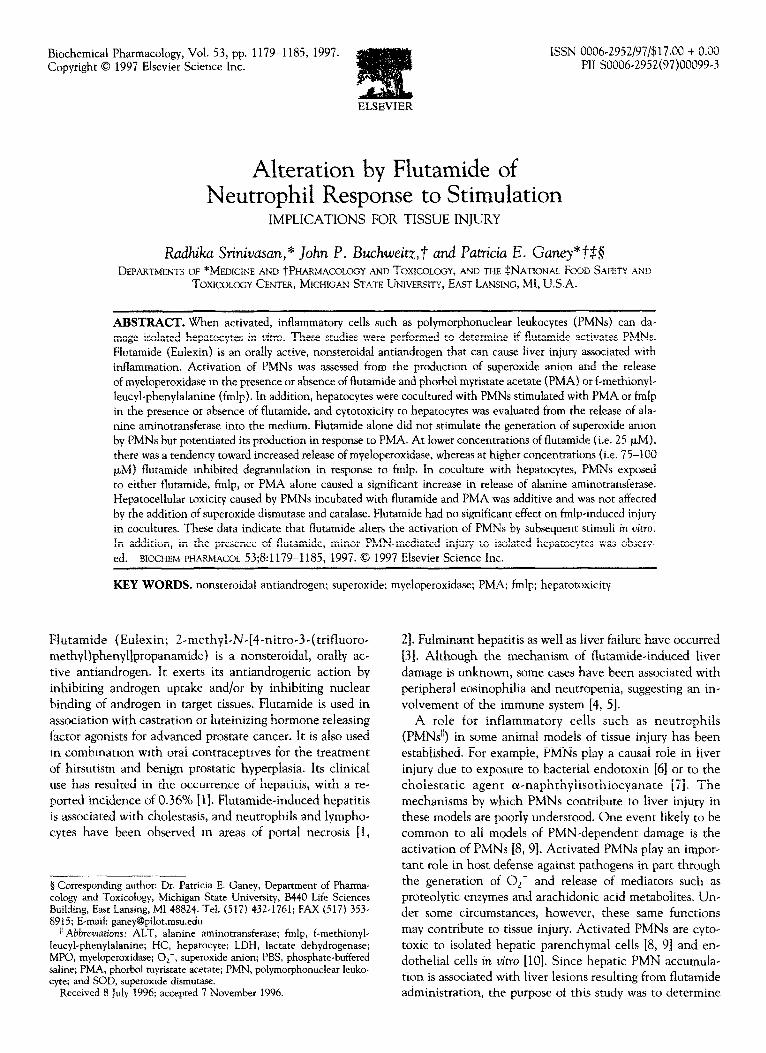
In the absence of stimulation, PMNs did not produce O,-
(Fig. 1). Flutamide alone at concentrations up to 100 p,M did not cause significant generation of O,-. Two concen-
trations of PMA were used: 2 ng/mL of PMA alone did not cause O,- production, whereas 20 ng/mL stimulated signifi- cant generation of O,-. In PMNs exposed to 2 ng/mL PMA, the addition of flutamide at concentrations a50 PM resulted in significant superoxide production. This en- hanced response occurred at lower concentrations of flu- tamide (25 FM) in the presence of 20 ng/mL PMA.
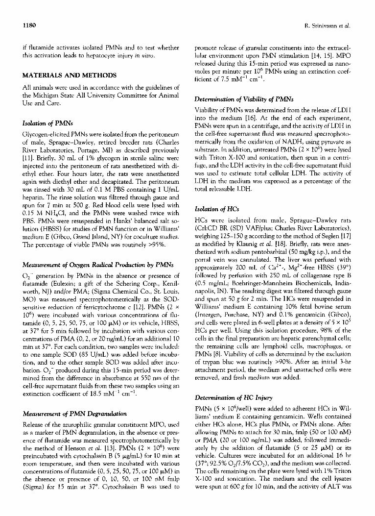
In the absence of stimulation, PMNs did not undergo degranulation, and little MPO was released (Fig. 2). Expo-
sure to flutamide in concentrations up to 100 (LM did not cause statistically significant release of MPO, although there was a trend toward increased activity in the medium
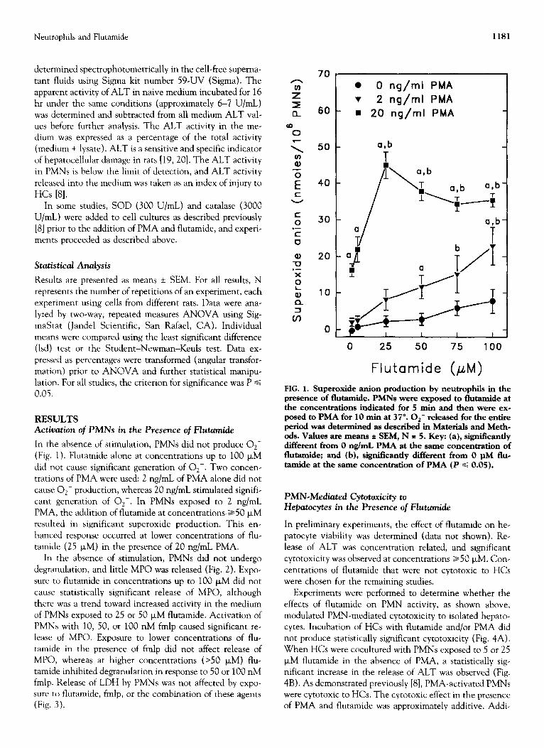
of PMNs exposed to 25 or 50 p,M flutamide. Activation of PMNs with 10, 50, or 100 nM fmlp caused significant re- lease of MPO. Exposure to lower concentrations of flu- tamide in the presence of fmlp did not affect release of MPO, whereas at higher concentrations (>50 PM) flu- tamide inhibited degranulation in response to 50 or 100 nM fmlp. Release of LDH by PMNs was not affected by expo- sure to flutamide, fmlp, or the combination of these agents (Fig. 3).
70
60
50
40
30
20
10
0
1181
l 0 ng/ml PMA v 2 ng/ml PMA n 20 ng/ml PMA
a,b
25 50
Flutamide ;EM,
100
FIG. 1. Superoxide anion production by neutrophils in the presence of flutamide. PMNs were exposed to flutamide at the concentrations indicated for 5 min and then were exe posed to PMA for 10 min at 37”. Or- released for the entire period was determined as described in Materials and Meth- ods. Values are means * SEM, N = 5. Key: (a), significantly different from 0 ng/mL PMA at the same concentration of flutamide; and (b), significantly different from 0 @I flue tamide at the same concentration of PMA (P s 0.05).
PMN-Mediated Cytotoxicity to Hepatocytes in the Presence of Flutamide
In preliminary experiments, the effect of flutamide on he-
patocyte viability was determined (data not shown). Re- lease of ALT was concentration related, and significant cytotoxicity was observed at concentrations a50 (LM. Con-
centrations of flutamide that were not cytotoxic to HCs were chosen for the remaining studies.
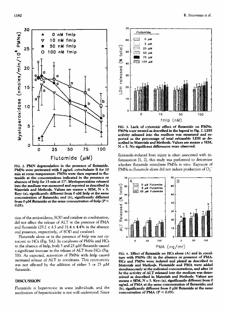
Experiments were performed to determine whether the effects of flutamide on PMN activity, as shown above, modulated PMN-mediated cytotoxicity to isolated hepato- cytes. Incubation of HCs with flutamide and/or PMA did not produce statistically significant cytotoxicity (Fig. 4A). When HCs were cocultured with PMNs exposed to 5 or 25 FM flutamide in the absence of PMA, a statistically sig- nificant increase in the release of ALT was observed (Fig. 4B). As demonstrated previously [8], PMA-activated PMNs were cytotoxic to HCs. The cytotoxic effect in the presence of PMA and flutamide was approximately additive. Addi-
A 0 nM fmlp
v 10 nM fmlp
II 50 nM fmlp
0 100 nM fmlp
I 1 I I I
70
60
0 25 50 75 100
Flutamide (PM)
FIG. 2. PMN degranulation in the presence of flutamide. PMNs were pretreated witb 5 pg/mL cytochalasiu B for 10 min at room temperature. PMNs were then exposed to flue tamide at tbe concentrations indicated in the presence or absence of fmlp for 15 min at 37”. Myeloperoxidase released into the medium was measured and reported as described in M’aterials and Methods. Values are means t SEM, N = 5. Key: (a), sign&~&y different from 0 nM fmlp at the same concentration of flutamide; and (b), signiikantiy different from 0 pM flutamide at the same concentration of fmlp (P c 0.05).
tion of the antioxidanrs, SOD and catalase in combination, did not affect the release of ALT in the presence of PMA and flutamide (29.2 f 4.5 and 3 1.4 + 4.4% in the absence and presence, respectively, of SOD and catalase).
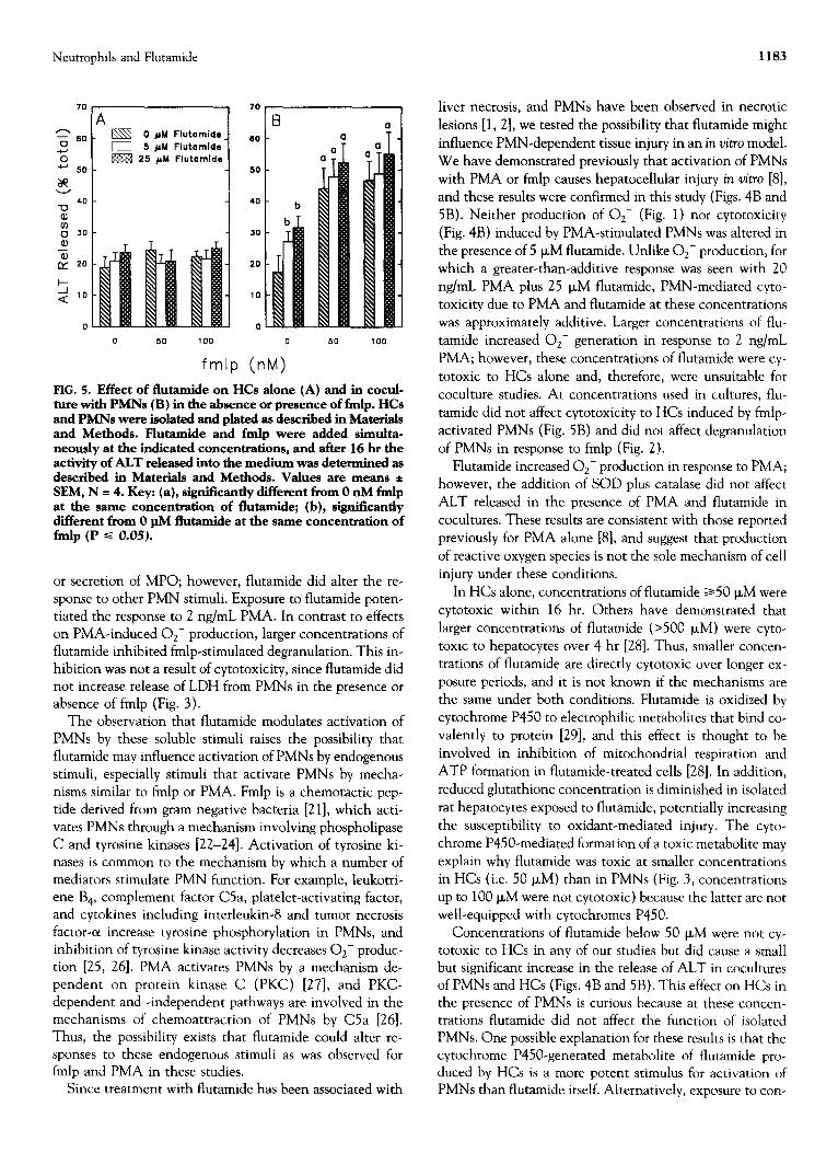
Flutamide alone or in the presence of fmlp was not cy totoxic to HCs (Fig, 5A). In cocultures of PMNs and HCs in the absence of fmlp, both 5 and 25 FM flutamide caused a significant increase in the release of ALT from HCs (Fig. 5B). As expected, activation of PMNs with fmlp caused increased release of ALT in cocultures. This cytotoxicity was not affected by the addition of either 5 or 25 PM fhrtamide.
DISCUSSION
Flutamide is hepatotoxic in some individuals, and the mechanism of hepatotoxicity is not well understood. Since
R. Srinivasan et al.
0 10 50 100
fmlp (nM)
FIG. 3. Lack of cytotoxic effect of flummide on PMNs. PMNs were treated as described in the legend to Fig. 2. LDH activity released into the medium was measured and re- ported as the percentage of total releasable LDH as de- scribed in Materials and Methods. Values are means * SEM, N = 5. No significant differences were observed.
flutamide-induced liver injury is often associated with in- flammation [l, 21, this study was performed to determine whether flutamide stimulates PMNs in vitro. Exposure of PMNs to flutamide alone did not induce production of O,-
70
z 60
-G +J 50
itp
4o u
: 0 30 e,
-z CY 20
!I Q. lo
0 fi)rl Flutomids 6 CM Flutsmide
25 ptrl Fiutamids
20 100 0 20 100
PMA (ng/ml)
FIG. 4. Effect of flutamide on HCs alone (A) and in coctd- ture witb PMNs (B) in the absence or presence of PMA. HCs and PMNs were isolated and plated as described in Materials and Methods. Hutamide and PMA were added simultaneously at the indicated concentrations, and after 16 ht the activity of ALT released into the medium was deter- mined as described in Materials and Methods. Values are means * SEM, N t 5. Key: (a), sign&antly different from 0 ng/mL of PMA at the same concentration of flutamide; and (b), signiScantly different from 0 $4 flutamide at the same concentration of PMA (P s 0.05).
Neutrophils and Flutamide 1183
80 m 0 pM Flutamide 0 5 pM Flutamids
25 pM Flutamlde
fmlp (nM)
FIG. 5. Effect of flutamide on HCs alone (A) and in cocul- ture with PMNs (B) in the absence or presence of fmlp. HCs and PMNs were isolate-d and plated as described in Materials and Methods. Flutamide and fmlp were added simulta- neously at the indicated concentrations, and after 16 hr the activity of AL.T released into the medium was determined as described in Materials and Methods. Values are means * SEM, N = 4. Key: (a), significantly different from 0 nM fmlp at the same concentration of flutamide; (b), significantly different from 0 pM flutamide at the same concentration of fmlp (P S 0.05).
or secretion of MPO; however, flutamide did alter the re- sponse to other PMN stimuli. Exposure to flutamide poten- tiated the response to 2 ng/mL PMA. In contrast to effects on PMA-induced Oz- production, larger concentrations of flutamide inhibited fmlp-stimulated degranulation. This in- hibition was not a result of cytotoxicity, since flutamide did not increase release of LDH from PMNs in the presence or absence of fmlp (Fig. 3).
The observation that flutamide modulates activation of PMNs by these soluble stimuli raises the possibility that flutamide may influence activation of PMNs by endogenous stimuli, especially stimuli that activate PMNs by mecha- nisms similar to fmlp or PMA. Fmlp is a chemotactic pep- tide derived from gram negative bacteria [21], which acti- vates PMNs through a mechanism involving phospholipase C and tyrosine kinases [22-241. Activation of tyrosine ki- nases is common to the mechanism by which a number of mediators stimulate PMN function. For example, leukotri- ene B,, complement factor C5a, platelet-activating factor, and cytokines including interleukin-8 and tumor necrosis factor-o increase tyrosine phosphorylation in PMNs, and inhibition of tyrosine kinase activity decreases Oz- produc- tion [25, 261. PMA activates PMNs by a mechanism de- pendent on protein kinase C (PKC) [27], and PKC- dependent and -independent pathways are involved in the mechanisms of chemoattraction of PMNs by C5a [26]. Thus, the possibility exists that flutamide could alter re- sponses to these endogenous stimuli as was observed for fmlp and PMA in these studies.
Since treatment with flutamide has been associated with
liver necrosis, and PMNs have been observed in necrotic
lesions [l, 21, we tested the possibility that flutamide might influence PMN-dependent tissue injury in an in vitro model. We have demonstrated previously that activation of PMNs
with PMA or fmlp causes hepatocellular injury in vitro [8], and these results were confirmed in this study (Figs. 4B and
5B). Neither production of Oz- (Fig. 1) nor cytotoxicity (Fig. 4B) induced by PMA-stimulated PMNs was altered in the presence of 5 PM flutamide. Unlike Oz- production, for which a greater-than-additive response was seen with 20 ng/mL PMA plus 25 p,M flutamide, PMN-mediated cyto-
toxicity due to PMA and flutamide at these concentrations was approximately additive. Larger concentrations of flu-
tamide increased Oz- generation in response to 2 ng/mL PMA; however, these concentrations of flutamide were cy
totoxic to HCs alone and, therefore, were unsuitable for
coculture studies. At concentrations used in cultures, flu- tamide did not affect cytotoxicity to HCs induced by fmlp-
activated PMNs (Fig. 5B) and did not affect degranulation of PMNs in response to fmlp (Fig. 2).
Flutamide increased O,- production in response to PMA; however, the addition of SOD plus catalase did not affect ALT released in the presence of PMA and flutamide in
cocultures. These results are consistent with those reported previously for PMA alone [8], and suggest that production
of reactive oxygen species is not the sole mechanism of cell injury under these conditions.
In HCs alone, concentrations of flutamide 250 p,M were cytotoxic within 16 hr. Others have demonstrated that
larger concentrations of flutamide (>500 JLM) were cyto- toxic to hepatocytes over 4 hr [28]. Thus, smaller concen- trations of flutamide are directly cytotoxic over longer ex-
posure periods, and it is not known if the mechanisms are the same under both conditions. Flutamide is oxidized by
cytochrome P450 to electrophilic metabolites that bind co- valently to protein [29], and this effect is thought to be involved in inhibition of mitochondrial respiration and
ATP formation in flutamide-treated cells [28]. In addition,
reduced glutathione concentration is diminished in isolated rat hepatocytes exposed to flutamide, potentially increasing
the susceptibility to oxidant-mediated injury. The cyto- chrome P450-mediated formation of a toxic metabolite may explain why flutamide was toxic at smaller concentrations
in HCs (i.e. 50 FM) than in PMNs (Fig. 3, concentrations up to 100 FM were not cytotoxic) because the latter are not well-equipped with cytochromes P450.
Concentrations of flutamide below 50 p,M were not cy- totoxic to HCs in any of our studies but did cause a small but significant increase in the release of ALT in cocultures of PMNs and HCs (Figs. 4B and 5B). This effect on HCs in the presence of PMNs is curious because at these concen- trations flutamide did not affect the function of isolated PMNs. One possible explanation for these results is that the cytochrome P450-generated metabolite of flutamide pro- duced by HCs is a more potent stimulus for activation of PMNs than flutamide itself. Alternatively, exposure to con-
1184 R. Srinivasan et al.
centrations of flutamide or its metabolites which are not overtly toxic may alter hepatocellular membranes to make HCs targets of PMN attack.
Variability in response is observed in humans treated with flutamide: the incidence of liver injury is only about 0.360/o, indicating that most patients do not experience liver toxicity. Furthermore, serum transaminases are el- evated but return to normal in some patients without al- teration of treatment, whereas for other patients reduction
of flutamide dose is required f30, 311. It is interesting to speculate that PMN-mediated injury may contribute to the
variability in response. The concentrations of flutamide used in these studies were larger than would be achieved in
plasma in humans undergoing treatment, but it is possible, for example, that in some individuals flutamide simulta- neously renders HCs more susceptible to oxidant-mediated
injury, initiates infiltration of PMNs into the liver, and increases PMN responsiveness to endogenous activators.
In summary, flutamide altered the response of PMNs to other stimuli but did not by itself activate PMNs. Despite the lack of effect of flutamide alone on PMN O,- produc- tion or degranulation, flutamide was cytotoxic to HCs in
the presence of PMNs at concentrations that were not toxic to HCs alone. PMN-mediated toxicity to HCs in the pres-
ence of flutamide and PMA was additive, whereas flutamide
did not alter cytotoxicity to HCs induced by fmlp-activated PMNs.
This work was supported by NIEHS Grant ES05722. 20.
References 21.
1.
2.
3.
4.
5.
6.
7.
8.
9.
10.
Gomez J, DuPont A, Cusan L, Tremblay M, Suburu R, Lemay M and Labrie B, Incidence of liver toxicity associated wirh the use of flutamide in prostate cancer patients. AmJ Med 92: 465-470, 1992. Rosman AS, Frissora-Rodeo C, Marshall AT, Reiter BP and Paronetto F, Cholestatic hepatitis following flutamide. Dig Dis sci 38: 1756-1759, 1993. Moller S, Iversen P and Franzmann M, Flutamide-induced liver failure. J Hepatol 10: 346-349, 1990. McDonnell ND and Livingston RB, Severe reversible neutro- penia following treatment of prostate cancer with flutamide. J UroE 151: 1353-1354, 1994. Hart W and Stricker BHC, Flutamide and hepatitis. Ann Intern Med 110: 943-944, 1989. Hewett JA, Schultze AE, VanCise S and Roth RA, Neutro- phi1 depletion protects against liver injury from bacterial en- dotoxin. Lab Invest 66: 347-361, 1992. Dahm LJ, Schultze AE and Roth RA, An antibody to neu- trophils attenuates ff-naphthylisothi~yanate-induced liver in- jury. _I Phlox Exp Ther 256: 412-420, 1991. Ganey PE, Bailie MB, VanCise S, Colligan ME, Madhukar BV, Robinson JP and Roth RA, Activated neutrophils from rat injured isolated hepatocytes. Lab Invest 70: 53-60, 1994. Guigui B, Rosenbaum J, Preaux A-M, Martin N, Zafrani ES, Dhumeaux D and Mavier P, Toxicity of phorboi myristate acetate-stimulated pol~o~honuclear neutrophils against rat hepatocytes. Demonstration and mechanism. Lab Invest 59: 83 l-837, 1988. Varani J, Ginsburg I, Schuger L, Gibbs DF, Bromberg J,
11.
12.
13.
14.
15.
16.
17.
18.
19.
22.
23.
24.
25.
26.
27.
Johnson KJ, Ryan US and Ward PA, Endothelial cell killing by neutrophils. Am .I Pathol 135: 435-438, 1989. Hewett JA and Roth RA, Dieldrin activates rat neutrophils in vitro. Toxicol Apex P~oI 96: 269-278, 1988. Babior BM, Kipnes RS and Cumutte JT, Biological defense mechanisms. The production by leukocytes of superoxide, a potential bactericidal agent. J Clin Invest 52: 741-744, 1973. Henson PM, Zanolari B, Schwartzman NA and Hong SR, Intracellular control of human neutrophil secretion. I. C5a- induced stimulus~speci~c desensitization and the effects of cytochalasin B. f ~~~~no~ 121: 851-855, 1978. Tissot M, Mathieu J, Mirossay L, Thuret A and Giroud JP, Polyphosphoinositide metabolism in polymorphonuclear cells from healthy and thermally injured rats: Effect of the im- munomodulator RU 41740. .I Leukoc Biol50: 607614, 1991. Bengtsson T, Dahlgren C, Stendahl 0 and Andersson T, Actin assembly and regulation of neutrophil function: Effects of cytochalasin B and tetracaine on chemotactic peptide- induced Oz- production and degranulation. J Leukoc Bio149: 236-244, 1991. Bergmeyer HV and Bernt E, Lactate dehydrogenase UV assay with pyruvate and NADH. In: Methods of Enzymatic Analysis (Ed. Bergmeyer HV), pp. 524-579. Academic Press, New York, 1974. Seglen PO, Preparation of rat liver cells. III. Enzymatic re- quirements for tissue dispersion. Exp Cell Res 82: 391-398, 1973. Klaunig JE, Goldblatt PJ, Hinton DE, Lipsky MM, Chacko J and Trump BF, Mouse liver cell culture. I. Hepatocyte isola- tion. In Vitro 17: 913-925, 1981. Loeb WF, Clinical biochemistry of laboratory rodents and rabbits. In: Clinical Biochemisrry of Domestic Animals (Ed. Kaneko JJ), pp. 866-875. Academic Press, San Diego, 1989. Kodavanti PRS and Mehendale HM, Biochemical methods of studying hepatotoxicity. In: Hepatotoxicology (Eds. Meeks RG, Harrison SD and Bull RJ), pp. 241-325. CRC Press, Boca Raton, 1991. Marasco WA, Phan SH, Krutzsch H, Showell HJ, Feltner DE, Nairn R, Becker EL and Ward PA, Purification and identifi- cation of formyl-methionyl-leucyl-phenylalanine as the major peptide neutrophil chemotactic factor produced by Escherichia coli. J Biol Chem 259: 5430-5439, 1994. Bagglioni M, Boulay F, Badwey JA and Cumutte JT, Activa- tion of neutrophil leukocytes: Chemoattractant receptors and respiratory burst. FASEB .I 7: 1004-1010, 1993. Cockcroft S, Barrowman MM and Gomperts BD, Breakdown and synthesis of polyphosphoinositides in fMetLeuPhe+ stimulated neutrophils. FEBS Lett 181: 2.59-263, 1985. Torres M, Hall FL and O’NeilI K, Stimulation of human neutrophils with fo~yl~methionyl.leu~yl-phenylalanine in- duces tyrosine phospho~lation and activation of two distinct mitogen-activated protein-kinases. .I Immunol 150: 1563- 1578, 1993. Laudanna C, Rossi F and Berton G, Effect of inhibitors of distinct signalling pathways on neutrophil O,- generation in response to tumor necrosis factor-o, and antibodies against CD18 and CDlla: Evidence for a common and unique pat- tern of sensitivity to wortmannin and protein tyrosine kinase inhibitors. Biochem Biophys Res Commun 190: 935-940,1993. Richard S, Farrell CA, Shaw AS, Showell HJ and Connelly PA, CSa as a model for chemotactic factor-stimulated tyro- sine phosphorylation in the human neutrophil. J ImmunoE 152: 2479-2487, 1994. Heyworth PG and Badwey JA, Protein phospho~lation asso- ciated with the stimulation of neutrophils. Modulation of superoxide production by protein kinase C and calcium. .I Bioenerg Biomembr 22: l-26, 1990.
Neutrophils and Flutamide 1185
28. Fau D, Eugene D, Berson A, Letteron P, Fromenty B, Fisch C and Pessayre D, Toxicity of the antiandrogen flutamide in isolated rat hepatocytes. J Pharmacol Exp Ther 269: 954-962, 1994.
29. Berson A, Wolf C, Chachaty C, Fisch C, Fau D, Eugene D, Loeper J, Gauthier J, Beaune P, Pompon D, Maurel P and Pessayre D, Metabolic activation of the nitroaromatic anti- androgen flutamide by rat and human cytochromes P-450,
including forms belonging to the 3A and 1A subfamilies. .I Phurrnacol Exp Ther 265: 366-372, 1993.
30. Wysowski DK, Freiman JP, Tourtelot JB and Horton ML III, Fatal and nonfatal hepatotoxicity associated with flutamide. Ann Intern Med 118: 860-864, 1993.
31. Dourakis SP, Alexopoulou AA and Hadziyannis SJ, Fulmi- nant hepatitis after flutamide treatment. J Hepatol 20: 350- 353, 1994.