Embed Size (px)
Citation preview
Published Ahead of Print 5 August 2013. 2013, 81(11):3966. DOI: 10.1128/IAI.00770-13. Infect. Immun.
Popov, Nisha Jain Garg and Lynn SoongEric D. Carlsen, Christie Hay, Calvin A. Henard, Vsevolod Neutrophil Microbicidal MechanismsTrigger Neutrophil Activation but Resist Leishmania amazonensis Amastigotes
http://iai.asm.org/content/81/11/3966Updated information and services can be found at:
These include:
SUPPLEMENTAL MATERIAL Supplemental material
REFERENCEShttp://iai.asm.org/content/81/11/3966#ref-list-1at:
This article cites 60 articles, 27 of which can be accessed free
CONTENT ALERTS more»articles cite this article),
Receive: RSS Feeds, eTOCs, free email alerts (when new
http://journals.asm.org/site/misc/reprints.xhtmlInformation about commercial reprint orders: http://journals.asm.org/site/subscriptions/To subscribe to to another ASM Journal go to:
on October 17, 2013 by IN
ST
ITU
TO
FE
DE
RA
L DO
SU
DE
ST
E D
E M
INA
S G
ER
AIS
http://iai.asm.org/
Dow
nloaded from
on October 17, 2013 by IN
ST
ITU
TO
FE
DE
RA
L DO
SU
DE
ST
E D
E M
INA
S G
ER
AIS
http://iai.asm.org/
Dow
nloaded from
on October 17, 2013 by IN
ST
ITU
TO
FE
DE
RA
L DO
SU
DE
ST
E D
E M
INA
S G
ER
AIS
http://iai.asm.org/
Dow
nloaded from
on October 17, 2013 by IN
ST
ITU
TO
FE
DE
RA
L DO
SU
DE
ST
E D
E M
INA
S G
ER
AIS
http://iai.asm.org/
Dow
nloaded from
on October 17, 2013 by IN
ST
ITU
TO
FE
DE
RA
L DO
SU
DE
ST
E D
E M
INA
S G
ER
AIS
http://iai.asm.org/
Dow
nloaded from
on October 17, 2013 by IN
ST
ITU
TO
FE
DE
RA
L DO
SU
DE
ST
E D
E M
INA
S G
ER
AIS
http://iai.asm.org/
Dow
nloaded from
on October 17, 2013 by IN
ST
ITU
TO
FE
DE
RA
L DO
SU
DE
ST
E D
E M
INA
S G
ER
AIS
http://iai.asm.org/
Dow
nloaded from
on October 17, 2013 by IN
ST
ITU
TO
FE
DE
RA
L DO
SU
DE
ST
E D
E M
INA
S G
ER
AIS
http://iai.asm.org/
Dow
nloaded from
on October 17, 2013 by IN
ST
ITU
TO
FE
DE
RA
L DO
SU
DE
ST
E D
E M
INA
S G
ER
AIS
http://iai.asm.org/
Dow
nloaded from
on October 17, 2013 by IN
ST
ITU
TO
FE
DE
RA
L DO
SU
DE
ST
E D
E M
INA
S G
ER
AIS
http://iai.asm.org/
Dow
nloaded from
Leishmania amazonensis Amastigotes Trigger Neutrophil Activationbut Resist Neutrophil Microbicidal Mechanisms
Eric D. Carlsen,a,b Christie Hay,b Calvin A. Henard,b Vsevolod Popov,c Nisha Jain Garg,b,c Lynn Soongb,c
M.D.-Ph.D. Combined Degree Program,a Department of Microbiology and Immunology,b and Department of Pathology,c University of Texas Medical Branch, Galveston,Texas, USA
Neutrophils are the first cells to infiltrate to the site of Leishmania promastigote infection, and these cells help to reduce parasiteburden shortly after infection is initiated. Several clinical reports indicate that neutrophil recruitment is sustained over thecourse of leishmaniasis, and amastigote-laden neutrophils have been isolated from chronically infected patients and experimen-tally infected animals. The goal of this study was to compare how thioglycolate-elicited murine neutrophils respond to L. ama-zonensis metacyclic promastigotes and amastigotes derived from axenic cultures or from the lesions of infected mice. Neutro-phils efficiently internalized both amastigote and promastigote forms of the parasite, and phagocytosis was enhanced inlipopolysaccharide (LPS)-activated neutrophils or when parasites were opsonized in serum from infected mice. Parasite uptakeresulted in neutrophil activation, oxidative burst, and accelerated neutrophil death. While promastigotes triggered the release oftumor necrosis factor alpha (TNF-�), uptake of amastigotes preferentially resulted in the secretion of interleukin-10 (IL-10)from neutrophils. Finally, the majority of promastigotes were killed by neutrophils, while axenic culture- and lesion-derivedamastigotes were highly resistant to neutrophil microbicidal mechanisms. This study indicates that neutrophils exhibit distinctresponses to promastigote and amastigote infection. Our findings have important implications for determining the impact ofsustained neutrophil recruitment and amastigote-neutrophil interactions during the late phase of cutaneous leishmaniasis.
Leishmania parasites are obligate intracellular protozoa thatcause leishmaniasis, a neglected tropical disease responsible for
extensive morbidity and mortality in the developing world. Infec-tion is initiated when metacyclic promastigotes are deposited intothe skin by the bite of a female sandfly, and parasitism of hostneutrophils, dendritic cells (DCs), and macrophages rapidly en-sues. In macrophages, promastigotes convert into amastigotes, theparasite stage that replicates in mammalian hosts. Leishmaniaamastigotes are able to modify macrophage functions and resistmacrophage microbicidal activity, resulting in the establishmentof an environment that is permissive for parasite growth (1–3).Parasite-mediated manipulation of multiple signaling pathwaysin other cell types, such as DCs, is also well established, and dis-ruption of innate immune cell function ultimately hinders theformation of a potent, effective T helper cell response. Conse-quently, amastigote replication continues unabated in the contextof low-grade inflammation and tissue damage (4, 5).
Neutrophils rapidly recruit to the site of infection after meta-cyclic promastigotes are delivered into the skin, either via theirnatural sandfly vector or by needle injection (6, 7). After contact-ing each other, Leishmania promastigotes and neutrophils mayeach undergo one of several fates. For example, L. major promas-tigotes can survive inside neutrophils and ultimately use these cellsas Trojan horses to facilitate silent infection of macrophages (8).In contrast, neutrophils respond to L. amazonensis promastigotesby undergoing several forms of cell death; many cells encounter-ing parasites rapidly die by NETosis, a specialized form of deaththat results in parasite entrapment and degradation (9), while theremaining neutrophils die largely by apoptosis (10).
In numerous mouse models, antibody-mediated neutrophildepletion has been extensively used to determine how these cellscontribute to the pathogenesis of various infectious diseases (11–13). However, there is currently a lack of consensus regarding thefunction of neutrophils during Leishmania promastigote infec-
tion, as these cells have been implicated in both promoting andinhibiting disease progression in different studies (14, 15). Despitereporting contradictory roles for neutrophils in controlling infec-tion, depletion studies nevertheless emphasize the importance ofthese cells in the early disease process of cutaneous leishmaniasis.
According to several clinical reports, neutrophil recruitment tothe site of infection is not limited to the promastigote-mediatedphase of disease but continues throughout the course of chronicleishmaniasis as well. In L. tropica-infected patients, neutrophilswere recovered from lesions ranging from 1 to 36 months in du-ration (16). Neutrophils were also observed in the ulcerated le-sions of patients chronically infected with L. major, and in somepatients, these cells were the predominant immune cell type at thesite of infection (17). Interestingly, BALB/c mice infected with L.major display a progressive increase in the number of intralesionalneutrophils throughout the first 6 weeks of infection (18), suggest-ing that persistent neutrophil recruitment may be a characteristicfeature of chronic cutaneous leishmaniasis.
Amastigote-laden neutrophils have been isolated from numer-ous infected hosts, including experimentally infected macaquesand naturally infected humans, dogs, and foxes (17, 19–21). How-ever, the immunological ramifications of amastigote-neutrophil
Received 22 June 2013 Returned for modification 13 July 2013Accepted 29 July 2013
Published ahead of print 5 August 2013
Editor: J. L. Flynn
Address correspondence to Lynn Soong, [email protected].
Supplemental material for this article may be found at http://dx.doi.org/10.1128/IAI.00770-13.
Copyright © 2013, American Society for Microbiology. All Rights Reserved.
doi:10.1128/IAI.00770-13
3966 iai.asm.org Infection and Immunity p. 3966–3974 November 2013 Volume 81 Number 11
interactions remain largely uncharacterized. We have recentlydemonstrated that L. amazonensis amastigotes are highly resistantto the antimicrobial effects of purified human histone proteins(22), which are known to be released together with other micro-bicidal agents when neutrophils undergo NETosis (23). Cur-rently, it is unclear whether neutrophils recognize amastigotes andinfluence amastigote clearance or persistence (24).
In this study, we aimed to examine the interaction between L.amazonensis amastigotes and peritoneal neutrophils obtainedfrom C57BL/6 mice. We demonstrate that neutrophils efficientlyinternalized both the amastigote and promastigote forms of theparasite, particularly when parasites were opsonized with Leish-mania-specific antibodies. Parasite uptake resulted in neutrophilactivation and oxidative burst, but neutrophils differed in theirresponses to amastigotes and promastigotes in several ways, in-cluding cytokine secretion and pathogen clearance. Specifically,neutrophils responded to promastigotes by releasing tumor ne-crosis factor alpha (TNF-�) and by killing the majority of para-sites. In contrast, neutrophils failed to efficiently kill amasti-gotes and preferentially released interleukin-10 (IL-10) inresponse to this stage of parasite. Therefore, the role of neutro-phils during leishmaniasis may differ depending on the stage ofparasite encountered. These findings have important implica-tions for understanding the pathogenic mechanisms of im-mune system dysfunction and chronic parasite persistenceduring experimental cutaneous leishmaniasis.
MATERIALS AND METHODSMice. Female C57BL/6 and BALB/c mice were purchased from TaconicFarms (Germantown, NY). C57BL/6 mice were the source of the majorityof neutrophils in this study, while BALB/c mice were predominately usedfor the maintenance of parasite infectivity and for isolating lesion-derivedamastigotes. Neutrophils from BALB/c mice were used as a control for theclearance of lesion-derived amastigotes shown in Fig. S4 in the supple-mental material. B6(Cg)-Ncf1m1J/J mice deficient in the gp47 subunit ofNADPH oxidase were obtained from The Jackson Laboratory (Bar Har-bor, ME) and bred on campus. Mice were maintained under specific-pathogen-free conditions and used at 6 to 12 weeks of age, according toprotocols approved by the Animal Care and Use Committee of the Uni-versity of Texas Medical Branch (Galveston, TX).
Parasite cultivation. The infectivity of L. amazonensis (strains RAT/BA/74/LV78 and MHOM/BR/77/LTB0016) was maintained by regularpassage through BALB/c mice. Strain RAT/BA/74/LV78 was used for allexperiments using promastigotes and amastigotes. Strain MHOM/BR/77/LTB0016 was used in the infection of mice to generate immune serum.Promastigotes were cultured at 26°C in M199 containing 40 mM HEPES,10% heat-inactivated fetal bovine serum (FBS), 0.1% hemin in 50:50 H2Oand triethanolamine (Frontier Scientific, Logan, UT), 0.1 mM adenine(pH 7.5), 5 mM L-glutamine, and 50 �g/ml gentamicin. Metacyclic pro-mastigotes were purified as described previously (25) by using the mono-clonal antibody 3A1, which was generously provided by Norma Andrews(University of Maryland). All experiments using promastigote groups uti-lized metacyclic promastigotes purified in this way. Axenic amastigoteswere cultured at 32°C in Grace’s insect cell culture medium (Invitrogen,Carlsbad, CA), pH 5.2, supplemented with 20% FBS and 25 �g/ml gen-tamicin. Lesion-derived amastigotes were collected from the footpads ofinfected BALB/c mice through mechanical tissue disruption, followed by3 washes and incubation in amastigote medium. Lesion-derived amasti-gotes were used within 48 h of isolation from infected footpads. Prior touse, lesion-derived amastigotes were washed an additional 3 times to re-move any residual tissue components. Fresh parasite lysates were pre-pared through 2 freeze-thaw cycles followed by sonication for 15 min.
Production of luciferase-expressing parasites. Circular pSP72-YNEO-�IR-LUC1.2 was generously provided by Barbara Papadopoulou(Laval University, Quebec, Canada). Logarithmic-phase RAT/BA/74/LV78 promastigotes were transfected with 35 �g plasmid, as reportedpreviously (26), resulting in episomal expression of firefly luciferase. Fol-lowing a 24-h rest period, selection for luciferase-expressing promasti-gotes was performed via titration of G418 (Invitrogen). Luciferase-ex-pressing amastigotes were derived from logarithmic-phase promastigotecultures. To maintain selective pressure, luciferase-expressing promasti-gotes and amastigotes were grown in normal parasite medium containingG418 (50 �g/ml).
Generation of immune serum and parasite opsonization. C57BL/6mice were infected with L. amazonensis MHOM/BR/77/LTB0016 pro-mastigotes in the rear footpads for 12 weeks. Infected mice and age- andsex-matched naive mice were subsequently sacrificed, and serum was col-lected, heat inactivated, and stored at �20°C. To ensure suitable anti-Leishmania antibody concentrations, antibody titers were determined viadirect enzyme-linked immunosorbent assay (ELISA). In experiments uti-lizing opsonized amastigotes, parasites were incubated in naive or im-mune serum (10%) for 20 min at room temperature prior to infection.
Neutrophil collection. Peritoneal exudate cells were obtained frommice 5 h after injection with 3% thioglycolate (Sigma-Aldrich, St. Louis,MO). Thioglycolate was removed, and neutrophils were purified via den-sity gradient centrifugation with Percoll (Sigma-Aldrich). Neutrophil pu-rity (�95%) was validated by fluorescence-activated cell sorting (FACS)and examination of morphology after staining; cell viability was routinely�95%, as monitored by trypan blue exclusion. Prior to treatment or co-culture with parasites, neutrophils were plated in tissue culture-treatedpolystyrene. Because L. amazonensis poorly tolerates high temperatures,all neutrophil-parasite cocultures were maintained at 32°C.
Neutrophil phagocytosis of parasites. Parasites were labeled with car-boxyfluorescein succinimidyl ester (CFSE) (Sigma-Aldrich), as describedpreviously (27). Neutrophils were cocultured with CFSE-labeled amasti-gotes or promastigotes at a multiplicity of infection (MOI) of 5 for 4 h at32°C with 5% CO2. Cells were collected, stained with allophycocyanin(APC)-conjugated anti-Ly6G (BD Biosciences, San Jose, CA), and ana-lyzed by FACS. Neutrophils were identified based on forward/side scattercharacteristics and Ly6G positivity. Parasite-carrying neutrophils wereidentified based on CFSE positivity. In some experiments, neutrophilswere treated with lipopolysaccharide (LPS), cytochalasin D (Sigma-Al-drich), or granulocyte-macrophage colony-stimulating factor (GM-CSF)(PeproTech, Oak Park, CA), and parasites were opsonized in heat-inacti-vated naive or immune serum prior to coculture. Data were collectedusing an Accuri C6 flow cytometer (Accuri Cytometers Inc., Ann Arbor,MI). Flow cytometry data were subsequently analyzed using CFlow ver-sion 1.0.227.4 (Accuri Cytometers Inc.) or FlowJo version 7.6.1 (Tree Star,Ashland, OR).
Electron microscopy (EM). Following 4 h of coculture with amasti-gotes, neutrophils were fixed in Ito’s fixative (2.5% formaldehyde pre-pared from paraformaldehyde, 0.1% glutaraldehyde, 0.03% CaCl2, and0.03% trinitrophenol in 0.05 M cacodylate buffer, pH 7.3) at room tem-perature for 15 min and then overnight at 4°C. After washing in 0.1 Mcacodylate buffer, samples were postfixed in 1% osmium tetroxide in thesame buffer for 1 h and en bloc stained with 1% aqueous uranyl acetate for20 min at 60°C. After dehydration in a graded series of ethanol solutions,samples were embedded in Poly/Bed 812 (Polysciences, Warrington, PA).Ultrathin sections were cut on a Leica EM UC7 ultramicrotome (LeicaMicrosystems, Buffalo Grove, IL), stained with lead citrate, and examinedusing a Philips 201 transmission electron microscope (Philips ElectronOptics, Eindhoven, The Netherlands) at 60 kV.
Measurement of neutrophil activation and oxidative burst. Neutro-phil-parasite cocultures were incubated for 4 h at 32°C and 5% CO2. After4 h, some neutrophils were blocked with anti-CD16/CD32 and stainedwith peridinin chlorophyll protein (PerCP)-Cy5.5-conjugated anti-CD11b (eBioscience, San Diego, CA) and APC-conjugated anti-Ly6G,
Leishmania Amastigotes Resist Killing by Neutrophils
November 2013 Volume 81 Number 11 iai.asm.org 3967
and samples were analyzed by FACS. Separate cell groups were stainedwith APC-conjugated anti-Ly6G and dihydrorhodamine 123 (1 �M; Sig-ma-Aldrich), which converts to fluorescent rhodamine 123 (Rho 123)when oxidized. In some experiments, 1 �M N-formyl-methionyl-leucyl-phenylalanine (fMLP) (Sigma-Aldrich) was added for the last 5 min ofincubation prior to measurement of oxidative burst. The oxidation reac-tion was stopped on ice, and neutrophil reactive oxygen species (ROS)production was analyzed by gating on Ly6G� cells and measuring themean fluorescence intensity (MFI) of Rho 123 by FACS. To determinewhether the parasite-mediated oxidative burst was restricted to infectedcells, amastigotes were labeled with PKH26 (Sigma-Aldrich) according tothe manufacturer’s instructions. The MFIs of Rho123 in PKH26� (in-fected) and PKH26� (bystander) neutrophils were then compared.
Neutrophil cytokine detection. To minimize protease activity, neu-trophils were treated with 50 �g/ml aprotinin (Sigma-Aldrich) prior totreatment with parasites at an MOI of 5. Supernatants were collected after24 h, and the cytokine concentration was measured via ELISA (eBiosci-ence). After treatment with tetramethylbenzidine substrate and stop so-lution, optical density (OD) values at 450 nm were measured with a Mul-tiskan Ascent ELISA reader (Labsystems, Helsinki, Finland).
Measurement of neutrophil apoptosis. Neutrophils were coculturedwith amastigotes in the presence or absence of GM-CSF (20 ng/ml). After18 h, neutrophils were collected and stained with APC-conjugated anti-Ly6G and the annexin V:FITC apoptosis detection kit I (BD Biosciences).Early apoptosis in Ly6G� neutrophils was quantified by FACS based onpositive staining for annexin V and negative staining for propidiumiodide (PI). To determine whether changes in apoptosis were restricted toinfected cells, CFSE-labeled amastigotes were cocultured with neutro-phils, followed by phycoerythrin (PE)-conjugated annexin V staining.
Parasite killing by neutrophils. Luciferase-expressing amastigotes orpromastigotes were cocultured with neutrophils at an MOI of 0.1. In someexperiments, to better simulate lesion-derived amastigotes, axenic para-sites were precoated with heat-inactivated serum from infected mice priorto coculture with neutrophils. At 0, 6, and 18 h postinfection, cocultureswere lysed and frozen at �80°C prior to analysis. Parasite burdens wereestimated by mixing lysates with luciferase assay substrate (Promega Cor-poration, Madison, WI) and measuring photon emission on a Veritasmicroplate luminometer (Turner BioSystems Inc., Sunnyvale, CA). Par-asite survival was estimated by comparing the baseline photon emission at0 h to the signal intensities at subsequent time points.
Statistical analysis. Differences between two groups were determinedby using the two-tailed Student’s t test. Graphs were prepared by usingGraphPad Prism 4.0 (GraphPad Software, San Diego, CA). The difference
between two groups was considered significant when the P value was�0.05.
RESULTSNeutrophils internalize L. amazonensis promastigotes andamastigotes. To investigate the interaction between neutrophilsand Leishmania amastigotes, we opted to use thioglycolate-elic-ited neutrophils from C57BL/6 mice and L. amazonensis amasti-gotes. We selected this particular system to dissect amastigote-neutrophil interactions for several reasons. First, C57BL/6 miceare traditionally viewed as a resistant strain in regard to Leishma-nia infection (28), and C57BL/6 neutrophils have been shown torespond to L. major by secreting biologically active IL-12p70 (29).Thioglycolate-elicited neutrophils can be isolated with high yieldand purity, which is advantageous for conducting a detailed anal-ysis of neutrophil function (30). Finally, we opted to examineneutrophil responses to L. amazonensis because these parasites areeasily propagated as amastigotes in vitro, induce a nonhealing dis-ease phenotype in both C57BL/6 and BALB/c mice, and have beenshown to have potent immunosuppressive effects on numerouscell types (31, 32).
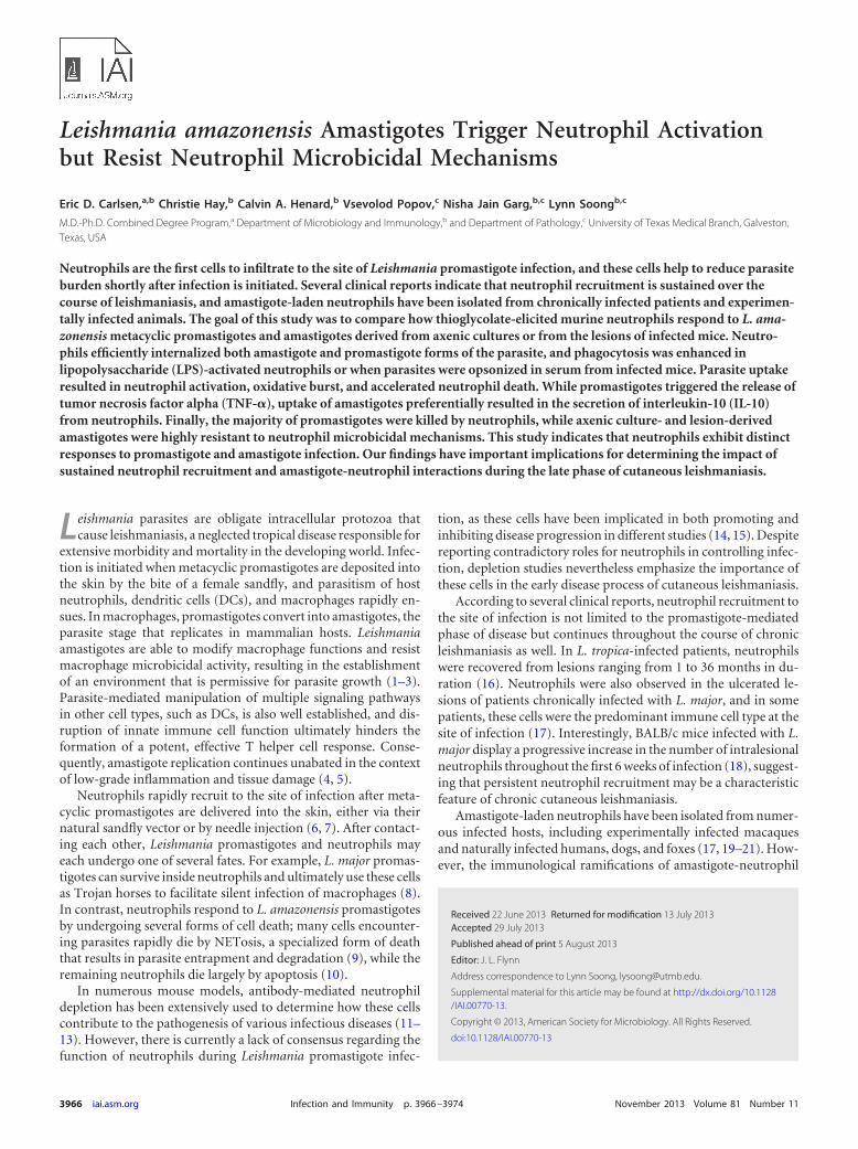
Neutrophil uptake of metacyclic promastigotes is a critical fea-ture of the initial phase of Leishmania infection. As infection pro-gresses, neutrophils may also encounter amastigotes liberatedfrom ruptured macrophages. However, despite the popularity ofmurine models of cutaneous and visceral leishmaniasis, reports ofamastigote uptake by mouse neutrophils are largely absent fromthe literature. We compared neutrophil phagocytosis of CFSE-labeled axenic amastigotes and metacyclic promastigotes. After 4h of coculture, we observed that approximately 8.6% of neutro-phils engulfed amastigotes, while 7.9% of neutrophils internalizedpromastigotes. Phagocytosis of parasites was inhibited in neutrophilsthat were pretreated with cytochalasin D (20 �M), confirming thatparasite uptake was mediated via an actin polymerization-dependentmechanism. Of note, parasite opsonization in heat-inactivated se-rum collected from L. amazonensis-infected mice markedly en-hanced neutrophil phagocytosis of both promastigotes and amas-tigotes (Fig. 1A). Opsonization with naive mouse serum alsoenhanced parasite uptake, but to a lesser extent that that with
FIG 1 Neutrophil phagocytosis of L. amazonensis parasites. (A) Thioglycolate-elicited peritoneal neutrophils were cocultured with CFSE-labeled axenicamastigotes (AxAm) or metacyclic promastigotes (Pm) for 4 h. In some groups, neutrophils were pretreated with cytochalasin D (Cyto D) (20 �M) or parasiteswere opsonized in serum from infected mice. Neutrophils were identified by forward/side scatter characteristics and by Ly6G positivity. CFSE positivity in theboxes shown represents Ly6G� neutrophils carrying parasites. Values are mean percentages of CFSE� cells � 1 standard deviation (SD). (B) Percentages ofCFSE� neutrophils (PMN) carrying amastigotes after 4 h in medium alone (Med) or in the presence of LPS (100 ng/ml) or GM-CSF (200 ng/ml). Data are pooledfrom 3 independent repeats and are shown as means � standard errors. *, statistically significant differences (P � 0.05) between the groups.
Carlsen et al.
3968 iai.asm.org Infection and Immunity
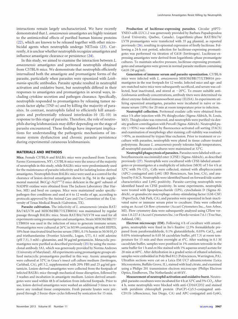
serum from infected animals (see Fig. S1 in the supplemental ma-terial). Phagocytosis of amastigotes was also significantly en-hanced in neutrophils treated with LPS (100 ng/ml) or GM-CSF(200 ng/ml) (Fig. 1B). Electron microscopy was used to confirmamastigote internalization. Ultrastructural analysis indicated thatinternalized amastigotes were housed within tight, membrane-bound vacuoles (Fig. 2). The presence of intact flagellar remnantsin some internalized amastigotes (Fig. 2, arrow) suggested thatparasites were not damaged by neutrophils during phagocytosis.In some instances, neutrophils carrying 4 or more parasites werealso observed (image not shown).
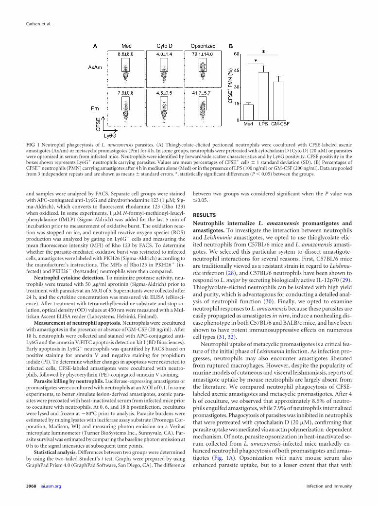
Amastigote infection triggers neutrophil activation and oxi-dative burst. Activated neutrophils characteristically upregulateCD11b on their surface, which reflects their ability to execute anumber of important functions, including phagocytosis, degran-ulation, apoptosis, and oxidative burst (33–36). As shown in Fig.3A, neutrophil coculture with axenic amastigotes or promasti-gotes resulted in an appreciable upregulation of surface CD11b oninfected neutrophils. However, the extent of CD11b upregulationdid not differ for neutrophils cocultured with promastigotes oramastigotes. In contrast, neutrophil coculture with lesion-derivedamastigotes resulted in a significant increase in CD11b upregula-tion over that of axenic amastigotes (Fig. 3A).
Because reactive oxygen species (ROS) are a critical compo-nent of the microbicidal armament of neutrophils and becausepromastigote-induced ROS production in neutrophils has beenreported (37), we investigated whether amastigotes also trigger aneutrophil oxidative burst. To do this, we labeled resting and par-asite-laden neutrophils with dihydrorhodamine 123, a cell-per-meative dye that converts into fluorescent rhodamine 123 (Rho123) when oxidized (38). As shown in Fig. 3B, neutrophil cocul-
ture with amastigotes or promastigotes for 4 h resulted in a signif-icant increase in rhodamine 123 fluorescence compared to that incells resting in medium. While promastigotes tended to elicitmore ROS, the difference between the amastigote- and promas-tigote-mediated oxidative bursts was not significant, regardless ofwhether the entire neutrophil population (Fig. 3B) or theRho123� population (Fig. 3C) was examined. In contrast, lesion-derived amastigotes did elicit significantly more neutrophil oxida-tive burst than their axenically cultured counterparts (Fig. 3B).Dihydrorhodamine 123-labeled parasites had no detectable dyeoxidation, confirming that the ROS measured in our assay wasneutrophil-derived (data not shown).
We examined whether an oxidative burst was occurring in in-fected or bystander neutrophils by coculturing cells with PKH26-labeled amastigotes. After 4 h of infection, neutrophils could beclearly gated based on PKH26 positivity, and we observed thatamastigote-laden (PKH26hi) cells were the major producers ofROS (Fig. 3D). The neutrophil oxidative burst in response to par-asites was dependent upon the presence of intact parasites, as par-asite lysates failed to increase ROS production above control levels(see Fig. S2A in the supplemental material). These findings collec-tively indicate that parasite internalization is required for a neu-trophil oxidative burst in response to L. amazonensis.
FIG 2 Ultrastructural analysis of amastigote uptake by neutrophils. Neutro-phils were cocultured with serum-coated amastigotes for 4 h, fixed, and pre-pared for analysis via electron microscopy. A characteristic neutrophil isdepicted, exhibiting a multilobular nucleus (N), electron-dense granules (ar-rowheads), and 2 intracellular amastigotes (asterisks). A flagellar remnant isalso clearly visible in the amastigote on the left (arrow). Bar, 2 �m.
FIG 3 Neutrophil activation and oxidative burst after contact with parasites.(A) Mean fluorescence intensity (MFI) of CD11b on neutrophils resting inmedium (Med) or cocultured with metacyclic promastigotes (Pm), axenicamastigotes (AxAm), or lesion-derived amastigotes (Am). (B) MFI of rhoda-mine 123 (Rho 123) in dihydrorhodamine 123-labeled Ly6G� cells after 4 h ofcoculture with parasites. (C) Oxidative burst in Ly6G� Rho123� neutrophils,indicating that the extent of burst on a per-cell basis does not differ for neu-trophils cocultured with promastigotes or amastigotes. (D) ROS production inneutrophils infected with PKH26-labeled axenic amastigotes for 4 h, showingthat the majority of ROS was generated in PKH26hi (amastigote-laden) cells.All data are pooled from at least 2 independent experiments and shown asmeans � standard errors. * (P � 0.05), ** (P � 0.01), and *** (P � 0.001)indicate statistically significant differences between groups. NS, not signifi-cant.
Leishmania Amastigotes Resist Killing by Neutrophils
November 2013 Volume 81 Number 11 iai.asm.org 3969
To determine whether parasites were able to alter the oxidativeburst elicited by an external signal such as N-formyl-methionyl-leucyl-phenylalanine (fMLP), we compared neutrophil produc-tion of ROS in response to amastigotes, fMLP, or both stimulicombined. Coculture with parasites plus fMLP treatment resultedin substantially greater ROS production than amastigote or fMLPtreatment alone (see Fig. S2B in the supplemental material). Toensure that the observed increase in ROS was due to phagocyteNADPH oxidase rather than mitochondrial damage or other ROSsources, we compared ROS production in neutrophils from wild-type (WT) mice and mice deficient in the gp47 subunit of theNADPH oxidase complex. gp47�/� neutrophils produced appre-ciably less ROS in response to fMLP or amastigotes than WT neu-trophils, confirming that the majority of the ROS detected in ourassay was derived from the NADPH oxidase (see Fig. S2C in thesupplemental material).
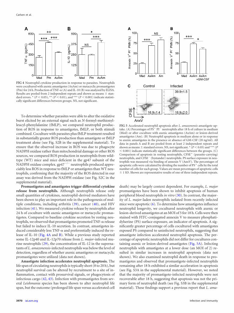
Promastigotes and amastigotes trigger differential cytokinerelease from neutrophils. Although neutrophils release onlysmall quantities of cytokines, neutrophil-derived mediators havebeen shown to play an important role in the pathogenesis of mul-tiple conditions, including arthritis (39), cancer (40), and HIVinfection (41). We measured cytokine release by neutrophils after24 h of coculture with axenic amastigotes or metacyclic promas-tigotes. Compared to baseline cytokine secretion by resting neu-trophils, we observed that promastigotes promoted TNF-� releasebut failed to induce IL-10 secretion. In contrast, amastigotes in-duced considerably less TNF-� and preferentially induced the re-lease of IL-10 (Fig. 4A and B). While a previous study reportedsome IL-12p40 and IL-12p70 release from L. major-infected mu-rine neutrophils (29), the concentration of IL-12 in the superna-tants of L. amazonensis-infected neutrophils was below the level ofdetection, regardless of whether axenic amastigotes or metacyclicpromastigotes were utilized (data not shown).
Amastigote infection accelerates neutrophil apoptosis. Thelife span of circulating neutrophils is typically short (8 to 20 h), butneutrophil survival can be altered by recruitment to a site of in-flammation, contact with prosurvival signals, or phagocytosis ofinfectious cargo (42, 43). Infection with promastigotes from sev-eral Leishmania species has been shown to alter neutrophil lifespan, but the outcome (prolonged life span versus accelerated cell
death) may be largely context dependent. For example, L. majorpromastigotes have been shown to inhibit apoptosis of humanperipheral blood neutrophils in vitro (30). In contrast, the major-ity of L. major-laden neutrophils isolated from recently infectedmice were apoptotic (6). To determine how amastigotes influenceneutrophil longevity, we cocultured neutrophils with axenic orlesion-derived amastigotes at an MOI of 5 for 18 h. Cells were thenstained with FITC-conjugated annexin V to measure phosphati-dylserine (PS) surface exposure, an indicator of apoptosis. A sig-nificantly greater percentage of cells cocultured with amastigotesexposed PS compared to uninfected neutrophils, suggesting thatamastigote infection accelerated neutrophil apoptosis. The per-centage of apoptotic neutrophils did not differ for cocultures con-taining axenic or lesion-derived amastigotes (Fig. 5A). Infectingneutrophils with amastigotes at a lower dose (an MOI of 2) re-sulted in similar increases in neutrophil apoptosis (data notshown). We also examined neutrophil death in response to pro-mastigotes and observed that promastigote-infected neutrophilsremaining after 18 h exhibited a similar acceleration in apoptosis(see Fig. S3A in the supplemental material). However, we notedthat the majority of promastigote-infected neutrophils were notrecoverable after 18 h, suggesting that apoptosis was not the pri-mary form of neutrophil death (see Fig. S3B in the supplementalmaterial). These findings support a previous report that L. ama-
FIG 4 Neutrophil cytokine production in response to parasites. Neutrophilswere cocultured with axenic amastigotes (AxAm) or metacyclic promastigotes(Pm) for 24 h. Production of TNF-� (A) and IL-10 (B) was analyzed by ELISA.Results are pooled from 2 independent repeats and shown as means � stan-dard errors. * (P � 0.05), ** (P � 0.01), and *** (P � 0.001) indicate statisti-cally significant differences between groups. NS, not significant.
FIG 5 Accelerated neutrophil apoptosis after L. amazonensis amastigote up-take. (A) Percentages of PS� PI� neutrophils after 18 h of culture in medium(Med) or after coculture with axenic amastigotes (AxAm) or lesion-derivedamastigotes (Am). (B) Neutrophil apoptosis in medium alone or in responseto axenic amastigotes in the presence or absence of GM-CSF (20 ng/ml). Alldata in panels A and B are pooled from at least 2 independent repeats andshown as means � standard errors. NS, not significant. * (P � 0.05) and *** (P� 0.001) indicate statistically significant differences between the groups. (C)Comparison of apoptosis in resting neutrophils, CFSE� (parasite-carrying)neutrophils, and CFSE� (bystander) neutrophils. PS surface exposure in neu-trophils was measured via binding of annexin V (AnxV). The percentages ofapoptotic cells were calculated by dividing the number of PS� cells by the totalnumber of cells for each group. Values are mean percentages of apoptotic cells� 1 SD. Shown are representative results of one of three independent repeats.
Carlsen et al.
3970 iai.asm.org Infection and Immunity
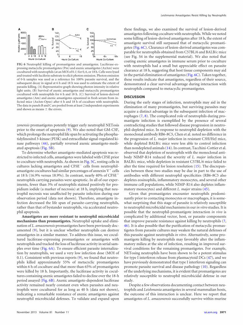
zonensis promastigotes potently trigger early neutrophil NETosisprior to the onset of apoptosis (9). We also noted that GM-CSF,which prolongs the neutrophil life span by activating the phospha-tidylinositol 3-kinase (PI3K) and extracellular signal-regulated ki-nase pathways (44), partially reversed axenic amastigote-medi-ated apoptosis (Fig. 5B).
To determine whether amastigote-mediated apoptosis was re-stricted to infected cells, amastigotes were labeled with CFSE priorto coculture with neutrophils. As shown in Fig. 5C, resting cells incultures lacking amastigotes and CFSE� cells from neutrophil-amastigote cocultures had similar percentages of annexin V� cellsat 18 h (16.9% versus 18.9%). In contrast, nearly 40% of CFSE�
neutrophils carrying parasites were apoptotic. In all of our exper-iments, fewer than 3% of neutrophils stained positively for pro-pidium iodide (a marker of necrosis) at 18 h, implying that neu-trophil necrosis was unaffected by parasite infection during ourobservation period (data not shown). Therefore, amastigote in-fection decreased the life span of parasite-carrying neutrophils,rather than that of bystander neutrophils, via accelerated neutro-phil apoptosis.
Amastigotes are more resistant to neutrophil microbicidalmechanisms than promastigotes. Neutrophil uptake and elimi-nation of L. amazonensis promastigotes have been previously doc-umented (9), but it is unclear whether neutrophils can destroyamastigotes in a similar manner. To address this issue, we cocul-tured luciferase-expressing promastigotes or amastigotes withneutrophils and tracked the loss of luciferase activity in serial sam-ples over time (Fig. 6A). To ensure efficient parasite internaliza-tion by neutrophils, we used a very low infection dose (MOI of0.1). Consistent with previous reports (9), we found that neutro-phils killed approximately 55% of metacyclic promastigoteswithin 6 h of coculture and that more than 65% of promastigoteswere killed by 18 h. Importantly, the luciferase activity in cocul-tures containing axenic amastigotes failed to decline over the 18-hperiod assayed (Fig. 6B). Axenic amastigote-dependent luciferaseactivity remained nearly constant even when parasites and neu-trophils were cocultured for as long as 40 h (data not shown),indicating a remarkable resistance of axenic amastigotes againstneutrophil microbicidal defenses. To validate and expand upon
these findings, we also examined the survival of lesion-derivedamastigotes following coculture with neutrophils. While we notedsome killing of lesion-derived amastigotes after 18 h, the extent ofamastigote survival still surpassed that of metacyclic promasti-gotes (Fig. 6C). Clearance of lesion-derived amastigotes was com-parable for neutrophils obtained from C57BL/6 and BALB/c mice(see Fig. S4 in the supplemental material). We also noted thatcoating axenic amastigotes in immune serum prior to coculturewith neutrophils had a small but appreciable effect on parasiteclearance at 18 h, suggesting that host tissue components may aidin the partial elimination of amastigotes (Fig. 6C). Taken together,these results indicate that amastigotes, regardless of their source,demonstrated a clear survival advantage during interaction withneutrophils compared to metacyclic promastigotes.
DISCUSSION
During the early stages of infection, neutrophils may aid in theelimination of many promastigotes, but surviving parasites mayacquire a distinct advantage in the subsequent infection of mac-rophages (7, 8). The complicated role of neutrophils during pro-mastigote infection is exemplified by the presence of severalcontradicting studies that followed disease progression in neutro-phil-depleted mice. In response to neutrophil depletion with themonoclonal antibody RB6-8C5, Chen et al. noted no difference inthe progression of L. major infection in resistant C3H/HeJ mice,while depleted BALB/c mice were less able to control infectionthan nondepleted animals (14). In contrast, Tacchini-Cottier et al.observed that depletion of neutrophils with the monoclonal anti-body NIMP-R14 reduced the severity of L. major infection inBALB/c mice, while depletion in resistant C57BL/6 mice failed toalter the time required for lesion resolution (15). The discrepan-cies between these two studies may be due in part to the use ofantibodies with different neutrophil specificities (RB6-8C5 alsodepletes eosinophils, inflammatory monocytes, and several otherimmune cell populations, while NIMP-R14 also depletes inflam-matory monocytes) and different L. major strains (45).
Given that promastigotes encounter neutrophils predomi-nantly prior to contacting monocytes or macrophages, it is some-what surprising that this stage of parasite is relatively susceptibleto neutrophil microbicidal mechanisms in our in vitro studies. It ispossible that the neutrophil-promastigote interaction in vivo iscomplicated by additional vector, host, or parasite componentsthat improve parasite resistance against killing by neutrophils (7,46). It is also possible that the purification of metacyclic promas-tigotes from parasite cultures may weaken the natural defenses ofthis parasite against neutrophils in vitro. Alternatively, some pro-mastigote killing by neutrophils may favorably alter the inflam-matory milieu at the site of infection, resulting in improved sur-vival conditions for the remaining promastigotes. For example,NETosing neutrophils have been shown to be a potent stimulusfor type I interferon release from plasmacytoid DCs (47), and wehave previously demonstrated that type I interferon signaling canpromote parasite survival and disease pathology (10). Regardlessof the underlying mechanisms, it is evident that promastigotes arerelatively susceptible to neutrophil microbicidal defense in ourstudy.
Despite a few observations documenting contact between neu-trophils and Leishmania amastigotes in several mammalian hosts,the outcome of this interaction is unclear. Here we report thatamastigotes of L. amazonensis successfully survive within murine
FIG 6 Neutrophil killing of promastigotes and amastigotes. Luciferase-ex-pressing metacyclic promastigotes (Pm) and axenic amastigotes (AxAm) werecocultured with neutrophils (at an MOI of 0.1) for 0, 6, or 18 h. Cells were lysedand treated with luciferin substrate to elicit photon emission. Photon emissionof 0-h samples was used as a reference for 100% parasite survival, and thesubsequent decay in signal at 6 h and 18 h was used to estimate the extent ofparasite killing. (A) Representative graph showing photon intensity in relativelight units. (B) Survival of axenic amastigotes and metacyclic promastigotescocultured with neutrophils for 6 h and 18 h. (C) Survival of lesion-derivedamastigotes (Am) and axenic amastigotes opsonized in fresh serum from in-fected mice (AxAm-Opso) after 6 h and 18 h of coculture with neutrophils.The data in panels B and C are pooled from at least 2 independent experimentsand shown as means � the errors.
Leishmania Amastigotes Resist Killing by Neutrophils
November 2013 Volume 81 Number 11 iai.asm.org 3971
neutrophils despite triggering neutrophil activation and apopto-sis. L. amazonensis is particularly adept at modifying the mamma-lian immune response to establish chronic persistence. This is bestexemplified by the uncommon clinical manifestation of infectionknown as diffuse cutaneous leishmaniasis (DCL). DCL patientsexhibit selective anergy against Leishmania antigens and cannotcontrol parasite replication and dissemination throughout theskin, resulting in the appearance of multiple, disfiguring lesionsthat are often refractory to treatment (31). L. amazonensis infec-tion in mice is similarly characterized by progressively growinglesions, poor T helper cell responses, and unchecked parasitegrowth, even in mouse strains (e.g., C57BL/6 and C3H) that aregenetically resistant to Leishmania major (48). Unlike many otherspecies, L. amazonensis amastigotes thrive under axenic condi-tions, permitting extensive opportunities to study this stage ofparasite in vitro. These characteristics make L. amazonensis anexcellent tool to study pathogenesis and immunoevasion in thecontext of chronic infection (31).
We have previously demonstrated a pathogenic role for anti-bodies and B cells during L. amazonensis infection (49). It has alsobeen reported that macrophage uptake of antibody-coated L.mexicana amastigotes can result in the robust secretion of IL-10,contributing to parasite immunoevasion (50). Similarly, micelacking IgG (due to a deletion of the Ig heavy chain) are moreresistant to L. major infection, and passive transfer of parasite-specific antibodies to IgG-deficient animals resulted in increasedIL-10 production, larger lesions, and increased parasite burden(51). Here, we demonstrate that coating of L. amazonensis amas-tigotes in serum from infected mice greatly enhanced parasite up-take by neutrophils (Fig. 1A), but serum coating had only mar-ginal effects on neutrophil-mediated killing of axenic amastigotesin vitro (Fig. 6C). These results provide further evidence to sup-port the notion that parasite-specific antibodies are not a majorprotective component of the immune response during Leishma-nia amastigote infection.
Phagocytosis of certain pathogens and subsequent respiratoryburst can result in a Mac-1-dependent acceleration in neutrophilapoptosis through a process known as phagocytosis-induced celldeath (PICD) (52). In contrast, many pathogens such as Franci-sella tularensis, Mycobacterium tuberculosis, and Chlamydia pneu-moniae can prolong the neutrophil life span as a part of theirimmunoevasion strategy (53–55). In this study, we observed thatL. amazonensis amastigotes do not utilize an antiapoptotic strat-egy when infecting neutrophils (Fig. 5A). The mechanism that L.amazonensis parasites employ to accelerate neutrophil apoptosisremains undetermined. We are currently investigating whetherparasites trigger neutrophil apoptosis through a PICD-like mech-anism by utilizing anti-CD11b and anti-CD18 antibodies andmice deficient in the phagocyte respiratory burst (such as gp47�/�
mice).The ability of neutrophils to modify the functions of other
immune cell types is an essential area for future investigation,particularly in the context of Leishmania infection. Importantly,neutrophils can display antigen-presenting functions and prime Tcells (56, 57). L. amazonensis-infected DCs are particularly poor atpriming and activating T cells and fail to trigger a strong adaptiveimmune response (5). However, parasite infection does ultimatelyresult in the generation of antigen-specific T cells and B cells,possibly indicating that antigen presentation may proceedthrough alternative or atypical mechanisms. The ability of neutro-
phils to present parasite antigens and prime T cells directly islargely unexplored at this time.
There is ample evidence that neutrophils can modulate DCfunction during leishmaniasis. For example, early DC recruitmentduring L. major infection was dependent upon CCL3 secretion byneutrophils, and CCL3 blockade delayed the development of aprotective adaptive response (58). Additionally, L. major-loadedneutrophils isolated from infected mice were efficiently internal-ized by dermal DCs, and parasites delivered through this mecha-nism were less efficient in activating DCs and priming T cells thanfree parasites (6).
Neutrophils can also aid or hinder macrophages in clearingLeishmania infection. Murine macrophages infected with L. ama-zonensis displayed enhanced microbicidal activity when cocul-tured with neutrophils (59). In contrast, delivery of L. major tomacrophages via apoptotic human neutrophils can result in anti-inflammatory cytokine production, favoring parasite growth (8).These findings suggest that neutrophils can positively or nega-tively affect the function of other immune cell types and that theoutcome of this interaction may vary greatly depending uponneutrophil activation and life status.
It is interesting to note some discrepancies between our find-ings and those from several previous reports. Specifically, thenewly described method for culturing axenic L. major amastigotesyields parasites that are not readily internalized by human neutro-phils (60). Additionally, it has been shown that L. donovani amas-tigotes derived from infected hamsters are degraded by humanneutrophils (24). These disparities suggest that differences in thehost and parasite species, as well the methods used to isolate par-asites, may contribute to dramatic differences in the interactionbetween Leishmania amastigotes and host neutrophils.
While this study is an important first step in improving ourunderstanding of neutrophil-amastigote interactions, it must beemphasized that there are some noteworthy limitations to thework presented here. Specifically, the in vitro nature of our exper-iments makes it difficult to fully understand the role of neutro-phils at the site of parasite infection. It is well established thatneutrophils are highly sensitive and responsive to the inflamma-tory environment around them (42), and this environment is ob-viously difficult to simulate in vitro. Therefore, we are currentlyinvestigating alterations in amastigote infection and diseasepathogenesis in neutropenic mice.
It is important to consider that the skin is a unique organ withspecialized cells and architecture, and it stands to reason that cellsresponding to an infection in the skin may behave differently thanthose responding to infection elsewhere. For this study, we usedneutrophils derived from the peritoneal cavities of thioglycolate-stimulated mice because this technique allows for the isolation ofsufficient neutrophils to conduct detailed functional analyses. Atthis time, it is difficult to determine whether the antiparasitic re-sponse of peritoneal neutrophils closely resembles the behavior ofneutrophils responding to parasites in the skin in vivo.
It was surprising to observe that lesion-derived amastigotestriggered more neutrophil activation and were more susceptible toneutrophil-mediated killing than their axenically cultured coun-terparts (Fig. 6C). We suspect that these differences are largely dueto host components that remain associated with lesion-derivedamastigotes (anti-Leishmania antibodies, complement compo-nents, etc.). However, it is currently unclear which host compo-nents are responsible for the improvement in lesion-derived
Carlsen et al.
3972 iai.asm.org Infection and Immunity
amastigote clearance that we observed. Additionally, the mechan-ical process of isolating amastigotes from the footpads of infectedmice is relatively vigorous, and lesion-derived parasites may re-quire additional time to fully recover and prime their antineutro-phil defenses.
This study, for the first time, examines in detail how murineneutrophils respond to Leishmania amastigotes and promasti-gotes. Here, we provide evidence that both promastigotes andamastigotes are efficiently internalized by mouse neutrophils andsimilarly trigger neutrophil activation and an oxidative burst.However, we observed that amastigotes are highly resistant toneutrophil microbicidal mechanisms and induce anti-inflamma-tory IL-10 release, while promastigotes trigger more TNF-� secre-tion and are more susceptible to killing by neutrophils. Collec-tively, this study supports and expands upon our previousunderstanding of the role of neutrophils during leishmaniasis (10,22) and highlights the possible cross talk between neutrophils andother immune cells involved in parasite recognition and clear-ance.
ACKNOWLEDGMENTS
This study was supported by NIH grant R56AI043003 to L. Soong. E.Carlsen was supported by NIH predoctoral training grant T32AI007526(principal investigator, A. Barrett) and the UTMB McLaughlin Endow-ment Pre-Doctoral Fellowship. C. Henard was supported by NIH post-doctoral training grant T32AI00753613 (principal investigator, C.White, Jr.).
We greatly appreciate the gift of monoclonal antibody 3A1 fromNorma Andrews (University of Maryland). We also thank E. Carlsen’sDissertation Supervisory Committee members (Peter Melby, Janice End-sley, Gustavo Valbuena, Judy Aronson, and Stephanie Watowich) forhelpful discussions and Mardelle Susman for assisting in manuscriptpreparation.
REFERENCES1. Wanasen N, Soong L. 2008. L-Arginine metabolism and its impact on
host immunity against Leishmania infection. Immunol. Res. 41:15–25.2. Sacks D, Noben-Trauth N. 2002. The immunology of susceptibility and
resistance to Leishmania major in mice. Nat. Rev. Immunol. 2:845– 858.3. Osorio y Fortea J, Prina E, de La Llave E, Lecoeur H, Lang T, Milon G.
2007. Unveiling pathways used by Leishmania amazonensis amastigotes tosubvert macrophage function. Immunol. Rev. 219:66 –74.
4. Mougneau E, Bihl F, Glaichenhaus N. 2011. Cell biology and immunol-ogy of Leishmania. Immunol. Rev. 240:286 –296.
5. Soong L. 2008. Modulation of dendritic cell function by Leishmania par-asites. J. Immunol. 180:4355– 4360.
6. Ribeiro-Gomes FL, Peters NC, Debrabant A, Sacks DL. 2012. Efficientcapture of infected neutrophils by dendritic cells in the skin inhibits theearly anti-Leishmania response. PLoS Pathog. 8:e1002536. doi:10.1371/journal.ppat.1002536.
7. Peters NC, Egen JG, Secundino N, Debrabant A, Kimblin N, KamhawiS, Lawyer P, Fay MP, Germain RN, Sacks D. 2008. In vivo imagingreveals an essential role for neutrophils in leishmaniasis transmitted bysand flies. Science 321:970 –974.
8. van Zandbergen G, Klinger M, Mueller A, Dannenberg S, Gebert A,Solbach W, Laskay T. 2004. Neutrophil granulocyte serves as a vector forLeishmania entry into macrophages. J. Immunol. 173:6521– 6525.
9. Guimaraes-Costa AB, Nascimento MT, Froment GS, Soares RP, Mor-gado FN, Conceicao-Silva F, Saraiva EM. 2009. Leishmania amazonensispromastigotes induce and are killed by neutrophil extracellular traps.Proc. Natl. Acad. Sci. U. S. A. 106:6748 – 6753.
10. Xin L, Vargas-Inchaustegui DA, Raimer SS, Kelly BC, Hu J, Zhu L, SunJ, Soong L. 2010. Type I IFN receptor regulates neutrophil functions andinnate immunity to Leishmania parasites. J. Immunol. 184:7047–7056.
11. Shi C, Hohl TM, Leiner I, Equinda MJ, Fan X, Pamer EG. 2011. Ly6G�
neutrophils are dispensable for defense against systemic Listeria monocy-togenes infection. J. Immunol. 187:5293–5298.
12. Archer NK, Harro JM, Shirtliff ME. 2013. Clearance of Staphylococcusaureus nasal carriage is T cell dependent and mediated through interleu-kin-17A expression and neutrophil influx. Infect. Immun. 81:2070 –2075.
13. Ribes S, Regen T, Meister T, Tauber SC, Schutze S, Mildner A, Mack M,Hanisch UK, Nau R. 2013. Resistance of the brain to Escherichia coli K1infection depends on MyD88 signaling and the contribution of neutro-phils and monocytes. Infect. Immun. 81:1810 –1819.
14. Chen L, Zhang ZH, Watanabe T, Yamashita T, Kobayakawa T, KanekoA, Fujiwara H, Sendo F. 2005. The involvement of neutrophils in theresistance to Leishmania major infection in susceptible but not in resistantmice. Parasitol. Int. 54:109 –118.
15. Tacchini-Cottier F, Zweifel C, Belkaid Y, Mukankundiye C, Vasei M,Launois P, Milon G, Louis JA. 2000. An immunomodulatory functionfor neutrophils during the induction of a CD4� Th2 response in BALB/cmice infected with Leishmania major. J. Immunol. 165:2628 –2636.
16. Dabiri S, Hayes MM, Meymandi SS, Basiri M, Soleimani F, MousaviMR. 1998. Cytologic features of “dry-type” cutaneous leishmaniasis. Di-agn. Cytopathol. 19:182–185.
17. Daboul MW. 2010. Role of neutrophils in cutaneous leishmaniasis. EastMediterr. Health J. 16:1055–1058.
18. Lopez Kostka S, Dinges S, Griewank K, Iwakura Y, Udey MC, vonStebut E. 2009. IL-17 promotes progression of cutaneous leishmaniasis insusceptible mice. J. Immunol. 182:3039 –3046.
19. Souza-Lemos C, de-Campos SN, Teva A, Corte-Real S, Fonseca EC,Porrozzi R, Grimaldi G, Jr. 2008. Dynamics of immune granulomaformation in a Leishmania braziliensis-induced self-limiting cutaneous in-fection in the primate Macaca mulatta. J. Pathol. 216:375–386.
20. Tenorio MD, Oliveira e Sousa L, Paixao MD, Alves MF, Paulan SD,Lima FL, Jusi MM, Tasca KI, Machado RZ, Starke-Buzetti WA. 2011.Visceral leishmaniasis in a captive crab-eating fox Cerdocyon thous. J. ZooWildl. Med. 42:608 – 616.
21. Vercosa BL, Melo MN, Puerto HL, Mendonca IL, Vasconcelos AC.2012. Apoptosis, inflammatory response and parasite load in skin of Leish-mania (Leishmania) chagasi naturally infected dogs: a histomorphometricanalysis. Vet. Parasitol. 189:162–170.
22. Wang Y, Chen Y, Xin L, Beverley SM, Carlsen ED, Popov V, Chang KP,Wang M, Soong L. 2011. Differential microbicidal effects of humanhistone proteins H2A and H2B on Leishmania promastigotes and amasti-gotes. Infect. Immun. 79:1124 –1133.
23. Lu T, Kobayashi SD, Quinn MT, Deleo FR. 2012. A NET outcome.Front. Immunol. 3:365.
24. Chang KP. 1981. Leishmanicidal mechanisms of human polymorphonu-clear phagocytes. Am. J. Trop. Med. Hyg. 30:322–333.
25. Courret N, Prina E, Mougneau E, Saraiva EM, Sacks DL, GlaichenhausN, Antoine JC. 1999. Presentation of the Leishmania antigen LACK byinfected macrophages is dependent upon the virulence of the phagocyto-sed parasites. Eur. J. Immunol. 29:762–773.
26. Roy G, Dumas C, Sereno D, Wu Y, Singh AK, Tremblay MJ, OuelletteM, Olivier M, Papadopoulou B. 2000. Episomal and stable expression ofthe luciferase reporter gene for quantifying Leishmania spp. infections inmacrophages and in animal models. Mol. Biochem. Parasitol. 110:195–206.
27. Vargas-Inchaustegui DA, Xin L, Soong L. 2008. Leishmania braziliensisinfection induces dendritic cell activation, ISG15 transcription, and thegeneration of protective immune responses. J. Immunol. 180:7537–7545.
28. Perez H, Arredondo B, Gonzalez M. 1978. Comparative study of Amer-ican cutaneous leishmaniasis and diffuse cutaneous leishmaniasis in twostrains of inbred mice. Infect. Immun. 22:301–307.
29. Charmoy M, Megnekou R, Allenbach C, Zweifel C, Perez C, Monnat K,Breton M, Ronet C, Launois P, Tacchini-Cottier F. 2007. Leishmaniamajor induces distinct neutrophil phenotypes in mice that are resistant orsusceptible to infection. J. Leukoc. Biol. 82:288 –299.
30. Baron EJ, Proctor RA. 1982. Elicitation of peritoneal polymorphonuclearneutrophils from mice. J. Immunol. Methods 49:305–313.
31. Soong L. 2012. Subversion and utilization of host innate defense by Leish-mania amazonensis. Front. Immunol. 3:58.
32. Xin L, Li K, Soong L. 2008. Down-regulation of dendritic cell signalingpathways by Leishmania amazonensis amastigotes. Mol. Immunol. 45:3371–3382.
33. Lau D, Mollnau H, Eiserich JP, Freeman BA, Daiber A, Gehling UM,Brummer J, Rudolph V, Munzel T, Heitzer T, Meinertz T, Baldus S.2005. Myeloperoxidase mediates neutrophil activation by association withCD11b/CD18 integrins. Proc. Natl. Acad. Sci. U. S. A. 102:431– 436.
Leishmania Amastigotes Resist Killing by Neutrophils
November 2013 Volume 81 Number 11 iai.asm.org 3973
34. Li Z. 1999. The alphaMbeta2 integrin and its role in neutrophil function.Cell Res. 9:171–178.
35. Coxon A, Rieu P, Barkalow FJ, Askari S, Sharpe AH, von Andrian UH,Arnaout MA, Mayadas TN. 1996. A novel role for the beta 2 integrinCD11b/CD18 in neutrophil apoptosis: a homeostatic mechanism in in-flammation. Immunity 5:653– 666.
36. Schymeinsky J, Mocsai A, Walzog B. 2007. Neutrophil activation viabeta2 integrins (CD11/CD18): molecular mechanisms and clinical impli-cations. Thromb Haemost. 98:262–273.
37. Horta MF, Mendes BP, Roma EH, Noronha FS, Macedo JP, Oliveira LS,Duarte MM, Vieira LQ. 2012. Reactive oxygen species and nitric oxide incutaneous leishmaniasis. J. Parasitol. Res. 2012:203818.
38. Rothe G, Emmendörffer A, Oser A, Roesler J, Valet G. 1991. Flowcytometric measurement of the respiratory burst activity of phagocytesusing dihydrorhodamine 123. J. Immunol. Methods 138:133–135.
39. Katayama M, Ohmura K, Yukawa N, Terao C, Hashimoto M, YoshifujiH, Kawabata D, Fujii T, Iwakura Y, Mimori T. 2013. Neutrophils areessential as a source of IL-17 in the effector phase of arthritis. PLoS One8:e62231. doi:10.1371/journal.pone.0062231.
40. Tecchio C, Scapini P, Pizzolo G, Cassatella MA. 2013. On the cytokinesproduced by human neutrophils in tumors. Semin. Cancer Biol. 23:159 –170.
41. Scapini P, Lapinet-Vera JA, Gasperini S, Calzetti F, Bazzoni F, Cassa-tella MA. 2000. The neutrophil as a cellular source of chemokines. Immu-nol. Rev. 177:195–203.
42. Luo HR, Loison F. 2008. Constitutive neutrophil apoptosis: mechanismsand regulation. Am. J. Hematol. 83:288 –295.
43. Colotta F, Re F, Polentarutti N, Sozzani S, Mantovani A. 1992. Mod-ulation of granulocyte survival and programmed cell death by cytokinesand bacterial products. Blood 80:2012–2020.
44. Klein JB, Rane MJ, Scherzer JA, Coxon PY, Kettritz R, Mathiesen JM,Buridi A, McLeish KR. 2000. Granulocyte-macrophage colony-stimulating factor delays neutrophil constitutive apoptosis through phos-phoinositide 3-kinase and extracellular signal-regulated kinase pathways.J. Immunol. 164:4286 – 4291.
45. Ribeiro-Gomes FL, Sacks D. 2012. The influence of early neutrophil-Leishmania interactions on the host immune response to infection. Front.Cell Infect. Microbiol. 2:59.
46. Wanderley JL, Pinto da Silva LH, Deolindo P, Soong L, Borges VM,Prates DB, de Souza AP, Barral A, Balanco JM, do Nascimento MT,Saraiva EM, Barcinski MA. 2009. Cooperation between apoptotic andviable metacyclics enhances the pathogenesis of Leishmaniasis. PLoS One4:e5733. doi:10.1371/journal.pone.0005733.
47. Garcia-Romo GS, Caielli S, Vega B, Connolly J, Allantaz F, Xu Z,Punaro M, Baisch J, Guiducci C, Coffman RL, Barrat FJ, Banchereau J,Pascual V. 2011. Netting neutrophils are major inducers of type I IFN
production in pediatric systemic lupus erythematosus. Sci. Transl. Med.3:73ra20. doi:10.1126/scitranslmed.3001201.
48. Ji J, Masterson J, Sun J, Soong L. 2005. CD4�CD25� regulatory T cellsrestrain pathogenic responses during Leishmania amazonensis infection. J.Immunol. 174:7147–7153.
49. Wanasen N, Xin L, Soong L. 2008. Pathogenic role of B cells and anti-bodies in murine Leishmania amazonensis infection. Int. J. Parasitol. 38:417– 429.
50. Thomas BN, Buxbaum LU. 2008. Fc�RIII mediates immunoglobulinG-induced interleukin-10 and is required for chronic Leishmania mexi-cana lesions. Infect. Immun. 76:623– 631.
51. Miles SA, Conrad SM, Alves RG, Jeronimo SM, Mosser DM. 2005. Arole for IgG immune complexes during infection with the intracellularpathogen Leishmania. J. Exp. Med. 201:747–754.
52. Zhang B, Hirahashi J, Cullere X, Mayadas TN. 2003. Elucidation ofmolecular events leading to neutrophil apoptosis following phagocytosis:cross-talk between caspase 8, reactive oxygen species, and MAPK/ERKactivation. J. Biol. Chem. 278:28443–28454.
53. Schwartz JT, Barker JH, Kaufman J, Fayram DC, McCracken JM, AllenLA. 2012. Francisella tularensis inhibits the intrinsic and extrinsic path-ways to delay constitutive apoptosis and prolong human neutrophil lifes-pan. J. Immunol. 188:3351–3363.
54. Blomgran R, Desvignes L, Briken V, Ernst JD. 2012. Mycobacteriumtuberculosis inhibits neutrophil apoptosis, leading to delayed activation ofnaive CD4 T cells. Cell Host Microbe 11:81–90.
55. van Zandbergen G, Gieffers J, Kothe H, Rupp J, Bollinger A, Aga E,Klinger M, Brade H, Dalhoff K, Maass M, Solbach W, Laskay T. 2004.Chlamydia pneumoniae multiply in neutrophil granulocytes and delaytheir spontaneous apoptosis. J. Immunol. 172:1768 –1776.
56. Ashtekar AR, Saha B. 2003. Poly’s plea: membership to the club of APCs.Trends Immunol. 24:485– 490.
57. Tillack K, Breiden P, Martin R, Sospedra M. 2012. T lymphocyte prim-ing by neutrophil extracellular traps links innate and adaptive immuneresponses. J. Immunol. 188:3150 –3159.
58. Charmoy M, Brunner-Agten S, Aebischer D, Auderset F, Launois P,Milon G, Proudfoot AE, Tacchini-Cottier F. 2010. Neutrophil-derivedCCL3 is essential for the rapid recruitment of dendritic cells to the site ofLeishmania major inoculation in resistant mice. PLoS Pathog. 6:e1000755.doi:10.1371/journal.ppat.1000755.
59. de Souza Carmo EV, Katz S, Barbieri CL. 2010. Neutrophils reduce theparasite burden in Leishmania (Leishmania) amazonensis-infected macro-phages. PLoS One 5:e13815. doi:10.1371/journal.pone.0013815.
60. Wenzel UA, Bank E, Florian C, Forster S, Zimara N, Steinacker J,Klinger M, Reiling N, Ritter U, van Zandbergen G. 2012. Leishmaniamajor parasite stage-dependent host cell invasion and immune evasion.FASEB J. 26:29 –39.
Carlsen et al.
3974 iai.asm.org Infection and Immunity