Embed Size (px)
Citation preview
AHL Transcription & Translation (7.2-7.3)IB Diploma Biology
An SEM micrograph of Ribosomes
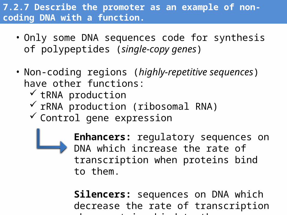
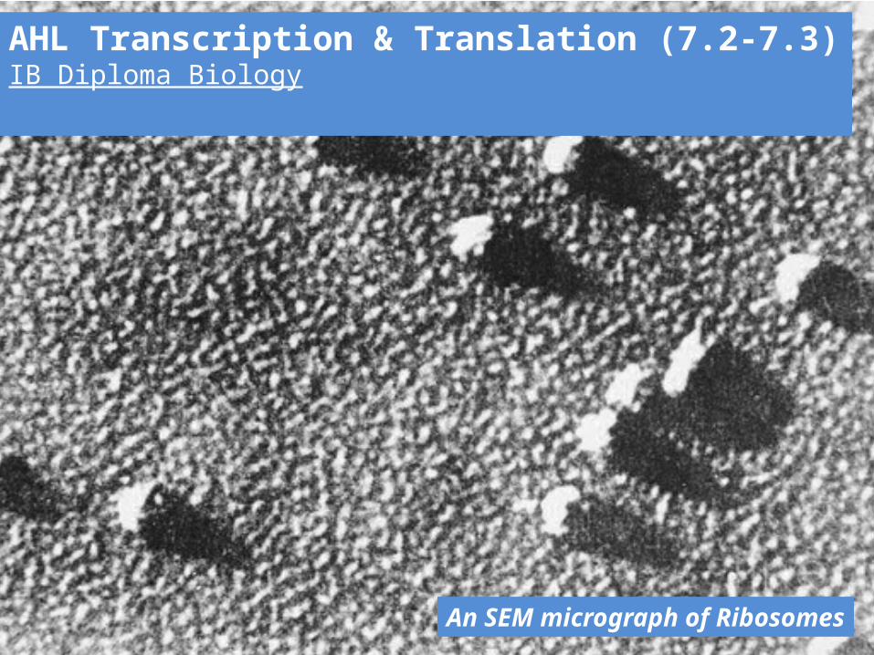
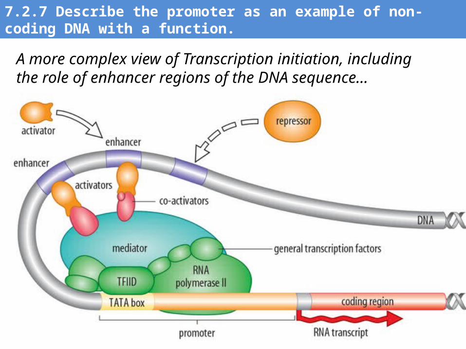
7.2.7 Describe the promoter as an example of non-coding DNA with a function.
• Only some DNA sequences code for synthesis of polypeptides (single-copy genes)
• Non-coding regions (highly-repetitive sequences) have other functions: tRNA production rRNA production (ribosomal RNA) Control gene expression
Enhancers: regulatory sequences on DNA which increase the rate of transcription when proteins bind to them.
Silencers: sequences on DNA which decrease the rate of transcription when proteins bind to them.
The Promoter is located near a gene’s location. It is the binding site of RNA polymerase--the enzyme that constructs mRNA from the DNA template during Transcription.
7.2.7 Describe the promoter as an example of non-coding DNA with a function.
7.2.7 Describe the promoter as an example of non-coding DNA with a function.
A more complex view of Transcription initiation, including the role of enhancer regions of the DNA sequence…
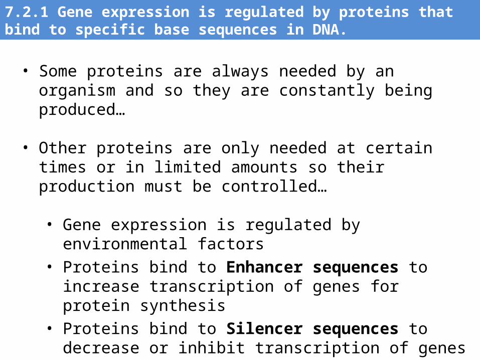
• Some proteins are always needed by an organism and so they are constantly being produced…
• Other proteins are only needed at certain times or in limited amounts so their production must be controlled…
• Gene expression is regulated by environmental factors• Proteins bind to Enhancer sequences to increase transcription
of genes for protein synthesis• Proteins bind to Silencer sequences to decrease or inhibit
transcription of genes for protein synthesis
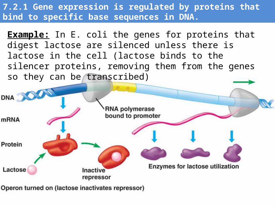
7.2.1 Gene expression is regulated by proteins that bind to specific base sequences in DNA.
7.2.1 Gene expression is regulated by proteins that bind to specific base sequences in DNA.
Example: In E. coli the genes for proteins that digest lactose are silenced unless there is lactose in the cell (lactose binds to the silencer proteins, removing them from the genes so they can be transcribed)
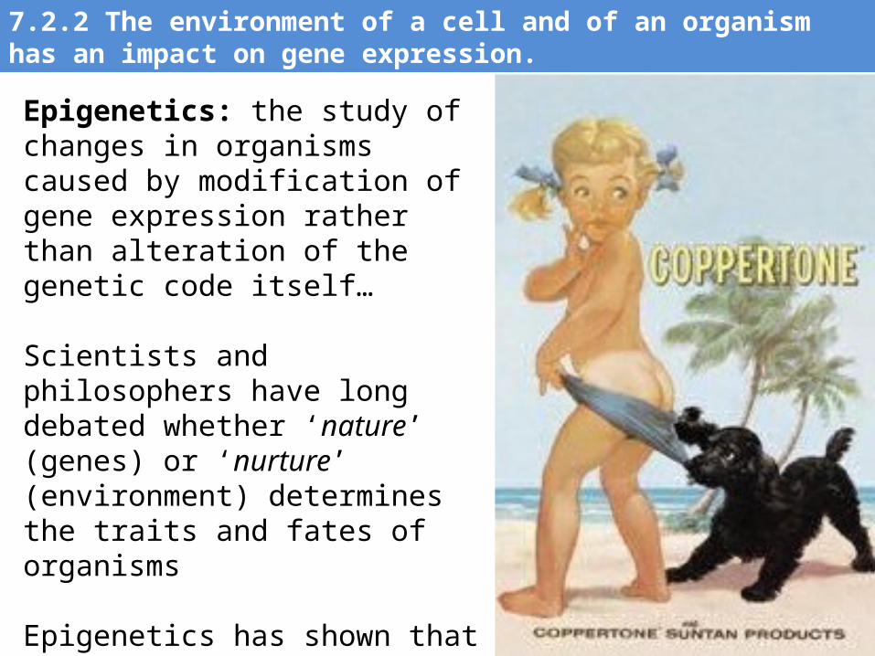
Epigenetics: the study of changes in organisms caused by modification of gene expression rather than alteration of the genetic code itself…
Scientists and philosophers have long debated whether ‘nature’ (genes) or ‘nurture’ (environment) determines the traits and fates of organisms
Epigenetics has shown that both play a substantial role as gene expression is clearly impacted by a cell’s environment (ex. human skin cells producing more melanin in high-sun environments…
7.2.2 The environment of a cell and of an organism has an impact on gene expression.
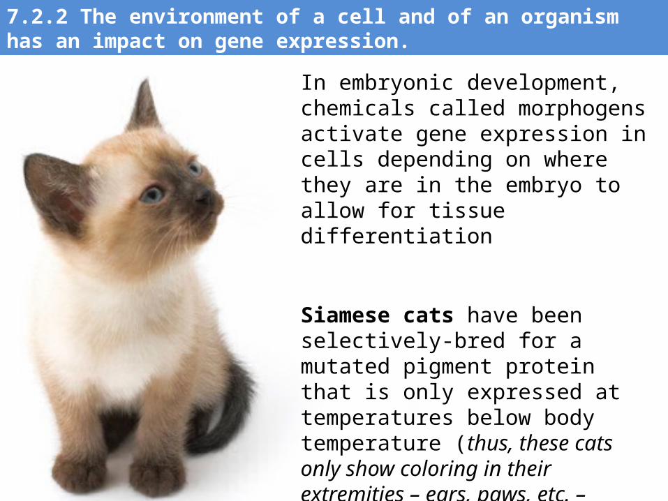
7.2.2 The environment of a cell and of an organism has an impact on gene expression.
In embryonic development, chemicals called morphogens activate gene expression in cells depending on where they are in the embryo to allow for tissue differentiation
Siamese cats have been selectively-bred for a mutated pigment protein that is only expressed at temperatures below body temperature (thus, these cats only show coloring in their extremities – ears, paws, etc. – where temperatures are lower)
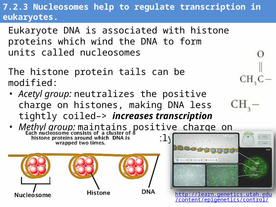
Eukaryote DNA is associated with histone proteins which wind the DNA to form units called nucleosomes
The histone protein tails can be modified: • Acetyl group: neutralizes the positive charge on histones,
making DNA less tightly coiled–> increases transcription • Methyl group: maintains positive charge on histones,
making DNA tightly coiled –> decreases transcription
http://learn.genetics.utah.edu/content/epigenetics/control/
7.2.3 Nucleosomes help to regulate transcription in eukaryotes.
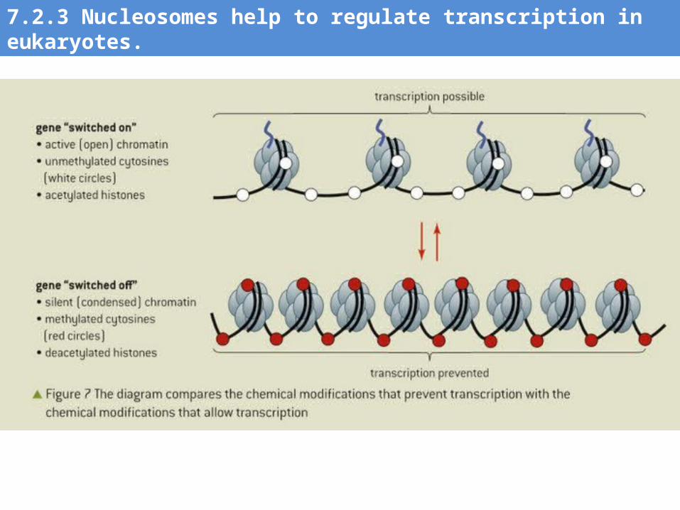
7.2.3 Nucleosomes help to regulate transcription in eukaryotes.
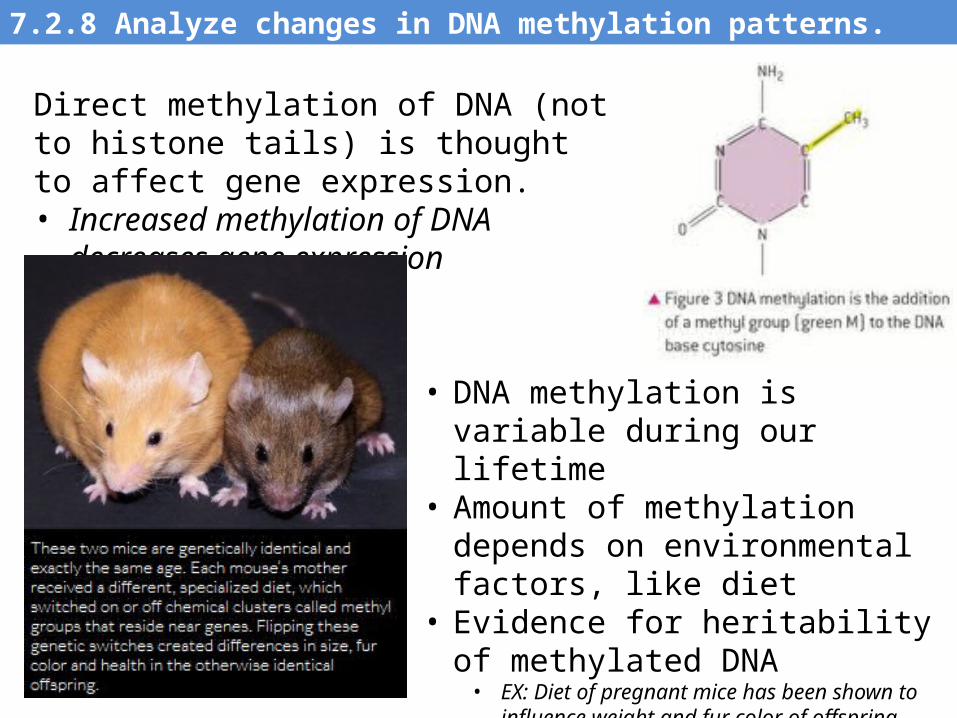
Direct methylation of DNA (not to histone tails) is thought to affect gene expression. • Increased methylation of DNA decreases
gene expression
• DNA methylation is variable during our lifetime
• Amount of methylation depends on environmental factors, like diet
• Evidence for heritability of methylated DNA• EX: Diet of pregnant mice has been shown to
influence weight and fur color of offspring (left)
7.2.8 Analyze changes in DNA methylation patterns.
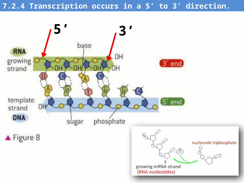
7.2.4 Transcription occurs in a 5’ to 3’ direction.
5’ 3’
http://bcs.whfreeman.com/thelifewire/content/chp14/1401s.swf
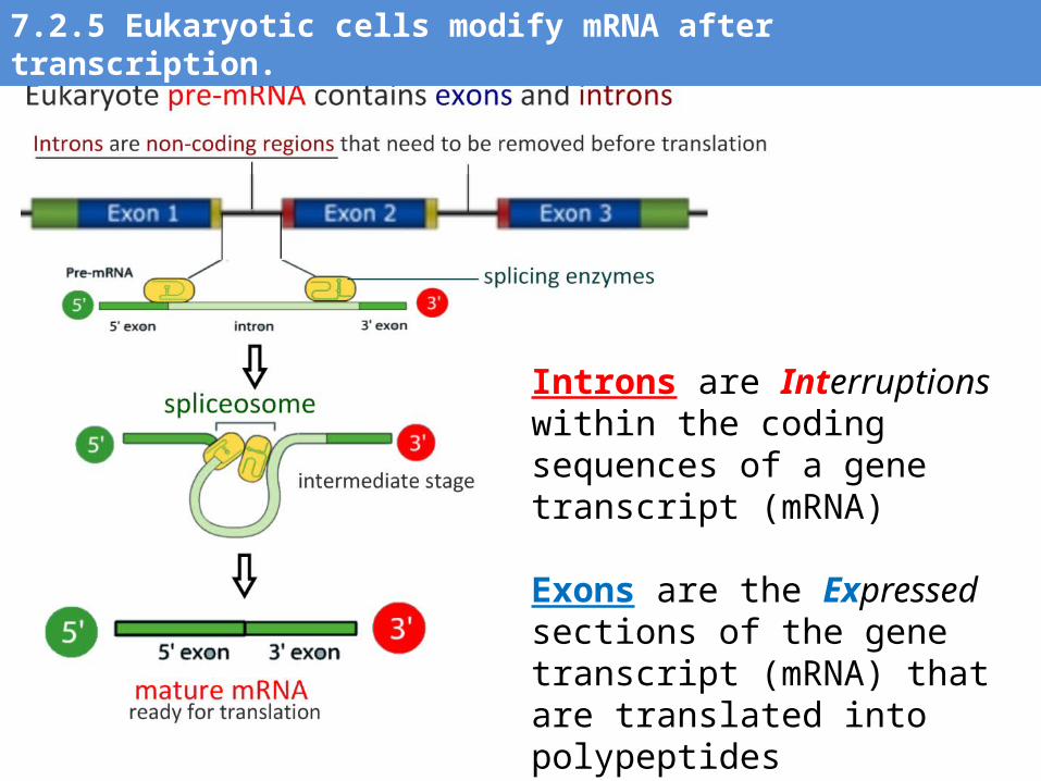
Introns are Interruptions within the coding sequences of a gene transcript (mRNA)
Exons are the Expressed sections of the gene transcript (mRNA) that are translated into polypeptides
7.2.5 Eukaryotic cells modify mRNA after transcription.
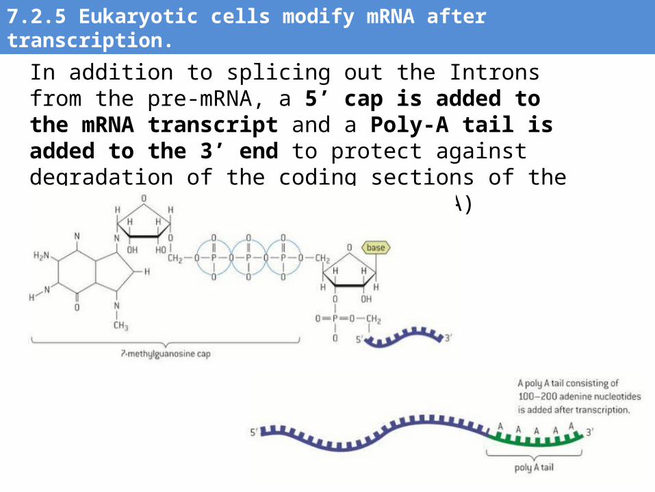
In addition to splicing out the Introns from the pre-mRNA, a 5’ cap is added to the mRNA transcript and a Poly-A tail is added to the 3’ end to protect against degradation of the coding sections of the mRNA (similar to telomeres in DNA)
7.2.5 Eukaryotic cells modify mRNA after transcription.
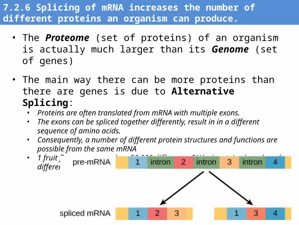
• The Proteome (set of proteins) of an organism is actually much larger than its Genome (set of genes)
• The main way there can be more proteins than there are genes is due to Alternative Splicing:• Proteins are often translated from mRNA with multiple exons.• The exons can be spliced together differently, result in in a different sequence of
amino acids. • Consequently, a number of different protein structures and functions are possible
from the same mRNA• 1 fruit fly gene can produce 38,000 different mRNAs / proteins based on the
different ways it’s exons can be spliced together!
7.2.6 Splicing of mRNA increases the number of different proteins an organism can produce.
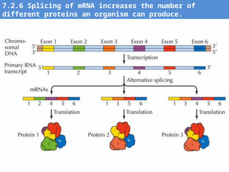
7.2.6 Splicing of mRNA increases the number of different proteins an organism can produce.
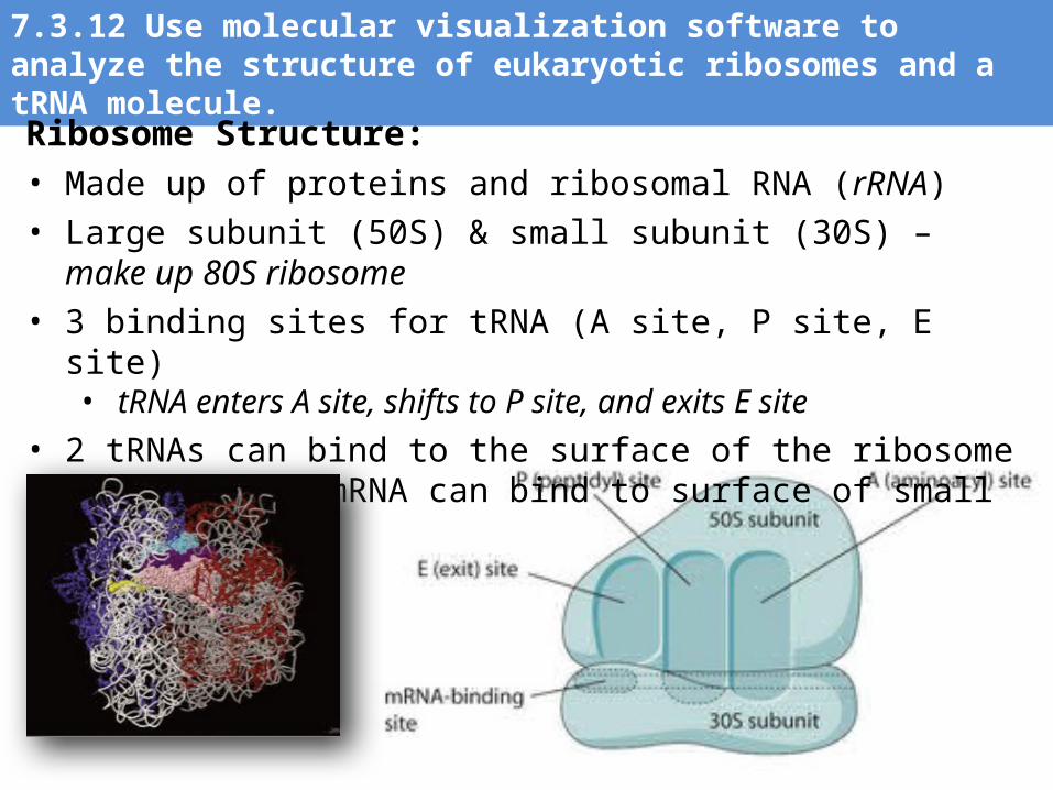
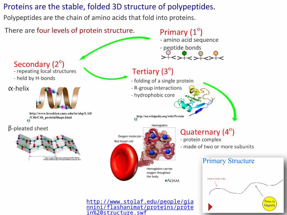
7.3.12 Use molecular visualization software to analyze the structure of eukaryotic ribosomes and a tRNA molecule.
Ribosome Structure: • Made up of proteins and ribosomal RNA (rRNA)• Large subunit (50S) & small subunit (30S) – make up 80S ribosome• 3 binding sites for tRNA (A site, P site, E site)
• tRNA enters A site, shifts to P site, and exits E site• 2 tRNAs can bind to the surface of the ribosome at a time, 1 mRNA
can bind to surface of small subunit
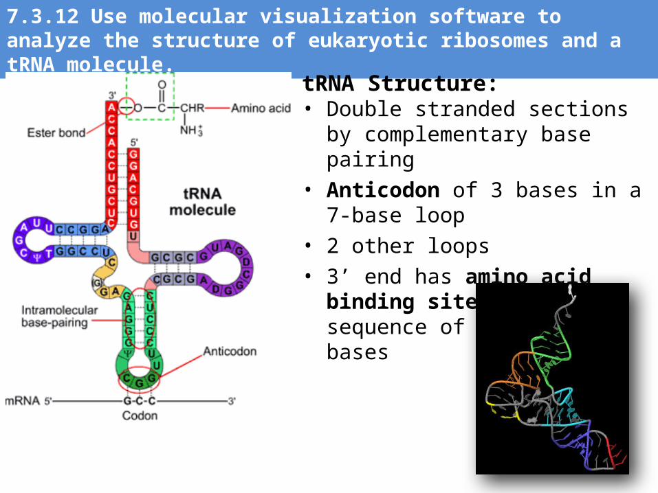
7.3.12 Use molecular visualization software to analyze the structure of eukaryotic ribosomes and a tRNA molecule.
tRNA Structure: • Double stranded sections by
complementary base pairing• Anticodon of 3 bases in a 7-base loop• 2 other loops• 3’ end has amino acid binding site
with CCA sequence of unpaired bases
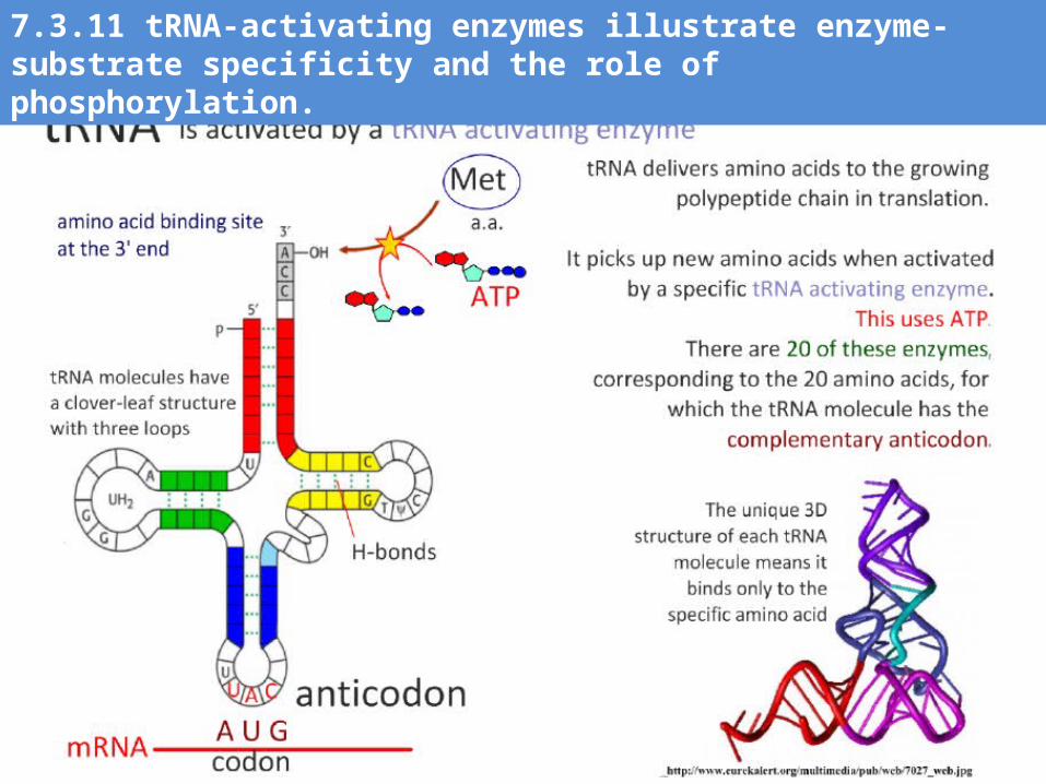
7.3.11 tRNA-activating enzymes illustrate enzyme-substrate specificity and the role of phosphorylation.
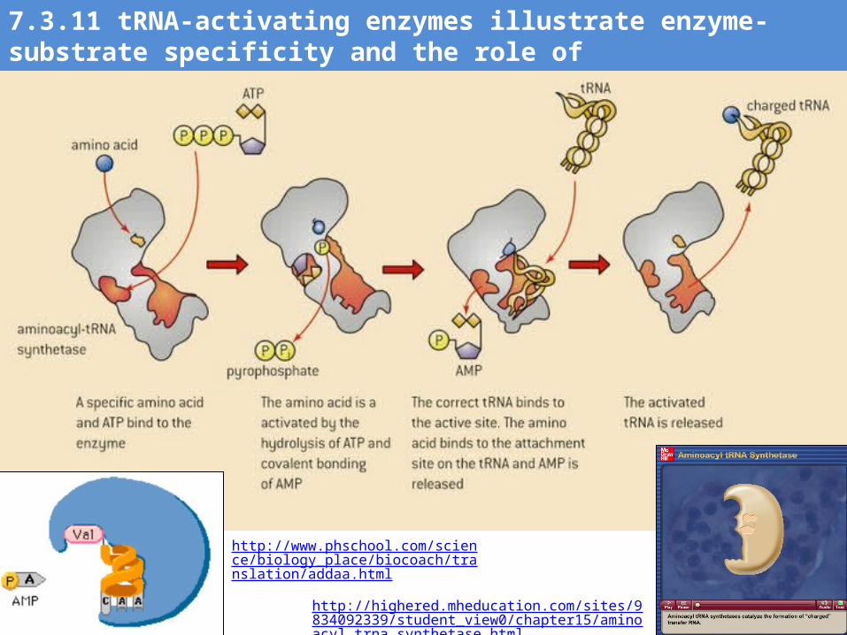
http://highered.mheducation.com/sites/9834092339/student_view0/chapter15/aminoacyl_trna_synthetase.html
7.3.11 tRNA-activating enzymes illustrate enzyme-substrate specificity and the role of phosphorylation.
http://www.phschool.com/science/biology_place/biocoach/translation/addaa.html
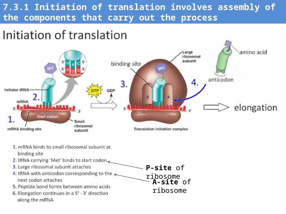
7.3.1 Initiation of translation involves assembly of the components that carry out the process
P-site of ribosome
A-site of ribosome
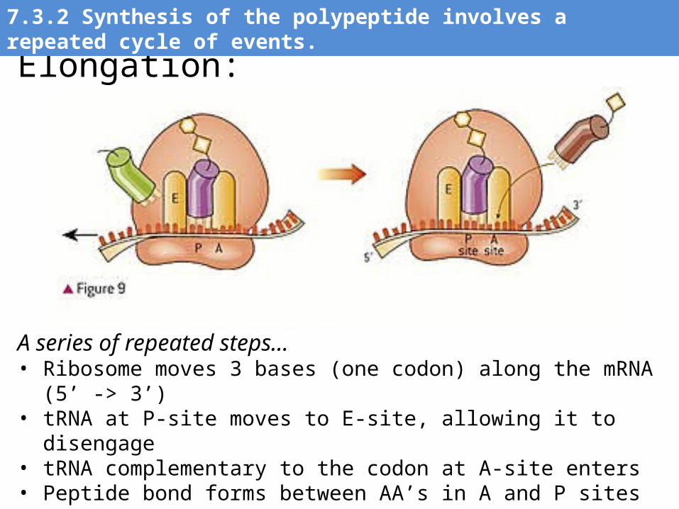
Elongation:
A series of repeated steps…• Ribosome moves 3 bases (one codon) along the mRNA (5’ -> 3’)• tRNA at P-site moves to E-site, allowing it to disengage• tRNA complementary to the codon at A-site enters• Peptide bond forms between AA’s in A and P sites• Process continues many times
7.3.2 Synthesis of the polypeptide involves a repeated cycle of events.
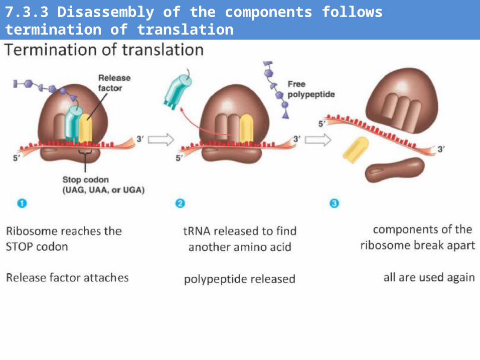
7.3.3 Disassembly of the components follows termination of translation
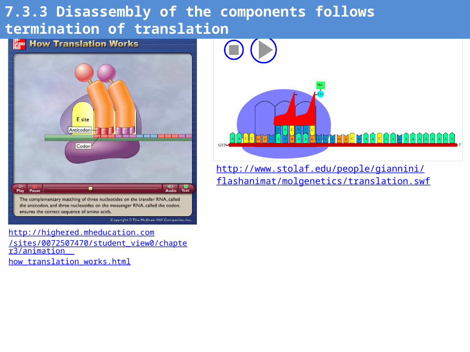
http://highered.mheducation.com/sites/0072507470/student_view0/chapter3/animation__how_translation_works.html
http://www.stolaf.edu/people/giannini/flashanimat/molgenetics/translation.swf
7.3.3 Disassembly of the components follows termination of translation
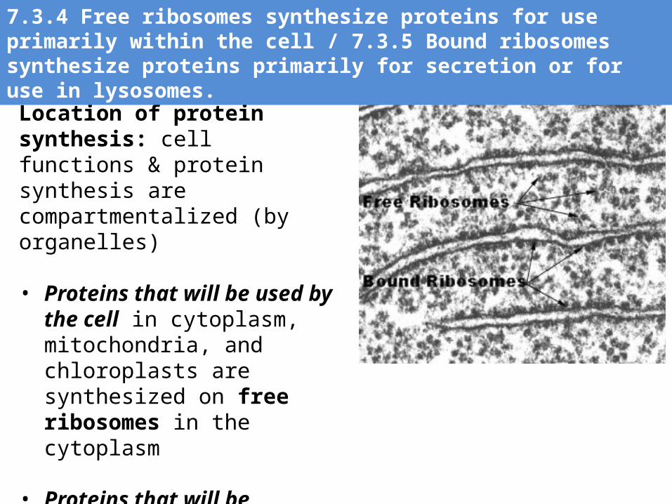
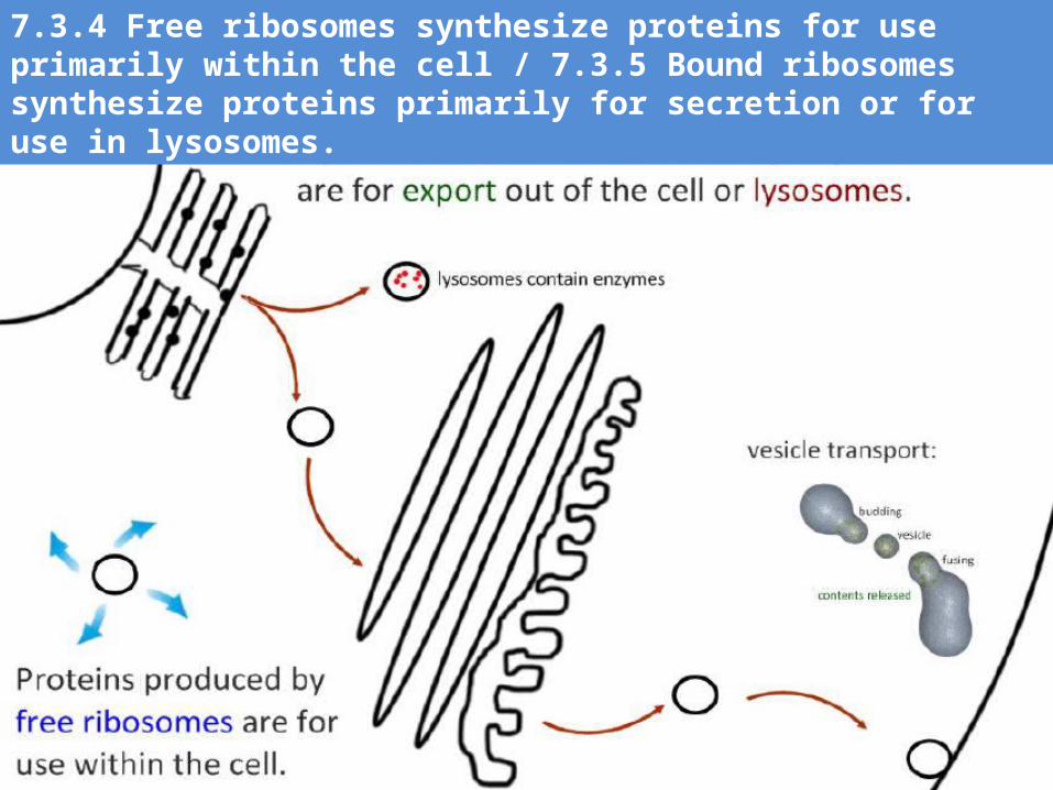
Location of protein synthesis: cell functions & protein synthesis are compartmentalized (by organelles)
• Proteins that will be used by the cell in cytoplasm, mitochondria, and chloroplasts are synthesized on free ribosomes in the cytoplasm
• Proteins that will be secreted or used by lysosomes are synthesized on bound ribosomes found on the RER
7.3.4 Free ribosomes synthesize proteins for use primarily within the cell / 7.3.5 Bound ribosomes synthesize proteins primarily for secretion or for use in lysosomes.
7.3.4 Free ribosomes synthesize proteins for use primarily within the cell / 7.3.5 Bound ribosomes synthesize proteins primarily for secretion or for use in lysosomes.
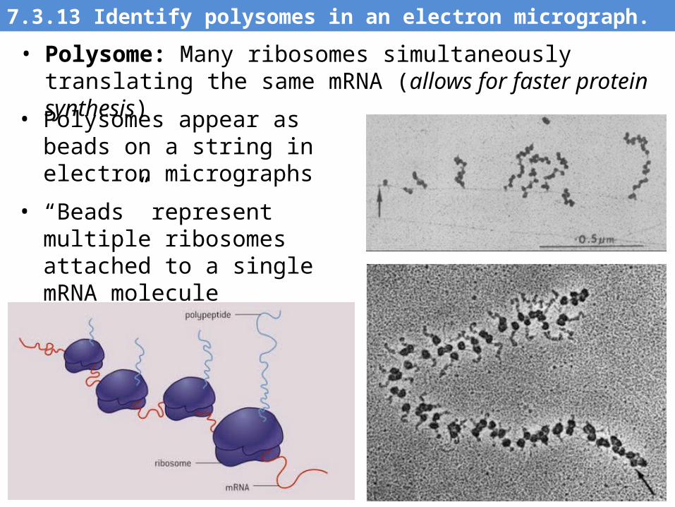
• Polysomes appear as beads on a string in electron micrographs
• “Beads” represent multiple ribosomes attached to a single mRNA molecule
• Poly = many, some = ribosome
7.3.13 Identify polysomes in an electron micrograph.
• Polysome: Many ribosomes simultaneously translating the same mRNA (allows for faster protein synthesis)
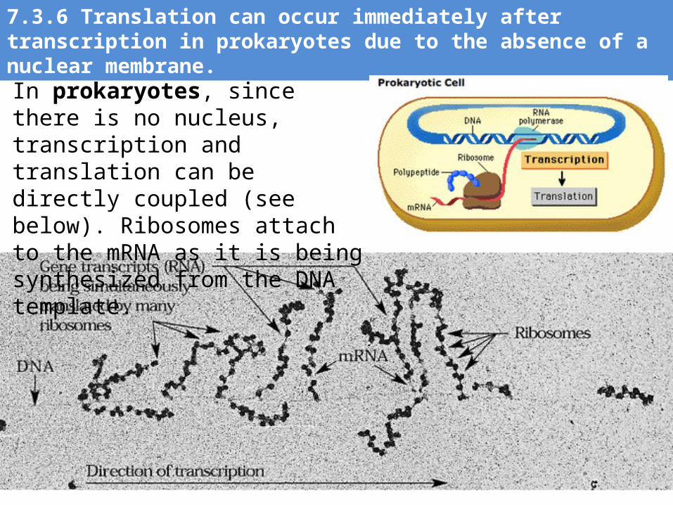
7.3.6 Translation can occur immediately after transcription in prokaryotes due to the absence of a nuclear membrane.
In prokaryotes, since there is no nucleus, transcription and translation can be directly coupled (see below). Ribosomes attach to the mRNA as it is being synthesized from the DNA template.
http://www.stolaf.edu/people/giannini/flashanimat/proteins/protein%20structure.swf
Bibliography / Acknowledgments
Darren Aherne
Chris Paine