Embed Size (px)
Citation preview
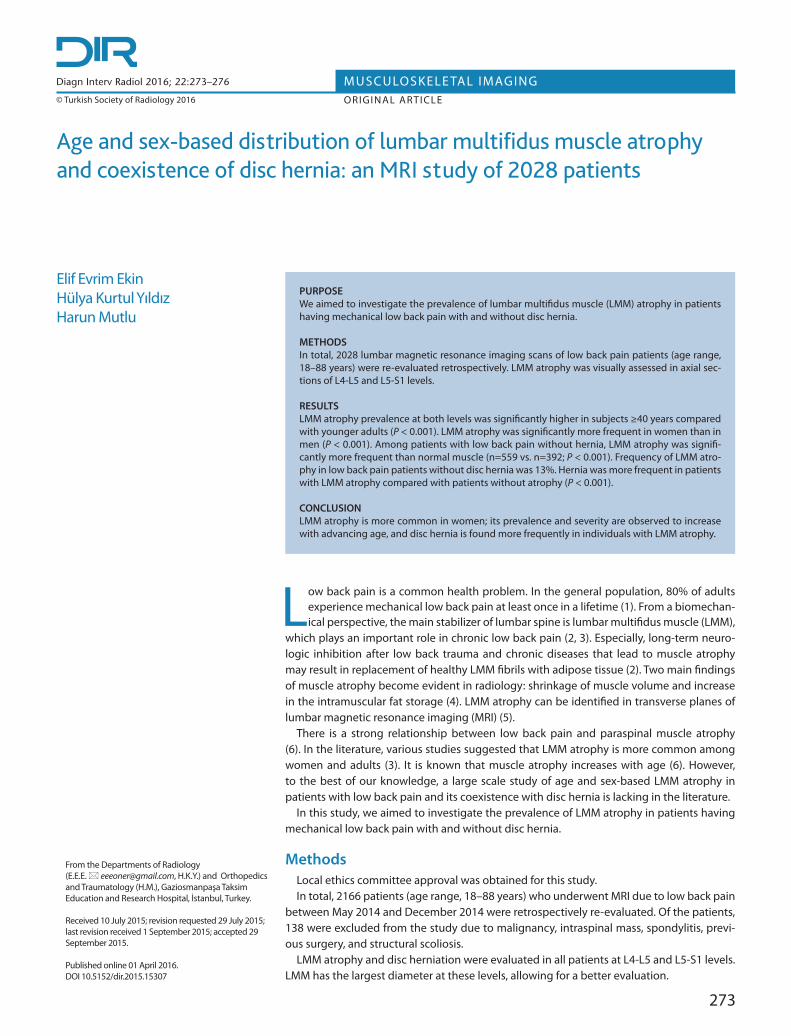
Age and sex-based distribution of lumbar multifidus muscle atrophy and coexistence of disc hernia: an MRI study of 2028 patients
Elif Evrim EkinHülya Kurtul Yıldız Harun Mutlu
Low back pain is a common health problem. In the general population, 80% of adults experience mechanical low back pain at least once in a lifetime (1). From a biomechan-ical perspective, the main stabilizer of lumbar spine is lumbar multifidus muscle (LMM),
which plays an important role in chronic low back pain (2, 3). Especially, long-term neuro-logic inhibition after low back trauma and chronic diseases that lead to muscle atrophy may result in replacement of healthy LMM fibrils with adipose tissue (2). Two main findings of muscle atrophy become evident in radiology: shrinkage of muscle volume and increase in the intramuscular fat storage (4). LMM atrophy can be identified in transverse planes of lumbar magnetic resonance imaging (MRI) (5).
There is a strong relationship between low back pain and paraspinal muscle atrophy (6). In the literature, various studies suggested that LMM atrophy is more common among women and adults (3). It is known that muscle atrophy increases with age (6). However, to the best of our knowledge, a large scale study of age and sex-based LMM atrophy in patients with low back pain and its coexistence with disc hernia is lacking in the literature.
In this study, we aimed to investigate the prevalence of LMM atrophy in patients having mechanical low back pain with and without disc hernia.
MethodsLocal ethics committee approval was obtained for this study.In total, 2166 patients (age range, 18–88 years) who underwent MRI due to low back pain
between May 2014 and December 2014 were retrospectively re-evaluated. Of the patients, 138 were excluded from the study due to malignancy, intraspinal mass, spondylitis, previ-ous surgery, and structural scoliosis.
LMM atrophy and disc herniation were evaluated in all patients at L4-L5 and L5-S1 levels. LMM has the largest diameter at these levels, allowing for a better evaluation.
273
From the Departments of Radiology (E.E.E. [email protected], H.K.Y.) and Orthopedics and Traumatology (H.M.), Gaziosmanpaşa Taksim Education and Research Hospital, İstanbul, Turkey.
Received 10 July 2015; revision requested 29 July 2015; last revision received 1 September 2015; accepted 29 September 2015.
Published online 01 April 2016.DOI 10.5152/dir.2015.15307
Diagn Interv Radiol 2016; 22:273–276
© Turkish Society of Radiology 2016
MUSCULOSKELE TAL IMAGINGORIGINAL ARTICLE
PURPOSE We aimed to investigate the prevalence of lumbar multifidus muscle (LMM) atrophy in patients having mechanical low back pain with and without disc hernia.
METHODSIn total, 2028 lumbar magnetic resonance imaging scans of low back pain patients (age range, 18–88 years) were re-evaluated retrospectively. LMM atrophy was visually assessed in axial sec-tions of L4-L5 and L5-S1 levels.
RESULTSLMM atrophy prevalence at both levels was significantly higher in subjects ≥40 years compared with younger adults (P < 0.001). LMM atrophy was significantly more frequent in women than in men (P < 0.001). Among patients with low back pain without hernia, LMM atrophy was signifi-cantly more frequent than normal muscle (n=559 vs. n=392; P < 0.001). Frequency of LMM atro-phy in low back pain patients without disc hernia was 13%. Hernia was more frequent in patients with LMM atrophy compared with patients without atrophy (P < 0.001).
CONCLUSIONLMM atrophy is more common in women; its prevalence and severity are observed to increase with advancing age, and disc hernia is found more frequently in individuals with LMM atrophy.

274 • May–June 2016 • Diagnostic and Interventional Radiology Ekin et al.
MRI procedure1.5 T MRI unit (Signa HDxt; General
Electric) was used with body surface coil. Lumbar spine was evaluated at L1-S1 levels, and L4-L5 and L5-S1 levels were re-evaluated. Scan sequences included sagittal T1-weighted fast spin-echo (FSE), T2-weighted FSE and an axial T2-weighted FSE (3680/128 repetition time/echo time, 180×256 matrix, 280 mm field of view and 4 mm section thickness, NEX 2).
LMM atrophy was visually examined at L4-L5 and L5-S1 levels. Based on the studies of Parkkola et al. (7) and Kader et al. (8), fatty atrophic changes in LMM were divided into three grades: grade 0, fatty atrophy <10% (Fig. 1a); grade 1, fat infiltration 10%–50% (Fig. 1b); grade 2, fat infiltration >50% (Fig. 1c). All images were evaluated by two expe-rienced radiologists.
Statistical analysis Normal distribution of the data was test-
ed by univariate Kolmogorov Smirnov test and a histogram graphic was prepared. Nor-mality was obtained by logarithmic trans-formation of age (log base 10).
Data were presented as mean, standard
deviation, minimum, maximum, frequency, and percent value based on various char-acteristics. Intergroup comparisons were made by using independent samples t-test or univariate analysis of variance. Post hoc comparisons after ANOVA were made by Tukey HSD test.
Comparison of nominal variables were made by chi-square test. The effects of atro-phy and hernia at L4-L5 and L5-S1 levels were tested by two-way analysis of variance.
Two tailed significance level was adjusted to P < 0.05. All statistical analyses were performed using the NCSS10 software (NCSS, LLC).
Results The study group consisted of 1263 wom-
en (62.3%) and 765 men (37.7%) with low back pain. The mean age was 43.4±13.7 years (range, 18–88 years).
At L4-L5, 1197 patients had disc hernia and 646 patients had grade 1 or 2 LMM atrophy. At L5-S1, 1077 patients had disc hernia and 1131 patients had grade 1 or 2 LMM atrophy. Hernia was more frequent in patients with LMM atrophy at both levels compared with patients without atrophy (P < 0.001, for both levels).
Occurrence of LMM atrophy at L4-L5 lev-el was 37% in women and 23.4% in men,
while occurrence of LMM atrophy at L5-S1 level was 73.9% in women and 52% in men (Table 1). At both levels, LMM atrophy was significantly more frequent in women than in men (P < 0.001, for both levels).
LMM atrophy in women and men was significantly more frequent at L5-S1 than at L4-L5 (P < 0.001). All patients with atrophy at L4-L5 had atrophy at L5-S1 as well. How-ever, of patients with LMM atrophy at L5-S1, only 33.8% had coexisting atrophy at L4-L5.
Results of LMM atrophy evaluation in subjects <40 years and ≥40 years of age are presented in Table 2. LMM atrophy at both L4-L5 and L5-S1 levels was significantly more frequent among men and women ≥40 years compared with younger ages (P <0.001).
At L4-L5, LMM atrophy was significantly associated with age (P < 0.001), while disc hernia was not (P = 0.085); on the other hand, coexistence of hernia and atrophy at L4-L5 was significantly associated with age (P = 0.048). At L5-S1, both atrophy and her-nia were significantly associated with age (P < 0.001), while their coexistence was not (P = 0.796).
Table 3 presents the mean ages of pa-tients according to the grade of LMM atro-phy at L4-L5 and L5-S1 levels, excluding the patients with disc hernia. At both levels, the mean age was significantly different among
Main points
• The origin of low back pain is not clear, but it is known that lumbar multifidus atrophy (LMM) is strongly associated with low back pain. LMM atrophy may be the only MRI finding in some low back pain patients.
• LMM atrophy is more common in women than in men, and its prevalence and severity increase with age. Forty years of age is especially important for LMM atrophy.
• Disc hernia is more frequent in patients with LMM atrophy.
Table 1. Distribution of LMM atrophy at L4-L5 and L5-S1 levels in females and males
Females (n=1263) Males (n=765)
Grade 0 Grades 1+2 Grade 0 Grades 1+2 P
L4-L5 level 796 (63) 467 (37) 586 (76.6) 179 (23.4) <0.001
L5-S1 level 330 (26.1) 933 (73.9) 367 (48) 398 (52) <0.001
Data are presented as n (%).LMM, lumbar multifidus muscle; Grade 0, no atrophy; Grade 1, mild atrophy; Grade 2, advanced atrophy.
Figure 1. a–c. Examples of lumbar multifidus muscle (LMM) atrophy as seen on axial T2-weighted images. Panel (a) shows normal condition (grade 0); panel (b) shows grade 1 atrophy with slight fat infiltration (10%–50%); panel (c) shows grade 2 atrophy with severe fat infiltration (>50%).
a b c

patients with different grades of LMM atro-phy (P < 0.001, for both levels). We observed that the severity of LMM atrophy increased with age (Table 3).
When low back pain patients without disc hernia at L5-S1 (n=951) were evalu-ated for LMM atrophy at the same level, grade 0, 1, and 2 LMM atrophy were pres-ent in 392, 497, and 62 patients, respec-tively. Among patients without disc hernia, significantly more patients had LMM atro-phy compared with normal LMM (n=559 vs. n=392; P < 0.001).
DiscussionIn this study we found that among pa-
tients with low back pain, women and pa-tients ≥40 years of age were more likely to have LMM atrophy. Disc herniation was frequently found together with LMM atro-phy; there were only two patients who had herniation without atrophy. On the other hand, LMM atrophy was present as the only radiologic finding in a significant subset of patients with low back pain (n=265).
The underlying etiology and the origin of pain are not clear in most patients with
low back pain (9). In order to diagnose me-chanical low back pain, conditions such as malignancy, inflammatory diseases, infec-tious processes and referred pain of other organs should be excluded (10). Accord-ingly, we excluded these pathologies to identify patients with mechanical low back pain.
In biomechanical assessment of low back pain, muscular stabilization of “neu-tral zone” in low back region becomes im-portant. It has been well understood that LMM is the key factor in neutral stabiliza-tion (11) and LMM dysfunction is strongly correlated with low back pain (12, 13). Fat involution in LMM muscle leads to muscle dysfunction. The etiology of muscular at-rophy includes nutritional disorders, lack of adequate physical activity, immobility, and long-term systemic diseases. Muscle mass starts to decrease progressively after 40 years of age and the reduction in muscle mass is about 8% in each decade (14). In our study, LMM atrophy was significantly high-er in subjects ≥40 years of age, regardless of the sex. In order to exclude long-term nerve compression-induced LMM atrophy, we
examined patients without disc hernia: the mean ages of Grade 0, 1 and 2 LMM atro-phy groups at L5-S1 level were 34.6±10.28 years, 42.90±12.17 years, and 52.21±12.94 years, respectively (Table 3). Severity of LMM atrophy and advancing decades were directly proportional according to our grad-ing system.
Prevalence of LMM atrophy was signifi-cantly higher in women than in men. LMM atrophy might be responsible for majority of the mechanical low back pain in women, and it affects female population more than male population.
Kader et al. (8), reported that muscle at-rophy in chronic low back pain patients mostly affected the LMM, which is the larg-est medial muscle in lumbosacral region of lumbar spine. Mobility in lumbar region occurs mostly at L5-S1 level, followed by L4-L5 level (8). Therefore, we assessed LMM muscle at L4-L5 and L5-S1 levels to associ-ate with low back pain. LMM atrophy was significantly more frequent at L5-S1 than L4-L5 in both sexes.
Lumbar disc hernia was found more frequently in patients with LMM atrophy compared with patients without atrophy. However, coexistence of LMM atrophy and disc hernia at L5-S1 level was not associat-ed with age. We found disc sequestration without LMM atrophy in two patients (Fig. 2). On the other hand, 265 patients had isolated LMM atrophy without disc her-nia (Figs. 3, 4). In younger patients, lack of muscle atrophy in acute disc hernia may be explained by the longer time period re-quired for fat replacement; but our study is limited as the duration of pain was not in-cluded. There were significantly more low back pain patients with grade 1 or 2 LMM atrophy without hernia than patients with normal muscle without hernia. In our study, LMM atrophy at L4-L5 and L5-S1 levels was the single pathologic MRI finding in 13% of patients with mechanical low back pain. Can LMM atrophy be instructive in low back pain patients without disc hernia, ad-vanced degeneration, or spondylolisthesis? Can LMM atrophy explain low back pain in those patients? This problem may be elu-cidated by a new study investigating the prevalence of LMM atrophy in the general population.
Our study has a number of limitations. We did not include the duration of pain in our analysis. Thus, we could not analyze acute and chronic pain subgroups separately. In addition, there was no age and sex-
Lumbar multifidus muscle atrophy and disc hernia • 275
Table 2. LMM atrophy at L4-L5 and L5-S1 levels in male and female subjects
L4-L5 level <40 years (n=937) ≥40 years (n=1091) P
Females Grade 0 457 (83.2) 339 (47.4) <0.001
Grade 1+2 92 (16.7) 375 (52.5)
Males Grade 0 360 (9.2) 226 (59.9) <0.001
Grade 1+2 28 (7.2) 151 (40)
L5-S1 level
Females Grade 0 225 (41) 105 (14.7) <0.001
Grade 1+2 324 (59) 609 (85.3)
Males Grade 0 255 (65.7) 112 (29.7) <0.001
Grade 1+2 133 (34.3) 265 (70.3)
Data are presented as n (%).LMM, lumbar multifidus muscle; Grade 0, no atrophy; Grade 1, mild atrophy; Grade 2, advanced atrophy.
Table 3. Age comparison between patients without hernia according to grade 0–2 LMM atrophy at L4-L5 and L5-S1 levels
L4-L5 L5-S1
n Age (years) P* n Age (years) P*
Grade 0 696 34.99±9.97 <0.001 392 34.6±10.28 <0.001
Grade 1 132 45.95±11.56 497 42.90±12.17
Grade 2 3 68.67±12.50 62 52.21±12.94
Data are presented as mean±standard deviation. LMM, lumbar multifidus muscle; Grade 0, no atrophy; Grade 1, mild atrophy; Grade 2, advanced atrophy.* One-way ANOVA (Posthoc Tukey HSD test).

276 • May–June 2016 • Diagnostic and Interventional Radiology Ekin et al.
matched control group in our study, due to the difficulty of establishing an adequate control group in such a large sample.
In conclusion, LMM atrophy is more fre-quent in women than in men, and its prev-alence and severity increase with age. For-
ty years of age appears to be a significant threshold for development of LMM atrophy. Hernia is found more frequently in individ-uals with LMM atrophy. We suggest that LMM atrophy may be the only MRI finding in low back pain patients, since 13% of our patients had LMM atrophy without disc her-nia. However, further studies on the general population are needed to support this hy-pothesis.
Conflict of interest disclosureThe authors declared no conflicts of interest.
References1. Rubin DI. Epidemiology and risk factors for spine
pain. Neurol Clin 2007; 25:353–371. [CrossRef]2. Freeman MD, Woodham MA, Woodham AW.
The role of the lumbar multifidus in chronic low back pain: a review. PM&R 2010; 2:142–146. [CrossRef]
3. Kjaer P, Bendix T, Sorensen JS, Korsholm L, Leb-oeuf-Yde C. Are MRI-defined fat infiltrations in the multifidus muscles associated with low back pain? BMC Med 2007; 5:2. [CrossRef]
4. Dannells LA, Vanderstraeten GG, Cambier DC, Witvrouw EE, De Cuyper HJ. CT imaging of trunk muscles in chronic low pain patients and healthy control subjects. Eur Spine J 2000; 9:266–272. [CrossRef]
5. Woodham M, Woodham A, Skeate JG, Freeman M. Long-term lumbar multifidus muscle atro-phy changes documented with magnetic reso-nance imaging: a case series. J Radiol Case Rep 2014; 8:27–34.
6. Beneck GJ, Kulig K. Multifidus atrophy is local-ized and bilateral in active persons with chron-ic unilateral low back pain. Arch Phys Med Re-habil 2012; 300–306. [CrossRef]
7. Parkkola R, Rytokoski U, Kormano M. Magnetic resonance imaging of the discs and trunk mus-cles in patients with chronic low back pain and healthy control subjects. Spine 1993; 18:830–836. [CrossRef]
8. Kader DF, Wardlaw D, Smith FW. Correlation between the MRI changes in the lumbar mul-tifidus muscles and leg pain. Clin Radiol 2000; 55:145–149. [CrossRef]
9. Karataş M. Lomber omurganın fiziksel özel-likleri ve fonksiyonel biyomekaniği. In: Beya-zova M, Gökçe Kutsal Y. eds. Fiziksel tıp ve rehabilitasyon. Ankara: Güneş Kitabevi, 2000; 459–480.
10. Özcan Yıldız E. Bel ağrısı. In: Beyazova M, Gökçe Kutsal Y. eds. Fiziksel tıp ve rehabilitasyon. An-kara: Güneş Kitabevi, 2000; 1465–1483.
11. Moseley GL, Hodges PW, Gandevia SC. Deep and superficial fibers of the lumbar multifidus mus-cle are differentially active during voluntary arm movements. Spine 2002; 27:29–36. [CrossRef]
12. Wu WW, Hu ZJ, Fan SW, et al. Influencing of chronic low back pain on multifidus muscle at-rophy. Zhongguo Gu Shang 2014; 27:207–212.
13. Battié MC, Niemelainen R, Gibbons LE, Dhillon S. Is level and side specific multifidus asymme-try a marker for lumbar disc pathology? Spine J 2012; 12:932–939. [CrossRef]
14. Grimby G, Saltin B. The ageing muscle. Clin Physiol 1983; 3:209–218.[CrossRef]
Figure 2. a, b. A 30-year-old female patient with disc hernia without LMM atrophy. Midsagittal T2-weighted image (a) shows sequestrated disc hernia at L5-S1 level; dehydration is seen in the disc. Axial T2-weighted image at L5-S1 level (b), shows right lateral compression of the sequestrated disc into the dural sac. Multifidus muscle was considered normal (grade 0).
a b
Figure 3. a, b. A 40-year-old female patient with grade 1 LMM atrophy without disc hernia. Midsagittal image (a) shows normal L4-L5 and L5-S1 discs; no significant loss of height or disc hernia is seen. Axial T2-weighted image (b) shows grade 1 LMM atrophy at L5-S1 level.
a b
Figure 4. a, b. A 36-year-old male patient with grade 1 LMM atrophy without disc hernia. Midsagittal image (a) shows normal L4-L5 and L5-S1 discs; no significant loss of height or disc hernia is seen. Axial T2-weighted image (b) shows grade 1 LMM atrophy at L5-S1 level.
a b