Embed Size (px)
Citation preview
Age and Growth
School of Marine Sciences, Dept. of Marine Biology, Microbiology and Biochemistry, CUSAT 169
66
AAGGEE AANNDD GGRROOWWTTHH
6.1 Introduction
6.2 Otolith
6.3 Materials and Methods
6.4 Results
6.5 Discussion
6.1 Introduction
With the growth, the fish attains age, and the age and growth are very
closely related.
The determination of fish age and growth is fundamental in fisheries
biology and management. Such age-determined parameters as mortality and
growth underlie the population dynamics models used in fishery analyses.
Age studies can furnish other basic data such as stock age structure, age at
first maturity, spawning frequency, individual and stock responses to
changes in the habitat, recruitment success, etc. Age and growth data also
permit the determination of population changes due to fishing activities.
According to Qasim (1973b), the main purpose of the study of growth is to
determine the amount of fish that can be produced in terms of quantity
(weight) in a body of water in relation to time.
One of the significant aspects for getting accurate data on fish
biology and population dynamics is to determine the age with lowest error
(Chalanchuk, 1984). There are two methods by which the age of fish can be
Con
tent
s
Chapter-6
School of Marine Sciences, Dept. of Marine Biology, Microbiology and Biochemistry, CUSAT 170
determined. The first is the direct method, which is accomplished through
direct measurements of growth rate of specific specimens extrapolated to
the stock as a whole. By rearing the fish in artificial enclosures or by
marking them in their natural environment and subsequent recapture (tag-
recapture studies- Morales- Nin, 1992; Das, 1994) are the two direct
estimation method. Age data estimated by this method is definite and
reliable; furthermore, it generally establishes a basis for validation studies
(Beamish and Mcfarlane, 1983). The marking method is limited in use due
to it being costly and time-consuming.
The second one, indirect method is accomplished in two ways which
are used as counterchecks to each other. One method in age determination
is by Peterson’s or the length-frequency analysis. However, it is considered
a reliable method when the samples are representative of a fish population
which has a short life, fast growth and reproduction once in a year
(Casselman, 1987; Baker and Timmons, 1991). The other method used in
the aging of fish is the examination of bony structures such as scales,
otoliths, vertebral centra, dorsal and pectoral spines etc. (anatomical
method) at least three times by multiple readers. Both methods have been
widely employed for growth and ageing studies of temperate fishes and
yielded good results. Until recently, these techniques were not employed
for tropical fish growth studies.
Calcified structures have been used to estimate growth and age for a
great diversity of fishes (Bagenal and Tesch, 1978; Pannella, 1980; Secor
et al., 1995). In temperate regions, growth checks on hard structures usually
correlate with responses to changes in water temperature or photoperiod
(Beckman and Wilson, 1995). At tropical latitudes, variation in temperature
and photoperiod is less pronounced and it is presumed that tropical fish
Age and Growth
School of Marine Sciences, Dept. of Marine Biology, Microbiology and Biochemistry, CUSAT 171
lack skeletal growth characters amenable to age and growth analyses.
Consequently, there are few reported studies of tropical fish growth based
on skeletal structures, and most of these are studies are on fishes of marine
origin (Fowler, 1995).
Determination of age by examination of the hard anatomical
structures is the preferred method as it is least prone to subjective
interpretation and tagging artifacts (Brothers, 1982). In temperate regions,
fisheries management largely relies upon this technique for determining
age, and the otoliths have emerged as the most-often preferred anatomical
structure (Bagenal, 1974; Manooch, 1982; Pentilla and Dery, 1988). Until
recently, the methods were not used for tropical fish growth studies for two
reasons. First, tropical fish were assumed to lack seasonal growth patterns.
This was thought to result in poorly developed growth marks in the hard
parts (Brothers, 1980). Second, tropical fishes were believed to lack
seasonality in recruitment. Protracted recruitment would result in skewed
and bimodal length distributions of cohorts. In the absence of a distinct
recruitment season, it is difficult to employ the length frequency method for
growth studies. However, Longhurst and Pauly (1987) pointed out that in
most tropical fishes growth follows predictable seasonal patterns, which
can be detected using length frequency data, or by the analysis of seasonal
bands in the otoliths.
Among the numerous hard structures used for age estimation (scale,
otolith, vertebrae, etc.), the otolith is generally considered to be the best for
estimating the age of many teleost fishes (Campana and Thorrold, 2001).
However, annulus formation in otoliths requires validation before it can be
used to estimate the age of the fish (Beamish and McFarlane, 1983;
Campana, 2001).

Chapter-6
School of Marine Sciences, Dept. of Marine Biology, Microbiology and Biochemistry, CUSAT 172
The physiological functions of fish follow dial environmental
changes and individuals undergo cycles of rest and activity that can be
registered on their otoliths (Radtke and Morales- Nin, 1989).
Discontinuities in otoliths also appear to be related to ontogeny and habitat
changes (Pannella, 1971). Otoliths continue to grow under conditions of
food deprivation (Marshall and Parker, 1982; Campana, 1983; Neilson and
Geen, 1984) while fish stressed by exertion (Campana, 1983) or by
exposure to low pH (Geen et al., 1985) do not show evidence of otolith
resorption. Otoliths are often the first calcified structures that appear during
the early development of teleosts.
Otoliths have several advantages over scales and spines for
estimating age because otoliths are not subjected to resorption and their
growth is acellular rather than by ossification (Secor et al., 1995). Higher
precision of age estimates from otoliths has led several authors to
recommended otoliths over other structures for estimating age (Boxrucker,
1986; Sharp and Bernard, 1988; Hoxmeier et al., 2001).
Otoliths have been used to estimate age in fish such as
channichthyids which lack scales (Kock and Everson, 1998). However,
the growth rings are known to be more distinct and clear on the otolith
than in other structure of the fish. Therefore, they are extensively used to
determine the age in fishes (Jennings et al., 2001; David and Pancharatna,
2003). This method of aging is based on the fact that successive rings are
formed as the fish grows in age. Moreover, otolith elemental analyses
give an exact idea on the migratory behaviour of the fishes and
environmental stress experienced by it due to pollution and / or other
factors. Such data can be incorporated into the fishery management plans
Age and Growth
School of Marine Sciences, Dept. of Marine Biology, Microbiology and Biochemistry, CUSAT 173
and appropriate conservation strategies can be put forth for those fishes
facing serious threat of extinction (Radhakrishnan et al., 2009).
Since there is definite periodicity in the seasons of the temperate
region, the growth rings or checks are more or less clear and easy to
interpret. Similar growth checks have been noticed in hard skeletal parts of
tropical and subtropical fishes and among the main references on the
subject are the works of Radhakrishnan (1954, 1957), Seshappa and
Bhimachar (1955), Pantulu and Singh (1962), Qasim (1973b), Buesa
(1987), Fowler (1990), Seshappa (1999), Pilling et al. (2000), Ambrosio
et al. (2003), Caldow and Wellington (2003), Pilling et al. (2003), Ismen
(2005), Grandcourt et al. (2006), Nolf et al. (2006), Zekeria et al. (2006),
Lin and Tzeng (2009) and Shamsan and Ansari (2010a). This work
represents the first comprehensive study on age, growth, and mortality of a
population of C. dussumieri from Cochin estuary based on age estimates
from otoliths and contributes to management of these stocks.
6.2 Otolith
The use of otoliths as indicators of fish age has now reached the
century landmark, starting with Reibisch’s observations of otolith annuli in
1899. In 1971, Pannella’s discovery of daily growth increments helped
propel the interpretation of otolith microstructure into the mainstream of
fish biology.
Otoliths (“ear stones”) are structures located in the inner ear cavity of
all teleost fish. All teleost fishes have three pairs of otoliths: the asteriscae,
lapillae and sagittae. The sagitta is the largest of the three otoliths and is
generally used in studies of age estimation in teleosts. Each sagittal otolith
is enclosed in a fluid filled sac called the sacculus within the otic capsule of
Chapter-6
School of Marine Sciences, Dept. of Marine Biology, Microbiology and Biochemistry, CUSAT 174
the head. The otic capsules are situated on either side of the posterior
portion of the brain. Otoliths are acellular and avascular. Otoliths are
complex polycrystalline bodies, composed of needle shaped crystals of
calcium carbonate in the form of aragonite, radiating outwards in three
dimensions from a centrally located nucleus and passing through a network
of fibrous collagen like protein called otolin. Otoliths act as sound receptors
and also play a role in balance and orientation. Otolith size and shape,
particularly of the sagittae, varies greatly among species. Size and shape of
the sagittae are related to their function of sound detection in fish (Popper
and Lu, 2000).
Seasonal changes in the fish’s growth rate are reflected in the otolith.
Materials are deposited on the otolith from the endolymph, the fluid that
surrounds the otolith. The deposits are formed in bands of alternating
optical density, which appear either opaque or hyaline (translucent) under
reflected light. The alternating zones on the otolith are due to the
differences in the amount of protein (otolin) in the zones and shape of the
aragonite crystals. The opaque zones in the otoliths are formed during the
period of fast growth and the hyaline zone is laid down usually during the
period of slow growth. These zones are also called rings.
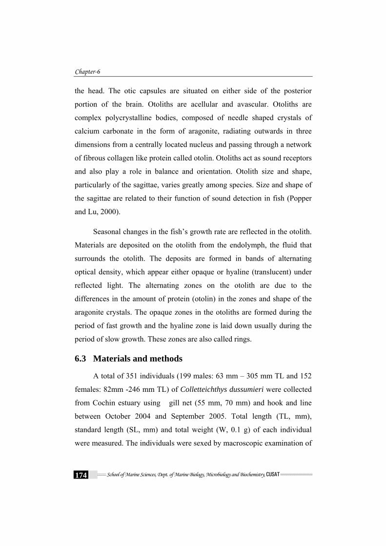
6.3 Materials and methods
A total of 351 individuals (199 males: 63 mm – 305 mm TL and 152
females: 82mm -246 mm TL) of Colletteichthys dussumieri were collected
from Cochin estuary using gill net (55 mm, 70 mm) and hook and line
between October 2004 and September 2005. Total length (TL, mm),
standard length (SL, mm) and total weight (W, 0.1 g) of each individual
were measured. The individuals were sexed by macroscopic examination of

Age and Growth
School of Marine Sciences, Dept. of Marine Biology, Microbiology and Biochemistry, CUSAT 175
the gonads as males and females. The otoliths were removed from the
cranium by means of a transverse incision through the head using a sharp
knife. Once the incision has been made, and the semi-circular canals
exposed, the sagitta was removed with a fine forceps. Sagittal otoliths were
removed, cleaned and stored dry in code-numbered vials.
Otoliths were placed in glycerine for one to two weeks to clear the
otolith which will allow for improved reading. The left sagitta was mainly
used for age determination; the right sagitta was used only if the left one was
damaged. Otoliths were placed in water and were observed with a compound
microscope (4x - 10x) using reflected light on a black background. Otolith
exhibited alternating opaque (broad, white) and hyaline or translucent
(narrow, clear) bands. The distal portion of each translucent zone was
considered the annulus. The areas of complete growth for each year were
represented by the combination of one opaque zone and a subsequent
translucent zone. The total number of translucent zones was recorded in
order to assign an estimated age to each fish. The linear measurements of
each otolith were taken with an ocular micrometer and it included: the otolith
length (OL: the longest dimension between the anterior and posterior edges),
the dorsal otolith radius (OR: the dimension from the focus (F) to the dorsal
edge), the width (WL: the dimension from the dorsal to the ventral edge) and
the distance from the focus to the outer margin of each translucent zone (ring
radius, rn ) on the dorsal radius (Fig. 6.1). All measurements were made with
the concave side of otoliths facing down, to ensure that the anterior and
posterior edge of the otolith were in the same focal plane. Otoliths were
weighed (OW) to the nearest 0.01 mg. Each otolith was examined thrice at
different times and an accurate count was taken for the analysis of growth
characteristics.

Chapter-6
School of Marine Sciences, Dept. of Marine Biology, Microbiology and Biochemistry, CUSAT 176
The relationship between fish total length-at- capture (TLc), otolith
length (OL), and otolith weight (OW) were statistically assessed using
regression analysis and calculating the correlation coefficient ‘r’ (Zar,
2005). The relationship of fish total length-at-capture (TLc ) and otolith
radius- at- capture (ORc) were analyzed with linear regression using
untransformed data. The linear equation was TLc = a + b ORc.
Characterization of the otolith edge was used for inferences on the
period of annulus formation (Newman and Dunk, 2003). Marginal
increment analysis was used to validate the periodicity of the formation of
the growth increments as well as the number of rings formed. The
proportion of samples with opaque / translucent margins was calculated for
each month and used to infer the timing and periodicity of increment
formation. The marginal increment ratio (MIR) is expressed as the
following equation and was used to establish the period of ring formation:
MIR = (RC – RL) x (RL – RL-1 ) -1
Where,
RC = otolith radius
RL = distance from the focus to the outer edge of outermost
transparent zone
RL-1 = distance from the focus to the outer edge of the penultimate
translucent zone.
The mean MIR and the standard deviation were computed for each
month by sex for all ages combined and also for each age separately. An
analysis of variance (ANOVA) was used to detect significant differences
by sex, age group and month of capture. The Post- Hoc multiple
comparison test was used to detect which pair wise differences among

Age and Growth
School of Marine Sciences, Dept. of Marine Biology, Microbiology and Biochemistry, CUSAT 177
treatments were significant. Means (± S.D.) were plotted against month of
capture, the minima indicating the month of annulus formation.
The back-calculated total lengths at each age were determined from
the body proportional equation (Francis, 1990):
LA = [(a+bRA)/ (a+bRC)] LC
Where,
LA = back- calculated total length to annulus A
RA = otolith radius to annulus
RC = total otolith radius at a time of capture
LC = total length at time of capture
a = intercept from the linear total length-otolith radius regression
b = slope from the linear total length- otolith radius regression.
Theoretical growth parameters for females and males were estimated
by fitting the back-calculated length-at-age to the von Bertalanffy (1938)
growth function (VBGF).
Lt = L∞ [1-exp (-K (t-t0))]
Lt = Length at age t
L∞ = Predicted asymptotic length (maximum length which the
fish tend to attain)
e = base of Neparian or natural logarithm
k = Brody growth coefficient, a measure of the rate at which
length approaches L∞
t = age of the fish
to = arbitrary origin of growth curve or theoretical age when fish
length = 0.

Chapter-6
School of Marine Sciences, Dept. of Marine Biology, Microbiology and Biochemistry, CUSAT 178
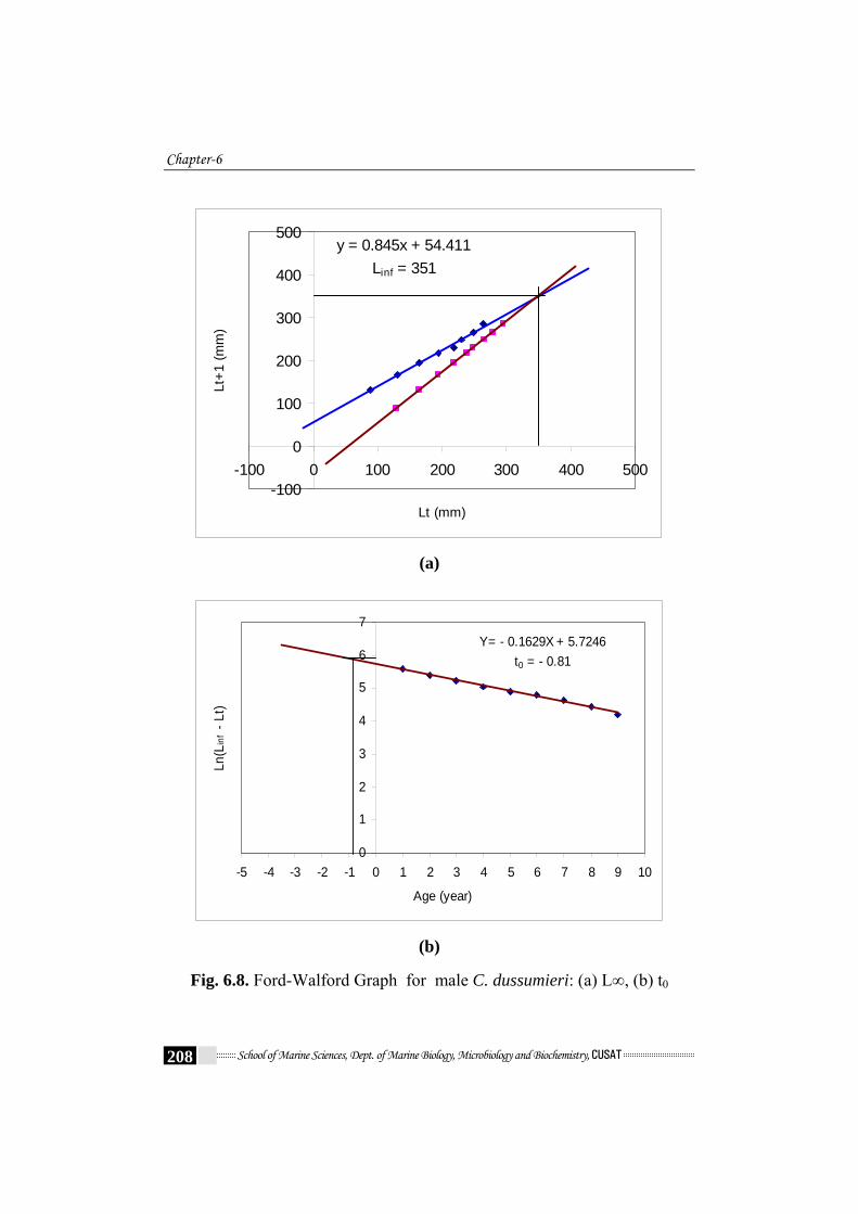
Growth parameters (L∞, K and t0) were estimated graphically by plotting
the Ford (1933) - Walford (1946) graph as described in Ricker (1975).
Differences in von Bertalanffy growth curves were tested by means
of the Chow test (Chow, 1960). This test is an application of the F-test,
commonly used to test structural change in a regression model attributable
to variations in some of or all the parameters. In the case of two groups, it
requires the sum of squared errors from three regressions, one for each
group (ess_1 and ess_2) and one for the pooled data (ess_c pool). Thus,
The formula for the “Chow test” of this constraint is
k*2N_2N_1ess_2ess_1
kess_2)(ess_1-ess_c
F
−++
+
=
Where,
ess_1 and ess_2 are the error sum of squares from the separate
regressions
ess_c is the error sum of squares from the pooled (constrained)
regression,
k is the number or estimated parameters
N_1 and N_2 are the number of observations in the two groups.
Longevity was calculated from Taylor’s equation (Taylor, 1958):
A0.95 = K996.2
t 0 +
Where A0.95 is the lifespan or age required to reach 95% of the final length
(L∞), and K and t0 are von Bertalanffy growth parameters.
Age and Growth
School of Marine Sciences, Dept. of Marine Biology, Microbiology and Biochemistry, CUSAT 179
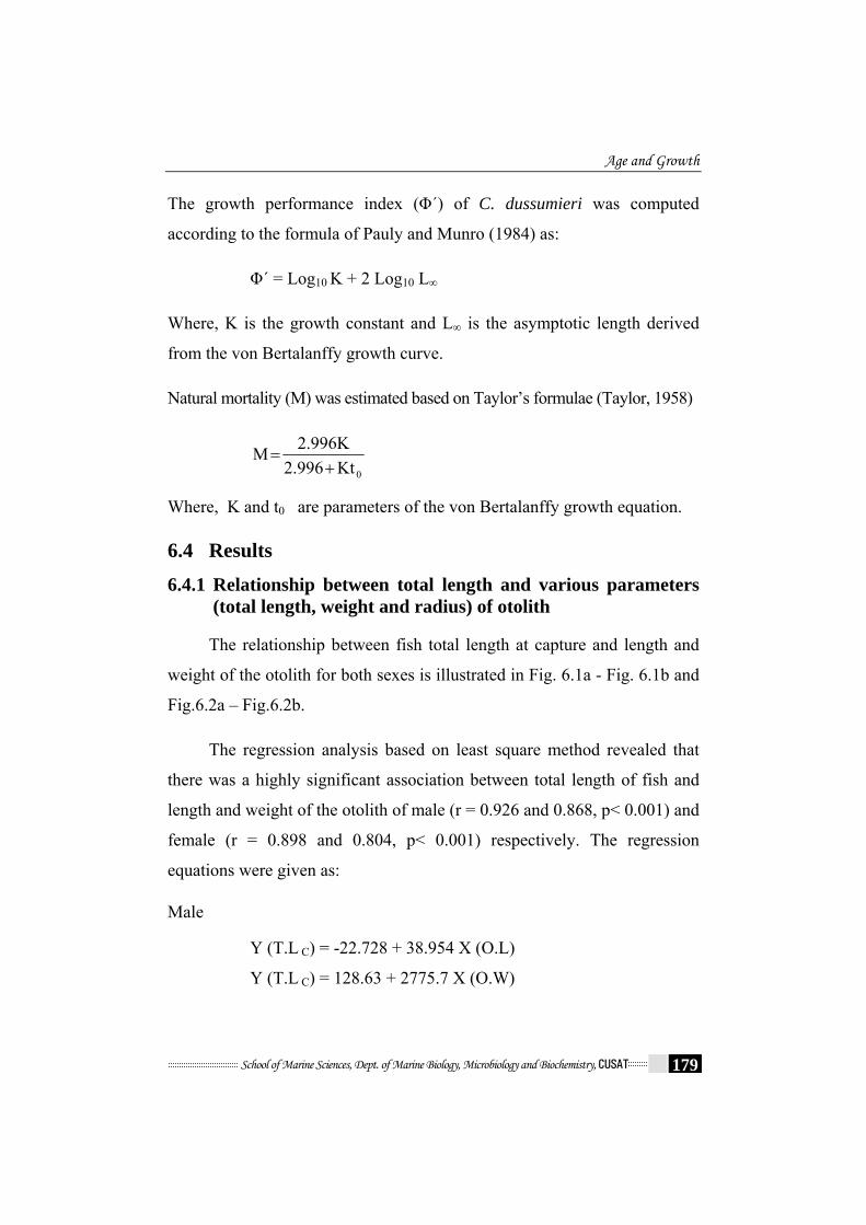
The growth performance index (Φ´) of C. dussumieri was computed
according to the formula of Pauly and Munro (1984) as:
Φ´ = Log10 K + 2 Log10 L∞
Where, K is the growth constant and L∞ is the asymptotic length derived
from the von Bertalanffy growth curve.
Natural mortality (M) was estimated based on Taylor’s formulae (Taylor, 1958)
0Kt2.996
2.996KM+
=
Where, K and t0 are parameters of the von Bertalanffy growth equation.
6.4 Results
6.4.1 Relationship between total length and various parameters (total length, weight and radius) of otolith
The relationship between fish total length at capture and length and
weight of the otolith for both sexes is illustrated in Fig. 6.1a - Fig. 6.1b and
Fig.6.2a – Fig.6.2b.
The regression analysis based on least square method revealed that
there was a highly significant association between total length of fish and
length and weight of the otolith of male (r = 0.926 and 0.868, p< 0.001) and
female (r = 0.898 and 0.804, p< 0.001) respectively. The regression
equations were given as:
Male
Y (T.L C) = -22.728 + 38.954 X (O.L)
Y (T.L C) = 128.63 + 2775.7 X (O.W)

Chapter-6
School of Marine Sciences, Dept. of Marine Biology, Microbiology and Biochemistry, CUSAT 180
Female
Y (T.L C) = -12.065 + 34.742 X (O.L)
Y (T.L C) = 130.69 + 2345.2 X (O.W)
Where T.L C is total length- at- capture, O.L is length of otolith and O.W
denoted the weight of otolith.
Somatic growth (T.L C) and otolith growth (O.L) were strongly linearly
related in both males (R2 = 0.858) and females (R2 = 0.806).
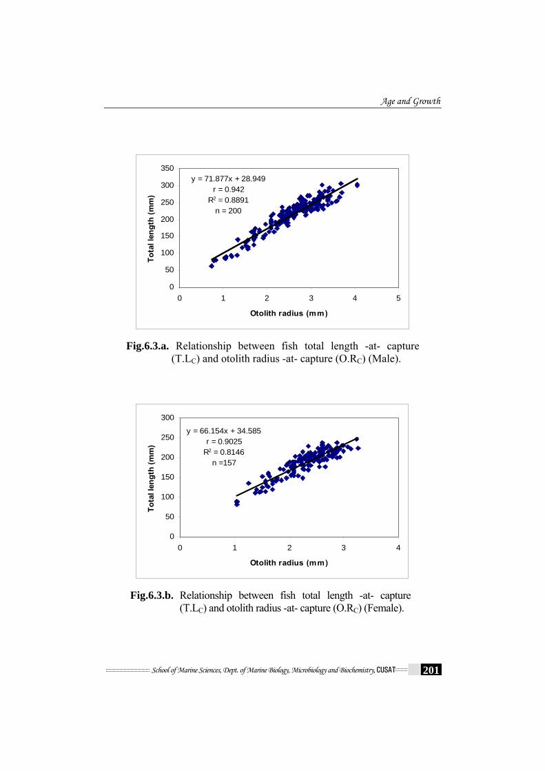
Fig.6.3a. and Fig.6.3b. shows the sex-specific relationship between fish
total length -at- capture (T.L C) and otolith radius -at- capture (O.R C).
Male Y (T.L C) = 28.949 + 71.877 X (O.R C) r = 0.942, R2 = 0.8891
Female Y (T.L C) = 34.585 + 66.154 X (O.R C) r = 0.903, R2 = 0.815
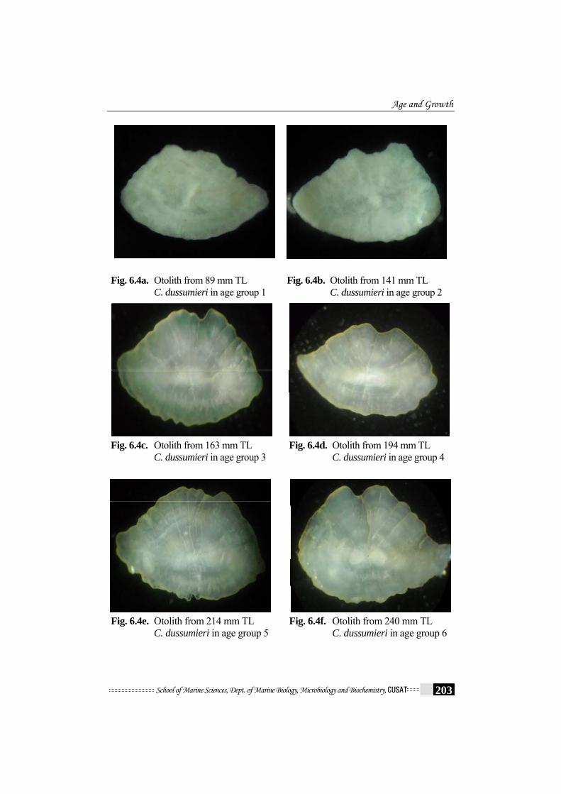
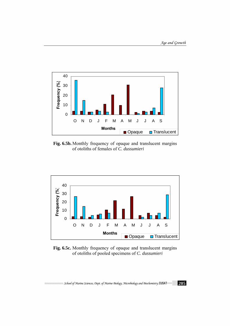
6.4.2 Growth check on otolith
Otolith displayed well-defined alternating thin translucent zones and
wide opaque zones under reflected light (Fig.6.4, Fig.6.4a – Fig.6.4i). An
opaque zone combined with a translucent zone was interpreted as one
year’s growth. Formation of growth increments followed a seasonal pattern
(Fig.6.5a - Fig.6.5c). The translucent zones begin to develop in June and
continued until February and the proportion of otolith with translucent
margins was highest in September to November, coinciding with peak
spawning (chapter 5). A marked decline in January and February, followed
by an absence of translucent margins from March to May indicates
cessation of translucent annulus formation and the onset of opaque annulus
formation beginning in June. The opaque zones were laid down mainly
from February to June when growth is faster. The data suggest that only

Age and Growth
School of Marine Sciences, Dept. of Marine Biology, Microbiology and Biochemistry, CUSAT 181
one opaque and one translucent zone are laid down per year and represent
valid annual growth increments.
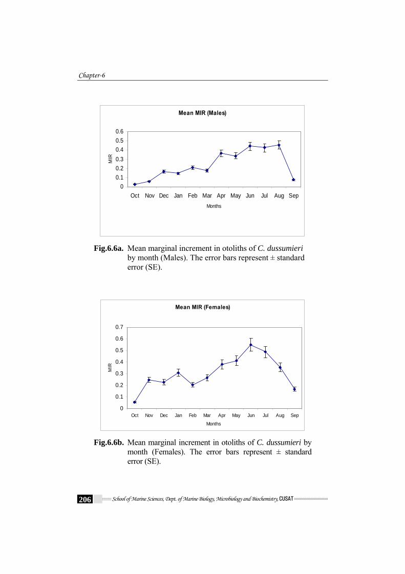
6.4.3 Marginal increment analysis
The changes with time of the rate of marginal increment (RC – RL) x
(RL – RL-1) -1 were measured to determine the time of annulus formation.
The monthly mean MIR (sexes and all ages combined) showed a single
minimum in September to November (Fig.6.6a - Fig.6.6c). The spawning
of this fish, judged from monthly changes in gonad indices, lasts from
September to January, with the highest intensity in October and November.
This pattern indicated the formation of one annulus per year in September
to October. Similar pattern was exhibited by monthly means of MIR when
plotted by sex, and for the different ages respectively. ANOVA detected
significant differences during the year (df = 11, 339; p<0.001) (Table 6.1.)
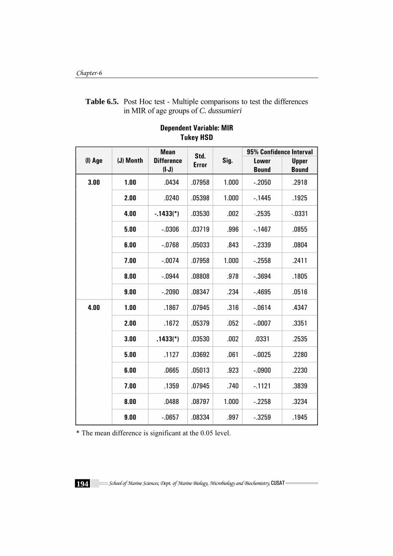
and for age-group (df = 8, 342; p<0.001) (Table 6.2.). The Post-Hoc Test
showed that the MIR in September – November was significantly lower
than the rest of the year, providing further evidence that annulus formation
occurred yearly in September – November (Table 6.4). Comparison of MIR
between age groups by means of Post Hoc Turkey HSD test revealed
differences between age groups 3 and 4 (Table 6.5). The t-test did not show
significant differences for the sexes. The number of annuli represents the
years from birth.
Age distributions for the sexes are exhibited in Fig.6.7. The
maximum age of the sampled toadfish was 9 years for males and 7 years
for females. In the present study, females less than 2 years were poorly
represented in the sample. Age distribution was almost similar for both
sexes and was skewed to the left. The population was dominated by 3 to 5

Chapter-6
School of Marine Sciences, Dept. of Marine Biology, Microbiology and Biochemistry, CUSAT 182
year old fishes. Fishes younger than 2 years and older than 6 years were
poorly represented. After age 4, males dominated the older age classes.
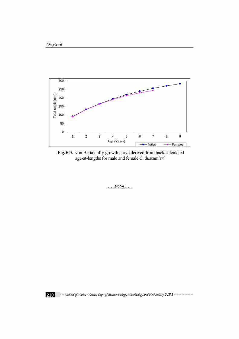
6.4.4 Back-calculated total length
The mean back – calculated lengths – at –age for 1 to 9 year old
specimens are listed in Table 6.6a and 6.6b. Growth increments were
higher during the first year of life (mean 39.9 mm and 41.0 mm LT for
males and females, respectively). After that, the growth rates of both sexes
slowed appreciably. Males and females showed similar mean lengths until
age 3; afterwards, males grew faster and consequently reached larger sizes
than females. The differences become more pronounced with increasing
age (Table 6.6a - Table 6.6b); Fig.6.9).
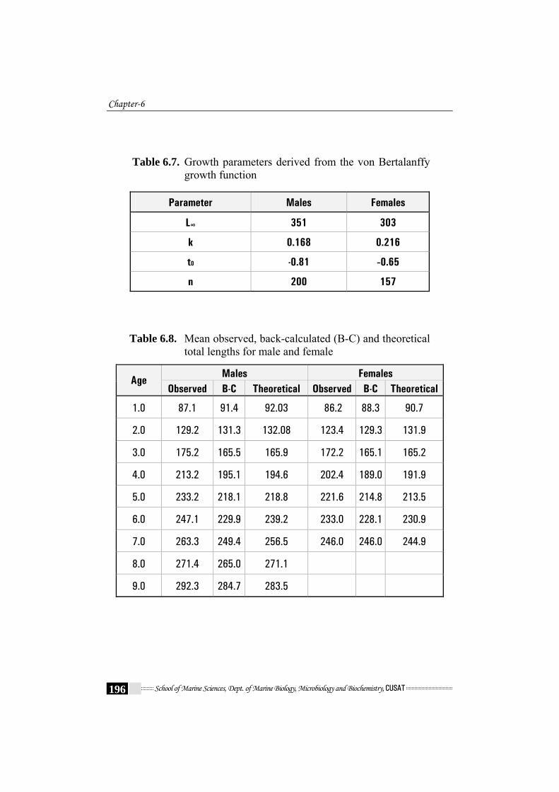
6.4.5 Estimation of growth parameters
Table 6.7. shows the various parameters of VBGE. The Various
parameters were found to be L∞ = 351 mm and t0 = -0.81 and k = 0.168 /year
and L∞ = 303mm and t0 = -0.65 and k = 0.216 /year were obtained by
graphical method (Fig. 6.8a - Fig. 6.8d) for males and females respectively.
The von Bertalanffy growth parameter estimates are listed in Table 6.7.
The von Bertalanffy (1938) growth equations, fitted to back-
calculated lengths- at- age for the last annulus were
Male : Lt = 351 [1-e (-0.168(t-(-0.81))]
Female : Lt = 303 [1-e (-0.216(t-(-0.65))]
The theoretical length at different ages as calculated by this equation
showed a very close agreement with those estimated by otolith method
(back-calculated lengths) (Table 6.8). Subsequently, the growth curve of
C. dussumieri is shown in Fig.6.9.
Age and Growth
School of Marine Sciences, Dept. of Marine Biology, Microbiology and Biochemistry, CUSAT 183
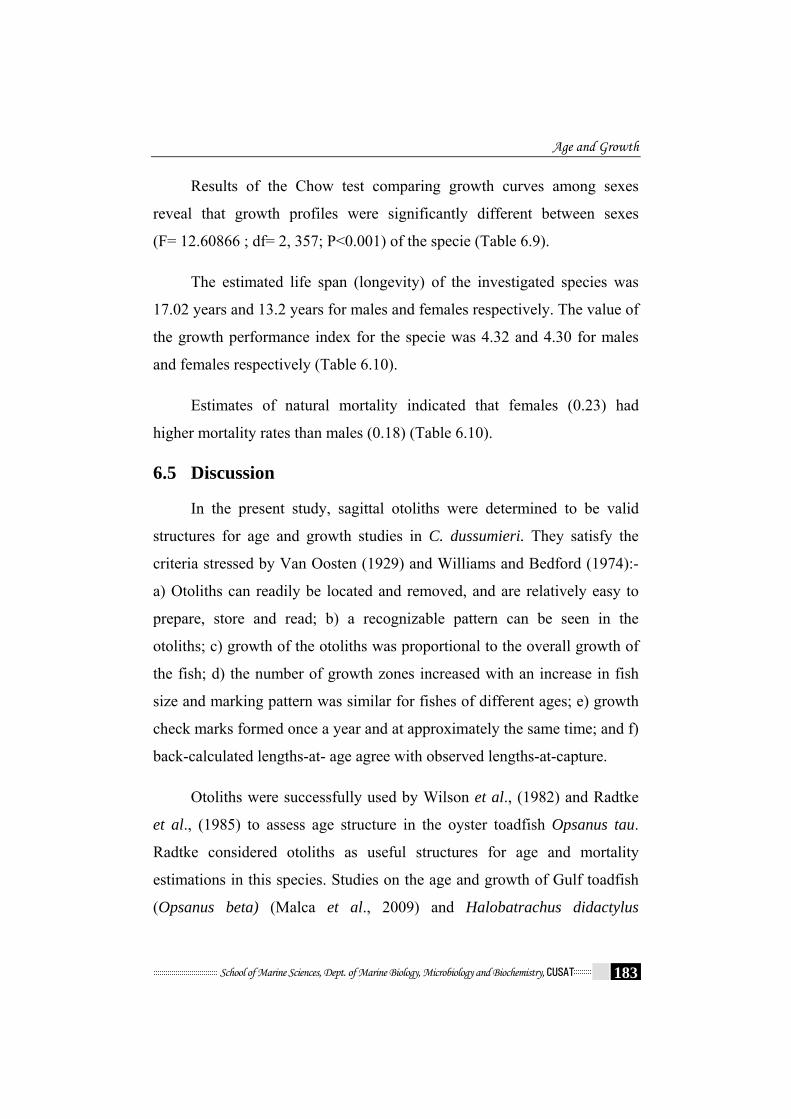
Results of the Chow test comparing growth curves among sexes
reveal that growth profiles were significantly different between sexes
(F= 12.60866 ; df= 2, 357; P<0.001) of the specie (Table 6.9).
The estimated life span (longevity) of the investigated species was
17.02 years and 13.2 years for males and females respectively. The value of
the growth performance index for the specie was 4.32 and 4.30 for males
and females respectively (Table 6.10).
Estimates of natural mortality indicated that females (0.23) had
higher mortality rates than males (0.18) (Table 6.10).
6.5 Discussion
In the present study, sagittal otoliths were determined to be valid
structures for age and growth studies in C. dussumieri. They satisfy the
criteria stressed by Van Oosten (1929) and Williams and Bedford (1974):-
a) Otoliths can readily be located and removed, and are relatively easy to
prepare, store and read; b) a recognizable pattern can be seen in the
otoliths; c) growth of the otoliths was proportional to the overall growth of
the fish; d) the number of growth zones increased with an increase in fish
size and marking pattern was similar for fishes of different ages; e) growth
check marks formed once a year and at approximately the same time; and f)
back-calculated lengths-at- age agree with observed lengths-at-capture.
Otoliths were successfully used by Wilson et al., (1982) and Radtke
et al., (1985) to assess age structure in the oyster toadfish Opsanus tau.
Radtke considered otoliths as useful structures for age and mortality
estimations in this species. Studies on the age and growth of Gulf toadfish
(Opsanus beta) (Malca et al., 2009) and Halobatrachus didactylus

Chapter-6
School of Marine Sciences, Dept. of Marine Biology, Microbiology and Biochemistry, CUSAT 184
(Palazon-Fernandez et al., 2010) revealed that sagittal otoliths as valid
structures for age and growth studies, similar to the results found in
Opsanus tau (Wilson et al., 1982). In C. dussumieri otoliths exhibited a
well- defined and consistent mark pattern consisting of one narrow
translucent zone deposited during slow growth and one broad opaque white
zone formed during fast growth.
Ring validation in age and growth studies is essential in regions
where climatic conditions are less variable, resulting in less clear otolith
marks (Beamish and McFarlane, 1983; Jepsen et al., 1999). The timing of
annulus formation in tropical fishes is not well understood, but Fowler
(1995) noted that most tropical marine species share a pattern similar to
temperate fishes; fast growth synchronous with opaque zone deposition.
Although the difficulty of resolving the margins of otolith increments for
MIR analysis may influence its validity (Campana, 2001), it has been
useful for validating the annual periodicity of the annulus in numerous fish
species (Takita et al., 1993; Colloca et al., 2003; Boudaya et al., 2008; Lin
and Tzeng, 2009; Palazon-Fernandez et al., 2010). Annual deposition of an
opaque and a hyaline band in the otoliths of tropical fishes has been
recorded for Lutjanus kasmira (Morales-Nin and Ralston, 1990),
surgeonfishes, parrotfishes (Choat and Axe, 1996) and chaetodon larvatus
(Zekeria, 2006). In reviews of otolith studies in tropical latitudes, Beckman
and Wilson (1995) found that opaque zone deposition occurs during period
of increased growth, whereas the corresponding translucent zone is
formed during periods of low metabolic activity. The marginal increment
analysis in this study showed that one opaque and one translucent zone
are formed each year. The thin translucent bands, considered annuli,
corresponding to the period of slow growth begins in June and is
Age and Growth
School of Marine Sciences, Dept. of Marine Biology, Microbiology and Biochemistry, CUSAT 185
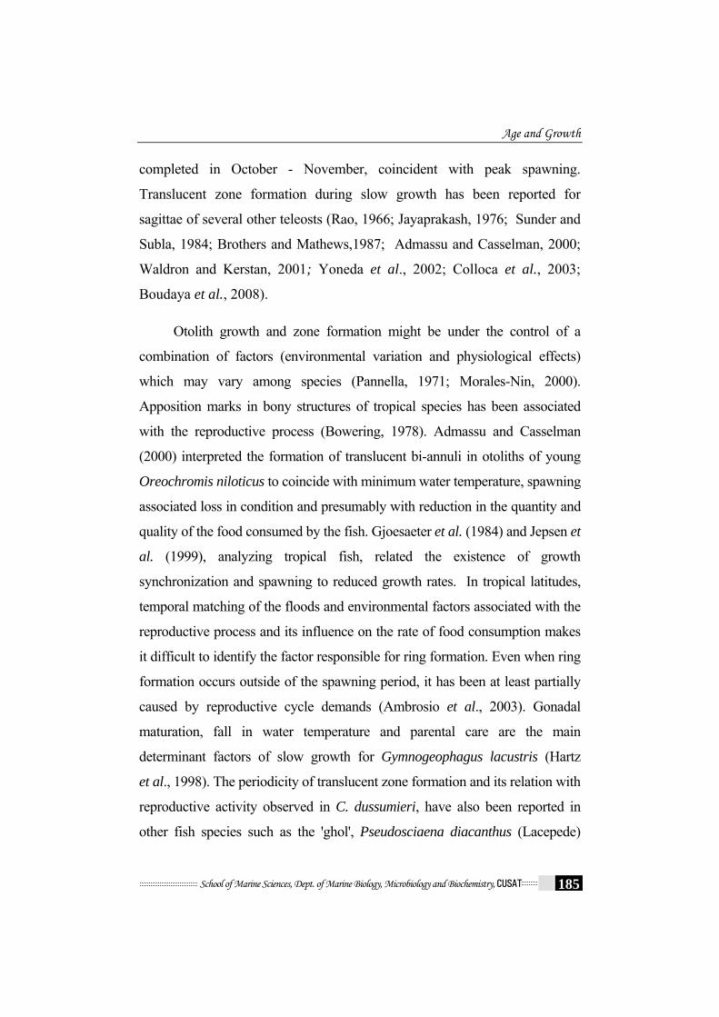
completed in October - November, coincident with peak spawning.
Translucent zone formation during slow growth has been reported for
sagittae of several other teleosts (Rao, 1966; Jayaprakash, 1976; Sunder and
Subla, 1984; Brothers and Mathews,1987; Admassu and Casselman, 2000;
Waldron and Kerstan, 2001; Yoneda et al., 2002; Colloca et al., 2003;
Boudaya et al., 2008).
Otolith growth and zone formation might be under the control of a
combination of factors (environmental variation and physiological effects)
which may vary among species (Pannella, 1971; Morales-Nin, 2000).
Apposition marks in bony structures of tropical species has been associated
with the reproductive process (Bowering, 1978). Admassu and Casselman
(2000) interpreted the formation of translucent bi-annuli in otoliths of young
Oreochromis niloticus to coincide with minimum water temperature, spawning
associated loss in condition and presumably with reduction in the quantity and
quality of the food consumed by the fish. Gjoesaeter et al. (1984) and Jepsen et
al. (1999), analyzing tropical fish, related the existence of growth
synchronization and spawning to reduced growth rates. In tropical latitudes,
temporal matching of the floods and environmental factors associated with the
reproductive process and its influence on the rate of food consumption makes
it difficult to identify the factor responsible for ring formation. Even when ring
formation occurs outside of the spawning period, it has been at least partially
caused by reproductive cycle demands (Ambrosio et al., 2003). Gonadal
maturation, fall in water temperature and parental care are the main
determinant factors of slow growth for Gymnogeophagus lacustris (Hartz
et al., 1998). The periodicity of translucent zone formation and its relation with
reproductive activity observed in C. dussumieri, have also been reported in
other fish species such as the 'ghol', Pseudosciaena diacanthus (Lacepede)

Chapter-6
School of Marine Sciences, Dept. of Marine Biology, Microbiology and Biochemistry, CUSAT 186
(Rao, 1966); two-banded seabream, Diplodus vulgaris (Pajuelo and Lorenzo,
2003); spotted seatrout, Cynoscion nebulosus (Ihde and Chittenden, 2003);
spotted goatfish, Pseudupeneus maculatus (Santana et al., 2006); striped
mullet, Mugil platanus (Castro et al., 2009). A possible explanation for this
would be that, during the spawning season, energy may be allocated primarily
for gamete production instead of somatic growth. Further more, during the
spawning season male toadfishes may decrease their feeding activity while
they guard nests and take care of the eggs and fry. This hypothesis, although
plausible for adult fish, cannot explain the formation of annuli in young
immature fish. So annulus formation is not necessarily a direct consequence of
the reproductive activity, and must also be related to seasonal patterns of
growth rather than reproduction. Similar observations are reported in other
toadfish species like Halobatrachus didactylus (Palazon-Fernandez et al., 2010
and Opsanus beta (Barimo et al., 2007; Malca et al., 2009). Many factors other
than spawning can affect growth rates of fish, including seasonal changes in
environmental factors, mainly water temperatures, photoperiod, feeding
regime and related effects on metabolism, as observed in other fish species
(Campana and Neilson, 1985; Campana, 1984; Morales – Nin and Ralston,
1990; Bullock et al., 1992; Newman et al., 2000; Pajuelo and Lorenzo, 2003).
Present results also imply that translucent bands coincide with a period of
decrease in temperature, reduced feeding (chapter 4) and lower body condition
(depletion of peritoneal fat reserves) (Chapter 7 and 8).
In some fish populations, when growth over the lifetime is measured,
stanzas are observed, with sudden changes in growth rate between the
stanzas. The most commonly reported growth rate stanzas are those related
to larval metamorphosis, with different growth patterns, pre- and post
metamorphosis. Other well-described stanzas are related to physiological

Age and Growth
School of Marine Sciences, Dept. of Marine Biology, Microbiology and Biochemistry, CUSAT 187
change or to maturation (Saborido-Rey et al., 2004). In C. dussumieri, the
growth rate stanzas precede and follow an age of 3 or 4 years (as revealed
from Table 6.5 and Fig.6.9). This can be attributed to maturation and
physiological change. In redfish, Sebastes mentella, the growth rate stanzas
were observed at an age of 5 or 6 years. Saborido-Rey et al. (2004) related
the alteration to progressive shift from a more pelagic to a more demersal
habitat with age and to change in feeding behaviour.
Of the 200 males and 157 females sampled, males were predominant
in specimens greater than 5 years old. The data also suggest a difference in
maximum length and age attained by the sexes. The oldest male collected
was 9 years old, whereas the oldest female was 7 years old. Examination of
the otoliths of oyster toadfish in South Carolina, revealed that ages ranged
from <1 to 8 year, with a median age of 3 year (Wilson et al., 1982). The
estimated ages of males and females of Opsanus beta ranged from <1 year
to 6 and 5 years, respectively (Malca et al., 2009). Schwartz and Dutcher
(1963) and Radtke et al. (1985) in Opsanus tau and Palazon-Fernandez et al.
(2010) in Halobatrachus didactylus reported maximum ages of 12 and 9
years for males and females toadfishes respectively. Radtke et al. (1985)
concluded that the differences in sex-specific numbers by age for O. tau
resulted from females experiencing a higher mortality rate as a result of the
increased energy investment for gametes in females.
Toadfishes are medium-lived fishes, and the maximum age for each
sex must be near the maximum age for the species owing to the low fishing
pressure on the population. C. dussumieri reach sexual maturity at 131 mm
TL for males and 141 mm TL for females (refer Chapter 5), which
correspond to 2 and >2 years of age for males and females respectively.
Similarly, Halobatrachus didactylus reach sexual maturity at 160 mm TL
Chapter-6
School of Marine Sciences, Dept. of Marine Biology, Microbiology and Biochemistry, CUSAT 188
for males and 191 mm TL for females, (Palazon-Fernandez et al., 2001),
which correspond to 2 and 3 years of age for males and females
respectively (Palazon-Fernandez et al., 2010). Gudger (1910) confirmed
that toadfish do not spawn until age 2. Similarly Wilson et al. (1982)
reported that O. tau became sexually matured between 2 to 7 years.
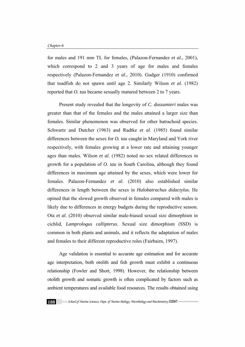
Present study revealed that the longevity of C. dussumieri males was
greater than that of the females and the males attained a larger size than
females. Similar phenomenon was observed for other batrachoid species.
Schwartz and Dutcher (1963) and Radtke et al. (1985) found similar
differences between the sexes for O. tau caught in Maryland and York river
respectively, with females growing at a lower rate and attaining younger
ages than males. Wilson et al. (1982) noted no sex related differences in
growth for a population of O. tau in South Carolina, although they found
differences in maximum age attained by the sexes, which were lower for
females. Palazon-Fernandez et al. (2010) also established similar
differences in length between the sexes in Halobatrachus didactylus. He
opined that the slowed growth observed in females compared with males is
likely due to differences in energy budgets during the reproductive season.
Ota et al. (2010) observed similar male-biased sexual size dimorphism in
cichlid, Lamprologus callipterus. Sexual size dimorphism (SSD) is
common in both plants and animals, and it reflects the adaptation of males
and females to their different reproductive roles (Fairbairn, 1997).
Age validation is essential to accurate age estimation and for accurate
age interpretation, both otolith and fish growth must exhibit a continuous
relationship (Fowler and Short, 1998). However, the relationship between
otolith growth and somatic growth is often complicated by factors such as
ambient temperatures and available food resources. The results obtained using

Age and Growth
School of Marine Sciences, Dept. of Marine Biology, Microbiology and Biochemistry, CUSAT 189
the back-calculation method are very satisfactory and demonstrate the validity
of using otoliths for estimating the growth of the flat toadfish. As ring
formation is regular, the otoliths can be used for age determination and
because the fish length and otolith size are closely correlated, it is judged as
valid to permit the use of measurements to previously formed marks to back
calculate the growth history (Campana, 1990; Francis, 1990). Similar results
were reported for O. tau by Radtke et al. (1985) and for Halobatrachus
didactylus by Palazon-Fernandez et al. (2010). They found a strong association
between otolith dimension and body growth and stated that otolith length
could be chosen as the best parameter for estimation of fish total length.
A method of validating growth parameters involves the comparison
of growth performance index (Φ́) in terms of growth in length with other
estimates obtained for the same or a closely similar species (Gayanilo and
Pauly, 1997). The von Bertalanffy growth parameter estimates of
C. dussumieri obtained in the present study were compared with the status
of those of other batrachoididae recorded in the previous studies (Table 6.11).
The growth performance index of C. dussumieri obtained in the present study
falls within the values mentioned of those estimated for other members of the
batrachoid family. Even though differences can be observed between the
population parameters, the similarity of the Φ́ values indicates the existence of
a similar growth pattern. According to Pauly and Munro (1984), related
species present similar values of Φ́ and each taxa may have a particular
distribution of values, different from other taxa, and can be described by its
mean value, i.e. the value of this parameter for species from a particular family
should be closer to its average than to the average value of another family. The
parameter Φ´ = Log10 K + 2 Log10 L∞ can be used as it has been found that
similar species with different growth parameters can have similar Φ́ estimates.

Chapter-6
School of Marine Sciences, Dept. of Marine Biology, Microbiology and Biochemistry, CUSAT 190
Moreover, differences in growth patterns can be the result of differences in
genetic structure and / or differences in temperature, density of food and
diseases (Pauly, 1994; Wootton, 1990).
The values of natural mortality (M) of C. dussumieri obtained in the
present study were similar to the figures recorded for other batrachiod
species in the previous studies (Table 6.11) except for O. tau females,
which have a higher mortality (M = 0.41) than males of the same species,
and to the estimations made for the rest of the batrachoids. The present
study estimated a higher natural mortality rate for females than for males.
This further substantiated the studies of Radtke et al. (1985, O. tau) and
Palazon-Fernandez et al. (2010, Halobatrachus didactylus). Although
variation in mortality can be explained by such diverse factors as age,
density, illnesses, parasites, food supply, abundance of predators, water
temperature, fishery pressure, sex and size (Vetter, 1988), according to
Palazon-Fernandez et al. ( 2010), this may be caused by increased
predation due to their smaller size, to behavioural or distributional
differences that make them more susceptible to predation or fishing effort,
or to the existence of higher environmental pressures acting on the
females. Another possibility is that they simply reach senescence before
males.
In conclusion, whole–view otolith examination can provide a precise
method for determining age and back-calculated lengths of C. dussumieri.
This study provides the first detailed estimates of age, growth and mortality
rate for the specie. Future research should focus on the environmental and
internal factors affecting their growth.

Age and Growth
School of Marine Sciences, Dept. of Marine Biology, Microbiology and Biochemistry, CUSAT 191
Table 6.1. Analysis of variance on monthly mean MIR of C. dussumieri
Descriptives (Monthwise) MIR
95% Confidence Interval for
Mean N Mean Std. Deviation
Std. Error
Lower Bound
Upper Bound
Min
imum
Max
imum
October 39 .0474 .09792 .01568 .0157 .0792 .00 .37
November 25 .1559 .16940 .03388 .0860 .2259 .00 .70
December 9 .1935 .20088 .06696 .0390 .3479 .00 .50
January 17 .2114 .17093 .04146 .1235 .2993 .00 .56
February 34 .2062 .22204 .03808 .1287 .2836 .00 1.08
March 50 .2100 .17347 .02453 .1607 .2593 .00 1.08
April 27 .3266 .19944 .03838 .2477 .4055 .03 .83
May 61 .3662 .22455 .02875 .3087 .4237 .05 .99
June 10 .4748 .30762 .09728 .2548 .6949 .10 1.13
July 20 .5332 .39310 .08790 .3492 .7172 .00 1.25
August 18 .3163 .22749 .05362 .2032 .4294 .00 .67
September 41 .1229 .19591 .03060 .0611 .1847 -.06 .67
Total 351 .2447 .24525 .01309 .2190 .2705 -.06 1.25
ANOVA MIR
Sum of Squares
df Mean
Square F Sig.
Between Groups 5.844 11 .531 11.844 <.001
Within Groups 15.207 339 .045
Total 21.052 350
Significant

Chapter-6
School of Marine Sciences, Dept. of Marine Biology, Microbiology and Biochemistry, CUSAT 192
Table 6.2. Analysis of variance on age group mean MIR of C. dussumieri
Descriptives (age –group) MIR
95% Confidence Interval for Mean N Mean Std.
DeviationStd. Error Lower
Bound Upper Bound M
inim
um
Max
imum
1.00 10 .1454 .09812 .03103 .0752 .2156 .03 .31
2.00 25 .1648 .15982 .03196 .0989 .2308 .00 .75
3.00 90 .1888 .17903 .01887 .1513 .2263 -.06 .69
4.00 93 .3321 .24556 .02546 .2815 .3827 .00 1.11
5.00 76 .2194 .25630 .02940 .1608 .2779 .00 1.08
6.00 30 .2656 .32197 .05878 .1453 .3858 .00 1.25
7.00 10 .1962 .32487 .10273 -.0362 .4286 .00 1.00
8.00 8 .2833 .39271 .13884 -.0451 .6116 .00 1.13
9.00 9 .3978 .23230 .07743 .2192 .5763 .00 .83
Total 351 .2447 .24525 .01309 .2190 .2705 -.06 1.25
ANOVA MIR
Sum of Squares
df Mean
Square F Sig.
Between Groups 1.558 8 .195 3.416 .001
Within Groups 19.494 342 .057
Total 21.052 350
Significant at 0.1% level
Table 6.3. T- test on MIR of males and females of C. dussumieri
Sex N Mean Std.
Deviation t df
Sig. (2-tailed)
Males 199 .2348 .24576 -.863 349 .389 MIR
Females 152 .2577 .24478
Not Significant
Age and Growth
School of Marine Sciences, Dept. of Marine Biology, Microbiology and Biochemistry, CUSAT 193
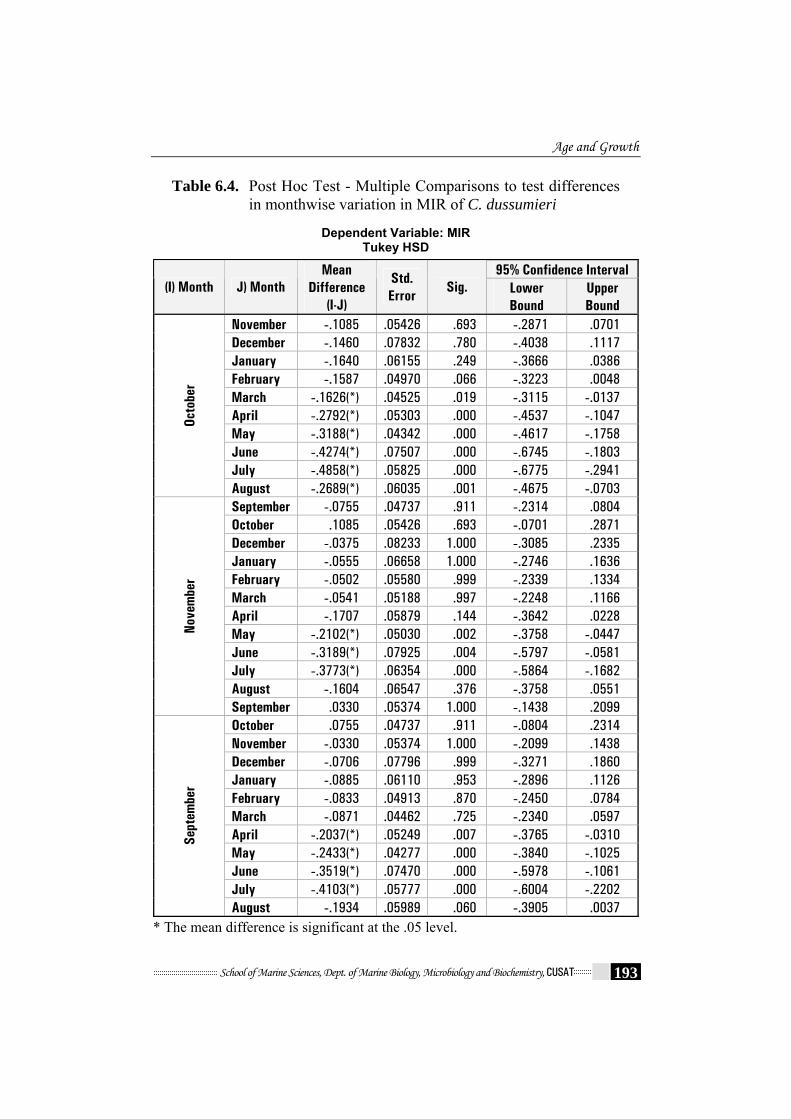
Table 6.4. Post Hoc Test - Multiple Comparisons to test differences in monthwise variation in MIR of C. dussumieri
Dependent Variable: MIR
Tukey HSD
95% Confidence Interval (I) Month J) Month
Mean Difference
(I-J)
Std. Error
Sig. Lower Bound
Upper Bound
November -.1085 .05426 .693 -.2871 .0701 December -.1460 .07832 .780 -.4038 .1117 January -.1640 .06155 .249 -.3666 .0386 February -.1587 .04970 .066 -.3223 .0048 March -.1626(*) .04525 .019 -.3115 -.0137 April -.2792(*) .05303 .000 -.4537 -.1047 May -.3188(*) .04342 .000 -.4617 -.1758 June -.4274(*) .07507 .000 -.6745 -.1803 July -.4858(*) .05825 .000 -.6775 -.2941
Octo
ber
August -.2689(*) .06035 .001 -.4675 -.0703 September -.0755 .04737 .911 -.2314 .0804 October .1085 .05426 .693 -.0701 .2871 December -.0375 .08233 1.000 -.3085 .2335 January -.0555 .06658 1.000 -.2746 .1636 February -.0502 .05580 .999 -.2339 .1334 March -.0541 .05188 .997 -.2248 .1166 April -.1707 .05879 .144 -.3642 .0228 May -.2102(*) .05030 .002 -.3758 -.0447 June -.3189(*) .07925 .004 -.5797 -.0581 July -.3773(*) .06354 .000 -.5864 -.1682 August -.1604 .06547 .376 -.3758 .0551
Nove
mbe
r
September .0330 .05374 1.000 -.1438 .2099 October .0755 .04737 .911 -.0804 .2314 November -.0330 .05374 1.000 -.2099 .1438 December -.0706 .07796 .999 -.3271 .1860 January -.0885 .06110 .953 -.2896 .1126 February -.0833 .04913 .870 -.2450 .0784 March -.0871 .04462 .725 -.2340 .0597 April -.2037(*) .05249 .007 -.3765 -.0310 May -.2433(*) .04277 .000 -.3840 -.1025 June -.3519(*) .07470 .000 -.5978 -.1061 July -.4103(*) .05777 .000 -.6004 -.2202
Sept
embe
r
August -.1934 .05989 .060 -.3905 .0037 * The mean difference is significant at the .05 level.
Chapter-6
School of Marine Sciences, Dept. of Marine Biology, Microbiology and Biochemistry, CUSAT 194
Table 6.5. Post Hoc test - Multiple comparisons to test the differences
in MIR of age groups of C. dussumieri
Dependent Variable: MIR Tukey HSD
95% Confidence Interval (I) Age (J) Month
Mean Difference
(I-J)
Std. Error
Sig. Lower Bound
Upper Bound
1.00 .0434 .07958 1.000 -.2050 .2918
2.00 .0240 .05398 1.000 -.1445 .1925
4.00 -.1433(*) .03530 .002 -.2535 -.0331
5.00 -.0306 .03719 .996 -.1467 .0855
6.00 -.0768 .05033 .843 -.2339 .0804
7.00 -.0074 .07958 1.000 -.2558 .2411
8.00 -.0944 .08808 .978 -.3694 .1805
3.00
9.00 -.2090 .08347 .234 -.4695 .0516
1.00 .1867 .07945 .316 -.0614 .4347
2.00 .1672 .05379 .052 -.0007 .3351
3.00 .1433(*) .03530 .002 .0331 .2535
5.00 .1127 .03692 .061 -.0025 .2280
6.00 .0665 .05013 .923 -.0900 .2230
7.00 .1359 .07945 .740 -.1121 .3839
8.00 .0488 .08797 1.000 -.2258 .3234
4.00
9.00 -.0657 .08334 .997 -.3259 .1945
* The mean difference is significant at the 0.05 level.

Age and Growth
School of Marine Sciences, Dept. of Marine Biology, Microbiology and Biochemistry, CUSAT 195
Table 6.6a. Mean observed and back-calculated lengths-at-ages
for male C. dussumieri
Annulus Age class (years)
N Observed Mean LT I II III IV V VI VII VIII IX
1 10 87.1 79.1 0.0 0.0 0.0 0.0 0.0 0.0 0.0 0.0
2 11 129.2 85.6 123.8 0.0 0.0 0.0 0.0 0.0 0.0 0.0
3 35 175.2 93.2 133.2 168.7 0.0 0.0 0.0 0.0 0.0 0.0
4 42 213.2 92.4 135.5 173.3 206.2 0.0 0.0 0.0 0.0 0.0
5 48 233.2 91.5 131.4 165.4 197.3 227.3 0.0 0.0 0.0 0.0
6 27 247.1 92.0 130.8 162.7 191.1 217.2 232.4 0.0 0.0 0.0
7 9 263.3 92.2 128.0 154.6 181.7 209.0 234.1 258.7 0.0 0.0
8 8 271.4 93.1 120.5 148.3 174.4 199.2 225.4 244.6 265.5 0.0
9 9 292.3 96.6 127.8 151.4 176.4 197.7 222.1 244.4 264.5 284.7
Mean 91.4 131.3 165.5 195.1 218.1 229.9 249.4 265.0 284.7
Increment 39.9 34.2 29.7 23.0 11.8 19.5 15.6 19.7
Table 6.6b. Mean observed and back-calculated lengths-at-ages
for female C. dussumieri
Annulus Age class (years)
N Observed Mean LT I II III IV V VI VII
1 5 86.2 78.7 0.0 0.0 0.0 0.0 0.0 0.0
2 14 123.4 83.7 118.2 0.0 0.0 0.0 0.0 0.0
3 55 172.2 88.1 130.3 166.3 0.0 0.0 0.0 0.0
4 51 202.4 90.5 132.2 167.2 192.3 0.0 0.0 0.0
5 27 221.6 89.5 128.8 160.7 185.1 216.5 0.0 0.0
6 4 233.0 90.4 121.6 156.1 178.3 208.5 230.6 0.0
7 1 246.0 92.7 115.6 145.6 166.5 191.4 217.8 246.0
Mean 88.3 129.3 165.1 189.0 214.8 228.1 246.0
Increment 41.0 35.8 23.9 25.7 13.3 17.9
Chapter-6
School of Marine Sciences, Dept. of Marine Biology, Microbiology and Biochemistry, CUSAT 196
Table 6.7. Growth parameters derived from the von Bertalanffy
growth function
Parameter Males Females
L∞ 351 303
k 0.168 0.216
t0 -0.81 -0.65
n 200 157
Table 6.8. Mean observed, back-calculated (B-C) and theoretical total lengths for male and female
Males Females Age
Observed B-C Theoretical Observed B-C Theoretical
1.0 87.1 91.4 92.03 86.2 88.3 90.7
2.0 129.2 131.3 132.08 123.4 129.3 131.9
3.0 175.2 165.5 165.9 172.2 165.1 165.2
4.0 213.2 195.1 194.6 202.4 189.0 191.9
5.0 233.2 218.1 218.8 221.6 214.8 213.5
6.0 247.1 229.9 239.2 233.0 228.1 230.9
7.0 263.3 249.4 256.5 246.0 246.0 244.9
8.0 271.4 265.0 271.1
9.0 292.3 284.7 283.5

Age and Growth
School of Marine Sciences, Dept. of Marine Biology, Microbiology and Biochemistry, CUSAT 197
Table 6.9. Results of the Chow test comparing growth curves among sexes
ANOVAb (Pooled)
Model Sum of Squares df Mean Square F Sig.
Regression 687991.288 1 687991.288 2167.454 .000a
Residual 113953.454 359 317.419 1
Total 801944.742 360
ANOVAb (Male)
Model Sum of Squares df Mean Square F Sig.
Regression 418301.159 1 418301.159 1166.576 .000a
Residual 70997.221 198 358.572 1
Total 489298.380 199
ANOVAb (Female)
Model Sum of Squares df Mean Square F Sig.
Regression 199894.849 1 199894.849 896.870 .000a
Residual 35437.995 155 222.880 1
Total 235332.845 156
Ess_c =113953.454, ess_1 = 70997.221 and ess_2= 35437.995, k=2 and N1=200, N2= 156
Hence f statistic is given by F= 12.60866
P value is given by 0.0000051. Hence we reject the null hypothesis that the
regression coefficients are equal and we need to consider separate
regression for both male and female.
Table 6.10. Estimated life span, growth performance index and natural mortality rates for male and female of C. dussumieri
Sex Life span Growth performance
index Natural
mortality Males 17.02 4.32 0.18 Females 13.2 4.30 0.23

Chapter-6
School of Marine Sciences, Dept. of Marine Biology, Microbiology and Biochemistry, CUSAT 198
Table 6.11. Comparisons of estimates of the von Bertalanffy growth parameters, Φ' value and natural mortality rates reported by various authors for some batrachoid species
Species L∞ K t0 Φ'1 M2 Investigators
Opsanus beta (♂&♀) 300 0.168 -0.09 4.18 0.17 Serafy et al., 1997
Opsanus beta (♂&♀) 300 0.218 -0.27 4.29 0.22 Serafy et al., 1997
Opsanus tau ♂ 407.5 0.15 -0.33 4.40 0.15 Radtke et al., 1985
Opsanus tau ♀ 271.5 0.39 -0.44 4.46 0.41 Radtke et al., 1985
Halobatrachus didactylus ♂ 477.1 0.15 -0.59 4.53 0.15 Palazon-Fernandez et al., 2010
Halobatrachus didactylus ♀ 363.7 0.20 -0.75 4.42 0.21 Palazon-Fernandez et al., 2010
C. dussumieri ♂ 351 0.168 -0.81 4.32 0.18 This study
C. dussumieri ♀ 303 0.216 -0.65 4.30 0.23 This study
1 values calculated from author’s data; 2 values calculated from author’s data using the equation of Taylor (1958)

Age and Growth
School of Marine Sciences, Dept. of Marine Biology, Microbiology and Biochemistry, CUSAT 199
y = 38.954x - 22.728r = 0.926
R2 = 0.8583n = 200
0
50
100
150
200
250
300
350
0 2 4 6 8 10
Otolith length (mm)
Tota
l len
gth
(mm
)
(a) Length of the otolith
y = 2775.7x + 128.63r = 0.868
R2 = 0.7549n = 200
0
50
100
150
200
250
300
350
400
0 0.02 0.04 0.06 0.08 0.1
Otolith weight (mg)
Tota
l len
gth
(mm
)
(b) Weight of the otolith
Fig. 6.1. Relationship between total length – at- capture of the fish (Male)

Chapter-6
School of Marine Sciences, Dept. of Marine Biology, Microbiology and Biochemistry, CUSAT 200
y = 34.742x - 12.065r = 0.8978
R2 = 0.8061n = 157
0
50
100
150
200
250
300
0 2 4 6 8
Otolith length (mm)
Tota
l len
gth
(mm
)
(a) Length of the otolith
y = 2345.2x + 130.69r = 0.8037 R2 = 0.646
n = 157
0
50
100
150
200
250
300
0 0.02 0.04 0.06 0.08
Otolith weight (mg)
Tota
l len
gth
(mm
)
(b) Weight of the otolith
Fig. 6.2. Relationship between total length – at- capture of the fish (Female)
Age and Growth
School of Marine Sciences, Dept. of Marine Biology, Microbiology and Biochemistry, CUSAT 201
y = 71.877x + 28.949r = 0.942
R2 = 0.8891n = 200
0
50
100
150
200
250
300
350
0 1 2 3 4 5
Otolith radius (mm)
Tota
l len
gth
(mm
)
Fig.6.3.a. Relationship between fish total length -at- capture
(T.LC) and otolith radius -at- capture (O.RC) (Male).
y = 66.154x + 34.585r = 0.9025
R2 = 0.8146n =157
0
50
100
150
200
250
300
0 1 2 3 4
Otolith radius (mm)
Tota
l len
gth
(mm
)
Fig.6.3.b. Relationship between fish total length -at- capture
(T.LC) and otolith radius -at- capture (O.RC) (Female).

Chapter-6
School of Marine Sciences, Dept. of Marine Biology, Microbiology and Biochemistry, CUSAT 202
Fig.6.4. Photomicrograph of a sagittal otolith of C. dussumieri
showing growth increments. OL – Otolith Length, OR- Otolith radius, rn- ring radius
Age and Growth
School of Marine Sciences, Dept. of Marine Biology, Microbiology and Biochemistry, CUSAT 203
Fig. 6.4a. Otolith from 89 mm TL C. dussumieri in age group 1
Fig. 6.4b. Otolith from 141 mm TL C. dussumieri in age group 2
Fig. 6.4c. Otolith from 163 mm TL C. dussumieri in age group 3
Fig. 6.4d. Otolith from 194 mm TL C. dussumieri in age group 4
Fig. 6.4e. Otolith from 214 mm TL C. dussumieri in age group 5
Fig. 6.4f. Otolith from 240 mm TL C. dussumieri in age group 6

Chapter-6
School of Marine Sciences, Dept. of Marine Biology, Microbiology and Biochemistry, CUSAT 204
05
101520253035
O N D J F M A M J J A S
Months
Freq
uenc
y (%
)
Opaque Translucent Fig. 6.5a. Monthly frequency of opaque and translucent margins
of otoliths of males of C. dussumieri
Fig. 6.4e. Otolith from 264 mm TL C. dussumieri in age group 7
Fig. 6.4f. Otolith from 270 mm TL C. dussumieri in age group 8
Fig. 6.4f. Otolith from 284 mm TL C. dussumieri in age group 9
Age and Growth
School of Marine Sciences, Dept. of Marine Biology, Microbiology and Biochemistry, CUSAT 205
0
10
20
30
40
O N D J F M A M J J A S
Months
Freq
uenc
y (%
)
Opaque Translucent
Fig. 6.5b. Monthly frequency of opaque and translucent margins of otoliths of females of C. dussumieri
0
10
20
30
40
O N D J F M A M J J A S
Months
Freq
uenc
y (%
)
Opaque Translucent
Fig. 6.5c. Monthly frequency of opaque and translucent margins of otoliths of pooled specimens of C. dussumieri
Chapter-6
School of Marine Sciences, Dept. of Marine Biology, Microbiology and Biochemistry, CUSAT 206
Mean MIR (Males)
00.10.20.30.40.50.6
Oct Nov Dec Jan Feb Mar Apr May Jun Jul Aug SepMonths
MIR
Fig.6.6a. Mean marginal increment in otoliths of C. dussumieri
by month (Males). The error bars represent ± standard error (SE).
Mean MIR (Females)
0
0.1
0.2
0.3
0.4
0.5
0.6
0.7
Oct Nov Dec Jan Feb Mar Apr May Jun Jul Aug Sep
Months
MIR
Fig.6.6b. Mean marginal increment in otoliths of C. dussumieri by
month (Females). The error bars represent ± standard error (SE).

Age and Growth
School of Marine Sciences, Dept. of Marine Biology, Microbiology and Biochemistry, CUSAT 207
Mean MIR (Pooled)
00.10.20.30.40.50.60.7
Oct Nov Dec Jan Feb Mar Apr May Jun Jul Aug Sep
Months
MIR
Fig.6.6c. Mean marginal increment in otoliths of C. dussumieri
by month (pooled). The error bars represent ± standard error (SE).
0
5
10
15
20
25
30
35
40
1 2 3 4 5 6 7 8 9
Age (years)
Freq
uenc
y (%
) MalesFemales
Fig.6.7. Age distribution for male and female C. dussumieri
Chapter-6
School of Marine Sciences, Dept. of Marine Biology, Microbiology and Biochemistry, CUSAT 208
y = 0.845x + 54.411Linf = 351
-100
0
100
200
300
400
500
-100 0 100 200 300 400 500
Lt (mm)
Lt+1
(mm
)
(a)
Y= - 0.1629X + 5.7246t0 = - 0.81
0
1
2
3
4
5
6
7
-5 -4 -3 -2 -1 0 1 2 3 4 5 6 7 8 9 10
Age (year)
Ln(L
inf -
Lt)
(b)
Fig. 6.8. Ford-Walford Graph for male C. dussumieri: (a) L∞, (b) t0

Age and Growth
School of Marine Sciences, Dept. of Marine Biology, Microbiology and Biochemistry, CUSAT 209
Y = 0.8058X + 58.866L inf = 303
-150-100-50
050
100150200250300350400
-100 0 100 200 300 400
Lt (mm)
Lt+1
(mm
)
(c)
Y = -0.215X + 5.5741t0 = - 0.646
0
1
2
3
4
5
6
7
-5 -4 -3 -2 -1 0 1 2 3 4 5 6 7 8 9 10
Age (year)
Ln(L
inf - L
t)
(d)
Fig. 6.8. Ford-Walford Graph for female C. dussumieri: (c) L∞, (d) t0
Chapter-6
School of Marine Sciences, Dept. of Marine Biology, Microbiology and Biochemistry, CUSAT 210
0
50
100
150
200
250
300
1 2 3 4 5 6 7 8 9Age (Years)
Tota
l leng
th (m
m)
Males Females Fig. 6.9. von Bertalanffy growth curve derived from back calculated
age-at-lengths for male and female C. dussumieri
….. …..