Embed Size (px)
Citation preview
Randall W. Ryan' James F. Lally
Zelimir Kozic
Received May 6, 1987; accepted after revision August 30, 1987.
'All authors: Department of Radiology, The Medical Center of Delaware, 4755 Stanton-Ogletown Rd ., Newark , DE 19718. Address reprint requests to R. W. Ryan.
AJNR 9:363-366, March/April 1988 0195-6108/88/0902-0363 © American Society of Neuroradiology
363
Asymptomatic Calcified Herniated Thoracic Disks: CT Recognition
Among 270 CT scans of the thorax obtained over a 7 -month period, four patients (1.5%) with calcified herniated thoracic disks were identified. Each of these patients presented with abnormal chest radiographs and had a CT examination for evaluation of suspected malignancy. None showed any signs or symptoms of thoracic spinal cord compression. The clinical significance of incidental thoracic disk protrusions is unknown. It may be that these patients are at risk for the later development of symptomatic disk disease.
Herniation of a thoracic intervertebral disk is an uncommon entity with a reported annual incidence of one patient per one million population [1]. Less than 4% of all disk herniations occur in the thoracic spine [2]. In our review of the literature, virtually all patients with thoracic disk protrusions presented with symptoms, including midline back and radicular pain, sensory and motor disturbances, and, occasionally, gastrointestinal or urogenital dysfunction. We have recently diagnosed incidental calcified thoracic disk herniations in four asymptomatic patients over a 7 -month period. Our experience suggests a higher incidence of thoracic disk herniation than has previously been reported. To our knowledge there have been no reports of the CT findings of asymptomatic calcified herniated thoracic disks.
Case Reports
CT examinations of the thorax were performed in 270 patients over a 7-month period. Incidental asymptomatic disk protrusions were identified in four of these patients. The clinical course and CT findings for these four patients are presented.
Case 1
A 47-year old woman presented with a 3-month history of increasing dyspnea and occasional hemoptysis. She had previously been in good health and denied fever or weight loss. A chest radiograph revealed a large right upper lobe mass. Bronchoscopy was negative, but a transthoracic needle biopsy revealed squamous cell carcinoma. CT examination of the thorax disclosed a large, non calcified pulmonary mass occupying the upper half of the right thorax with invasion of the mediastinum and metastatic spread to the contralateral lung. Further workup revealed metastases to the brain.
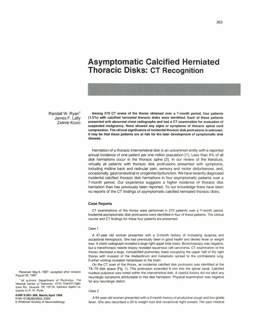
On the CT scan of the thorax , an incidental calci fied disk protrusion was identified at the T8-T9 disk space (Fig. 1). This protrusion extended 6 mm into the spinal canal. Calcified nucleus pulposus was noted within the intervertebral disk. A careful history did not elicit any neurologic symptoms attributable to this disk herniation. Physical examination was negative for any neurologic deficit.
Case 2
A 64-year-old woman presented with a 2-month history of productive cough and low-grade fever. She also described a 20 Ib weight loss and occasional night sweats . Her past medical
364 RYAN ET AL. AJNR:9 , March/April 19S5
1 2
history was significant for previous episodes of cholecystitis, pyelonephritis, and pancreatitis. She denied any neurologic symptoms. Physical examination revealed decreased breath sounds over the right upper thorax and cervical lymphadenopathy. No neurologic deficits were elicited. A chest radiograph depicted a large right upper lobe cavitating mass. Bronchoscopic biopsy was positive for adenocarcinoma. A CT scan of the thorax revealed right upper lobe consolidation without evidence of mediastinal abnormality. Also noted was herniation of a calcified thoracic disk at the T8-T9 disk space. Disk material protruded 7 mm into the spinal canal (Fig . 2). A similar but smaller disk protrusion was identified at the adjacent disk space above. Nonherniated nucleus pulposus calcification was seen at multiple thoracic levels.
Case 3
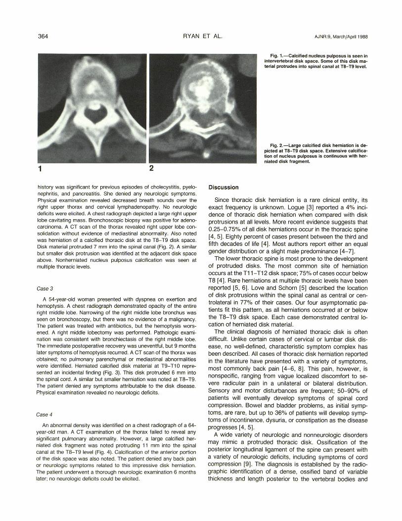
A 54-year-old woman presented with dyspnea on exertion and hemoptysis. A chest radiograph demonstrated opacity of the entire right middle lobe. Narrowing of the right middle lobe bronchus was seen on bronchoscopy, but there was no evidence of a malignancy. The patient was treated with antibiotics , but the hemoptysis worsened. A right middle lobectomy was performed. Pathologic examination was consistent with bronchiectasis of the right middle lobe. The immediate postoperative recovery was uneventful, but 9 months later symptoms of hemoptysis recurred. A CT scan of the thorax was obtained; no pulmonary parenchymal or mediastinal abnormalities were identified. Herniated calcified disk material at T9-T1 0 represented an incidental finding (Fig. 3). This disk protruded 6 mm into the spinal cord. A similar but smaller herniation was noted at T8-T9. The patient denied any symptoms attributable to the disk disease. Physical examination revealed no neurologic deficits.
Case 4
An abnormal density was identified on a chest radiograph of a 64-year-old man. A CT examination of the thorax failed to reveal any significant pulmonary abnormality. However, a large calcified herniated disk fragment was noted protruding 11 mm into the spinal canal at the T8-T9 level (Fig . 4). Calcification of the anterior portion of the disk space was also noted. The patient denied any back pain or neurologic symptoms related to this impressive disk herniation. The patient underwent a thorough neurologic examination 6 months later; no neurologic deficits could be elicited.
Discussion
Fig. 1.-Calcified nucleus pulposus is seen in intervertebral disk space. Some of this disk material protrudes into spinal canal at T8-T9 level.
Fig. 2.-Large calcified disk herniation is depicted at T8-T9 disk space. Extensive calcification of nucleus pulposus is continuous with herniated disk fragment.
Since thoracic disk herniation is a rare clinical entity, its exact frequency is unknown. Logue [3] reported a 4% incidence of thoracic disk herniation when compared with disk protrusions at all levels. More recent evidence suggests that 0.25-0.75% of all disk herniations occur in the thoracic spine [4, 5]. Eighty percent of cases present between the third and fifth decades of life [4]. Most authors report either an equal gender distribution or a slight male predominance [4-7] .
The lower thoracic spine is most prone to the development of protruded disks. The most common site of herniation occurs at the T11-T12 disk space; 75% of cases occur below T8 [4]. Rare herniations at multiple thoracic levels have been reported [5, 6] . Love and Schorn [5] described the location of disk protrusions within the spinal canal as central or centrolateral in 77% of their cases. Our four asymptomatic patients fit this pattern, as all herniations occurred at or below the T8-T9 disk space. Each case demonstrated central location of herniated disk material.
The clinical diagnosis of herniated thoracic disk is often difficult. Unlike certain cases of cervical or lumbar disk disease, no well-defined, characteristic symptom complex has been described. All cases of thoracic disk herniation reported in the literature have presented with a variety of symptoms, most commonly back pain [4-6, 8]. This pain, however, is nonspecific, ranging from vague localized discomfort to severe radicular pain in a unilateral or bilateral distribution. Sensory and motor disturbances are frequent; 50-90% of patients will eventually develop symptoms of spinal cord compression. Bowel and bladder problems, as initial symptoms, are rare, but up to 36% of patients will develop symptoms of incontinence, dysuria, or constipation as the disease progresses [4, 5].
A wide variety of neurologic and non neurologic disorders may mimic a protruded thoracic disk. Ossification of the posterior longitudinal ligament of the spine can present with a variety of neurologic deficits, including symptoms of cord compression [9]. The diagnosis is established by the radiographic identification of a dense, ossified band of variable thickness and length posterior to the vertebral bodies and
AJNR :9, March/April 1988 HERNIATED THORACIC DISKS 365
Fig. 3,-At T9-no level calcified nucleus pulpo!Ous is seen in continuity with calcified herniated disk fragment. This fragment extends 6 mm into spinal canal.
Fig. 4.-Large calcified herniated disk fragment protrudes 11 mm into spinal canal. Anterior disk space calcification depicts site of origination of protruded disk fragment. This patient had no neurologic symptoms.
3
intervertebral disks, seen most commonly in the cervical spine. The ossified band is often separated from the vertebral body by a radiolucent zone. This appearance is distinct from that of herniated thoracic disks. In our reported cases, protruded calcified thoracic disk was seen in direct continuity with intervertebral disk material. Resnick [9] states that the two diagnoses should not realistically be confused.
Posterior thoracic disk herniation and spinal cord compression have been described in association with Scheuermann disease. The radiographic appearance of this disorder is fairly typical [10, 11]. None of our patients displayed the usual radiographic findings of Scheuermann disease. Other bone disorders of the thoracic spine, spinal cord tumors, demyelinating disease of the spinal cord , and various upper abdominal disorders may mimic thoracic disk herniation.
Controversy exists over the usefulness of conventional radiographs in the diagnosis of calcified thoracic disk herniations. McAllister and Sage [6] reviewed the dorsal spine radiographs of 200 patients without spinal cord symptoms; disk space calcification was present in eight (4%). However, in their series of 20 surgically proved thoracic disk herniations, 70% demonstrated calcification of disk material at the appropriate level. They also described 11 patients (55%) with calcified disk fragments protruding into the spinal canal. Logue [3] described a similar frequency of disk space calcification in association with herniated disk material. Baker et al. [8] concludes that disk space calcification was helpful in diagnosing thoracic disk herniation in only a small percentage of cases. Calcified disk fragments were observed within the spinal canal in five (12%) of his 43 patients. This finding was strongly indicative of disk herniation; however, it was observed in only three patients before surgery. Shapiro [13] states that calcification of the nucleus pulposus occurs frequently in the absence of symptomatic disk herniation and therefore is not a helpful diagnostic sign.
In the past, myelography has been most valuable in diagnosing thoracic disk herniation. CT scanning has now assumed a more critical role . Several reports have illustrated the usefulness of CT in diagnosing both calcified and non calcified disk herniations [12, 14, 15]. Metrizamide-enhanced CT
4
scans can be helpful in delineating the extent of dural sac compression , especially in the presence of noncalcified disk protrusions.
Thoracic disk herniations are extremely rare, as detailed earlier in this paper. The frequency of asymptomatic disk protrusions is not known . There have been no reports in the literature describing asymptomatic thoracic disk herniation . Shapiro [13] stated that asymptomatic disk herniations are commonly seen at autopsy, but he offers no data in support of this statement. Since the natural history of asymptomatic disk herniation has not been defined, we are uncertain of the significance of this finding . In view of the impressive radiographic appearance of these disk herniations and the susceptible nature of the thoracic spinal cord to compressive injury, we would speculate that these patients are at risk for the development of future neurologic symptoms . We recommend that these patients be followed periodically to detect early signs and symptoms of neurologic dysfunction . It is in these early stages of compressive cord disease that surgery can be most beneficial [16].
Addendum
Since this manuscript was accepted for publication, we have identified two more patients with asymptomatic calcified thoracic disk protrusions on thoracic CT scans.
REFERENCES
1. Carson J, Gumpert J, Jefferson A. Diagnosis and treatment of thoracic intervertebral disc protrusions. J Neurol Neurosurg Psychiatry 1971 ;34 :68-77
2. Chin LS, Black KL, Hoff JT. Multiple thoracic disc herniations. J Neurosurg 1987;66 : 290-292
3. Logue V. Thoracic intervertebral disc prolapse with spinal cord compression. J Neurol Neurosurg Psychiatry 1952;15 : 227-241
4. Arce CA, Dohrmann GJ. Herniated thoracic disks. NeurolClin 1985 ;3 :383-392
5. Love JG, Schorn VG. Thoracic disk protrusions. JAMA 1965;191 :627-631 6. McAllister VL, Sage MR. The radiology of thoracic disc protrusion . Clin
Radio/ 1976;27:291-299
366 RYAN ET AL. AJNR:9, March/April 1988
7. Benjamin MV, Ransohoff J. Thoracic disc disease. In: Rothman RH , Simeone FA, eds. The spine. Philadelphia: Saunders, 1982:500-507
8. Baker HL, Love JG, Uihlein A. Roentgenologic features of protruded thoracic intervertebral disks. Radiology 1965;84: 1059-1065
9. Resnick D. Ossification of the posterior longitudinal ligament of the spine. In: Resnick D, Niwayama G, eds. Diagnosis of bone and joint disorders. Philadelphia: Saunders, 1981 :1453-1463
10. Van Landingham JH. Herniation of thoracic intervertebral discs with spinal cord compression in kyphosis dorsalis juvenilis (Scheuermann's disease). J Neurosurg 1954;11 :327-329
11 . Resnick D. The osteochondroses. In: Resnick D, Niwayama G, eds.
Diagnosis of bone and jOint disorders. Philadelphia: Saunders, 1981:2902-2906
12. Schimel S, Deeb ZL. Herniated thoracic intervertebral discs. J Comput Tomogr 1985;9 :141-143
13. Shapiro R. Myelography. Chicago: Year Book Medical, 1984:471-473 14. Arce CA, Dohrmann GJ. Thoracic disc herniation: improved diagnosis with
CT scanning and a review of the literature. Surg Neuro/1985;23 :356-361 15. Van Ameyden FC , Van Wiechen PJ . Herniation of calcified nucleus pulpo
sus in the thoracic spine. J Comput Assist Tomogr 1983;7 : 1122-1123 16. Terry AF, McSweeney T, Jones HWF. Paraplegia as a sequel to dorsal
disc prolapse. Paraplegia 1981 ; 19: 111-117